radiology in the information age - rsna 2015 · radiology in the information age ... system, you...
TRANSCRIPT
By Michael Hart
The Information Age has arrived at RSNA 2015. If the hallmark of RSNA’s first century was the ability to
deliver ever more precise and meaningful images, the Society’s second century will be defined by the development of innova-tive ways to manage those images and the associated information in ways that may ultimately improve patient care.
As radiology bridges these two eras, there are challenges – and not all of them are technical, according to Murray Reicher, M.D., chief medical officer at Merge Healthcare, an IBM company.
“Radiologists are drowning in images,” Dr. Reicher said. “We have entered an era where 1,000+ image CT, MRI, and PET exams are common. In academic environ-ments, it is not at all uncommon to see 3,000-, 4,000- and 5,000-image studies. In addition, radiologists now need to assimilate more clinical data than ever and take into account more scientific research in order to optimally perform their jobs. Radiolo-gists need to track recommendations as well as communicate with other physicians and patients. It’s a daunting task that has become overwhelming.”
“Let’s first look at job satisfaction,” Dr. Reicher said. “Part of enjoying work is get-ting into a mental state of flow. Job satisfac-tion requires a sense of mastery and creative control over one’s environment, as well as knowing that you are doing a diligent job and helping people. You can’t get into a mental state of flow and you can’t enjoy your work when pre-sented with an insurmountable task.”
There are ways to help resolve this dilemma, and Merge Healthcare, an IBM company, will address these topics at RSNA 2015.
“Cognitive computing holds great promise to help,” Dr. Reicher said, and he outlined three applications of computer technology Merge Healthcare believes may help radiologists efficiently navigate their workloads.
“First,” he said, “cognitive computing can be used to collect, compile, analyze, and present clinical data.”
In the future, he pointed out, prior to reading an exam, instead of inferring the pre-test probability of various diagnoses based on a keyhole view of the patient, he envisions that the radiologist will receive an accurate, concise presentation of exist-ing diagnoses and be able to identify options for treatment that consider the imaging studies in the context of other available patient data, such as doctors’ notes from a patient record or patient con-tributed data from wearable devices.
“Second, image analytics can be designed to analyze the images to help clinicians identify anomalies,” Dr. Reicher said.
As an example, he described the daily workload of a hypothetical radiologist who
must read 50 CTs a day (“Which is not very many,” he added.) If each of those CTs has 1,000 images, and there’s an old CT for comparison purposes, those 50 CTs trans-late to 100,000 images in a day’s work.
“Rather than a human triaging an insur-mountable number of images, perhaps tech-nology can enable the radiologist to instead focus on uniquely human and rewarding
tasks, such as evaluating relevant images, applying our experience to making diag-noses, directing further work-ups, consult-ing with referring doctors, supervising technologists, participating in selection of proper imaging procedures, and educating patients,” adds Dr. Reicher.
Finally, Dr. Reicher suggested, “there’s a third, under-emphasized area of research
and development, “perceptual design.” Per-ceptual design is informed by the study of human perception and cognition, and aims to create a radiologist’s reading environment that maximally leverages our human capabil-ities while helping us with our weaknesses.
All three applications of computer tech-nologies may help – more efficient and
D e c e m b e r 2 , 2 0 1 5 Product
Announcements Inside
w e d n e s d ay
Murray Reicher, M.D.
continued on page 6b
Radiology in the Information ageMerge Highlights Solutions for Radiologists’ Workloads
Making an accurate breast cancer diagnosis can be a complex process,
and time is always of the essence. Now, with the Coronis Uniti™ display
system, you can view 3D mammography, 2D mammography, breast
MRI and breast ultrasound all on the same screen in perfect grayscale
and precisely calibrated color. You’ll see the subtlest details with greater
clarity, increasing your ability to make faster, better clinical decisions.
The result: greater peace of mind for you and your patients.
Get the 100% solution for digital breast imaging.
So revealing
Discover the new reading room on www.barco.com/uniti
Visit Barco #2559 to discover the latest
breakthroughs in diagnostic imaging.

3bd a i l y b u l l e t i n • W e d n e s d a y , d e c e m b e r 2 , 2 0 1 5
COMPUTED TOMOGRAPHY
Leidel & KrachtBooth 6707
Multifunctional Knee-Joint support ProtectorsThis new LK Knee-Joint Support can be used for any kind of imaging, CT, MR, X-ray, nuclear medicine, or radiotherapy. The colors and the materials can be adapted to the customer’s requirements regarding design and comfort. If you want it soft and comfortable for a longer MR scan or stiff and precise for a quick CT, the right cushion is available. Leidel & Kracht has approved the geometry through many tests and feedback from some of our key customers. It can be used very comfort-ably by people of different weights and sizes. Biocompatibility and compliance to Restriction of Hazardous Substances and reach are of course a given as Leidel & Kracht 3D Foam Technologies is an origi-nal equipment manufacturer-supplier with many years of experience in the medical market.
Koning CorporationBooths 1737 and 1358
Low-dose CT scanner for Imaging entire BreastKoning Corporation introduces the first dedicated, low-dose CT scanner designed to image the entire breast. With a horizon-tal gantry, the Koning Breast CT (KBCT), allows comfortable prone patient position-ing without compression of the breast and captures hundreds of volumetric images in a 10-second rotation. This creates true isotropic 3D images and ultra-thin cross sectional slices, giving breast imaging experts an unprecedented view of an organ that is notoriously hard to visual-ize. With KBCT, images are clearer and higher contrast, 360-degree views are easily manipulated, and the ability to effectively and efficiently detect breast cancer is improved.
Block ImagingBooth 2507
new Online store for Order-ing Imaging equipment Parts Block Imaging launched a new e-commerce platform for ordering imag-ing equipment parts online. Radiology managers, biomeds and imaging engineers will enjoy the ability to buy imaging parts online and get the part needed next day. Thousands of parts for every modality, from CT and mammography to digital X-ray, are available within a few clicks. Online visitors will enjoy immediate access to real-time inventory lists, with pricing, live support chat and online pay-ment features designed to increase the efficiency of the parts ordering process. The Block Imaging Parts eCommerce site is the first of its kind in the industry. No more waiting as transactions can take place 24/7. Find the needed part fast. Buy it online. Get it next day.
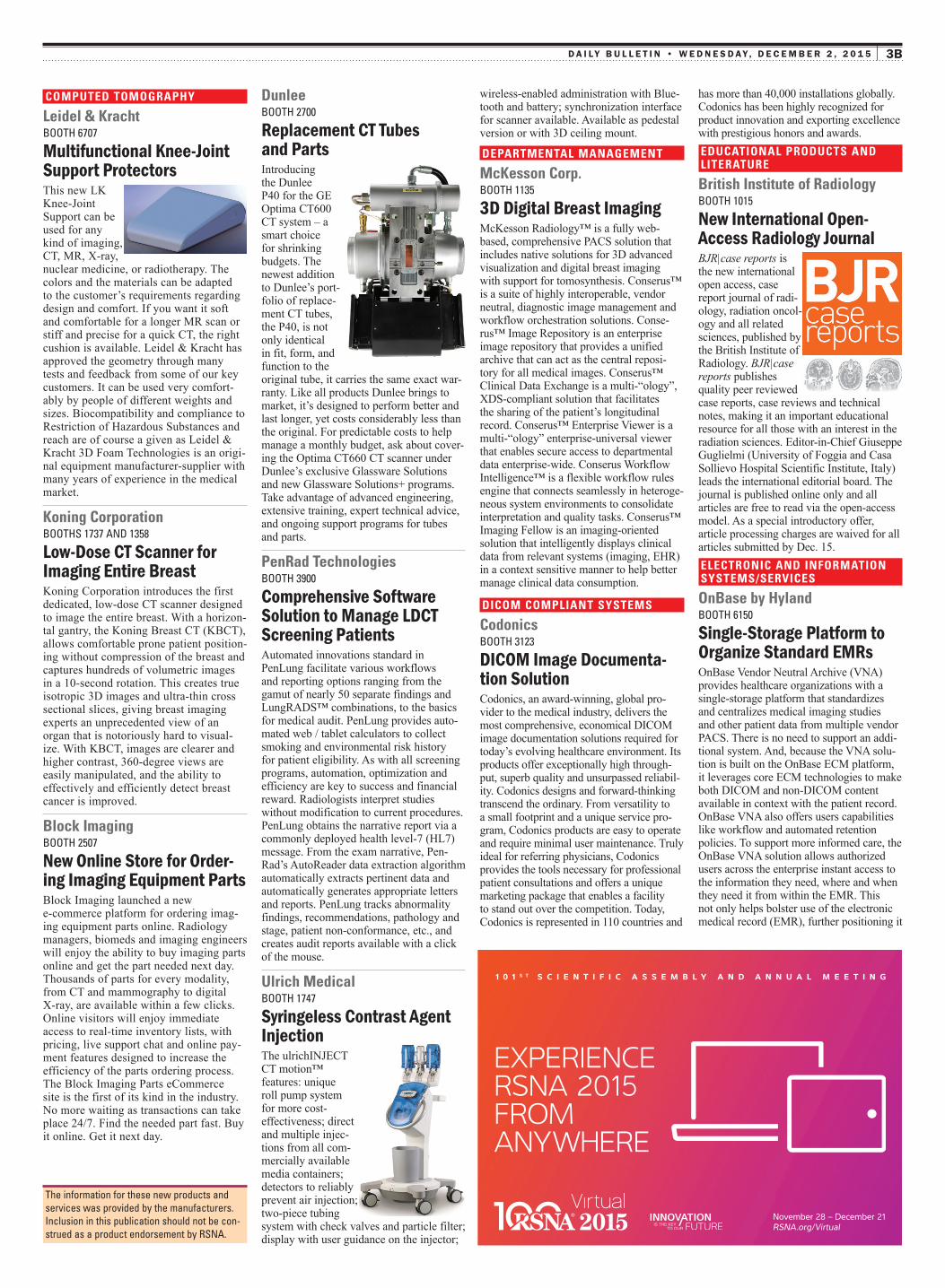
DunleeBooth 2700
Replacement CT Tubes and PartsIntroducing the Dunlee P40 for the GE Optima CT600 CT system – a smart choice for shrinking budgets. The newest addition to Dunlee’s port-folio of replace-ment CT tubes, the P40, is not only identical in fit, form, and function to the original tube, it carries the same exact war-ranty. Like all products Dunlee brings to market, it’s designed to perform better and last longer, yet costs considerably less than the original. For predictable costs to help manage a monthly budget, ask about cover-ing the Optima CT660 CT scanner under Dunlee’s exclusive Glassware Solutions and new Glassware Solutions+ programs. Take advantage of advanced engineering, extensive training, expert technical advice, and ongoing support programs for tubes and parts.
PenRad TechnologiesBooth 3900
Comprehensive software solution to Manage LdCT screening Patients Automated innovations standard in PenLung facilitate various workflows and reporting options ranging from the gamut of nearly 50 separate findings and LungRADS™ combinations, to the basics for medical audit. PenLung provides auto-mated web / tablet calculators to collect smoking and environmental risk history for patient eligibility. As with all screening programs, automation, optimization and efficiency are key to success and financial reward. Radiologists interpret studies without modification to current procedures. PenLung obtains the narrative report via a commonly deployed health level-7 (HL7) message. From the exam narrative, Pen-Rad’s AutoReader data extraction algorithm automatically extracts pertinent data and automatically generates appropriate letters and reports. PenLung tracks abnormality findings, recommendations, pathology and stage, patient non-conformance, etc., and creates audit reports available with a click of the mouse.
Ulrich MedicalBooth 1747
syringeless Contrast agent Injection The ulrichINJECT CT motion™ features: unique roll pump system for more cost-effectiveness; direct and multiple injec-tions from all com-mercially available media containers; detectors to reliably prevent air injection; two-piece tubing system with check valves and particle filter; display with user guidance on the injector;
wireless-enabled administration with Blue-tooth and battery; synchronization interface for scanner available. Available as pedestal version or with 3D ceiling mount.
DEPARTMENTAL MANAGEMENT
McKesson Corp.Booth 1135
3d digital Breast Imaging McKesson Radiology™ is a fully web-based, comprehensive PACS solution that includes native solutions for 3D advanced visualization and digital breast imaging with support for tomosynthesis. Conserus™ is a suite of highly interoperable, vendor neutral, diagnostic image management and workflow orchestration solutions. Conse-rus™ Image Repository is an enterprise image repository that provides a unified archive that can act as the central reposi-tory for all medical images. Conserus™ Clinical Data Exchange is a multi-“ology”, XDS-compliant solution that facilitates the sharing of the patient’s longitudinal record. Conserus™ Enterprise Viewer is a multi-“ology” enterprise-universal viewer that enables secure access to departmental data enterprise-wide. Conserus Workflow Intelligence™ is a flexible workflow rules engine that connects seamlessly in heteroge-neous system environments to consolidate interpretation and quality tasks. Conserus™ Imaging Fellow is an imaging-oriented solution that intelligently displays clinical data from relevant systems (imaging, EHR) in a context sensitive manner to help better manage clinical data consumption.
DICOM COMPLIANT SYSTEMS
CodonicsBooth 3123
dICOM Image documenta-tion solutionCodonics, an award-winning, global pro-vider to the medical industry, delivers the most comprehensive, economical DICOM image documentation solutions required for today’s evolving healthcare environment. Its products offer exceptionally high through-put, superb quality and unsurpassed reliabil-ity. Codonics designs and forward-thinking transcend the ordinary. From versatility to a small footprint and a unique service pro-gram, Codonics products are easy to operate and require minimal user maintenance. Truly ideal for referring physicians, Codonics provides the tools necessary for professional patient consultations and offers a unique marketing package that enables a facility to stand out over the competition. Today, Codonics is represented in 110 countries and
has more than 40,000 installations globally. Codonics has been highly recognized for product innovation and exporting excellence with prestigious honors and awards.EDUCATIONAL PRODUCTS AND LITERATURE
British Institute of RadiologyBooth 1015
new International Open-access Radiology JournalBJR|case reports is the new international open access, case report journal of radi-ology, radiation oncol-ogy and all related sciences, published by the British Institute of Radiology. BJR|case reports publishes quality peer reviewed case reports, case reviews and technical notes, making it an important educational resource for all those with an interest in the radiation sciences. Editor-in-Chief Giuseppe Guglielmi (University of Foggia and Casa Sollievo Hospital Scientific Institute, Italy) leads the international editorial board. The journal is published online only and all articles are free to read via the open-access model. As a special introductory offer, article processing charges are waived for all articles submitted by Dec. 15.ELECTRONIC AND INFORMATION SYSTEMS/SERVICES
OnBase by HylandBooth 6150
single-storage Platform to Organize standard eMRsOnBase Vendor Neutral Archive (VNA) provides healthcare organizations with a single-storage platform that standardizes and centralizes medical imaging studies and other patient data from multiple vendor PACS. There is no need to support an addi-tional system. And, because the VNA solu-tion is built on the OnBase ECM platform, it leverages core ECM technologies to make both DICOM and non-DICOM content available in context with the patient record. OnBase VNA also offers users capabilities like workflow and automated retention policies. To support more informed care, the OnBase VNA solution allows authorized users across the enterprise instant access to the information they need, where and when they need it from within the EMR. This not only helps bolster use of the electronic medical record (EMR), further positioning it
the information for these new products and services was provided by the manufacturers. Inclusion in this publication should not be con-strued as a product endorsement by Rsna.
November 28 – December 21RSNA.org/Virtual
1 0 1 S T S C I E N T I F I C A S S E M B L Y A N D A N N U A L M E E T I N G
EXPERIENCE RSNA 2015 FROM ANYWHERE
4b d a i l y b u l l e t i n • W e d n e s d a y , d e c e m b e r 2 , 2 0 1 5
as the system of record, but it helps health-care organizations move that much closer to the goal of “One patient. One record.”
IntrasenseBooth 6524
Full-Imaging software solu-tion for Oncology, Chronic diseases
Intrasense will unveil the brand-new version of its server software Myrian®, a full imaging solution for the diagnosis,
therapy planning and follow-up on oncology and chronic diseases. New functionalities include higher acces-sibility, a brand new user interface and structured reading features for unequalled ergonomics and time-savings. New pow-erful capabilities have also been added to most Myrian clinical modules including XP-Breast, XP-Prostate, XP-Liver and XT-Brain modules. Be part of this break-through and discover how the new Myrian can improve productivity.
Mint MedicalBooth 4761
standardized, Context-driv-en Read Procedures and structured Reports
With the new mint Lesion™ 3.1, Mint Medical created a simplified and uniform
user interface that focuses on the particular context of a current case and gently offers guidance according to the corresponding screening, staging, or response assess-ment criteria. mint Lesion knows about the intricacies of particular guidelines and automatically determines stage of disease and response to therapy, based on radio-logical multiparametric image annotations, classifications, and further clinical param-eters. The profiles include a variety of tumor-node-metastasis (TNM) tumor enti-ties, staging and transplant scoring of liver disease, screening and staging of prostate carcinoma, and numerous response evalu-ation criteria (RECIST, mRECIST HCC, Cheson, irRC, irRECIST, Choi, RANO, PCWG2, etc.). In addition, mint Lesion characterizes lesions also by their texture and highlights potential tipping points for clinical treatment decisions during the read process and in the automatically generated structured reports.
Imorgon Medical, LLCBooth 6133
Ultrasound workflow and Reporting enhancementImorgon, the industry leader in ultrasound workflow and reporting enhancement, is pleased to announce the latest system upgrade to version 3.0. This ambitious release reflects Imorgon’s commitment to its customers and its mission to streamline ultrasound workflow. Dynamic clips are critical to quality ultrasound diagnosis and Imorgon’s ability to display clips is unpar-alleled. This core feature is turbocharged with faster loading of images and clips, full support of unlimited length clips, creation of sub-clips, flagged frames and the ability to save a frame as a static image. Images and clips can be acquired on-the-fly for presentations in a variety of formats (MP4, MOV, AVI) with new flexibility to select only a region of the image or clip for export. Images can be re-ordered to stream-line the dictation process. New quality control (QC) tools enhance patient safety.
Laurel Bridge SoftwareBooth 1125
enterprise Medical Imaging workflow solutionsLaurel Bridge Software, a provider of enterprise medical imaging workflow solutions, recently added health level-7 (HL7) workflow capability, and 64-bit support to its Compass™ DICOM solution. These new enterprise workflow capabili-ties can significantly enhance a healthcare organization’s ability to automate its (increasingly) sophisticated diagnostic, clinical and patient centric workflows that often span diverse healthcare IT systems. The Compass image and HL7 workflow management solution now supports a level of granularity not previously available for automating the ingestion and distribution of imaging studies and HL7 messages among disparate clinical imaging and archiving systems. Regardless of organizational or workflow complexity, Compass can ensure that imaging studies are identified and associated with the correct patient, originating location and study description to enable creation of complex, site-specific workflows that span technology domains and expedited workflow integration, PACS consolidation and VNA implementation.
Technical exhibiTion booTh KeySouth Building, Hall ABooths 1000 – 5999North Building, Hall BBooths 6000 – 8599
the information for these new products and services was provided by the manufacturers. Inclusion in this publication should not be con-strued as a product endorsement by Rsna.
• Earn a Quality Essentials Certificate (QEC) by scoring 80% or higher on the SAM test at either of the following quality sessions: MSQI32 and MSQI33 on Tuesday, December 1 (Room S406B).• Learn more about online opportunities for earning QECs and how QECs can lead to an Advanced Level Quality Certificate at RSNA.org/Quality-Improvement.
New! QI Storyboard Poster Walk, Tuesday December 1, 4:30-5:30 PM, Learning Center, Quality Storyboard SectionJoin David Larson, MD, and Paul Nagy, PhD, experts in quality improvement in radiology, as they walk through the QI storyboards, highlighting examples of great work and sound methodology. Bring your walking shoes and come prepared for an interactive session. Those who are interested in leading and publishing QI projects in the coming months and years will find this especially valuable.
RSNA QUALITY ESSENTIALS AND ADVANCED LEVEL QUALITY CERTIFICATES
Questions? Please email [email protected] for more information.
5bd a i l y b u l l e t i n • W e d n e s d a y , d e c e m b e r 2 , 2 0 1 5
Velox RIS SolutionsBooth 8138
Customizable, Fully Integrated Practice Management solutionVelox RIS boosts profitability and increases quality of care by reducing costs, mini-mizing turnaround times and optimizing workflows. Velox RIS Solutions provides imaging centers and radiology groups with a cost-optimized, customizable and fully integrated practice management solution. Velox intuitive RIS Suite includes Velox RIS, billing integration, operational man-agement and analytical tools, and referring physician portal.
INFINITT North America, Inc.Booth 2548
Vna solution and Universal Viewer Healthcare PlatformThe new INFINITT Healthcare Platform (IHP) centralized VNA solution provides open, standards-based storage and manages DICOM and non-DICOM data over their life cycle regardless of where the data orig-inated. This VNA consolidates storage and unlocks proprietary formats from disparate PACS or siloed storage architectures, and makes it easier and more cost-efficient to comply with retention policies and security regulations. This fully scalable archive solution includes INFINITT’s universal viewer, a zero download viewer that sup-ports all types of data from any HTML5 browser-based device. IHP supports intel-ligent information lifecycle management and integrates with any major EMR. It is offered as a software-only option or can be packaged with enterprise-class hardware from INFINITT’s Certified Solution Part-ners. INFINITT will work with providers to develop an enterprise medical data strat-egy that reflects the organization’s overall needs, and can help them leverage their existing infrastructure.FILM AND IMAGE MANAGEMENT
StatRadBooth 2006
PaCs-Integrated, Cloud-Based Image Management systemCreated by radiologists, RadConnect is a cloud-based medical image management solution that integrates with PACS to enable intuitive, quick and secure sharing of DICOM images. It is the easy way to upload, view, share and store all the medical imaging needs. Simply: 1. Upload images into RadConnect using a modern web browser; 2. View the images with StatRad’s diagnostic quality, zero footprint HTML5 viewer; 3. Share and collaborate by sending a simple, secure email link. No more CDs, manual tracking, courier fees, or the manpower required to manage it. RadConnect’s cloud-based solution lets an entire organization – and patients and referring partners – view unbelievably clear, diagnostic quality images on any screen at any time, even on tablets and smartphones. RadConnect’s sharing fea-ture can help you prepare for new patient appointments and transfers, and it can help prevent repeat exams as ordering clinicians have access to their patients’ prior images.
Intelemage, LLCBooth 8344
Medical Image exchange and workflow PlatformIntelemage, LLC announces the release of InteleGRID Core 4.0, the latest version of its industry leading, SaaS based, medical image exchange and workflow platform. Since 2007 InteleGRID has been power-ing clinical trial and life science work-flows and today powers the top medical device companies’ pre-surgical planning platforms. InteleGRID Core 4.0 boasts the most secure, reliable and easily configu-rable medical image exchange along with the convenience of native mobile apps for iOS and Android devices. At RSNA 2015, Intelemage will present the latest cases in video enabled tele-health, health
level-7 (HL7) interface modules (includ-ing RefMD auto-share) and its unlimited user / unlimited volume secure image exchange platform.
INTERVENTIONAL RADIOLOGY & SPECIAL PROCEDURES
Laurane Medical, LLCBooth 7208
Bone access and Biopsy device
Laurane Medical LLC is proud to announce the preliminary launch of the new gen-eration of its bone access and biopsy
Technical exhibiTion booTh KeySouth Building, Hall ABooths 1000 – 5999North Building, Hall BBooths 6000 – 8599
Technical exhibiTion hoursSouth Building, Hall A and North Building, Hall Bsunday – Wednesday. . . .10:00 a.m. – 5:00 p.m.thursday . . . . . . . . . . . . . . .10:00 a.m. – 2:00 p.m.
Full exhibiTor lisTingto see complete company profiles and product information, visit RSNA2015.RSNA.org/Exhibit.
6b d a i l y b u l l e t i n • W e d n e s d a y , d e c e m b e r 2 , 2 0 1 5
complete presentation of patient clinical information, computer-aided image analyt-ics, and enhanced perceptual design.
“Merge Healthcare will discuss all three topics at RSNA,” Reicher said.
For more information on the RSNA Tech-nical Exhibits, see the RSNA 2015 Meet-ing App, RSNA.org/ExhibitingCompanies and the Technical Exhibits Guide.
Radiology in the Information agecontinued from page 1b
address the challenges of delivering qual-ity public healthcare and crisis response care in remote environments.ULTRASONOGRAPHY
SuperSonic ImagineBooth 6339Already the pioneers of ShearWave™ Elastography, SuperSonic Imagine now introduces the next wave in ultrasound innovation – AngioPLUS* (PLanewave UltraSensi-tive) imaging. AngioPLUS is a significant advancement in color Dop-pler imaging. Conventional Doppler is limited in its ability to show microvascular slow flow. AngioPLUS sig-nificantly improves color sensitivity and spatial resolution, resulting in the next level of microvascular imaging to visual-ize flows that couldn’t be seen before. Soon available on Aixplorer®, this innovation will provide highly detailed real-time information to physicians, which is key in diagnosing cancerous lesions. Lesion microvascularization and vessel flow are important indicators of a potential malignancy in areas such as breast, lymph nodes, thyroid and liver. This technique is also valuable for mus-culoskeletal assessments to help identify low-grade tendon inflammation. * Pend-ing regulatory approval.
CIVCO Medical SolutionsBooth 2317
next Generation Ultrasound needle GuideCIVCO Medical Solutions will unveil the latest innovation in needle guidance technology at RSNA 2015. A leader in the development of guidance systems for minimally invasive ultrasound procedures, CIVCO will demonstrate its next genera-tion guidance system designed to provide clinicians with accurate guidance and confident outcomes for today’s environ-ment. The new guide offers advanced versatility, featuring a wider range of compatible instruments, expanded target depths to accommodate multiple clinical applications, and improved stability and functionality to meet a wide range of diagnostic and therapeutic procedures. The device has not received clearance by the U.S. Food and Drug Administration, and is targeted to launch on leading ultrasound equipment in 2016.
while the specimen is sent directly to pathology.
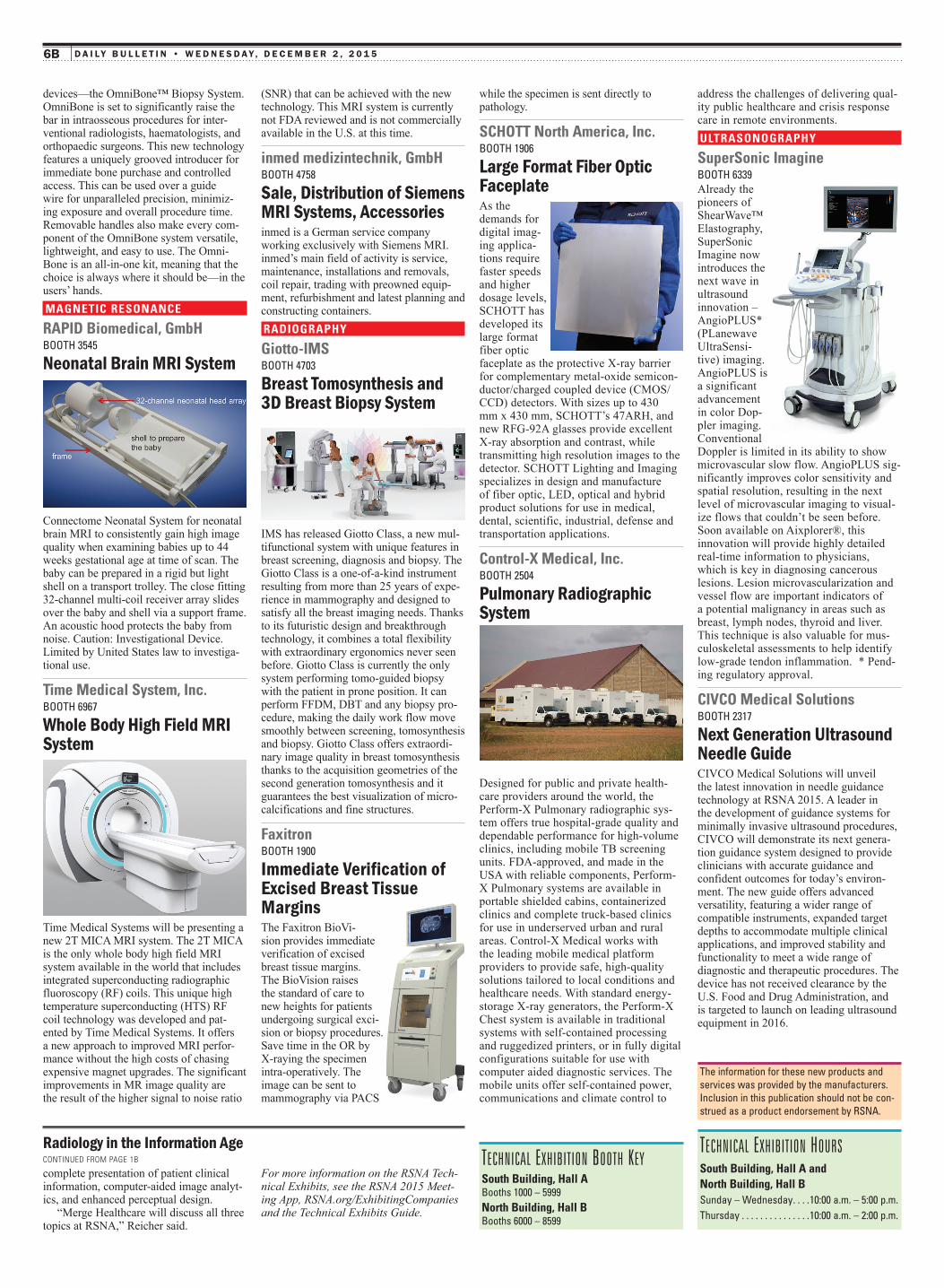
SCHOTT North America, Inc.Booth 1906
Large Format Fiber Optic FaceplateAs the demands for digital imag-ing applica-tions require faster speeds and higher dosage levels, SCHOTT has developed its large format fiber optic faceplate as the protective X-ray barrier for complementary metal-oxide semicon-ductor/charged coupled device (CMOS/CCD) detectors. With sizes up to 430 mm x 430 mm, SCHOTT’s 47ARH, and new RFG-92A glasses provide excellent X-ray absorption and contrast, while transmitting high resolution images to the detector. SCHOTT Lighting and Imaging specializes in design and manufacture of fiber optic, LED, optical and hybrid product solutions for use in medical, dental, scientific, industrial, defense and transportation applications.
Control-X Medical, Inc. Booth 2504
Pulmonary Radiographic system
Designed for public and private health-care providers around the world, the Perform-X Pulmonary radiographic sys-tem offers true hospital-grade quality and dependable performance for high-volume clinics, including mobile TB screening units. FDA-approved, and made in the USA with reliable components, Perform-X Pulmonary systems are available in portable shielded cabins, containerized clinics and complete truck-based clinics for use in underserved urban and rural areas. Control-X Medical works with the leading mobile medical platform providers to provide safe, high-quality solutions tailored to local conditions and healthcare needs. With standard energy-storage X-ray generators, the Perform-X Chest system is available in traditional systems with self-contained processing and ruggedized printers, or in fully digital configurations suitable for use with computer aided diagnostic services. The mobile units offer self-contained power, communications and climate control to
(SNR) that can be achieved with the new technology. This MRI system is currently not FDA reviewed and is not commercially available in the U.S. at this time.
inmed medizintechnik, GmbHBooth 4758
sale, distribution of siemens MRI systems, accessoriesinmed is a German service company working exclusively with Siemens MRI. inmed’s main field of activity is service, maintenance, installations and removals, coil repair, trading with preowned equip-ment, refurbishment and latest planning and constructing containers.RADIOGRAPHY
Giotto-IMSBooth 4703
Breast Tomosynthesis and 3d Breast Biopsy system
IMS has released Giotto Class, a new mul-tifunctional system with unique features in breast screening, diagnosis and biopsy. The Giotto Class is a one-of-a-kind instrument resulting from more than 25 years of expe-rience in mammography and designed to satisfy all the breast imaging needs. Thanks to its futuristic design and breakthrough technology, it combines a total flexibility with extraordinary ergonomics never seen before. Giotto Class is currently the only system performing tomo-guided biopsy with the patient in prone position. It can perform FFDM, DBT and any biopsy pro-cedure, making the daily work flow move smoothly between screening, tomosynthesis and biopsy. Giotto Class offers extraordi-nary image quality in breast tomosynthesis thanks to the acquisition geometries of the second generation tomosynthesis and it guarantees the best visualization of micro-calcifications and fine structures.
FaxitronBooth 1900
Immediate Verification of excised Breast Tissue MarginsThe Faxitron BioVi-sion provides immediate verification of excised breast tissue margins. The BioVision raises the standard of care to new heights for patients undergoing surgical exci-sion or biopsy procedures. Save time in the OR by X-raying the specimen intra-operatively. The image can be sent to mammography via PACS
devices—the OmniBone™ Biopsy System. OmniBone is set to significantly raise the bar in intraosseous procedures for inter-ventional radiologists, haematologists, and orthopaedic surgeons. This new technology features a uniquely grooved introducer for immediate bone purchase and controlled access. This can be used over a guide wire for unparalleled precision, minimiz-ing exposure and overall procedure time. Removable handles also make every com-ponent of the OmniBone system versatile, lightweight, and easy to use. The Omni-Bone is an all-in-one kit, meaning that the choice is always where it should be—in the users’ hands.MAGNETIC RESONANCE
RAPID Biomedical, GmbHBooth 3545
neonatal Brain MRI system
Connectome Neonatal System for neonatal brain MRI to consistently gain high image quality when examining babies up to 44 weeks gestational age at time of scan. The baby can be prepared in a rigid but light shell on a transport trolley. The close fitting 32-channel multi-coil receiver array slides over the baby and shell via a support frame. An acoustic hood protects the baby from noise. Caution: Investigational Device. Limited by United States law to investiga-tional use.
Time Medical System, Inc. Booth 6967
whole Body High Field MRI system
Time Medical Systems will be presenting a new 2T MICA MRI system. The 2T MICA is the only whole body high field MRI system available in the world that includes integrated superconducting radiographic fluoroscopy (RF) coils. This unique high temperature superconducting (HTS) RF coil technology was developed and pat-ented by Time Medical Systems. It offers a new approach to improved MRI perfor-mance without the high costs of chasing expensive magnet upgrades. The significant improvements in MR image quality are the result of the higher signal to noise ratio
the information for these new products and services was provided by the manufacturers. Inclusion in this publication should not be con-strued as a product endorsement by Rsna.
Technical exhibiTion booTh KeySouth Building, Hall ABooths 1000 – 5999North Building, Hall BBooths 6000 – 8599
Technical exhibiTion hoursSouth Building, Hall A and North Building, Hall Bsunday – Wednesday. . . .10:00 a.m. – 5:00 p.m.thursday . . . . . . . . . . . . . . .10:00 a.m. – 2:00 p.m.


Invest in the Campaign at RSNA.org/Campaign
Keep radiology vital. Join the Campaign for Funding Radiology’s Future®. We are raising $17.5 million to ensure the future of radiology. Your investment will inspire promising researchers and drive innovation.
MTG655 R&E Invest Ad Full Page TE Focus.indd 1 11/4/15 3:13 PM