radiology - the supply · pdf file• tear-drop fracture. old sp fracture of t1 ......
TRANSCRIPT
5/2/14
1
Hector RiveraMelo DC, DACBR [email protected]
Radiographic Imaging
� Benefits � Visualization of significant osseous and
articular pathology � Risks
� No safe amount of ionizing radiation � Limitations
� Articular cartilage � Soft tissues � Early osseous and articular changes
Cervical Spine Imaging
� Standard radiographic series � AP lower cervical � AP open mouth � Lateral
Standard Cervical Spine Series AP Lower Cervical Lateral
Standard Cervical Spine Series AP Open Mouth
Cervical Spine Imaging
� Accessory radiographic views � Obliques � Flexion/Extension
5/2/14
2
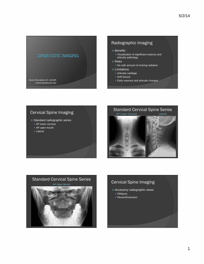
Accessory Cervical Spine Views Anterior Obliques
Accessory Cervical Spine Views
Flexion
Extension
Cervical Spine Imaging
� Common pathology seen on X-rays � Osteoarthritis ○ Degenerative disc disease ○ Facet arthrosis ○ Uncovertebral arthrosis
� DISH � Congenital annomalies � Hangman’s fracture (C2) � Unilateral facet dislocation
Cervical Spine Imaging
� Common pathology not seen on X-rays � Disc herniations � Chiari malformations � Facet fractures*
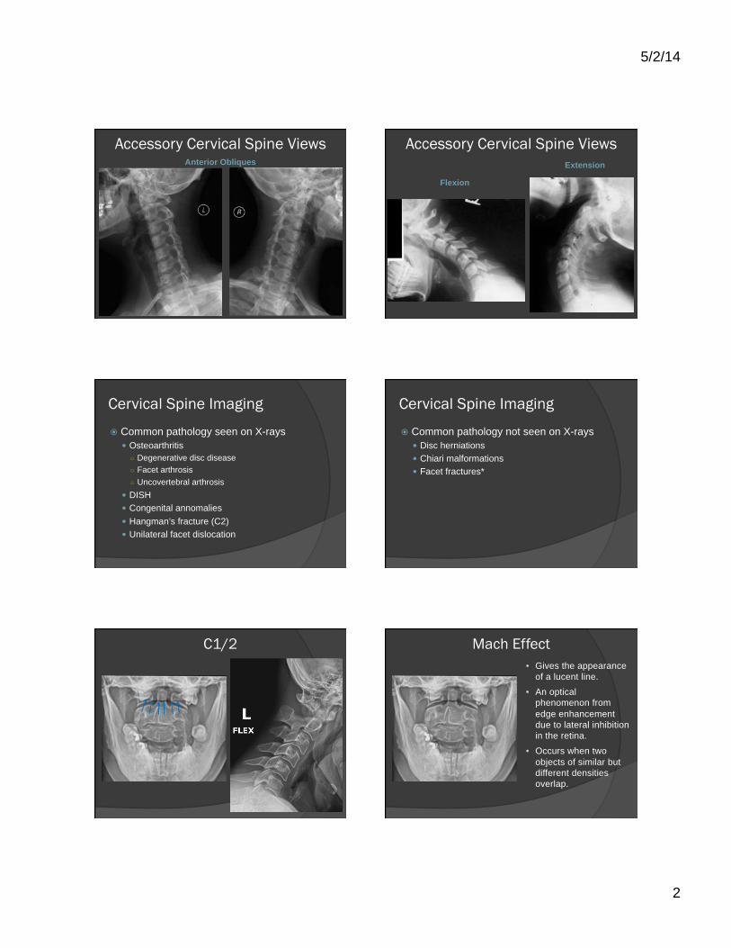
C1/2 Mach Effect • Gives the appearance
of a lucent line. • An optical
phenomenon from edge enhancement due to lateral inhibition in the retina.
• Occurs when two objects of similar but different densities overlap.
5/2/14
3
C1/2 Odontoid Fracture
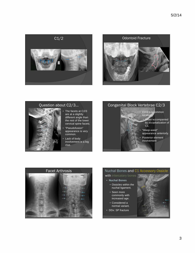
Question about C2/3… • The facets at C2/3
are at a slightly different angle than the rest of the lower cervical spine facets.
• “Pseudofusion” appearance is very common.
• Lack of body involvement is a big clue.
Congenital Block Vertebrae C2/3 • C2/3 is a common
location.
• Often accompanied by occipitalization of C1.
• “Wasp-waist” appearance anteriorly.
• Posterior element involvement
Facet Arthrosis
• Radiographic Features:
• Hypertrophy (enlargement) of the articular processes
• Sclerosis • Anterior or posterior
translations (advanced)
Nuchal Bones and C1 Accessory Ossicle with intercalary bones • Nuchal Bones
• Ossicles within the nuchal ligament.
• Seen more commonly with increased age.
• Considered a normal variant.
• DDx: SP fracture
5/2/14
4
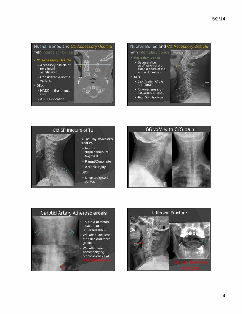
Nuchal Bones and C1 Accessory Ossicle with intercalary bones
• C1 Accessory Ossicle • Accessory ossicle of
no clinical significance.
• Considered a normal variant.
• DDx: • HADD of the longus
colli • ALL calcification
Nuchal Bones and C1 Accessory Ossicle with intercalary bones • Intercalary Bones
• Degenerative calcification of the anterior fibers of the intervertebral disc.
• DDx: • Calcification of the
ALL (DISH). • Atherosclerosis of
the carotid arteries. • Tear-Drop fracture.
Old SP fracture of T1
• AKA: Clay-shoveler’s fracture
• Inferior displacement of fragment
• Parent/Donor site
• A stable injury • DDx:
• Ununited growth center.
66 yoM with C/S pain
Carotid Artery Atherosclerosis • This is a common
location for atherosclerosis.
• Will often look less tube-like and more globular.
• Will often see accompanying atherosclerosis of other large arteries.
Jefferson Fracture
Bonus: Tear-Drop Fracture
5/2/14
5
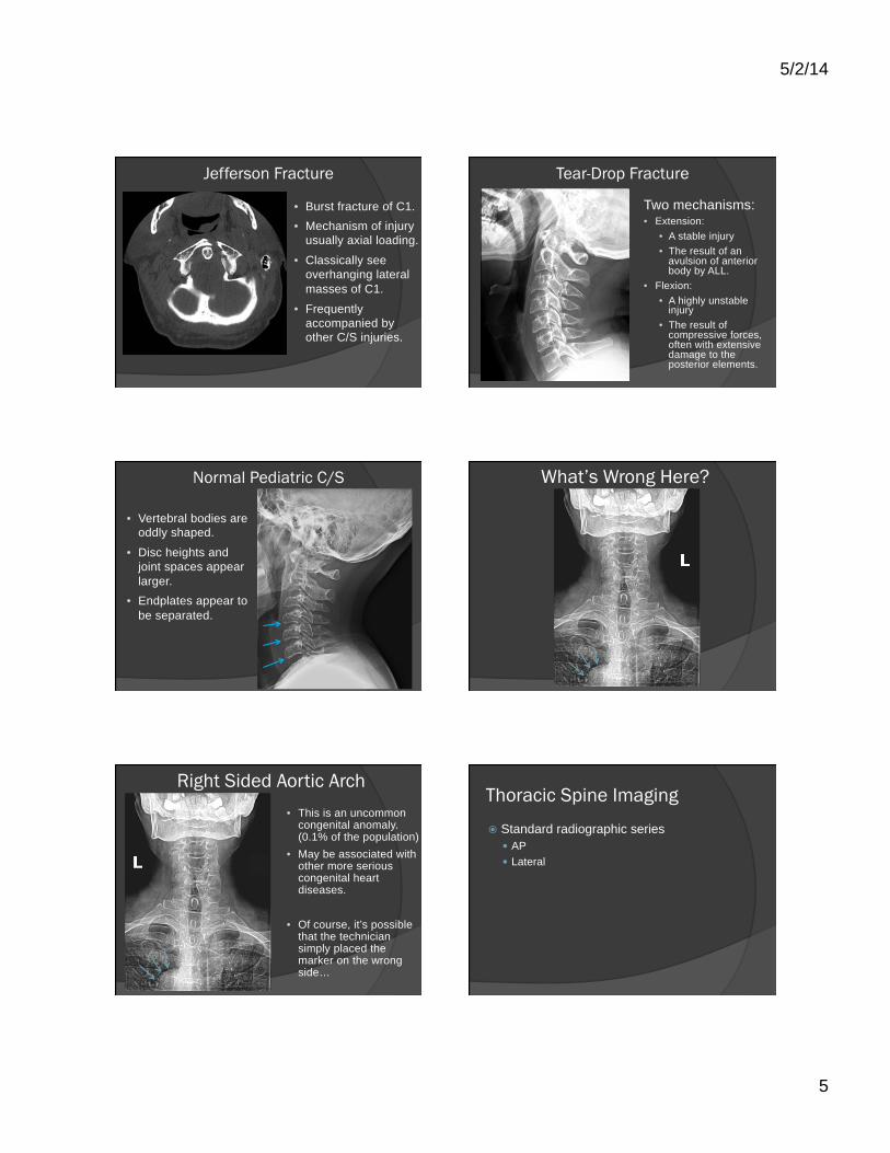
Jefferson Fracture
• Burst fracture of C1. • Mechanism of injury
usually axial loading.
• Classically see overhanging lateral masses of C1.
• Frequently accompanied by other C/S injuries.
Tear-Drop Fracture
Two mechanisms: • Extension:
• A stable injury • The result of an
avulsion of anterior body by ALL.
• Flexion: • A highly unstable
injury • The result of
compressive forces, often with extensive damage to the posterior elements.
Normal Pediatric C/S
• Vertebral bodies are oddly shaped.
• Disc heights and joint spaces appear larger.
• Endplates appear to be separated.
What’s Wrong Here?
Right Sided Aortic Arch • This is an uncommon
congenital anomaly. (0.1% of the population)
• May be associated with other more serious congenital heart diseases.
• Of course, it’s possible that the technician simply placed the marker on the wrong side…
Thoracic Spine Imaging
� Standard radiographic series � AP � Lateral
5/2/14
6
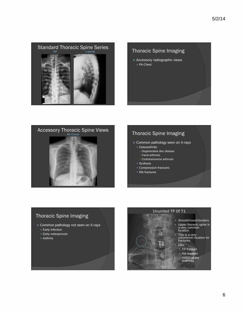
Standard Thoracic Spine Series AP Lateral Thoracic Spine Imaging
� Accessory radiographic views � PA Chest
Accessory Thoracic Spine Views PA Chest Thoracic Spine Imaging
� Common pathology seen on X-rays � Osteoarthritis ○ Degenerative disc disease ○ Facet arthrosis ○ Costotransverse arthrosis
� Scoliosis � Compression fractures � Rib fractures
Thoracic Spine Imaging
� Common pathology not seen on X-rays � Early Infection � Early osteoporosis � Asthma
Ununited TP Of T1
T1
• Smooth/round borders • Upper thoracic spine is
a very common location.
• This is a very uncommon location for fractures.
• DDx: • TP fracture • Rib fracture • HADD of the
scalenes

5/2/14
7
20 yof with c/s and t/s pain Hair Artifacts
What happened here?
• Multiple metallic curvilinear densities.
• Seen outside the chest.
• Here’s another patient with the same mystery…
• Acupuncture needles!!
What went wrong? • This is an unaltered
(not cropped) image taken at a chiropractic office.
• Intended to be a lateral thoracic spine view.
• What went wrong? • Cassette not pushed
in all the way. • Even digital
machines use cassettes (CR)
QUIZ
1) Which of the following levels o6en gives the false appearance of facet fusion? a) C1/2 b) C2/3 c) C3/4 d) C5/6
2) Which of the following is considered a sign of pathology?
a) Nuchal bones b) Intercalary bones c) Mach bands d) Ununited growth
centers
Lumbar Spine Imaging
� Standard radiographic series � AP (or PA) � Lateral
5/2/14
8
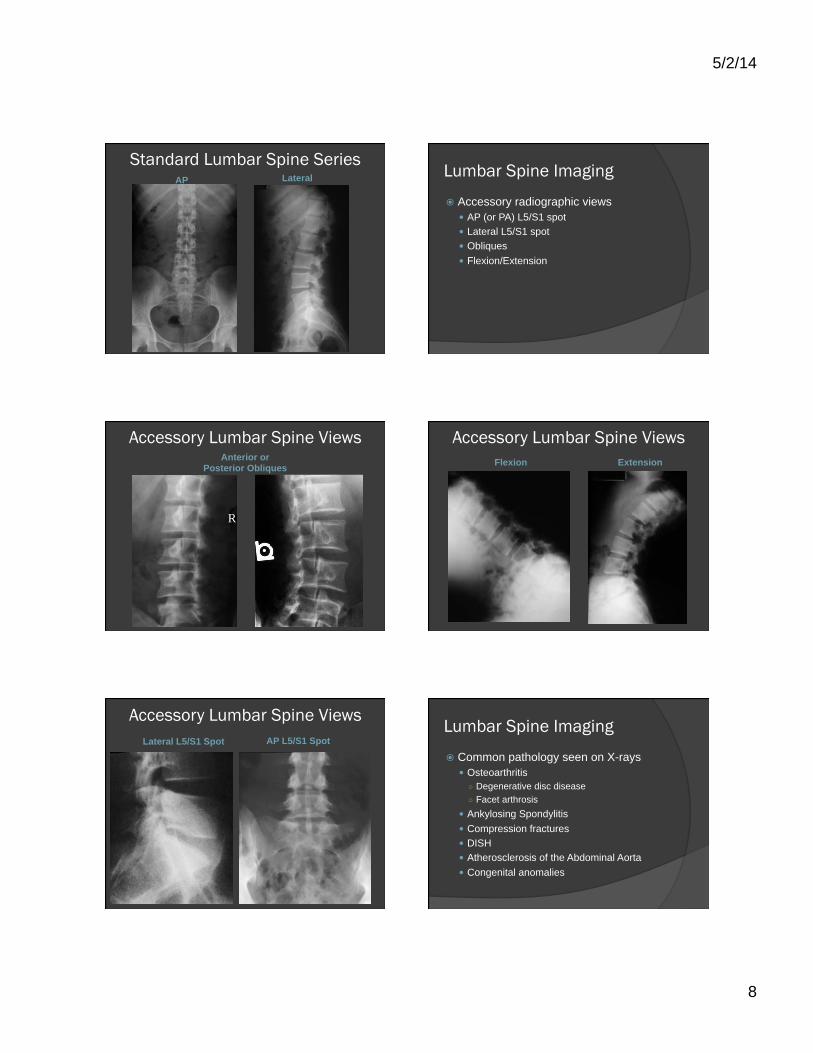
Standard Lumbar Spine Series AP Lateral Lumbar Spine Imaging
� Accessory radiographic views � AP (or PA) L5/S1 spot � Lateral L5/S1 spot � Obliques � Flexion/Extension
Accessory Lumbar Spine Views Anterior or
Posterior Obliques
R
Accessory Lumbar Spine Views Flexion Extension
Accessory Lumbar Spine Views Lateral L5/S1 Spot AP L5/S1 Spot
Lumbar Spine Imaging
� Common pathology seen on X-rays � Osteoarthritis ○ Degenerative disc disease ○ Facet arthrosis
� Ankylosing Spondylitis � Compression fractures � DISH � Atherosclerosis of the Abdominal Aorta � Congenital anomalies
5/2/14
9
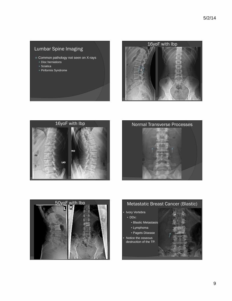
Lumbar Spine Imaging
� Common pathology not seen on X-rays � Disc herniations � Sciatica � Piriformis Syndrome
16yoF with lbp
16yoF with lbp Normal Transverse Processes
50yoF with lbp Metastatic Breast Cancer (Blastic) • Ivory Vertebra
• DDx: • Blastic Metastasis
• Lymphoma • Pagets Disease
• Notice the osseous destruction of the TP.

5/2/14
10
56yom with lbp 48yom with lbp
Limbus Bones
• Very common in the lumbar spine.
• Represent intravertebral disc herniations.
• Typically asymptomatic (especially if anterior).
38yof with lbp
R
Gall Stones
• Located in the right upper quadrant.
• Tend to be more dense around the periphery.
• Common in females over 40.
• May or may not be symptomatic.
39yom with lbp following MVA
5/2/14
11
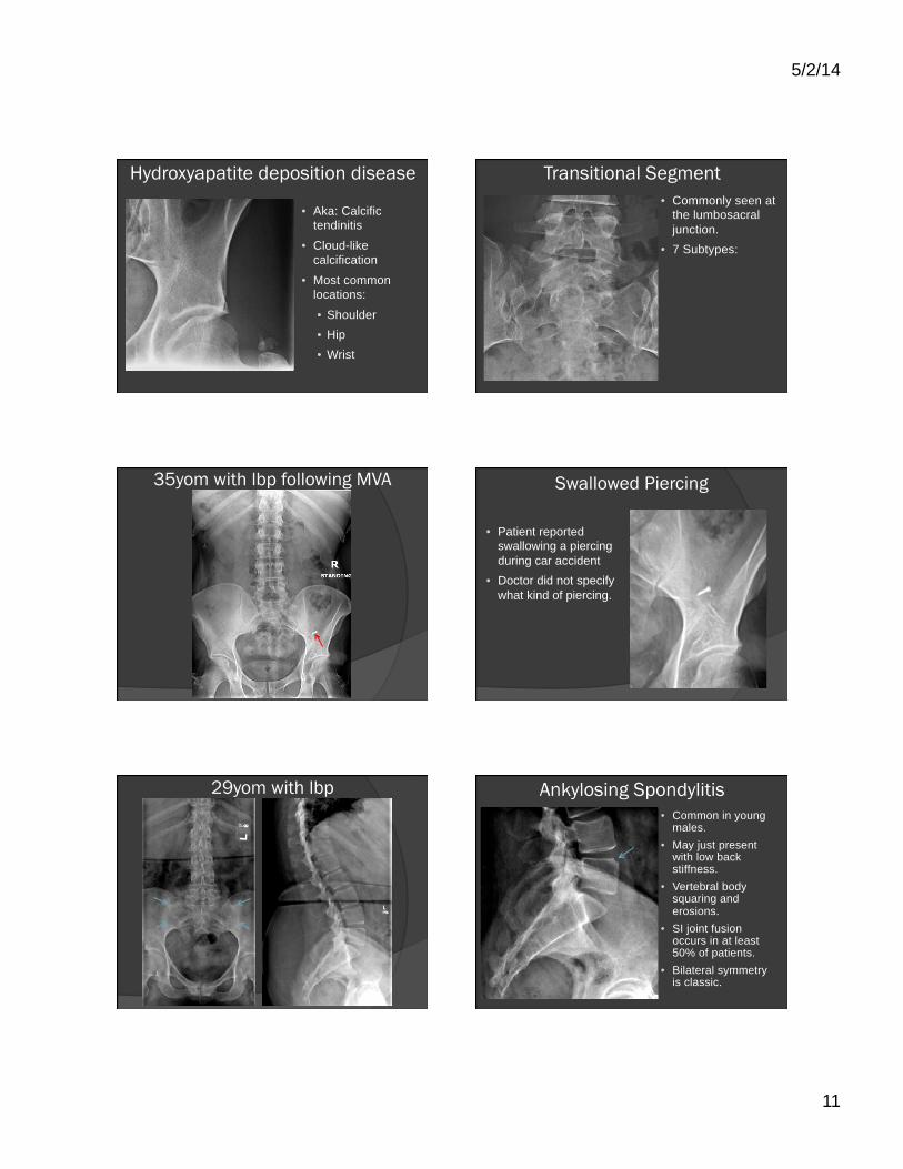
Hydroxyapatite deposition disease
• Aka: Calcific tendinitis
• Cloud-like calcification
• Most common locations:
• Shoulder • Hip • Wrist
Transitional Segment • Commonly seen at
the lumbosacral junction.
• 7 Subtypes:
35yom with lbp following MVA Swallowed Piercing
• Patient reported swallowing a piercing during car accident
• Doctor did not specify what kind of piercing.
29yom with lbp Ankylosing Spondylitis • Common in young
males. • May just present
with low back stiffness.
• Vertebral body squaring and erosions.
• SI joint fusion occurs in at least 50% of patients.
• Bilateral symmetry is classic.

5/2/14
12
Upper extremity
Hector RiveraMelo, DC, DACBR [email protected]
Extremity Imaging
� Common indications for extremity imaging � Unresponsive to care after 4 weeks � Significant activity restriction >4 weeks � Non-mechanical pain � Osteoarthritis unrelieved by conservative care � Suspected/known inflammatory arthritis � Significant trauma � Suspected physical abuse in children
Extremity Imaging
� Red flag indicators � Signs/symptoms or history of cancer � Red skin, fever, immunosuppressed � History of non-investigated trauma � Unexplained significant sensory/motor deficit
Shoulder Imaging
� Standard radiographic series � AP internal rotation � AP external rotation � Baby arm
Standard Shoulder Series AP Internal
Rotation AP External
Rotation
Standard Shoulder Series Baby Arm

5/2/14
13
Shoulder Imaging
� Accessory radiographic views � Axial � Trans-scapular ‘Y’
Accessory Shoulder Views Axial
Accessory Shoulder Views Trans-scapular ‘Y’ Shoulder Imaging
� Common pathology seen on X-rays � Hydroxyapatite Deposition Disease (HADD) � AC injuries (grades II-III) � Glenohumeral dislocations � Osteoarthritis (especially AC) � Clavicular fractures
Shoulder Imaging
� Common pathology not seen on X-rays � Rotator cuff injury � Adhesive capsulitis � Impingement syndrome � Labral injury � AC injury (grade I)
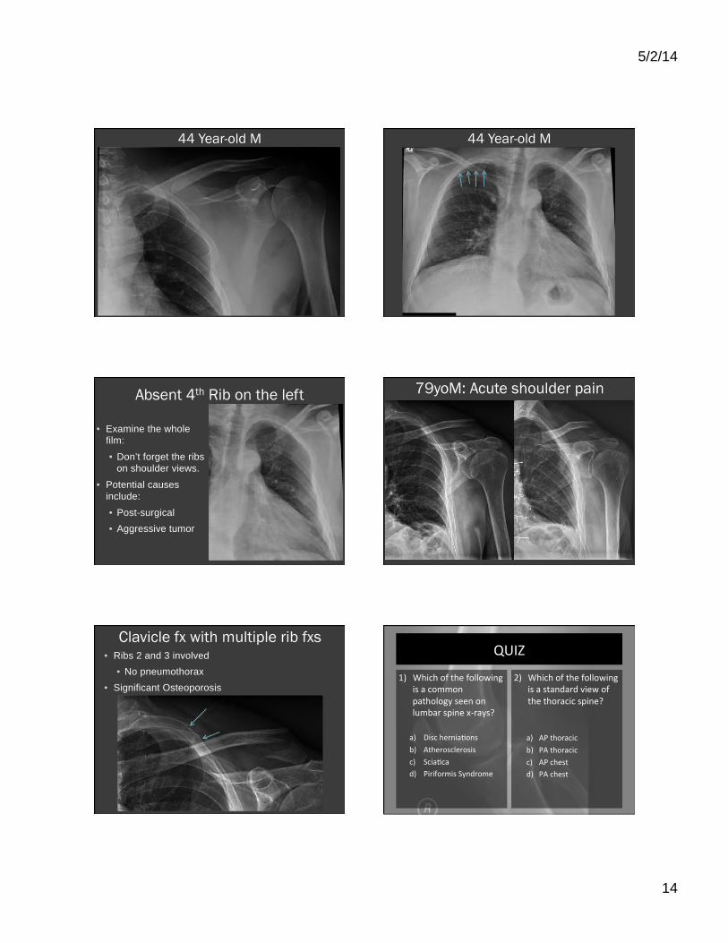
44 Year-old M
5/2/14
14
44 Year-old M 44 Year-old M
Absent 4th Rib on the left
• Examine the whole film:
• Don’t forget the ribs on shoulder views.
• Potential causes include:
• Post-surgical • Aggressive tumor
79yoM: Acute shoulder pain
Clavicle fx with multiple rib fxs • Ribs 2 and 3 involved
• No pneumothorax • Significant Osteoporosis
QUIZ
1) Which of the following is a common pathology seen on lumbar spine x-‐rays? a) Disc herniaLons b) Atherosclerosis c) SciaLca d) Piriformis Syndrome
2) Which of the following is a standard view of the thoracic spine?
a) AP thoracic b) PA thoracic c) AP chest d) PA chest

5/2/14
15
Wrist Imaging
� Standard radiographic series � PA � Lateral � Medial oblique � PA ulnar deviation
Standard Wrist Series PA Lateral
Standard Wrist Series Medial Oblique PA Ulnar Deviation
Wrist Imaging
� Accessory radiographic views � Carpal Tunnel � Angulated Scaphoid
Accessory Wrist Views Carpal Tunnel Angulated Scaphoid
Wrist Imaging
� Common pathology seen on X-rays � Osteoarthritis (especially 1st CMC joint) � Scaphoid fractures � Distal radial fractures � Instability � Lunate dislocations � Inflammatory arthritis � Avascular necrosis
5/2/14
16
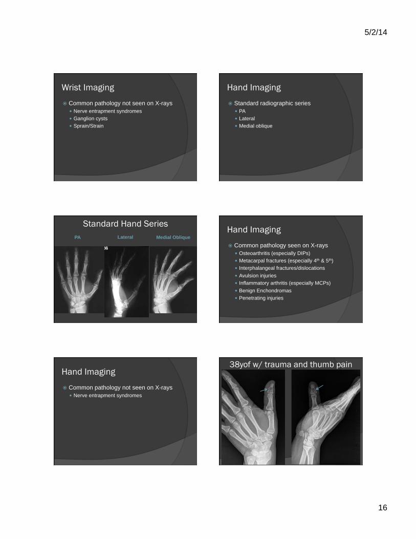
Wrist Imaging
� Common pathology not seen on X-rays � Nerve entrapment syndromes � Ganglion cysts � Sprain/Strain
Hand Imaging
� Standard radiographic series � PA � Lateral � Medial oblique
Standard Hand Series PA Lateral Medial Oblique
Hand Imaging
� Common pathology seen on X-rays � Osteoarthritis (especially DIPs) � Metacarpal fractures (especially 4th & 5th) � Interphalangeal fractures/dislocations � Avulsion injuries � Inflammatory arthritis (especially MCPs) � Benign Enchondromas � Penetrating injuries
Hand Imaging
� Common pathology not seen on X-rays � Nerve entrapment syndromes
38yof w/ trauma and thumb pain
5/2/14
17
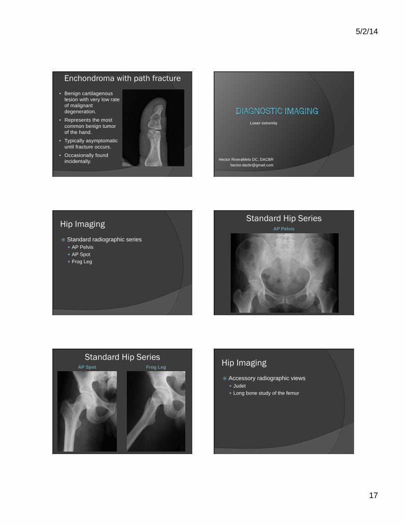
Enchondroma with path fracture
• Benign cartilagenous lesion with very low rate of malignant degeneration.
• Represents the most common benign tumor of the hand.
• Typically asymptomatic until fracture occurs.
• Occasionally found incidentally.
Lower extremity
Hector RiveraMelo DC, DACBR [email protected]
Hip Imaging
� Standard radiographic series � AP Pelvis � AP Spot � Frog Leg
Standard Hip Series AP Pelvis
Standard Hip Series AP Spot Frog Leg Hip Imaging
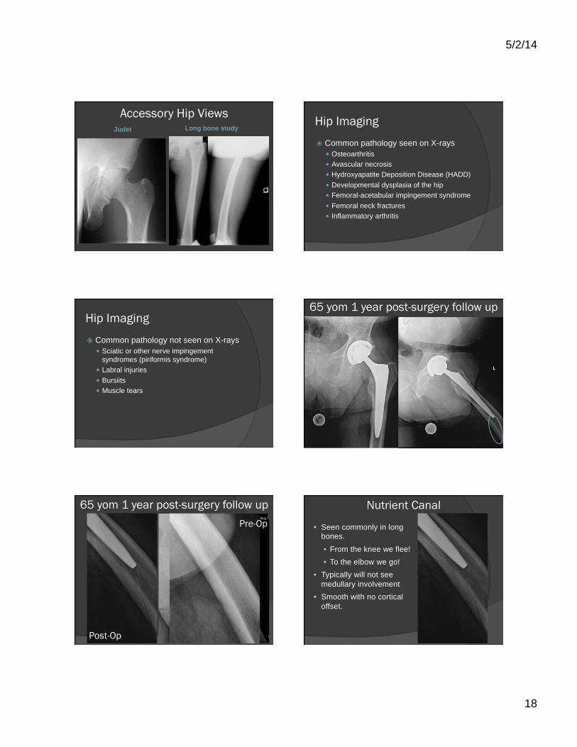
� Accessory radiographic views � Judet � Long bone study of the femur
5/2/14
18
Accessory Hip Views Judet Long bone study
Hip Imaging
� Common pathology seen on X-rays � Osteoarthritis � Avascular necrosis � Hydroxyapatite Deposition Disease (HADD) � Developmental dysplasia of the hip � Femoral-acetabular impingement syndrome � Femoral neck fractures � Inflammatory arthritis
Hip Imaging
� Common pathology not seen on X-rays � Sciatic or other nerve impingement
syndromes (piriformis syndrome) � Labral injuries � Bursiits � Muscle tears
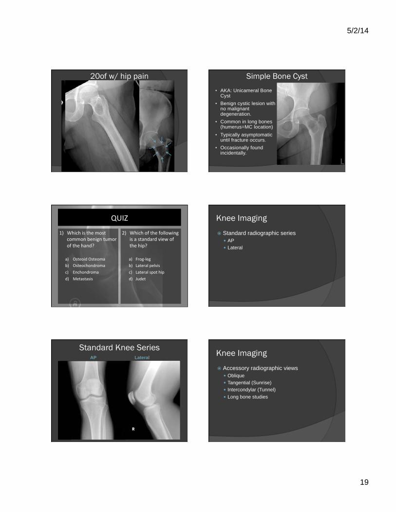
65 yom 1 year post-surgery follow up
65 yom 1 year post-surgery follow up Pre-Op
Post-Op
Nutrient Canal
• Seen commonly in long bones.
• From the knee we flee! • To the elbow we go!
• Typically will not see medullary involvement
• Smooth with no cortical offset.
5/2/14
19
20of w/ hip pain Simple Bone Cyst • AKA: Unicameral Bone
Cyst • Benign cystic lesion with
no malignant degeneration.
• Common in long bones (humerus=MC location)
• Typically asymptomatic until fracture occurs.
• Occasionally found incidentally.
QUIZ
1) Which is the most common benign tumor of the hand?
a) Osteoid Osteoma b) Osteochondroma c) Enchondroma d) Metastasis
2) Which of the following is a standard view of the hip?
a) Frog-‐leg b) Lateral pelvis c) Lateral spot hip d) Judet
Knee Imaging
� Standard radiographic series � AP � Lateral
Standard Knee Series AP Lateral
Knee Imaging
� Accessory radiographic views � Oblique � Tangential (Sunrise) � Intercondylar (Tunnel) � Long bone studies

5/2/14
20
Knee Accessory Views
Tangential (Sunrise)
Intercondylar (Tunnel)
Knee Accessory Views Medial Oblique Long bone study
� Catch high percentage of fractures of the knee � Order x-rays of the knee if
there is trauma to the knee and any of the following: ○ Age ≥55 ○ Isolated tenderness at:
� Head of the fibula � Patella
○ Inability to flex knee >90° ○ Inability to walk 4 weight-
bearing steps at presentation or at presentation
Knee Imaging: Ottawa Knee Rules Knee Imaging
� Common pathology seen on X-rays � Osteoarthritis � Osteochondral defects (femur) � Calcium pyrophosphate deposition disease
(CPPD) � Proximal fibular/tibial fractures � Aggressive and benign 1°bone tumors
Knee Imaging
� Common pathology not seen on X-rays � Patellar tendinitis (Jumpers knee) � Meniscal injury � ACL, PCL, MCL, LCL injury � Sprain/Strain � Chondromalacia Patella � Osgood-Schlatter disease*
59 yof with bilateral knee pain

5/2/14
21
49 yof with bilateral knee pain Osteoarthritis of the knees
• Age range is typically <45 • Typically asymmetric
involvement.
• Prominent osteophytes • Medial joint compartment
typically affected 1st.
31 yom with right knee pain Fabella
• A sesamoid bone. • Located within the
tendon of the lateral head of the gastrocnemious muscle.
• A very common normal variant.
41 yof with right knee pain Cyamella
• A sesamoid bone, similar to the fabella.
• Located within the tendon of the popliteus muscle.
• A normal variant

5/2/14
22
Meniscal Ossicle
• An accessory ossicle. • Located within the
meniscus.
• Typically seen posteriorly and medially
• May be triangular. • An uncommon
normal variant.
66 yoM with left knee pain Popliteal Artery Atherosclerosis • This is a common
location for atherosclerosis.
• The popliteal artery is the most common location for peripheral aneurysms.
• Will often see accompanying atherosclerosis of other large arteries.
44 yom with bilateral knee pain 44 yom with bilateral knee pain
5/2/14
23
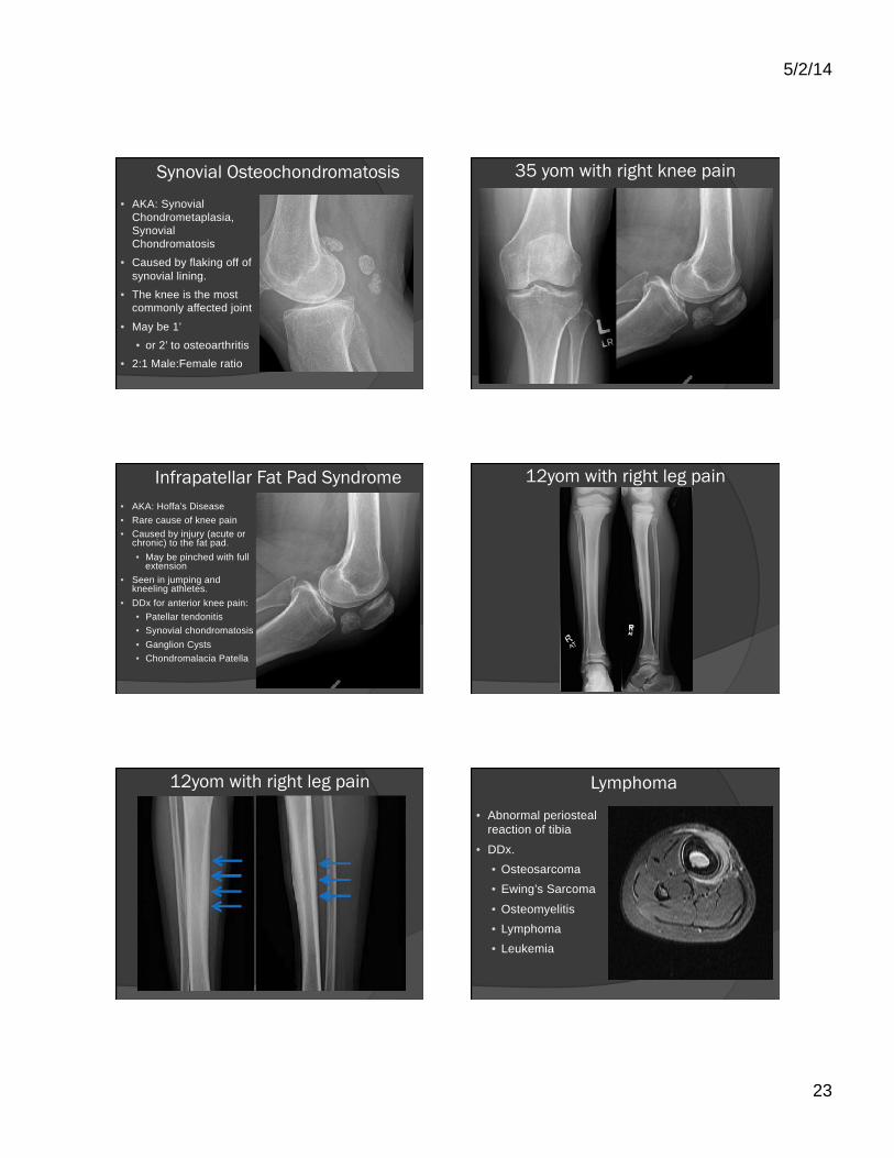
Synovial Osteochondromatosis • AKA: Synovial
Chondrometaplasia, Synovial Chondromatosis
• Caused by flaking off of synovial lining.
• The knee is the most commonly affected joint
• May be 1’ • or 2’ to osteoarthritis
• 2:1 Male:Female ratio
35 yom with right knee pain
Infrapatellar Fat Pad Syndrome • AKA: Hoffa’s Disease • Rare cause of knee pain • Caused by injury (acute or
chronic) to the fat pad. • May be pinched with full
extension • Seen in jumping and
kneeling athletes. • DDx for anterior knee pain:
• Patellar tendonitis • Synovial chondromatosis • Ganglion Cysts • Chondromalacia Patella
12yom with right leg pain
12yom with right leg pain Lymphoma
• Abnormal periosteal reaction of tibia
• DDx. • Osteosarcoma • Ewing’s Sarcoma
• Osteomyelitis • Lymphoma • Leukemia

5/2/14
24
More Acupuncture Needles!! Foot Imaging
� Standard radiographic series � DP � Lateral � Medial oblique
Standard Foot Series DP Medial Oblique
Standard Foot Series Lateral
Foot Imaging
� Accessory radiographic views � Tangential Calcaneus (Harris-Beath)
Accessory Foot Views Tangential Calcaneus
(Harris-Beath)
5/2/14
25
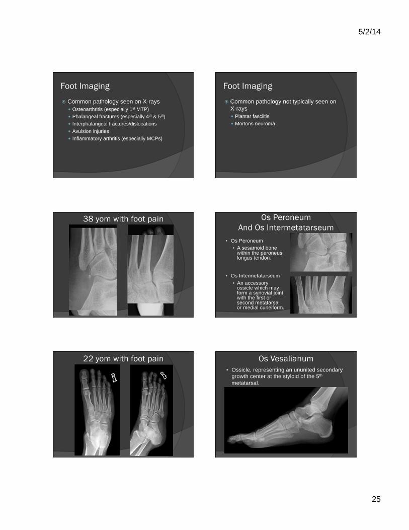
Foot Imaging
� Common pathology seen on X-rays � Osteoarthritis (especially 1st MTP) � Phalangeal fractures (especially 4th & 5th) � Interphalangeal fractures/dislocations � Avulsion injuries � Inflammatory arthritis (especially MCPs)
Foot Imaging
� Common pathology not typically seen on X-rays � Plantar fasciitis � Mortons neuroma
38 yom with foot pain Os Peroneum And Os Intermetatarseum
• Os Peroneum • A sesamoid bone
within the peroneus longus tendon.
• Os Intermetatarseum • An accessory
ossicle which may form a synovial joint with the first or second metatarsal or medial cuneiform.
22 yom with foot pain Os Vesalianum • Ossicle, representing an ununited secondary
growth center at the styloid of the 5th metatarsal.
5/2/14
26
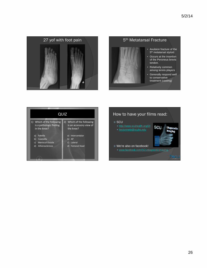
27 yof with foot pain 5th Metatarsal Fracture
• Avulsion fracture of the 5th metatarsal styloid.
• Occurs at the insertion of the Peroneus brevis tendon.
• Relatively common among tennis players
• Generally respond well to conservative treatment (casting)
QUIZ
1) Which of the following is a pathologic finding in the knee? a) Fabella b) Cyamella c) Meniscal Ossicle d) Atherosclerosis
2) Which of the following is an accessory view of the knee?
a) Intercondylar b) AP c) Lateral d) Femoral Head
How to have your films read:
� SCU � http://www.scuhealth.org/DI � [email protected]
� We’re also on facebook! � www.facebook.com/SCUdiagnosticimaging