rahul singh
TRANSCRIPT

TISSUE FIXATIVE,MODES OF FIXATION&HISTOCHEMICAL STAINING TECHNIQUES FOR IDENTIFICATION OF
DIFFERENT ELEMENTS OF LESION
Submitted by- RAHUL SINGH M.V.Sc Scholar ROLL-5455
MAJOR CREDIT SEMINARON

TISSUE FIXATION-DEFINITION
A chemical process by which biological tissues preserved from decay ,either through autolysis, putrefaction.
It terminates any on going biochemical reaction &may also increase the mechanical strength or stability.

PURPOSE OF FIXATION
Inhibition of autolysis & putrefaction.Preserve sample (tissue) as close to natural
state.Disable intrinsic biomolecules - proteolytic
enzymes.Protect sample from extrinsic damage.Increase their mechanical strength& rigidity.

CONTI-
To aid in optical differentiationTo aid in the better staining propertyExample-
Poor fixed tissueGood fixed tissue
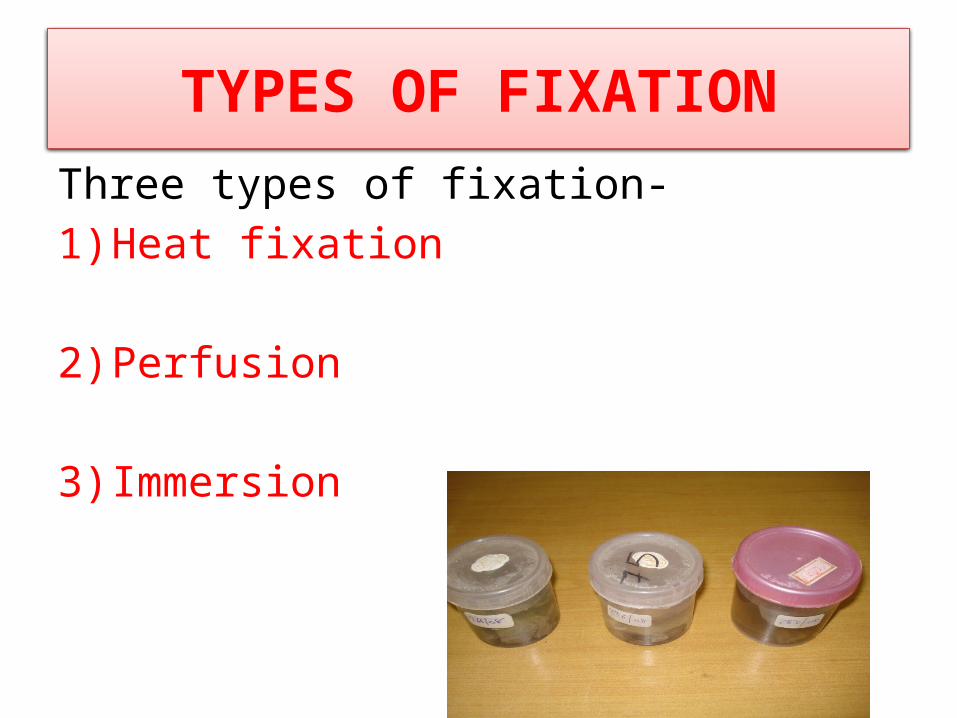
TYPES OF FIXATIONThree types of fixation-1) Heat fixation
2) Perfusion
3) Immersion

FIXATIVES OR FIXATION SOLUTION (Classification)
I Simple Fixatives
II Compound fixatives

Simple Fixatives
•Formalin – used as single reagent•Mercuric chloride•Picric acid•Acetone•Ethyl alcohol•Osmic acid•Osmium tetroxide : Used for fat preservation

Compound fixatives
Microanatomical fixativeFormal saline
Neutral buffered formalinZenker’s fluid
Bouin fluid
Cytological FixativesI Nuclear fixative
e.g.Carnoy’s fluidII Cytoplasmic fixaives
e.g. Champy’s fluid
Histochemical fixativesCold acetone Ethyl alcohol

MODE OF ACTION OF FIXATIVES
I. Non coagulating cross-linking
II. Coagulating fixatives


10% FORMALIN
• Formaldehyde is a gas sold as a solution in water (approx. 40% in content ), in which form it is known as formalin.
• SOLUTION
• Formaldehyde mainly preserves proteins.
(40%Formaldehyde ) formalin 10.0 m
Distilled water 90.0ml

Conti-
Formaldehyde –Cross-linking fixatives act by creating covalent chemical bonds between proteins in tissue.
This anchors soluble proteins to the cytoskeleton, and lends additional rigidity to the tissue.

Conti-
Conti-
• Long stored formalin produces formic acid .
• formic acid show deposition of brown colour formic acid pigment in the section.
• To remove this formic acid pigment the sections are treated with alkali (ammonia) water.

10% NEUTRAL BUFFERED FORMALINpH - 7.0
Formulation 40% formaldehyde: - 100 ml Distilled water: - 900 ml Sodium dihydrogen phosphate monohydrate: - 4 g Disodium hydrogen phosphate anhydrous - 6.5 g The solution should have a pH of -7.0 Fixation time: - 12 – 24 hours
most widely used formaldehyde-based fixative for routine histopathology.
The buffer tends to prevent the formation of formalin pigment

Formal saline
• Formulation• 40% formaldehyde: 100 ml• Sodium chloride: 9 g• Distilled water: 900 ml• Fixation time: 12 – 24 hours
• It often produces formalin pigment

BOUIN’S FLUID
Used for staining of glycogen & Masson’s trichrome stain
Composition-
Sat.acq.soln. of picric acid……………………….750ml
Formalin…………………………………………250ml
Glacial acetic acid………………………………...50ml

ZENKER’S FLUID
• Used for bone marrow & spleen fixation.• Composition-
Mercuric chloride……………………….50g
Potassium dichromate…………………..25g
Sodium sulphate………………………..10g
Distill. Water upto…………………….1000ml
Glacial acetic acid……………………..50ml

OSMIUM TETROXIDE
In solution, usually 1% aqueous solution, osmium tetroxide.
Fats & other lipids reduce osmium tetroxide or combine with it to form a dark compound
Osmium tetraoxide is used for fat preservation

ETHYL ALCOHOL
• Ethyl alcohol hardens tissue but causes serious shrinkage.
• It is strong cytoplasmic coagulant.
Precipitating (or denaturing) fixatives act by reducing the solubility of protein molecules.
disrupting the hydrophobic interactions that give
many proteins their tertiary structure

PICRIC ACID
• Picric acid is usually used in a saturated aqueous solution , 0.9-1.2%
• It is an excellent protein coagulant.• forming protein picrates having a strongly
affinity for acid dyes.• It is recommended as a fixative for glycogen
CYTOLOGICAL FIXATIVE
Nuclear fixative-Carnoy’s fluid- This is used for biopsy of cancerous tissue. Quickest fixative most of the cytoplasmic
details .• Composition- Absolute alcohol……………………………60ml Chloroform…………………………………..30ml Glacial acetic acid…………………………10ml

HISTOCHEMICAL FIXATIVE
• Used for staining of enzymes, carbohydrate, fat, minerals, nucleic acid, RNA, DNA, proteins etc.
• Examples-
1) Cold acetone
2) Absolute alcohol-used for glycogen & uric acid crystals.
3) Formal saline
Factors affecting fixation
pH
Size of specimen
Volume of the fixative
Temperature
Duration

GOLDEN RULES
SIZE OF TISSUE- about 3mm.
VOLUME OF FIXATIVE-20x.
TIME OF FIXATION-24hour in formalin

DECALCIFICATION
Bone, Tubercle nodules, calcified tissue.
• 5% Aqueous Nitric acid3-5 days
• Electrophoretic decalcifying instrument

How to know that all calcium has removed?
By piercing the needle
By X ray
Testing the solution of HNO3 for calcium

Histochemical techniques for demonstration of different Biomolecules
Fat &GLYCOGEN
VON KOSSA'S METHOD – CALCIUM
PRUSSIAN BLUE REACTION – (FOR HAEMOSIDERIN)
DE GALANTHA'S METHOD FOR URATE CRYSTALS.
Congo Red Stain For Amyloid

FAT1. Oil Red O2. Sudan a. Sudan II b. Sudan III c. Sudan IV d. Sudan Black B3. Osmium Tetroxide
GLYCOGEN1. PAS2. Best’s carmine

Oil Red O STAIN FOR FAT
PRINCIPLE: Oil Red O is a lysochrome (fat-soluble dye) diazo dye used for staining of neutral triglycerides and lipids on frozen sections and some lipoproteins on paraffin sections.
Method
1. Cut frozen sections at 8 to10mm, air dry the sections to the slides
2. Fix in formalin, briefly wash with running tap water 1-10 mins
3. Rinse with 60% isopropanol
4. Stain with freshly prepared Oil Red O working solution 15 mins
5. Rinse with 60% isopropanol
6. Lightly stain nuclei with alum haematoxylin 5 dips
7. Rinse with distilled water
8. Mount in aqueous mountant or glycerine jelly.
Results:- Lipid......................................Red
Nuclei....................................Blue

Fatty change of liver Oil Red-O Stain.
Fatty change of liver HE Stain.

Lipid in skin was demonstrated by Oil Red O method

SUDAN BLACK STAIN FOR FAT
PRINCIPLE: Sudan Black is slightly basic dye and will combine withacidic groups in compound lipids, thus staining phospholipids
PROCEDURE:1. Pick-up frozen sections on clean glass slides if fresh, albuminized slides if fixed.2. Fix slides in 10% formalin if fresh.3. Wash well it tap, rinse in distilled, drain off excess water.4. Propylene glycol, two changes, 5 minutes each.5. Sudan Black, 7 minutes, agitate.6. 85% Propylene glycol, 3 minutes.7. Rinse in distilled water.8. Nuclear Fast Red, 3 minutes.9. Wash in water.10. Wash in tap water, rinse in distilled.11. Mount with aqueous mounting media, Glycerin Jelly.
RESULTS: Fat ..…………….Black

Sudan black stained fat in muscle

Fat accumulation in perimycium stained with sudan black


OSMIUM TETROXIDE STAIN FOR FAT
Introduction : Osmium Tetroxide is a good fixative and excellent stain for lipids in membranous structures and vesicles Procedure1. Before starting, dilute the 4% OsO4 to a final concentration of 0.1%.2. Start with a fixed, sliced tissue sample.3. Wash the sample three times with double distilled water.4. Incubate the sample with 0.1% OsO4 for 30 minutes. 5. Wash four times with double distilled water.
RESULTS: Fat ..…………….Black

Osmium tetra oxide stained fat in liver

Osmium tetra oxide stain: the lipid-rich myelin sheath of nerve fibers is heavily colored. Peripheral nerve: 100x and 400x

Adipose Tissue fixed and stained by Osmium Tetroxide

PRINCIPLE Tissue containing vicinal glycol groups or their amino or alkylamino derivatives are oxidized by periodic acid to form dialdehydes, which combine with Schiff's reagent to form an insoluble magenta compound.
Procedure: 1. Deparaffinize and hydrate to water. 2. Oxidize in 0.5% periodic acid solution for 5 minutes. 3. Rinse in distilled water. 4. Place in Schiff reagent for 15 minutes.5. Wash in lukewarm tap water for 5 minutes.6. Counterstain in Mayer's hematoxylin for 1 minute. 7. Wash in tap water for 5 minutes. 8. Dehydrate and coverslip using a synthetic mounting medium.
RESULTS: Glycogen…………………….Magenta Nuclei ………………………Blue
PAS STAIN for glycogen

Liver with Pas staining

H&E stain:- liver of dog
PAS stain shows intense staining of hepatocytes (arrowheads) and basement membranes of bile ducts (arrow

PAS staining of formalin fixed, paraffin embedded
mouse lung section showing purple colored glycogen
staining.
PAS staining mouse lung section showing purple colored glycogen staining.

Ilium with glycogen droplets stained red pas

PRINCIPLE This staining technique demonstrates glycogen by hydrogen bond formation between OH groups on the glycogen, and H atoms of the carminic acid. Fibrin and neutral mucin stain weakly with this method.
PROCEDURE 1. Dewax test + positive control sections and hydrate to water. 2. Duplicate sections may be treated with diastase if desired (See6.2).3. Stain nuclei of all sections well using one of the iron haematoxylin
solutions eg. Weigerts iron haematoxylin (See 2.4) 4. Treat with carmine solution for 10 mins.5. Transfer slides quickly to a Coplin jar of Differentiating solution.6. Wash in alcohol (NOT water), clear in histoclear and mount.
RESULTS: Glycogen……………Bright red Nuclei…………….....Blue
BEST'S CARMINE

Best’s carmine method for glycogen

Best’s carmine stain Rabbit liver

VON KOSSA'S METHOD - CALCIUMPRINCIPLE-Tissue sections are treated with silver nitrate solution, thecalcium is reduced by the strong light and replaced with silver deposits ,visualized as metallic silver.
REAGENTS:5% Silver Nitrate Solution:
Silver nitrate 25.0 gmDistilled water 500.0 ml 5% Sodium thiosulfate[hypo] Nuclear fast red
FIXATION: 95% ethyl alcohol or 10% buffered neutral formalin

Staining Procedure Deparaffinize and hydrate to 95% alcohol and air dry the
slides. Place slides on a staining rack and flood the sections with 5%
silver nitrate. Expose the slides to an ultraviolet lamp for 10 minutes.
Rinse with four changes of distilled water. Sodium thiosulfate solution for 1 minute. Rinse with four changes of distilled water. Nuclear fast red solution for 3 minutes. Rinse with three changes of distilled water. Dehydrate through graded alcohols. Mount with synthetic resin.

Staining ResultsCalcium salts ------------------------------------------------------- black
Nuclei -------------------------------------------------------------- red
Cytoplasm ---------------------------------------------------------- light pink
DIAGNOSIS APPLICATION -Calcification in tuberculosis -Atherosclerosis -Myeloid tumours -Dead(necrotic) or dying tissues

1 &2black deposits in the walls of this lung that was calcified . This lung was stained with VON KOSSA STAIN
1 2
3
3- H&E Stain calcification tissue stain blue colour

Thick calcified rings - that could be observed macroscopically - were found along the aortas of animals with massive calcification. Von Kossa stained ...

PRUSSIAN BLUE REACTION – (FOR HAEMOSIDERIN)
PRINCIPLE:
FIXATIVE: 10%buffered neuttral formalin
TECHNIQUE: Cut paraffin sections 6μ
Pearl’s Prussian blue reaction is that potassium ferrocyanide will form ferric ferrocyanide (Prussian blue) with reactive ferric salts in an acid solution. Dilute hydrochloric acid liberates loosely bound ferric iron from protein.

Staining Procedure•Deparaffinize and hydrate to distilled water.
•Place the slides in the hydrochloric acid-potassium ferrocyanide solution for 30 minutes at room temperature.
•Rinse with five changes of distilled water.
•Nuclear fast red solution for 3 minutes.
•Rinse with three changes of distilled water.
•Dehydrate in graded alcohols.
•Clear in xylene, three or four changes.
•Mount with synthetic resin.

RESULTS:Iron (hemosiderin) - blue
Nuclei - red
Background - pink
Diagnostic Application: Hemochromatosis, esp., liver. excess iron deposition is stained as blue granules
Gastric ulcer 2nd to iron overdose:

Dog liver show overdose ironHemochomatosis
Gastric ulcer 2nd to iron overdose

DE GALANTHA'S METHOD FOR URATE CRYSTALS
Fixation: Absolute Alcohol. If specimen contains bone, decalcify in Nitric AcidAlcohol (F-96). Wash in Absolute Alcohol for 24 hours.
Sections: Paraffin @ 8-10 microns.
Staining:
Xylene Absolute Alcohol, two changes each.
Expose sections in Silver Nitrate Solution to strong sunlight for 3 hours(until urates are bright rose).
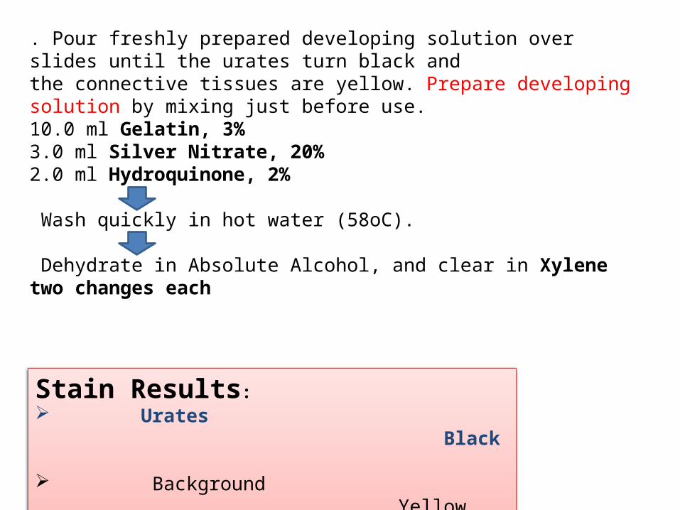
. Pour freshly prepared developing solution over slides until the urates turn black andthe connective tissues are yellow. Prepare developing solution by mixing just before use.10.0 ml Gelatin, 3%3.0 ml Silver Nitrate, 20% 2.0 ml Hydroquinone, 2%
Wash quickly in hot water (58oC).
Dehydrate in Absolute Alcohol, and clear in Xylene two changes each
Stain Results: Urates Black
Background Yellow

.
. Section of liver from chick diedof group IV showing deposition of uric acidcrystals in hepatic parenchyma. H & E X300
Section of kidney from chickdied of showing deposition of blackuric acid crystals in renal parenchyma. Dgalantha's X 300e.

Section of kidney from chickdied of showing marked depositionof needle shaped uric acid crystals in renalparenchyma.
. Section of kidney from chickdied of showing marked intertubular haemorrhages, deposition of uricacid crystals degeneration and necrosis ofrenal tubules. H & E X 300

Congo Red Stain For Amyloid
PRINCIPLE-The Congo red dye is a linear molecule, and this configuration permits hydrogen bonding of the azo and amine groups of the dye to similarly spaced hydroxyl radicals of the amyloid.
Fixation:-10% buffered neutral FormalinReagents:1.1% Congo redCongo red : 1.0 gmD.W. : 100.0 ml2. 1% Sodium hydroxideSodium hydroxide: 1.0 gmD.W. : 100.0 ml3. Alkaline Alcohol Solution1% Sodium hydroxide: 1.0 ml50% Alcohol : 99 ml4.Hematoxylin

Procedure Deparaffinize and hydrate sections to water.
.Stain in Congo red solution for 1 hr.
.Rine in distilled water.
Differentitate quickly in alkaline alcohol solution.
Rinse in tap water for 5 minutes.
Counterstain with Hematoxylin for 5 minutes.
Rinse in tap water for 10 minutes.
Dehydrate through 95% alcohol ,2 changes of 100% alcohol.
Clear in xylene
Mounting
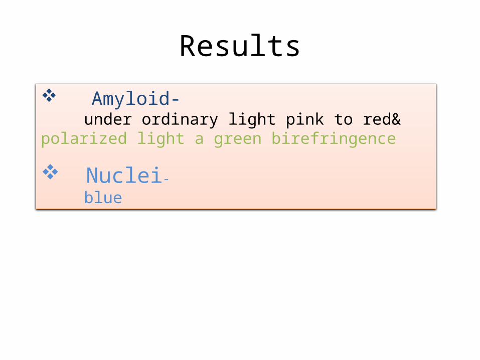
Results
Amyloid- under ordinary light pink to red& polarized light a green birefringence
Nuclei- blue

section of renal glomerular amyloidosis in a dog. (A) Light eosinophilic amorphousdeposits partly effaced the glomerular architecture (H&E stain). The deposit stained lightred with Congo red stain (B) and exhibited green birefringence under polarized light (C).The amyloid deposit failed to stain with Congo red (D) after pretreatment with PotassiumPermanganate solution, indicating that the amyloid is AA-amyloid

Kidney: Multiple Glomeruli: Polarized light and Congo Red stain- Apple green bi-refringence = amyloid
Glomerulus with multiple deposits of amyloid. Congo red staining
amyloidosis kidney HE stain

Histological features of skin of a dog with nodular cutaneous amyloidosis, which stained light eosinophilic with haematoxylin-eosin (A) and red with Congo red stain (B). The amyloid showed yellow-green birefringence (C) illuminated with polarized light, characteristic of amyloid deposits

Histochemical techniques for demonstration of connective tissue
Mallory stainingMasson trichrome staining
Gomori’s method for reticulum fibres

Mallory staining
• Principle- Presence of phosphomolybdic acid on the fibres (collagen) act as colourless acid dye. It act as dye excluder with acid dye such as basic fuchin exclding them from collagen. Then the aniline blue stains collagen exclusively
• Fixation- Mercuric chloride• Procedure-1. Deparafinize and hydrate slides to water label2. Stain in Mallory I:- 15 sec3. Rinse in distill water:- 10 sec4. Treat with phosphomolybdic acid:- 1-5min

5. Rinse briefly in distill water6. Stain in Mallory II:- 2 min7. Rinse in distill water8. Differentiate aniline blue in 90% ethyl alcohol9. Dehydrate in absolute alcohol, clear and mount• Result:
Nuclei- redMuscle and some cytoplasmic elements- red to orangeCollagen- dark blueConnective tissue and hyaline substance- blue

• Characteristics - cells and extracellular matrix (ECM)• Distribution - lies between basal lamina of epithelia and external lamina of
muscle and nerve supporting cells
Epiglottis (Mallory-Azan stain)

Mallory trichrome staining of stomach
Mallory trichrome staining of renal cortex

Masson trichrome staining
• Purpose: Used to differentiate between collagen and smooth muscle in tumours, and the increase of collagen in diseases such as cirrhosis & hence routinely used stain for liver and kidney biopsies.
• Principle: Three dyes are employed selectively staining muscle, collagen fibers & fibrin. The general rule in trichrome staining is that the less porous tissues are coloured by the smallest dye molecule.

• Fixative: Bouin's fluid is preferred, 10% formalin. • Procedure-1. Deparafinize and hydrate slides to water label2. Mordant with iron alum: 5 min @40-50˚C3. Wash in running water: 5 min4. Stain in haematoxylene: 5 min @40-50˚C5. Wash in running water: 5 min6. Differentiate in saturated aquous picric acid7. Wash thoroughly in running water: 10-20 min8. Stain in Masson A: 5 min9. Rinse in distill water

10. Treat with Masson B: 10 min11. Stain in Masson C: 2-5 min12. Differentiate in acetic acid rinse: 1-2 min13. Dehydrate quickly 70% & 95% alcohol14. Dehydrate, absolute alcohol, 2 changes: 3 minutes each15. Clear and mount• Result:
Nuclei- blackCollagen, - blue or greenCytoplasmic element, keratin, muscle- red

Chronic active hepatitis with collapse in liver, trichrome stain.
77
Cerebral abscess in brain, trichrome stain.
Scleroderma with fibrosis of submucosa in stomach, trichrome stain.

Mammary gland (Masson stain)

Gomori’s method for reticulum fibres
Fixation :
10% buffered neutral formalinTechnique : Cut paraffin sections – 6µ thickStains :
Ammoniacal silver solution 0.5% Potassium permanganate solution 2% Potassium metabisulfite solution 2% Ferric ammonium sulfate solution 20% Formalin solution 0.2% Gold chloride solution 2% Sodium thiosulfate solution

Reticulum fibres Black
Background Grey
Result :

CONCLUSION
• Tissue fixation is one of the most important determinants of the quality of histological sections.
• Incomplete fixation can lead to unsatisfactory and poorly reproducible results
• It follows that the best results often require a close interaction between the investigator and Histology Lab.
• Histopathological method as an aid in diagnosis of diseases .

REFERENCES
p 16 Cook H C, Manual of Histological Demonstration Techniques p 188 Bancroft JD & Stevens A, Theory and Practice of Histological Techniques, 2nd ed
Garvey, W. et al: Combined modified periodic acid-Schiff and batch staining method. J. Histotechnol. 15:117-120, 1992.
McManus, J.F.A. and Mowry, R.W.: Staining Methods Histologic and Histochemical, New York, Paul B. Hoeber, 1960, pp. 126-128.
Preece A, A manual for Histologic Technicians, 3rd Ed, 1972, Little,Brown and Co, Boston
Crookham,J, Dapson,R, Hazardous Chemicals in the Histopathology Laboratory, 2nd ED, 1991, Anatech
Luna, L.: Manual of Histologic Staining Methods of the Armed Forces Institute of Pathology, 3rd edition,
Lillie, R.D.: Histopathologic Technic and Practical Histochemistry, 4th ed., New York, McGraw-Hill, 1976, p. 507
