ranzcr mri safety guidelines
TRANSCRIPT

THE ROYAL AUSTRALIAN AND NEW ZEALAND COLLEGE OF RADIOLOGISTS®
MRI SAFETY GUIDELINES
FACULTY OF CLINICAL RADIOLOGY

The
Roy
al A
ustra
lian
and
New
Zea
land
Col
lege
of R
adio
logi
sts
Elig
ibilit
y to
Atte
mpt
the
Rad
iolo
gy P
art 1
Exa
min
atio
n &
Succ
essf
ul C
ompl
etio
n of
the
Part
1 Ex
amin
atio
n
2
Name of document and version: MRI Safety Guidelines, Version 2.0
Approved by: Faculty of Clinical Radiology Council
Date of approval: 8 December 2017
ABN 37 000 029 863
Copyright for this publication rests with The Royal Australian and New Zealand College of Radiologists ®
The Royal Australian and New Zealand College of RadiologistsLevel 9, 51 Druitt StreetSydney NSW 2000, Australia
Email: [email protected]: www.ranzcr.comTelephone: + 61 2 9268 9777Facsimile: + 61 2 9268 9799
Disclaimer: The information provided in this document is of a general nature only and is not intended as a substitute for medical or legal advice. It is designed to support, not replace, the relationship that exists between a patient and his/her doctor.
Page 2 of 34
MR
I Saf
ety
Gui
del
ines
Ver
sion
2.0
| ©
The
Ro
yal A
ustr
alia
n an
d N
ew Z
eala
nd C
olle
ge o
f Rad
iolo
gist
s® |
Dec
embe
r 20
17
Document name MRI Safety Guidelines
Description The MRI Safety Guidelines provide the Magnetic Resonance imaging team (radiographers, technologists and scientists) with advice in addressing MRI safety issues and requirements.
Created By MRI Reference Group
Date Created 2007
Maintained By Faculty of Clinical Radiology
Version Number Modifications Made Date Modified
1.0 Document published Apr 2007
2.0 Document updated Dec 2017

Page 3 of 34
MR
I Saf
ety
Gui
del
ines
Ver
sion
2.0
| ©
The
Ro
yal A
ustr
alia
n an
d N
ew Z
eala
nd C
olle
ge o
f Rad
iolo
gist
s® |
Dec
embe
r 20
17
TABLE OF CONTENTS
1. Introduction 5
2. Definitions and Abbreviations 5
3. Administrative Aspects 5
4. MRI Equipment – General 7
5. MRI Equipment – Special Cases 10
6. Site Design 11
7. Magnetic Resonance Imaging Personnel 14
8. Screening of Patients and Others 16
9. Management of Implants and Foreign Bodies 18
10. Entry to Scan Room 21
11. Patient Management 21
12. Contrast agents 23
13. Noise Protection 24
14. Thermal Injury: Burns and Excessive SAR 25
15. Special Patient Groups 27
16. Occupational Exposure 28
17. Exposure of the public 28
18. Infection Control 29
19. Acknowledgements 29
20. Appendices 29
Appendix A: IEC SAR LIMITS 30
Appendix B: Guidelines for Management of Patients with Particular Foreign Bodies 31
Appendix C: Useful References: MRI Safety Issues 33

Page 4 of 34
MR
I Saf
ety
Gui
del
ines
Ver
sion
2.0
| ©
The
Ro
yal A
ustr
alia
n an
d N
ew Z
eala
nd C
olle
ge o
f Rad
iolo
gist
s® |
Dec
embe
r 20
17
About the College
The Royal Australian and New Zealand College of Radiologists (RANZCR) is a not-for-profit association of members that delivers skills, knowledge, insight, time and commitments to promote the science and practice of the medical specialties of clinical radiology (diagnostic and interventional) and radiation oncology in Australia and New Zealand.
The Faculty of Clinical Radiology, RANZCR, is the peak bi-national body for setting, promoting and continuously improving the standards of training and practice in diagnostic and interventional radiology for the betterment of the people of Australia and New Zealand.
Our Vision
RANZCR as the peak group driving best practice in clinical radiology and radiation oncology for the benefit of our patients.
Our Mission
To drive the appropriate, proper and safe use of radiological and radiation oncological medical services for optimum health outcomes by leading, training and sustaining our professionals.
Our Values
Commitment to Best Practice
Exemplified through an evidence-based culture, a focus on patient outcomes and equity of access to high quality care; an attitude of compassion and empathy.
Acting with Integrity
Exemplified through an ethical approach: doing what is right, not what is expedient; a forward thinking and collaborative attitude and patient-centric focus.
Accountability
Exemplified through strong leadership that is accountable to members; patient engagement at professional and organisational levels.
Code of Ethics
The Code defines the values and principles that underpin the best practice of clinical radiology and radiation oncology and makes explicit the standards of ethical conduct the College expects of its members.

Page 5 of 34
MR
I Saf
ety
Gui
del
ines
Ver
sion
2.0
| ©
The
Ro
yal A
ustr
alia
n an
d N
ew Z
eala
nd C
olle
ge o
f Rad
iolo
gist
s® |
Dec
embe
r 20
17
1. INTRODUCTION
1.1 Purpose and Scope
The MRI Safety Guideline is intended to assist The Royal Australian and New Zealand College of Radiologists® (ABN 37 000 029 863) (RANZCR) its staff, Fellows, members and other individuals involved in the Magnetic Resonance imaging team (radiographers, technologists and scientists) in addressing MRI safety issues and requirements.
1.2 The Intent of this Document
a) To provide information and guidance on the safe clinical use and research of MRI
b) To assist practices in developing appropriate protocols and procedures to support their use of MRI
c) To help prevent adverse patient outcomes in relation to MRI.
1.3 RANZCR Mission
The mission of The Royal Australian and New Zealand College of Radiologists is to drive the appropriate, proper and safe use of radiological and radiation oncological medical services for optimum health outcomes by leading, training and sustaining our professionals.
2. DEFINITIONS AND ABBREVIATIONS
In this MRI Safety Guideline:
RANZCR means The Royal Australian and New Zealand College of Radiologists.
Member means a member of the RANZCR.
IEC means International Electrotechnical Commission.
MRI means Magnetic Resonance Imaging.
3. ADMINISTRATIVE ASPECTS
3.1 Designated Responsible Person
Responsibility for the safe operation of the MRI site must be explicitly assigned to a nominated medical practitioner (typically titled the Medical Director of MRI). This person shall be responsible for the formulation and application of policies and procedures that ensure the safety of patients, MRI workers, and others in the MRI environment. This person may be called upon to assess the balance of risk and benefit for unusual scanning situations, and should therefore be a medical practitioner with substantial experience in MRI.
3.1.1 MRI Safety Officer
It is recommended that the responsible person delegate some MRI safety-related tasks to an MRI Safety Officer, who may be responsible for the day-to-day implementation of the site’s safety policies. An MRI Safety Officer must be suitably trained and experienced in MRI and MRI safety, but need not be a medical practitioner.
The MRI Medical Director, MRI Safety Officer and other MRI staff should together, develop a consistent approach to ensure the safety of patients and others within the MRI suite.
3.1.2 MRI Safety Resources

Page 6 of 34
MR
I Saf
ety
Gui
del
ines
Ver
sion
2.0
| ©
The
Ro
yal A
ustr
alia
n an
d N
ew Z
eala
nd C
olle
ge o
f Rad
iolo
gist
s® |
Dec
embe
r 20
17
The MRI Safety Officer, and other MRI staff must have access to a wide range of safety information pertaining to implants likely to be encountered at the site. Such information may be available as some combination of printed reference publications, manufacturer product data sheets, records of previous in-house testing, or online data services.
The MRI site must maintain MRI safety screening information for all MRI and other staff who enter the MRI Department Zones III and IV. This should be updated annually (refer to section 6.1.1 for definition of zones).
3.1.3 MRI Safety Education
The MRI safety officer or designate should conduct annual refresher presentations to MRI Radiographers who work routinely in Zones III or IV on advanced MRI safety topics, such as:
o SAR and SAR control o Peripheral nerve stimulation o Device/implant conditionality and required procedures for assessing this o Reading spatial gradient maps o Understanding fringe field strengths and boundaries o Emergency procedures in the case of patient cardiac arrest or equipment
failure o Procedures in the event of a quench o Safe monitoring technique of the Zone IV region.
There should also be annual education sessions for staff/visitors who occasionally interact with the MRI department from other departments, explaining the MRI environmental Zones, basic MRI safety principles, and procedures for gaining access to appropriate parts of the MRI suite.
3.2 MRI Safety Expert
It is recommended that sites have access to expert third party MRI safety advice, an experienced MRI Medical Director or MRI Safety Officer from another practice may fill this role. The MRI Safety Expert may be invited to review existing policy documents, conduct external audits of procedures, advise on proposed building plans, etc.
3.3 MRI Safety Committee
Larger sites may wish to establish an MRI Safety Committee to assist with policy reviews and the management of incident reports. The Responsible Person, the Safety Officer (if appointed) and Safety Expert should be members of such a committee
3.4 Documentation
There must be a safe practice manual to include procedures for all aspects of scanning, with particular attention to emergency situations: cardiac arrest, contrast reaction, fire and quench (as a minimum). This should form part of a larger MRI or department-wide procedure manual. This should be reviewed periodically (at least annually, and with every hardware and major software modification).
There must also be an incident reporting system involving at least one of the Responsible Person or the Safety Officer (incidents reported within 24 hrs), reports to which must be monitored and reviewed periodically (with documented responses). Where it exists, the MRI Safety Committee shall review incidents, analyse their root causes, and implement recommendations for improvement.
Records of training provided and attended must be maintained.

Page 7 of 34
MR
I Saf
ety
Gui
del
ines
Ver
sion
2.0
| ©
The
Ro
yal A
ustr
alia
n an
d N
ew Z
eala
nd C
olle
ge o
f Rad
iolo
gist
s® |
Dec
embe
r 20
17
3.5 Records of Examinations
Key technical parameters (name, date, sequence identifier; slice no, FOV, thickness, contrast use) must be recorded on images (film, or electronic archive). For images provided in digital format, additional detail will be available from the DICOM header
* Examinations transmitted on film shall include appropriate reference (‘scout’, pilot’) images showing the location and orientation of 2D cross-sectional images relative to known anatomical landmarks (see RANZCR-SSA Joint Guidelines for Confirming Vertebral Levels in Spine Imaging1). Images transmitted electronically shall allow cross-referencing between sequences, to allow demonstration of the position and alignment of one cross-sectional image relative to another.
A record of all scans performed on each MRI system should be maintained. This may be a written logbook, an electronic record within the system console (appropriately backed up), or stored in the practice RIS (appropriately backed up).
For examinations in which a contrast agent is administered, a written or electronic record shall be kept of the name and dose of the agent administered, the route by which it was administered, and the authorising radiologist.
3.6 Internal Review
There must be periodic quality assurance activities and reviews of reported incidents.
Audits of the performance and accuracy of screening procedures may be appropriate. Such activities may be conducted by the MRI Safety Committee, if constituted, or by the Responsible Person and/or MRI Safety Officer.
Records of each of these activities shall be maintained, and reviewed annually by the Safety Committee and/or the Responsible Person.
3.7 Relationship with Servicing Organisation
Formal delineation of the responsibilities of the site and its service organisation(s) for safety before, during, and after service periods is strongly recommended.
3.8 Maintenance and Service
Records of maintenance and service records must be kept. These should comply with DIAS standards.2
4. MRI EQUIPMENT – GENERAL
All major manufacturers of MRI equipment have adopted the relevant Standard of the International Electrotechnical Commission (IEC), IEC 60601-2-33 – amendments 1 & 2, Edition 3.2 (version current as of November 2017)3 (refer to Appendix C). This Standard establishes basic safety and essential performance requirements for MRI equipment to provide protection for the patient and the MRI worker.
The equipment requirements of this Standard and its subsequent revisions have therefore been adopted for the RANZCR Guidelines.
The IEC Standard defines three conditions of operation for MRI equipment:
1 https://www.ranzcr.com/documents-download/professional-documents/guidelines/586-joint-ranzcr-ssa-guidelines-for-
confirming-vertebral-levels-in-spine-imaging/file 2 http://www.health.gov.au/internet/main/publishing.nsf/Content/F4405D11CDDCBB5BCA257EF3001842F0/$File/DIAS-
Practice-Accreditation-Standards-from-1-January-2016.pdf 3 https://webstore.iec.ch/publication/2647 (NB: requires purchase)

Page 8 of 34
MR
I Saf
ety
Gui
del
ines
Ver
sion
2.0
| ©
The
Ro
yal A
ustr
alia
n an
d N
ew Z
eala
nd C
olle
ge o
f Rad
iolo
gist
s® |
Dec
embe
r 20
17
1) Normal mode: “Mode of operation of the MRI equipment in which none of the outputs have a value that may cause physiological stress to patients”.
2) First-level controlled mode: “Mode of operation of the MRI equipment in which one or more outputs reach a value that may cause physiological stress to patients which needs to be controlled by medical supervision”.
Software allowing access to this mode must require specific acknowledgement by the operator that the first-level controlled mode has been entered
3) Second-level controlled mode: “Mode of operation of the MRI equipment in which one or more outputs reach a value that may produce significant risk for patients, for which explicit ethical approval is required (i.e. a Human studies protocol approved to local Requirements).
Software allowing access to this mode must be key or password protected.
MRI requires the use of three types of magnetic fields and as such there are three specific interactions between these fields and the patient that need to be considered.
4.1 Static Field
The main or static magnetic field (referred to as B0) produced by the system is of sufficient magnitude to establish a detectable net magnetisation within the patient. Current clinical systems range from 0.2 Tesla (T) to 3 T (up to 7 T in research) in field strength.
Transient effects such as ‘metallic taste ’and vertigo have been observed at field strengths of 3 T and above, and may be related to movement within the high field areas of the magnet. No long term biological effects have been proven at current clinical field strengths.
A more serious safety risk is the “projectile effect” which refers to the translational force experienced by ferromagnetic material placed in close proximity to the scanner. The magnitude of this effect is related to the force product, which is equal to the magnetic field strength multiplied by the spatial rate of change in this field (the fringe field spatial gradient) at a given location. The area immediately around the opening of the scanner bore has the highest force product, and is thus of particular concern. Fields below 3 mT (30 gauss) are generally insufficient to move unrestrained ferromagnetic objects.
Ferromagnetic materials and devices, whether implanted within the patient or lying outside the patient, will be potentially subject to both translational forces and torques (proportional to the square of the magnetic field, and related to the angle of the object with this field) and must be carefully screened for, and/or excluded from, the scan room. The torque is greatest on an elongated ferromagnetic object with its long axis perpendicular to the static field direction.
Regulated Parameter: Static field operating limit:
Normal mode <3 T (IEC, 2015)
First-level controlled mode >3 T, ≤8T (IEC, 2015)
Second-level controlled mode >8T (IEC, 2015)
Regulated Parameter: Static field movement limit: Limit for movement within the static stray field (for MRI workers)
3 Ts-1 (IEC, 2013)

Page 9 of 34
MR
I Saf
ety
Gui
del
ines
Ver
sion
2.0
| ©
The
Ro
yal A
ustr
alia
n an
d N
ew Z
eala
nd C
olle
ge o
f Rad
iolo
gist
s® |
Dec
embe
r 20
17
4.2 Time-Varying Field (dB-by-dt, gradient field, /dt or low frequency time-varying gradient field)
This refers to the linear change in the static field caused by the application of short duration electrical pulses through the gradient coils along each orthogonal axis. The amplitudes of these field changes are much smaller than the main field but the concomitant rate of change, characterised by the overall slew rate of the gradient, is sufficient to generate acoustic vibrations of the gradient coils and potentially harmful electric fields (resulting in nerve or muscle stimulation) within the patient.
IEC: median threshold for peripheral nerve stimulation (PNS) determined by numerical modelling or clinical human studies (cardiac stimulation requires much higher slew rates).
Normal mode: <80% of median PNS threshold
1st level controlled mode: 80-100% of median PNS threshold 2nd level controlled mode: 100-120% of median PNS threshold, requires ethics approval
Gradient parameters corresponding to these limits are:
1) determined from direct human studies submitted for regulatory approval; or 2) for whole-body systems, set to default limits: Median PNS threshold = 20(1 + 0.00036/dt)
T/s where dt is the duration of the changing field.
Regulated Parameter: Sound pressure level
Acoustic noise varies with particular imaging sequences. Threshold exposure limits have been defined as an equivalent sound level of between 85-99 dB(A), which will be exceeded on most systems. As such hearing protection is mandatory for all patients, and for any other personnel required to; be in the examination room during pulsing of the gradient coils.
IEC limits:
Absolute limit: 140 dB Hearing protection mandatory: >99 dB(A)
4.3 Radiofrequency (RF) Field (High frequency time-varying magnetic field)
This is the magnetic component of the oscillating electromagnetic field produced by the RF coils used to elicit an MRI signal from the patient’s tissues. Power dissipation within the patient causes tissue heating, and is a potential source of RF burns. The rate of power dissipation is quantified by the specific absorption rate (SAR) in Watts per kg of bodyweight.
At 3 T and above, the SAR effect increases and the shorter wavelength of the RF field results in a more non uniform distribution of RF heating in the body.
MRI systems provide an empirical measure of SAR based on patient weight and type of imaging sequence. In addition, the fractions of RF power that are reflected and transmitted may also be monitored. MRI systems should be capable of displaying a SAR monitor on the scanner console, but these may not be accurate, due to limitations of the underlying mathematical model, and variations in factors such as ambient temperature and humidity.
Regulated Parameter: Specific Absorption Rate (SAR), Values stated are averaged over 6 minutes and assume room temperature <24 oC, humidity <60%

Page 10 of 34
MR
I Saf
ety
Gui
del
ines
Ver
sion
2.0
| ©
The
Ro
yal A
ustr
alia
n an
d N
ew Z
eala
nd C
olle
ge o
f Rad
iolo
gist
s® |
Dec
embe
r 20
17
Limits apply to whole body, partial body, head, and local SAR; IEC whole-body limits are reproduced below, for others, see Appendix A.
Whole Body Head Normal mode 2 W/kg 3.2 W/kg 1st level controlled mode 4 W/kg 3.2 W/kg 2nd level controlled mode > 4 W/kg > 3.2 W/kg
SAR may be reduced by a number of factors including the use of lower flip angles, longer or fewer RF pulses, increasing the TR, and reducing the number of image slices or echoes or sat bands. Shorter RF pulses used in fast imaging increase SAR, and also necessitate the use of stronger gradient amplitudes, with an increased likelihood of electrical stimulation.
In addition, RF exposure is subject to limits in absorbed energy and temperature rise.
4.4 Regulated Parameter: Specific absorbed energy
For a long MRI examination, the maximum allowed specific absorbed energy is 14.4 kJ (joules)/kg or 240 W.min/kg.
4.5 Regulated Parameter: Body Temperature
IEC Normal mode, core body temperature rise <0.5 oC
Maximum local temperature: Head <38 oC, Torso <39 oC, Extremities <40 oC
1st level controlled mode, core body temperature rise <1 oC
Maximum local temperature: Head <38 oC, Torso <39 oC, Extremities <40 oC
2nd level controlled mode, core body temperature rise > 1 oC Maximum local temperature: Head >38 oC, Torso >39 oC, Extremities >40 oC,
5. MRI EQUIPMENT – SPECIAL CASES
5.1 Interventional MRI
Field plots of the static fringe field and information on its spatial gradient are required to guide equipment and staff placement.
Staff exposure monitoring (e.g. of time spent inside 0.5 mT line, which should be minimised as a precaution).
Requires clear definitions of roles and responsibilities of all staff entering procedure room. Cable positioning assigned to specific staff member(s), with specific guidelines. There should be a designated procedure safety officer. Instrument checklists before and after procedure (as for surgical operations in theatre) are
strongly recommended. Special attention to safety of accessories brought into the MRI environment is required, and
items must be labelled according to ASTM standard F2503.4 It may be helpful to demarcate the 3 mT / 30 gauss region. Regular monitoring (quality control – QC) of the geometric accuracy of the imaging system
is recommended.
4 ASTM F2503-13, Standard Practice for Marking Medical Devices and Other Items for Safety in the Magnetic Resonance
Environment, ASTM International, West Conshohocken, PA, 2013, www.astm.org

Page 11 of 34
MR
I Saf
ety
Gui
del
ines
Ver
sion
2.0
| ©
The
Ro
yal A
ustr
alia
n an
d N
ew Z
eala
nd C
olle
ge o
f Rad
iolo
gist
s® |
Dec
embe
r 20
17
5.2 Open Systems
Field plots of the static fringe field and information on its spatial gradient are required to inform on implant safety in the MRI environment, and the extent of the 0.5 mT fringe field must be designated.
Special consideration must be given to safety procedures at sites in which the static field cannot be abolished (i.e. those with permanent resistive magnets).
Geometric distortion must be monitored (quality control – QC) as B0 and gradient fields may be less linear than in other systems.
5.3 Mobile MRI Equipment
Clear definitions of the respective roles of unit staff and local facility staff are required. 0.5 mT line must be identified, and access beyond it allowed only to persons who have
undergone appropriate safety screening.
5.4 MRI Equipment Used for Radiotherapy Planning
Where MRI is to be used for treatment planning, particular attention should be placed on the accuracy of the alignment and positioning equipment e.g. external lasers, MRI bore lasers, table location.
Geometrical (system) distortions increase with distance from the isocentre and should be regularly measured using a suitably designed phantom covering an appropriate imaging field-of-view. Vendor correction algorithms should always be applied to planning images (3D if available).
Radiation therapy-specific equipment (e.g., RF coil supports, patient immobilisation masks, brachytherapy applicators and flat table inserts) must be appropriately MRI conditional, and ideally not compromise image quality.
Dedicated scanners within radiotherapy clinics may require special consideration in terms of siting and zoning requirements in departments otherwise unfamiliar with the use of high magnetic fields. It may be helpful to demarcate the 3 mT / 30 gauss region.
6. SITE DESIGN
6.1 Access Restriction and Zoning
A policy of restricting access to the scanning and control areas is mandatory. While the strict designation and use of the following zones may not always be practicable, locally agreed rules regarding access restriction should be followed which serve the following basic requirements:
1) Preventing persons potentially at risk from exposure to non-physiological magnetic fields from being exposed to fields greater than 0.5 mT (5 gauss).
2) Defining a “buffer zone” around the examination room which is free of potentially hazardous metal objects. This minimises the risk of accidental transport or carriage of a hazardous object into the scan room.
3) Use of the Four Zone system as defined by the American College of Radiology Expert Panel on MRI Safety (2013) is strongly advised. This is also recommended in the Australian Health Facility Guidelines5, and must be considered mandatory for all new builds.
5 Australian Health Facility Guidelines (Part B – Health Facility Briefing and Planning 0440 – Medical Imaging Unit), Australian
Health Infrastructure Alliance, 2016.

Page 12 of 34
MR
I Saf
ety
Gui
del
ines
Ver
sion
2.0
| ©
The
Ro
yal A
ustr
alia
n an
d N
ew Z
eala
nd C
olle
ge o
f Rad
iolo
gist
s® |
Dec
embe
r 20
17
6.1.1 Zones
6.1.1.1 Zone I
No restrictions; area open to free public access.
6.1.1.2 Zone II
Typically, a patient waiting area; supervised by clinical staff, but readily accessible to public.
safety screening usually occurs here. interception and safe storage of all removable ferromagnetic or potentially
ferromagnetic objects should take place in this area.
6.1.1.3 Zone III (‘the outer controlled area’)
Restricted access. Must include all areas where the fringe field >0.5 mT (5 gauss). Ideally one contiguous area, including all access routes to examination room. Single large door, self-closing, and which can be opened from the inside, for each
non-contiguous part of Zone III. Adjacent spaces (e.g., external gardens, roof space, MRI cabinet space etc.) lying
within the 0.5 mT / 5 gauss line must be secured against uncontrolled public access, with prominent warning signs displayed at the perimeter.
Door(s) closed except during patient and staff entry/exit. Access restricted to MRI-trained staff. Prominently labelled re hazards: pacemakers, projectile effects. Continuously supervised by senior MRI personnel. Strong recommendation for two MRI personnel at all times during scanning. Where this is not feasible, a lone operator must be able to attend fully and
continuously to the patient throughout the period for which the patient is within Zone IV. Lone operation must not be considered the norm.
6.1.1.4 Zone IV
The room containing the MRI scanner. appropriate warning signs indicating (a) the presence and permanency of a strong
magnetic field and (b) items for which entry is prohibited. All entrances in direct line of sight for the supervising person. An additional ‘inner controlled area’ may be highlighted within this zone, usually
defined as >3 mT (30 gauss), where the risk from projectile hazard is at its greatest.
6.1.2 Resuscitation Area
There shall be a designated site, outside Zone IV, and outside the 0.5 mT line, for patients requiring emergency treatment.
Preferably this will also be outside Zone III; if the resuscitation area is required to be in Zone III, it must be well separated from the entrance to Zone IV, and precautions must be in place to prevent accidental movement of people or objects from the resuscitation area into the scan room (colour-coded flooring, removable barriers/curtains, monitoring by nominated trained MRI staff member, etc.).
Appropriate transport (appropriately MRI conditional trolley) immediately available (if system does not use docking table).
In the event of cardiac or respiratory arrest during a scan, the patient must be immediately evacuated to the designated resuscitation area, with basic life support
Page 13 of 34
MR
I Saf
ety
Gui
del
ines
Ver
sion
2.0
| ©
The
Ro
yal A
ustr
alia
n an
d N
ew Z
eala
nd C
olle
ge o
f Rad
iolo
gist
s® |
Dec
embe
r 20
17
procedures, and emergency services (hospital arrest team, ambulance service, as appropriate) notified.
Access restrictions to Zone IV (and Zone III if the resuscitation area lies outside it) must be maintained during any arrest procedure.
A diagram illustrating the layout of the scanner in relation to these designated zones should be kept on record and shown to staff as part of their training/induction.
6.2 Emergency Quench Provisions
6.2.1 Superconducting Magnets
In the event of a magnet quench, the patient and all other personnel must be immediately evacuated from the examination room. No person shall be allowed access to the scan room until B0 has been shown to be near zero.
The (shielded) quench button should show typical time to abolish B0 at the site in question (typically 30-60 s).
There must be a fail-safe ventilation path for quenched helium; this must be protected from accidental obstruction by water, debris, or animals, and periodically inspected.
The quench pipe should discharge to an area from which all personnel are excluded, with boundaries of the “quench exclusion zone” marked prominently. If accessible from open space, the exclusion zone should be fenced (2.5-3 m), with warning signs attached. There should be no windows or vents opening into the quench exclusion zone.
An open waveguide from the examination room to an adjacent room (e.g., the equipment room), or an alternative means of pressure equalisation, is strongly recommended to prevent room overpressure during quench. For inward opening doors, a removable panel of a minimum area of 60 x 60 cm is recommended for emergency pressure equalisation (reference 1, of Appendix C).
Room design must ensure adequate ventilation of patient areas during a quench, even if the primary quench pipe fails. All scan rooms should contain an oxygen monitor alarm (preferably in conjunction with another system) to alert the displacement of oxygen by helium in the event of primary quench pipe failure.
In the (very rare) event of an explosion or serious fire within the MRI suite that threatens the MRI examination room, quenching should be initiated to avoid further complications from chilled helium gas, and to allow firefighters to enter the MRI room safely to fight the fire.
A deliberate quench should in general only be considered when there is immediate danger of life-threatening or other serious injury to patients or others due to the presence of the main magnetic field (e.g. from uncontrolled ferromagnetic objects in the examination room). In most resuscitation scenarios (see 6.1.2 above), a quench will NOT be required—the patient should be removed to a safer area.
6.3 Cryogen Storage
Locations where cryogens are stored (e.g. between delivery and magnet filling) must be adequately ventilated. Close cryogen monitoring to proactively prevent potential helium loss and/or equipment failure is recommended. Cryogens must only be handled by appropriately trained personnel.

Page 14 of 34
MR
I Saf
ety
Gui
del
ines
Ver
sion
2.0
| ©
The
Ro
yal A
ustr
alia
n an
d N
ew Z
eala
nd C
olle
ge o
f Rad
iolo
gist
s® |
Dec
embe
r 20
17
6.4 Fringe Fields
The field external to the magnet known as the fringe field is specific to magnet type, the presence of passive (room) shielding and the particular environment of the scanner.
Mapping of the fringe field in consultation with manufacturer site plans is recommended, using a hand-held gaussmeter. Particular attention should be paid to areas around the scan room and in areas of patient and public access. This should include any areas external to the building where there is potential for members of the public to be exposed to fields > 0.5 mT if access is not restricted by some other means (e.g., fencing).
A contour of 0.5 mT (5 g, ‘the 5 gauss line’) is used to define the perimeter for pacemaker safety (including MRI conditional pacemakers whose operation cannot be verified as being set in ‘MR mode’). This area should usually be confined to the scan room.
Fields above 3 mT (30 gauss) are generally considered to present a projectile risk, and will be within the scan room, in the vicinity of the magnet. Fields as low as 0.1mT (1 gauss) may affect peripheral electronic equipment, while the Earth’s background magnetic field measures approximately 0.05 mT (0.5 gauss).
Fringe field maps should be kept on record and shown to staff as part of training and induction.
6.5 Provision of Appropriate Ancillary Equipment
Equipment intended to be taken into the scan room must be at least MRI conditional (see section 9.2, and reference 3 of Appendix C), and must be labelled as MRI safe/MRI conditional/MRI unsafe (special precautions required for the latter).
The following are required:
MRI safe or appropriately conditional fire extinguishers. room temperature and humidity monitors. MRI safe or appropriately conditional patient trolleys and wheelchairs. MRI safe or appropriately conditional interventional equipment.
6.6 Patient Management
There must be a method (e.g., “patient alert button”) whereby the patient can immediately signal distress to the operator at any time during the examination. In addition, there must also be adequate MRI safe or appropriately conditional monitoring devices for patients requiring sedation and/or pain medicine.
Any area designated as an anaesthesia/sedation preparation area must lie outside Zone IV. If such an area is located in Zone III, special precautions must be in place to prevent inadvertent passage of unscreened or MRI unsafe objects into Zone IV—either by preventing their entry into Zone III (preferred), or by thorough screen of the patient and patient bed before transfer into Zone IV.
7. MAGNETIC RESONANCE IMAGING PERSONNEL
7.1 Training Status Definition
MRI Trained Personnel
“Junior” MRI personnel can work safely in the MRI environment. All zone III/IV workers must at least meet requirements for junior MRI personnel, all others require supervision.
“Senior” MRI personnel can safely supervise others in the MRI environment. All require documented training, and at least annual refresher.

Page 15 of 34
MR
I Saf
ety
Gui
del
ines
Ver
sion
2.0
| ©
The
Ro
yal A
ustr
alia
n an
d N
ew Z
eala
nd C
olle
ge o
f Rad
iolo
gist
s® |
Dec
embe
r 20
17
Non-MRI Personnel
Must be under Senior MRI Personnel supervision in Zone III/IV; i.e. visual and/or verbal contact. Formal transfer of supervision (to another Senior MRI Person) required at shift change, etc. “Non-MRI personnel” include patients, carers, volunteers, visitors, other hospital staff [medical, nursing, cleaners, etc.], emergency services workers, maintenance staff, medical researchers.
MRI Medical Director/ MRI Safety Expert/ MRI Safety Officer
The MRI Medical Director will be responsible for all aspects of MRI safety, including those associated with the use of gadolinium-based contrast agents.
The MRI Safety Officer will have day-to-day responsibility for supervising all aspects of MRI safety for staff, visitors and patients.
The MRI Safety Expert will be able to advice on all technical and engineering aspects of MRI safety and bio-effects of electromagnetic fields.
It is expected that each of the above positions will be held by an appropriately qualified/registered health professional (e.g. with RANZCR, ASMIRT, or ACPSEM).
7.2 Training
7.2.1 Target Populations
MRI unit staff—clinical radiologists, anaesthetists, technologists/radiographers, nurses, orderlies and site secretarial staff.
Potential referring clinicians. MRI researchers, engineers and physicists. Other institutional medical staff including medical students, hospital interns and residents. Institutional non-medical staff regularly in or near zones III/IV e.g. managers, security staff
and cleaners/maintenance staff. Emergency services: police, fire brigade, and institution’s emergency response teams.
7.2.2 Contents of Training
Level 1
Definitions, rules and procedures for Controlled Areas. Use of hearing protection. Emergency procedures. Quench procedures. Magnet safety screening.
Level II
Principles of electrical, static field, gradient field, and RF safety; Exposure limits; Cryogen hazards; and Implant conditions.
Level III
Bio-effects of magnetic fields. Risks associated with contrast agents.
Junior MRI personnel must be trained at least in Level I.
Senior MRI personnel must be trained in all of Levels I and II.

Page 16 of 34
MR
I Saf
ety
Gui
del
ines
Ver
sion
2.0
| ©
The
Ro
yal A
ustr
alia
n an
d N
ew Z
eala
nd C
olle
ge o
f Rad
iolo
gist
s® |
Dec
embe
r 20
17
Emergency services workers, cleaners, managers, referrers need training at least in the rules and procedures for controlled areas.
Workers involved with cryogen handling need training in the rules and procedures for controlled areas, and specific cryogen handling training.
The MRI Medical Director, MRI Safety Supervisor and MRI Safety Expert require knowledge of Levels I, II and III. Additionally, the MRI Safety Expert will have in-depth knowledge of the technical and engineering aspects of the equipment and its interaction with human tissues and implants.
7.3 Emergency Attendance out of Hours
There should be provision for an MRI-trained person (preferably a senior MRI trained person) to attend whenever the fire brigade or police are called to the site in the case of an emergency. Hence it may be prudent to train (and periodically pre-screen) security personnel to act in this role.
8. SCREENING OF PATIENTS AND OTHERS
8.1 Pre-Screening – Patients
Referrers should be required to confirm that no major contraindication to MRI is present, either by use of a specific MRI request form, or by direct statement on a generic request form.
A specific MRI request form listing major contra-indications will facilitate this. It is highly desirable that all previous imaging be available before the examination.
8.2 Structure
Three safety screenings are recommended, at least two should occur on site i. on acceptance of the booking—administrative, using referral form ii. on arrival of the patient—MRI personnel, using screening sheet iii. immediately prior to the patient entering the examination room—MRI personnel.
Verbal emphasis appropriate to the patient’s level of understanding should be placed on the importance of accurate responses to questions.
All three screenings apply in emergency situations, which may require a more extensive application of safety screening iii (e.g., physical examination of an unresponsive patient) by MRI personnel.
An unconscious/sedated/anaesthetized patient with a metallic implant will not respond if heating of the implant occurs. Particular attention to the requirements for conditional scanning of the implant is required, and caution should be exercised in scanning such patient.
Compliance of referrers with 8.1 should be monitored, to allow appropriate referrer education when needed.
8.3 Persons Other Than Patients
Persons other than patients include:
Patient companions Persons servicing site, incl. fire, police, security, cleaners Volunteers / research subjects.
These persons should have no access to Zone IV unless their presence is essential.

Page 17 of 34
MR
I Saf
ety
Gui
del
ines
Ver
sion
2.0
| ©
The
Ro
yal A
ustr
alia
n an
d N
ew Z
eala
nd C
olle
ge o
f Rad
iolo
gist
s® |
Dec
embe
r 20
17
Anyone who intends entering Zone IV must be fully screened by MRI personnel. If orbit radiography is necessary for a person other than a patient, informed consent for this may be required/appropriate.
Anyone who intends entering Zone III must be screened at least for a cardiac pacemaker. If such a person is not also screened for metallic foreign bodies, precautions must be taken to ensure the person does not approach the entrance to Zone IV.
All non-MRI persons in Zone III must be directly supervised by an MRI Person.
MRI staff should be screened at least annually, with documentation retained on file. MRI Personnel must report any procedure or event in which a ferromagnetic object or electronically activated device may have become implanted within them as soon as is practicable.
8.4 Screeners
At least the latter two of the three screens defined in Section 8.2 must be performed by MRI Personnel (at least one by Senior MRI Personnel).
8.5 Screening Information Sought
Verify patient identity, in conformity with the ACSQHC6 and DIAS Practice Accreditation Standards s2.47, check weight.
Checklist: metallic implants and foreign bodies: Current lists may have 30 + items; (examples are available from reference 2, of Appendix C and www.MRISafety.com); pregnancy, seizures, medications, allergies, asthma, diabetes, hypertension, renal disease.
Checklists should be prepared with consideration of both their comprehensiveness, and the possibility of reduced compliance with excessively long questionnaires.
Checklist reviewed with patient by one of the Senior MRI Personnel, who signs their initials on the form to document that it has been reviewed, and by whom.
8.5.1 Identification of High-Risk Patients
Medical History Incomplete or Potentially Inaccurate
Suspect when the patient:
Is unconscious; Is conscious but impaired—illness, pain, drugs; Has no common language (English or other) with MRI personnel; Is known or reasonably suspected to suffer psychiatric disturbance; Exhibits malicious intent; and/or Is a child—may be unwilling to disclose screening information in the presence of a
carer.
Recommended procedure:
Make use of information from family/guardian/carers. Perform physical exam targeted to implants. If not available already, obtain any previous SXR, CXR (or CT head/chest). Obtain additional radiographs or CT examinations as appropriate to clinical history
and findings on physical examination.
6 ACSQHC, 2012; NSQHS Standards https://www.safetyandquality.gov.au/publications/national-safety-and-quality-health-
service-standards/ 7 The Department of Health, 2016; Diagnostic Imaging Accreditation Scheme (DIAS) Practice Accreditation Standards
http://www.health.gov.au/internet/main/publishing.nsf/Content/F4405D11CDDCBB5BCA257EF3001842F0/$File/DIAS-Practice-Accreditation-Standards-from-1-January-2016.pdf

Page 18 of 34
MR
I Saf
ety
Gui
del
ines
Ver
sion
2.0
| ©
The
Ro
yal A
ustr
alia
n an
d N
ew Z
eala
nd C
olle
ge o
f Rad
iolo
gist
s® |
Dec
embe
r 20
17
It may be appropriate to screen a child both with and without the presence of parents and/or carers.
Identification of Patients at Increased Risk from the MRI Procedure
Any examination in “controlled mode”. Any experimental MRI technique. Medically unstable—including hypoxia, arrhythmia; many emergency patients will
fall into this category. Psychiatrically unstable. Impaired thermoregulation—potentially increased vulnerability to high SAR levels:
Neonates, pregnant, elderly, diabetes, cardio-vascular disease, renal impairment, obese, febrile.
Medications—beta-blockers, calcium blockers, vasodilators, diuretics. History of epilepsy (risk of seizure in bore). Nauseated patient (risk of aspiration). Previous contrast reaction (either to iodinated or gadolinium-based agents) IV therapy (monitoring potentially difficult). Retained wires (abandoned pacing leads, etc.)—possible risk of burns, induced
currents. Unable to communicate (more vulnerable to burn injuries)—neonate, non-English-
speaking, deaf, unconscious, sedated/anaesthetised.
Recommended procedure:
Ensure appropriate medical supervision and/or monitoring pertaining to the risk is directly managed throughout the time that this patient is in Zone IV.
9. MANAGEMENT OF IMPLANTS AND FOREIGN BODIES
Implantable devices or other foreign bodies may contraindicate MRI scanning and/or cause significant image artefacts. There is a growing number of medical devices and implants that are classified as ‘MRI conditional’, placing the responsibility for safety on the operator. It should be stressed that safety at a defined field strength or for a specific MRI system is no guarantee of safety at a higher (or lower) field strength, or a different MRI system at the same field strength.
If there is any doubt as to the nature of a device then a scan should only proceed after a careful assessment of the potential risks and benefits of the scan with the device in situ. The MRI Safety Expert can assist with identifying and quantifying the risks, but the decision to scan is a clinical one.
9.1 Documentation of Implant/Foreign Body Presence
Where the history leaves any doubt as to the presence of a potentially significant implant or foreign body, the following are recommended:
seek written documentation (operation notes, product ID forms) of: o the nature of the implant o any pre-implant testing performed at the implanting site.
If written documentation cannot be obtained, previous post-implant imaging will at least confirm or exclude the presence of a metallic implant (it is unlikely to specifically identify it).
If neither of the above is available, appropriately limited radiography (occasionally CT) of relevant anatomical areas should be requested. Again, this is unlikely to specifically identify any implant detected.
Scanning outside of the conditions must be in accordance with site policies approved by the MRI Medical Director, with informed consent sought from the patient.

Page 19 of 34
MR
I Saf
ety
Gui
del
ines
Ver
sion
2.0
| ©
The
Ro
yal A
ustr
alia
n an
d N
ew Z
eala
nd C
olle
ge o
f Rad
iolo
gist
s® |
Dec
embe
r 20
17
9.2 Documentation of Implant/Foreign Body MRI Compatibility Status
The IEC (reference 1, of Appendix C) and ASTM International (reference 5, of Appendix C) has recommended standardised labelling of implants and devices in each of the following categories.
Category Definition Sign
MRI safe Poses no known hazards in all MRI environments
MRI conditional Has been demonstrated to pose no known hazards in a specified MRI environment with specified conditions of use. Field conditions that define the specified MRI environment include field strength (eg 3 T or less), spatial gradient gradient (e.g. 7.2 T/m, which converts to 720 g/cm), dB/dt (time rate of change of the imaging gradient magnetic fields), radio frequency (RF) fields (µT), and specific absorption rate (SAR W/Kg). Additional conditions, including specific configurations of the item, may be required.
MRI unsafe Known to pose hazards in all MRI environments
9.2.1 Verification of Status
Scanner technical specifications/data sheet
Detailed information with regard to the fringe field of each specific scanner should be consulted. This is required under IEC60601-2-33 to be supplied by the scanner manufacturer for each particular system. The data should include contour maps for the magnetic field strength, spatial gradient and/or force product, or be tabulated in a sufficient manner for evaluating risk at various distances from the scanner. Consideration should be given to where the device will be positioned, both at patient set-up on the bed and during imaging.
Documented pre-implant testing—some standardised techniques have been defined by the ASTM.
Product information—the FDA requires all implants marketed in the US for human use to have MRI safety information available. There is a possibility of a similar requirement in Australia in the medium-term future. There is a similar requirement in the Australian Therapeutic Goods (Medical Devices) Regulations of 2002 – Schedule 1, part 2 s 13.4.3

Page 20 of 34
MR
I Saf
ety
Gui
del
ines
Ver
sion
2.0
| ©
The
Ro
yal A
ustr
alia
n an
d N
ew Z
eala
nd C
olle
ge o
f Rad
iolo
gist
s® |
Dec
embe
r 20
17
for information about any risk arising from the presence of a magnetic field associated with an MRI device to be included in the implant’s Instructions for Use, but such information may not always be available.
Independent literature regarding independent testing of same make/model of implant (e.g., reference 5 of Appendix C).
Objects without written documentation of their MRI safety status should be presumed unsafe, especially if there is an obvious metallic component.
Use of handheld magnet or threshold metal detector
A small handheld magnet (with field strength ≥ 0.2 T) can be used to test for the presence of metal objects. If used, care should be taken when applying them to sensitive devices.
Ferro-magnetic metal detectors set within the entrance of the scan room are increasingly available as an additional method of screening. However, their sensitivity is not well established.
Reliance on either of these types of devices as the sole means of screening patients is not recommended and should never replace the more comprehensive and traditional methods of patient screening.
Sites should have a written policy for implant acceptance/rejection (which may incorporate Appendix B). This should include provision for written documentation of the acceptance/exclusion of patients with implants, the reasons for this, and the name of the relevant supervising radiologist (this could be made part of the screening form).
Guidance in relation to some common implants is provided in Appendix B. Further guidance is available from standard references (e.g. Shellock, F.G., reference 3 of Appendix C.)
9.3 Ancillary Equipment
Procedure for items taken into zone IV with patient, on trolley, or with other staff:
All moveable ancillary equipment within Zones III and IV must be clearly labelled MRI Safe or MRI Conditional. If at all possible, no MRI unsafe items should be kept in Zone III (or IV).
Inspect patient, staff, bed/wheelchair (strip sheets, check clothing (e.g. for portable infusion pumps). For items of potential concern, e.g. oxygen cylinders, label with nature and safety status—use standard labelling (or unambiguous black on yellow)
Safe/conditional (see section 9.2 (a) for definitions). “Unknown” = unsafe in all MRI environments.
In exceptional instances, sites may make provision for admission of essential ferromagnetic items, with appropriate conditions (such as physical restraint of the item), direct supervision by Senior MRI Personnel, and by personnel familiar with the proper function of the item, and with prior approval of the MRSO/MRSE.
Incident reporting and auditing of any inappropriate entry of such equipment into the scan room is required.

Page 21 of 34
MR
I Saf
ety
Gui
del
ines
Ver
sion
2.0
| ©
The
Ro
yal A
ustr
alia
n an
d N
ew Z
eala
nd C
olle
ge o
f Rad
iolo
gist
s® |
Dec
embe
r 20
17
10. ENTRY TO SCAN ROOM
The door to the exam room should remain closed, except during patient/staff entry and exit. MRI personnel should monitor the doorway at all times when the door is open.
10.1 Removal of Ferromagnetic Objects
All persons entering Zone III must be required to remove mechanical watches, credit cards, ferromagnetic objects (hairpins etc.), magnetic storage media, and store these in an appropriate locker.
All patients should be changed into pocket-less gowns, and asked to remove all readily removable extraneous metal, incl. piercings, cosmetics, drug patches). It should be noted that some items of underwear and sportswear contain metallic threads, with potential for significant heating, with risk of burns, during an MRI examination. Patients should be specifically warned of this risk.
Persons accompanying the patient into the scan room must also remove all extraneous metal, and any electronic devices, but may be allowed to remain in street clothes after careful screening of these.
Prisoners: removal and replacement of handcuffs, RF tracking devices, etc., is the responsibility of the custodial agency. These activities must occur outside Zone IV.
10.2 Verification that Unsafe Objects have been removed from Patient and Transport Devices
Visual inspection of ALL persons (and accompanying equipment) entering exam room looking specifically for:
metal objects; and/or relevant scars e.g. from pacemaker insertion.
Third round of verbal screening
Any metallic object brought into exam room must be labelled “MRI safe/ MRI conditional/ MRI unsafe”.
11. PATIENT MANAGEMENT
11.1 Modification of scan parameters
Scan parameters of patients with retained conductors in sensitive locations, such as the brain or the myocardium, should be reviewed, in conjunction with any available MRI safety information concerning the implant; it may be appropriate to avoid sequences with high dB/dt and/ SAR or B1+rms {root mean square) values.
11.2 Monitoring
Routine minimum: visual (e.g., video/CCTV) and verbal plus patient-activated alarm (“panic button’).
Patient at risk (including those given IV contrast agents): consider adding pulse oximetry, with audible alarm.
Patients at risk who are unable to communicate must be monitored with pulse oximetry as a minimum.
Sedated patient: add pulse oximetry (minimum), with audible alarm, and blood pressure monitoring. Supervising medical practitioner must be immediately available to attend to patient, and is responsible for safety and stability of the sedated patient (refer to current ANZCA guidelines).
A designated, appropriately qualified staff member (not the MRI technologist conducting the scan) should be assigned to attend to the monitor, make periodic record of pulse,

Page 22 of 34
MR
I Saf
ety
Gui
del
ines
Ver
sion
2.0
| ©
The
Ro
yal A
ustr
alia
n an
d N
ew Z
eala
nd C
olle
ge o
f Rad
iolo
gist
s® |
Dec
embe
r 20
17
oxygen saturation, and blood pressure, and notify supervising medical practitioner of any abnormal reading.
Anaesthetised patient: multi-parameter physiologic monitoring ECG, pO2, BP. Check ECG leads/dots for heating frequently, especially if not fibre-optic.
Refer to RANZCR Standards of Practice for Diagnostic and Interventional Radiology, section 6.6 (reference 8 of Appendix C) for guidance on sedation and anaesthesia during medical imaging procedures in general.
11.3 Claustrophobia
Non-pharmaceutical management may include:
patient education allowing a patient companion to accompany the patient into the scan room continuous verbal contact with the patient patient headphones equipped with audio or video use of prone and/or feet first positioning use of a blindfold, fan, or bright lights; or aromas, relaxation, desensitisation, hypnosis
techniques employed by appropriately trained personnel.
The patient must have immediate access to a “panic button”, or other alarm system, at all times.
If examination without sedation is not feasible, the patient must be assessed for any risk factors relevant to sedation and, in the case of outpatients, arrangements made for a responsible adult to accompany the patient after discharge.
11.4 Sedation
At-risk groups (major organ disease, respiratory, cardiac, liver disease, diabetes, medications, allergies, previous adverse reactions, and children) need special consideration and may need anaesthetist supervision.
Patient preparation—provide appropriate information. Patient to have fasted 6 hrs from solids, 2 hrs from liquids. Sites should define a standard regime of appropriate sedative agents and doses. Supervision of the administration of sedation, and subsequent monitoring of the sedated
patient, must be by appropriately trained personnel (see RANZCR Standards of Practice for Diagnostic and Interventional Radiology, Section 6.6.1 (reference 8 of Appendix C)).
Sedated outpatients must be discharged in the care of a responsible adult, and warned of the risks of driving or operating heavy machinery following sedation.
11.5 General Anaesthesia
Refer to RANZCR Standards of Practice for Diagnostic and Interventional Radiology, section 6.6.2 (reference 8 of Appendix C).
The preparation area should be outside Zone III, if possible. If not, there must be precautions to minimise the risk of objects that are MRI unsafe, inappropriately conditional, or of unknown safety status being inadvertently taken into Zone IV (see section 5).
11.6 Unanticipated Metal Artefacts
If, during an examination, artefacts suggesting local magnetic field distortion by a previously unsuspected metallic foreign body are identified, the supervising radiologist must be advised, and must decide whether to continue the examination (which will often be an appropriate course), and if so, whether any modification of the protocol is required. Patient table and other movements should be kept slow and deliberate, and the patient should remain in the designated examination position, to minimise torque forces on any such foreign body.

Page 23 of 34
MR
I Saf
ety
Gui
del
ines
Ver
sion
2.0
| ©
The
Ro
yal A
ustr
alia
n an
d N
ew Z
eala
nd C
olle
ge o
f Rad
iolo
gist
s® |
Dec
embe
r 20
17
12. CONTRAST AGENTS
An MRI radiologist shall be responsible for assessing in each case whether contrast administration is appropriate, and for reviewing examinations in which the use of contrast material may be determined by the initial findings in non-contrast images. In some cases, the reviewing radiologist may not be the same person as the protocolling radiologist.
Appropriately experienced person is to administer agent if given, in accordance with the DIAS Practice Accreditation Standards s2.4.8
12.1 Patients at Increased Risk
Patients with an increased risk of an adverse reaction to contrast agents include those who:
Have had a previous reaction to a gadolinium chelate; Have had a previous reaction to iodinated contrast, Have had a previous reaction to other medical or non-medical substances; Have asthma; Are pregnant (see section 15.2); Are lactating (see section 15.2); and/or Are patients with end-stage, severe, and (possibly) moderate renal failure.
Biochemical screening of patients for renal impairment is not thought warranted, in the absence of relevant symptoms or history. Specific questions about renal disease should be included in the screening questionnaire.
12.2 Precautions in Patients with Significant Renal Impairment
In patients with severe or end-stage renal impairment, often defined as an eGFR of <30 mL/min/1.73msq, MRI examination without a gadolinium-based contrast agent, or with another modality, should be considered.
If the potential benefit of an MRI examination with contrast is thought to outweigh the risk of complications in such a patient (including the development of nephrogenic systemic fibrosis):
Informed consent should be obtained from the patient; Higher-risk agents (gadodiamide, gadoversetamide, and gadopentetate) should be
avoided, and minimum effective doses of other agents used; The examination should be monitored, with a view to avoiding the use of a contrast agent
if initial findings are sufficient to answer the clinical question, and to ensuring the most efficient use of any contrast administered;
In patients already on haemodialysis, the MRI examination should be scheduled immediately before a dialysis session and the possibility of a second session within 24 hours, and perhaps a third, additional, session should be considered (refer to Joint RANZCR-ANZ Society of Nephrology/Kidney Health Australia, Guideline of the Use of Gadolinium-containing Contrast Agents in Patients with Renal Impairment, 2013).9 This advice is based on theoretical considerations, benefit from haemodialysis in this context has not yet been established;
In patients not already on haemodialysis, the possibility of commencing haemodialysis will require individual consideration;
Clearance of gadolinium agents from the body in patients on peritoneal dialysis is poor, and it may be prudent to with-hold these agents altogether from these patients, unless haemodialysis would be clinically appropriate for other reasons.
8 The Department of Health, 2016; Diagnostic Imaging Accreditation Scheme (DIAS) Practice Accreditation
Standards http://www.health.gov.au/internet/main/publishing.nsf/Content/F4405D11CDDCBB5BCA257EF3001842F0/$File/DIAS-Practice-Accreditation-Standards-from-1-January-2016.pdf
9 https://www.ranzcr.com/college/document-library/gadolinium-containing-mri-contrast-agents-guidelines

Page 24 of 34
MR
I Saf
ety
Gui
del
ines
Ver
sion
2.0
| ©
The
Ro
yal A
ustr
alia
n an
d N
ew Z
eala
nd C
olle
ge o
f Rad
iolo
gist
s® |
Dec
embe
r 20
17
The level of risk in patients with moderate renal impairment (estimated creatinine clearance eGFR of 30-60 ml/min/1.73 m2) is extremely low; caution with higher-risk agents may be appropriate.
In patients with mild or moderate renal impairment, the use of gadolinium based chelates other than the higher risk agents would be prudent.
Cases of NSF should be reported to the TGA, and the International Registry (www.icfndr.org).
12.3 Precautions for Patients at Increased Risk – Other Groups
Review need for contrast agent. Consider different agent. Consider steroid pre-medication. If the patient is in dialysed renal failure, schedule examination before dialysis session. Closely monitor patient, and consider use of pulse oximetry.
12.4 Gadolinium Retention
Recent reports have shown that very small amounts of gadolinium are retained in the body after intravenous injection, even in patients with normal renal function. This is associated with the development of signal abnormalities in certain parts of the brain. Such retention appears to occur to a greater extent with the higher risk agents.
At this stage, it is not known whether such gadolinium retention causes any clinically significant effects. However, it is prudent to ensure that gadolinium is only given where its administration can reasonably be expected to yield additional clinically useful information.
Based on the clinical and scientific evidence, as at November 2017, use of the more stable gadolinium-based contrast agents (based on macrocyclic chelates), in preference to contrast agents containing linear chelates, is expected to result in lower levels of long-term gadolinium retention, and may therefore be preferred, unless there would be a clear clinical benefit from the use of a linear agent.
Given the rapid developments in this field, the RANZCR MRI Reference Group is monitoring the relevant literature closely, and will advise members if any change in policy is warranted.
Refer to RANZCR Statement on Gadolinium Retention, 2017.10
13. NOISE PROTECTION
13.1 Hearing Protection
Unless documented evidence exists that sound pressure levels (SPL) for all pulse sequences to be used in an examination are below 85 dB(A), all patients must wear ear protection.
earplugs and earmuffs decrease sound pressure levels (SPL) by 20 -30 dB at the most relevant (speech) frequencies; both together reduce SPL by approximately 30 – 50 dB (lesser reductions below 1 kHz).
Patients who refuse mandatory ear protection should not be scanned unless only examined with pulse sequences with documented SPL less than 85 dB(A).
Any person remaining within the examination room during scan acquisition must use ear protection.
10 https://www.ranzcr.com/whats-on/news-media/171-ranzcr-statement-on-gadolinium-retention

Page 25 of 34
MR
I Saf
ety
Gui
del
ines
Ver
sion
2.0
| ©
The
Ro
yal A
ustr
alia
n an
d N
ew Z
eala
nd C
olle
ge o
f Rad
iolo
gist
s® |
Dec
embe
r 20
17
13.2 Scan Techniques to Minimise Noise
In general, sequences using longer TR, larger FOV, and thicker slices will generate lower SPL. Some scanners offer low SAR RF pulses and low noise gradient pulses. Selection of these options will reduce acoustic noise. Some scanners are equipped with specialised ‘quiet’ sequences.
13.3 Foetal Exposure
A theoretical risk to the hearing of the foetus in utero has been proposed; however, no risk was found in a large retrospective study at 1.5 T.11,12 It is recommended that noise exposure of pregnant patients and staff be minimised.
14. THERMAL INJURY: BURNS AND EXCESSIVE SAR
Uneventful scanning at one field strength does not guarantee that a scan at a different field strength will not encounter thermal issues, since resonant frequencies vary with field strength/wave length. Implant safety at 3 T does not guarantee safety at 1.5 T.
Not all magnets at the same marketed field strength have exactly the same actual field strength or resonant frequency, so monitoring and safety precautions must be followed in all cases.
14.1 Patient Warning
Warn patient to IMMEDIATELY report any sensation of warmth.
14.2 Cable Precautions
Remove all unnecessary conductors from bore. Check lead condition prior to scan. Fibre-optic cables where possible, pads to keep cable off patient. Avoid loops (especially large ones) in conductors that must remain in the bore; there must
be no crossing or diagonal leads. Check conductors (cables, etc.) lying on unconscious patients frequently, for evidence of
heating.
14.3 Patient Positioning and Padding
Patients should be instructed not to clasp their hands or cross their legs in the magnet bore.
Pads between patient thighs and ankles, between arms and body, and between any other potentially apposed tissues (skin folds, etc.) may reduce the risk of burns.
Pads between body and magnet bore may be particularly important at high field, refer to the manufacturers’ recommendations.
Any necessary conductors (cables, etc.) should be insulated and padded, and possibly cooled; they must be run parallel to the z-axis, centrally in the magnet bore.
Patients (especially those who are unconscious) must be monitored for evidence of excessive heating around any conductors present.
14.4 Precautions for Metal in Skin
The patient should be warned of possible heating in skin staples, and some (iron oxide-containing) tattoos and eye-shadow. If required, cooling pads may be applied to relevant areas.
11 Strizek B. Jani JC, Mucyo E et al Radiology 2015; 275(2):530-537 12 Baker PN, Johnson IR, Harvey PR, Gowland PA, Mansfield P. A three-year follow-up of children imaged in utero with echo-
planar magnetic resonance. Am J Obstet Gynecol 1994; 170:32-33

Page 26 of 34
MR
I Saf
ety
Gui
del
ines
Ver
sion
2.0
| ©
The
Ro
yal A
ustr
alia
n an
d N
ew Z
eala
nd C
olle
ge o
f Rad
iolo
gist
s® |
Dec
embe
r 20
17
There are anecdotal reports of tattoo dye bleed associated with scans performed within 48 hours of tattoo ink injection.
If a patient has a tattoo which is under 6 weeks old, it is recommended that they sign on their safety questionnaire that they understand the theoretical risk for heating/tattoo bleeding, and are prepared to have the scan; the patient should be given means to alert the MRI operator if heating/discomfort were to occur.
14.5 Precautions for Internal Metal
All internal metal reported by patient, clinician or imaging must be specifically identified (make/model/serial no., where relevant).
Risk- benefit assessment required if conductive inserts (some Swan- Ganz catheters, Foley catheters with electrical leads, etc.) to be placed in RF field.
Large non-ferromagnetic prostheses may also undergo heating. Endo-coils should be tested prior to marketing.
14.6 RF Heating – Drug Delivery Patches
Some cutaneously applied drug delivery patches contain conductive metal mesh.
If clinically appropriate, such metal-containing patches should be removed immediately prior to the scan, after consultation with the prescribing doctor, and the patch replaced after the scan.
If temporary removal of the patch is thought unsafe, the patient must be warned to report any sensation of warmth, and consideration should be given to the pre-application of cooling packs over the patch. In this case it is recommended that patients sign on their safety questionnaire that they understand the theoretical risk for heating, and are prepared to have the scan; the patient should have means to alert the MRI operator if heating/discomfort were to occur.
14.7 RF Heating – SAR
14.7.1 Influence of Environmental Conditions
All regulatory criteria assume room temperature <24 ºC, humidity <60 %
System SAR monitor may not correct for variations from these values (refer to manufacturers’ instructions)
14.7.2 Patients at Greater Risk
Consider monitoring temperature, blood pressure, and heart rate, and restricting MRI system operation to “normal/uncontrolled” conditions, and/or those restricting temperature rise to <0.5 oC.
Those at greater risk from thermal stress are:
Neonates. Pregnant women. Elderly. Diabetics. Patients with cardiovascular disease. Obese patients. Febrile patients. Those on medications such as beta-blockers, calcium blockers, vasodilators,
diuretics. Those with retained wires (temporary pacing, etc.), and those with large non-
ferromagnetic implants.

Page 27 of 34
MR
I Saf
ety
Gui
del
ines
Ver
sion
2.0
| ©
The
Ro
yal A
ustr
alia
n an
d N
ew Z
eala
nd C
olle
ge o
f Rad
iolo
gist
s® |
Dec
embe
r 20
17
15. SPECIAL PATIENT GROUPS
15.1 Patients and Volunteers Participating in Research
Scan must form part of a study with human research ethics committee (HREC) approval. Patients must be medically assessed as fit for the study. Pregnancy must be excluded, except where authorisation to examine pregnant patients
has been granted by the institution’s HREC. Informed consent must be obtained from all such volunteers and patients. A limit should be set for the maximum total number of research scans per person per year Images obtained in such examinations shall be reported and appropriate clinical follow-up
provided.
15.2 Pregnancy
A theoretical risk of undergoing an MRI in pregnancy is teratogenesis due to exposure from electromagnetic waves. Follow up studies of children scanned at 1.5 T in utero did not demonstrate harmful foetal effects.13 14 However, the possibility of pregnancy must be included in the screening questionnaire, and where relevant, specifically addressed in the review of the questionnaire.
15.2.1 Pregnant Patients
The proposed scan must: o be needed before the end of the pregnancy, to guide management before the
end of the pregnancy o provide information not available by other non-ionising means o be justified by the balance of risk and benefit, with waiting not clinically
prudent It may be appropriate to formally obtain informed consent from the patient in these
circumstances. MRI in the first trimester is usually performed for maternal rather than foetal
indications, and in this setting MRI is preferable to imaging with ionising radiation. The risk-benefit and timing considerations discussed above still apply.
No adverse effect of gadolinium-based contrast agents on the mother or conceptus has been convincingly demonstrated, but there is limited evidence of their safety in this setting. It is known that intravenously injected gadolinium chelates reach the amniotic fluid, where their residence time is unknown, as are their effects, and the effects of any free gadolinium that may also be present. These agents should therefore be used only where warranted by the potential clinical benefit to the mother and/or conceptus, over and above any benefit from non-contrast MRI examination; the risks and benefits of gadolinium use must be discussed with the pregnant patient and referring clinician. Specific informed consent should be obtained. If a gadolinium based contrast agent is to be used, then an agent with a lower risk of NSF should be used, with as low a dose as possible.
When a pregnant patient is examined, carefully review the need to use parameters beyond “normal” mode.
At this time studies are reassuring, suggesting there it is no evidence of acoustic injury to the foetus. However, it is considered prudent to attempt to minimise noise exposure of the foetus (see section 13.3).15
13 Kok RD, de Vries MM, Heerschap A, van den Berg PP. Absence of harmful effects of magnetic resonance exposure at 1.5 T
in utero during the third trimester of pregnancy: a follow-up study. Magn Reson Imaging 2004; 22:851-854 14 Ray JG, Vermeulen MJ, Bharatha A, Montanera WJ, Park AL. Association between MRI exposure during pregnancy and fetal
and childhood outcomes. JAMA 2016; 316(9) 952-961 15 Baker PN, Johnson IR, Harvey PR, Gowland PA, Mansfield P. A three-year follow-up of children imaged in utero with echo-
planar magnetic resonance. Am J Obstet Gynecol 1994; 170: 32-33.

Page 28 of 34
MR
I Saf
ety
Gui
del
ines
Ver
sion
2.0
| ©
The
Ro
yal A
ustr
alia
n an
d N
ew Z
eala
nd C
olle
ge o
f Rad
iolo
gist
s® |
Dec
embe
r 20
17
15.2.2 Pregnant MRI Staff
Should not remain in exam room during scanning (on account of a possible risk to foetal hearing).
Otherwise may work normally.
15.2.3 Pregnant Persons who are not Patients or MRI Personnel
It would be prudent to exclude all such persons from the scan room completely. In special cases (e.g. mother of patient), presence during an MRI examination could be considered, but only with informed consent obtained.
15.3 Lactating Mothers
Gadolinium-based agents are avoided on grounds of prudence, but excretion of one such agent into breast milk has been shown to be minimal; less than 0.04 % of the intravascular dose is excreted in breast milk (references 15 and 16, of Appendix C). Therefore, gadolinium is not absolutely contra-indicated, and breast milk need not necessarily be discarded after gadolinium injection.
Temporary cessation of breast feeding may be adopted as an additional precaution of uncertain (probably minimal) value for 12-24 hours. The issues should be discussed with the patient, to allow her to make an informed choice; it may be appropriate to record this in a formal informed consent document.
16. OCCUPATIONAL EXPOSURE
Occupational exposure to magnetic fields in routine clinical MRI has been shown to be within international guidelines16.
In keeping with the ALARA principle, MRI workers should not be exposed unnecessarily, for example, in general MRI workers should not remain in the examination room during gradient pulsation unless their presence is essential to the procedure.
Hearing protection is required for staff remaining in the examination room during scan acquisition (see section 13).
A risk assessment should be undertaken for sites that perform interventional procedures, this may indicate the monitoring the exposures of MRI workers (see section 15.2.2 for management of pregnancy in MRI workers).
17. EXPOSURE OF THE PUBLIC
Free public access to MRI sites should be restricted to zones I and II (section 6.1 (1)), where static field strength is <0.5 mT.
All persons, including members of the public accompanying patients, etc., must be screened for ferromagnetic foreign bodies and active implanted devices (such as cardiac pacemakers) before exposure to static field strengths >0.5 mT (now often confined to Zone IV), but screening prior to entry to Zone III would be prudent.
16 McRobbie D.W. Occupational exposure in MRI. Br. J. Radiol.2012; 85:293-312.

Page 29 of 34
MR
I Saf
ety
Gui
del
ines
Ver
sion
2.0
| ©
The
Ro
yal A
ustr
alia
n an
d N
ew Z
eala
nd C
olle
ge o
f Rad
iolo
gist
s® |
Dec
embe
r 20
17
18. INFECTION CONTROL
See RANZCR Standards of Practice for Diagnostic and Interventional Radiology, Version 10.2 -2016, Standard 6.2.
19. ACKNOWLEDGEMENTS
This position paper was prepared in conjunction with the MRI Reference Group and MRI Safety Guidelines Working Group; A/Prof Nicholas Ferris (Chair), Ben Kennedy, Gary Liney, Dr Yong Han Lo, Dr Arthur David McKenna, Donald McRobbie in consultation with the Safety, Quality and Standards Committee and the Faculty of Clinical Radiology Council.
No funding or conflicts of interest are declared.
20. APPENDICES
Appendix A: IEC SAR Limits
Appendix B: Guidelines for Management of Patients with Particular Foreign Bodies.
Appendix C: Useful References: MRI Safety Issues
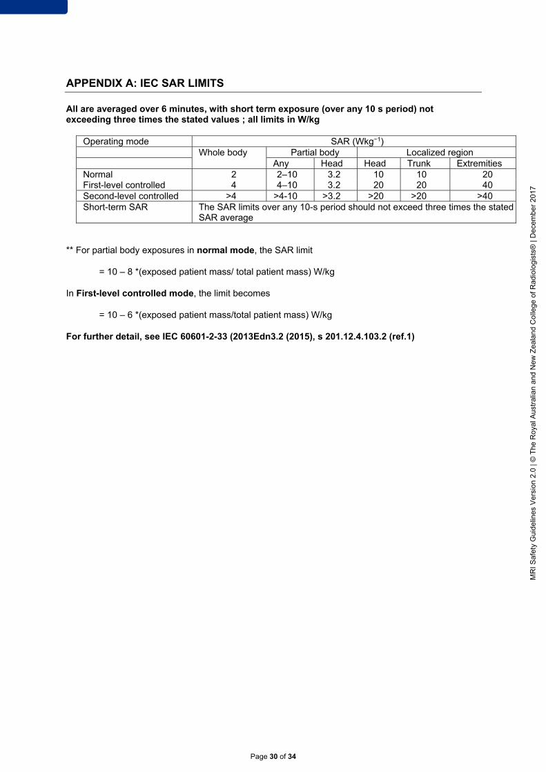
Page 30 of 34
MR
I Saf
ety
Gui
del
ines
Ver
sion
2.0
| ©
The
Ro
yal A
ustr
alia
n an
d N
ew Z
eala
nd C
olle
ge o
f Rad
iolo
gist
s® |
Dec
embe
r 20
17
APPENDIX A: IEC SAR LIMITS
All are averaged over 6 minutes, with short term exposure (over any 10 s period) not exceeding three times the stated values ; all limits in W/kg
Operating mode SAR (Wkg−1) Whole body Partial body Localized region Any Head Head Trunk Extremities Normal 2 2–10 3.2 10 10 20 First-level controlled 4 4–10 3.2 20 20 40 Second-level controlled >4 >4-10 >3.2 >20 >20 >40 Short-term SAR The SAR limits over any 10-s period should not exceed three times the stated
SAR average
** For partial body exposures in normal mode, the SAR limit
= 10 – 8 *(exposed patient mass/ total patient mass) W/kg
In First-level controlled mode, the limit becomes
= 10 – 6 *(exposed patient mass/total patient mass) W/kg
For further detail, see IEC 60601-2-33 (2013Edn3.2 (2015), s 201.12.4.103.2 (ref.1)

Page 31 of 34
MR
I Saf
ety
Gui
del
ines
Ver
sion
2.0
| ©
The
Ro
yal A
ustr
alia
n an
d N
ew Z
eala
nd C
olle
ge o
f Rad
iolo
gist
s® |
Dec
embe
r 20
17
APPENDIX B: GUIDELINES FOR MANAGEMENT OF PATIENTS WITH PARTICULAR FOREIGN BODIES
A. Orbital Foreign Bodies
Metal injury to eye:
If patient went to doctor and exam negative, or object fully removed, no screen required. Otherwise screen with at least one view of the orbits (unless more recent plain film/CT already available).
For non-patients, it may be appropriate to obtain informed consent for radiographic screening.
B. Cardiac Pacemakers and Implantable Cardioverter Defibrillators
Although older devices were generally strictly contra-indicated for MRI examination there are now several MRI conditional pacemakers and implantable defibrillators.
MRI conditional devices should be scanned in compliance with the stated conditions. These vary between models and between manufacturers, but will often involve adjustments to the device by an electrophysiologist before and after the scan, and a requirement for monitoring during the scan.
Some other devices can be scanned safely, but only with special precautions and where the risk is deemed acceptable after discussion amongst the MRI unit director, the managing cardiologist and other clinicians, and the patient (reference 19 of Appendix C).
Such an examination would require:
clinical risk-benefit assessment documented pacemaker type and model number, known to be suitable for supervised MR
examination documented lead types, known to be suitable for supervised MRI examination prior knowledge of lead configuration (from CXR if required) appropriately MRI-conditional ECG monitoring and (at least) pulse oximetry cardiac electrophysiological expertise in MRI suite throughout scan, with ability to promptly
re-program pacemaker device if necessary equipment and trained staff for Advance Life Support (ALS) expert check of pacemaker function before patient leaves the MRI suite after the
examination, with further follow-up at 1-6 weeks. specific informed consent (including risk of death)
C. Intracranial Aneurysm Clips
Confirm present. Signed, written documentation of type (mfr, model no, lot no, serial no.) +/- testing, and
manufacturer’s statement of MRI conditionality (or otherwise). If clip cannot be adequately characterised, proceed only on risk-benefit basis, after
discussion between patient, neurosurgeon, and MRI unit director, with informed consent specifically including risk of death. The MRI radiologist must authorise examination of the patient in writing.

Page 32 of 34
MR
I Saf
ety
Gui
del
ines
V2.
0 | ©
The
Ro
yal A
ustr
alia
n an
d N
ew
Zea
land
Col
lege
of R
adio
logi
sts®
| D
ecem
ber
2017
D. Stapes Implants
Document the manufacturer/model/lot no. Refer to product and independent literature; if prosthesis cannot be adequately
characterised, perform a thin-section CT to exclude metal—if positive, exclude patient; if negative, accept patient.
E. Programmable Hydrocephalus Shunts (Codman-Medos And Sophy)
These may be de-programmed by exposure to the field, and will require re-programming by appropriately trained personnel after the examination
F. Large Metal Implants (non-ferromagnetic joint prostheses, etc.)
May be MRI Conditional but undergo significant heating. At high field, Lenz law effects may result in repulsion of non- ferromagnetic conductors by the static field, as the implant is introduced into the bore. This is not necessarily a contra-indication to scanning. The effect may be reduced by reducing the speed of introduction of the patient and implant into the magnet bore. However, consideration should be given as to whether RF-induced artefacts may render the images non-diagnostic.

Page 33 of 34
MR
I Saf
ety
Gui
del
ines
Ver
sion
2.0
| ©
The
Ro
yal A
ustr
alia
n an
d N
ew Z
eala
nd C
olle
ge o
f Rad
iolo
gist
s® |
Dec
embe
r 20
17
APPENDIX C: USEFUL REFERENCES: MRI SAFETY ISSUES
1. Strizek B JJ, Mucyo E et al. Safety of MR Imaging at 1.5 T in Fetuses: A Retrospective Case-Control Study of Birth Weights and the Effects of Acoustic Noise. Radiology. 2015;275(2):530-7.
2. Ray JG VM, Bharatha A, Montanera WJ, Park AL. Association between MRI exposure during pregnancy and fetal and childhood outcomes. JAMA. 2016;316(9):952-61.
3. RANZCR. Accreditation standards for diagnostic and interventional radiology. Magnetic resonance imaging. Current version.
4. Protection ICoN-IR. Amendment to the ICNIRP “statement on medical magnetic resonance procedures: protection of patients.”. Health Physics. 2009;97:259-61.
5. Protection ICoN-IR. Guidelines on limits to exposure from static magnetic field. Health Physics. 2009;96:504-14.
6. NM H. Suspension of breast-feeding following gadopentetate dimeglumine administration. Radiology 2000;216(2):325-6.
7. Kok RD dVM, Heerschap A, van den Berg PP. Absence of harmful effects of magnetic resonance exposure at 1.5 T in utero during the third trimester of pregnancy: a follow-up study. Magn Reson Imaging. 2004;22:851-4.
8. JR G. Implantable pacemaker and defibrillator safety in the MR environment: New thoughts for the new millennium. In: E K, editor. Special Cross-Specialty Categorical Course in Diagnostic Radiology: Practical MR Safety Considerations for Physicians, Physicists, and Technologists Oak Brook, IL, USA2001. p. 69-76.
9. International A. Terminology of symbols and definitions relating to magnetic testing A0340-17. West Conshohocken, PA, USA2017.
10. Health TDo. Diagnostic Imaging Accreditation Scheme (DIAS) Practice Accreditation Standards. 2016.
11. FG S. Reference Manual for MR Safety. Salt Lake City: Amirsys Inc.; 2017. 12. FG S. Magnetic resonance safety: Recommendations and guidelines for pre- procedure
screening. In: E K, editor. Special Cross-Specialty Categorical Course in Diagnostic Radiology: Practical MR Safety Considerations for Physicians, Physicists, and Technologists Oak Brook, IL, USA2001. pp.33-9.
13. E K. Magnetic resonance safe practice guidelines of the University of Pittsburgh Medical Center. In: E K, editor. Special Cross-Specialty Categorical Course in Diagnostic Radiology: Practical MR Safety Considerations for Physicians, Physicists, and Technologists. Oak Brook, IL, USA. 2001. pp.155-63.
14. DW M. Occupational exposure in MRI. Br J Radiol. 2012;85:293-312. 15. Important information on anti-theft and metal detector systems and pacemakers, ICDs and spinal
cord stimulators [press release]. FDA safety advisory, 28 September 1998 1998. 16. Commission IE. IEC 60601-2-33. Medical Electrical Equipment – Part 2-33: Particular
Requirements for the Safety of Magnetic Resonance Equipment for Medical Diagnosis 3.2 ed. Geneva: IEC; 2017.
17. Baker PN JI, Harvey PR, Gowland PA, Mansfield P. . A three-year follow-up of children imaged in utero with echo-planar magnetic resonance. Am J Obstet Gynecol. 1994;170:32-3.
18. Australia JR-AsonKH. Guideline of the use of gadolinium-containing contrast agents in patients with renal impairment. 2013.
19. al. K-HRe. Gadopentetate dimeglumine excretion into human breast milk during lactation. Radiology 2000;216 (2):555-8.
20. Agency UMD. Guidelines for Magnetic Resonance DiagnosticEquipment in Clinical Use 4th ed. 2014.
21. Agency HP. Protection of patients and volunteers undergoing MRI procedures. In: RCE-7 HPA, editor. Chilton, United Kingdom2008.
22. (Australia) NHaMRC. Safety guidelines for magnetic resonance diagnostic facilities (1991) : approved at the 112th session of the National Health and Medical Research Council, Canberra, October 1991 / National Health and Medical Research Council. Canberra : Australian Govt. Pub. Service1992.
23. (ACR) ACoR. ACR White Paper on MRI Safety; and associated commentary. American Journal of Roentgenology. 2002;178(6):1335-47; 49-52.
24. MRI Bioeffects, Safety and Patient Management. Los Angeles: Biomedical Research Publishing Group; 2013. 718 p.
25. Care ACoSaQiH. National Safety and Quality Health Service Standards. 2012.

Page 34 of 34
MR
I Saf
ety
Gui
del
ines
V2.
0 | ©
The
Ro
yal A
ustr
alia
n an
d N
ew
Zea
land
Col
lege
of R
adio
logi
sts®
| D
ecem
ber
2017

THE ROYAL AUSTRALIAN AND NEW ZEALAND COLLEGE OF RADIOLOGISTS®