rapid assay format for multiplex detection of humoral ... manuscripts/corstjens... · with 100 ng...
TRANSCRIPT
Rapid Assay Format for MultiplexDetection of Humoral ImmuneResponses to Infectious DiseasePathogens (HIV, HCV, and TB)
PAUL L. A. M. CORSTJENS,a ZONGYUANG CHEN,b
MICHEL ZUIDERWIJK,a HAIM H. BAU,b WILLIAM R. ABRAMS,c
DANIEL MALAMUD,c R. SAM NIEDBALA,d AND HANS J. TANKEa
aDepartment of Molecular Cell Biology, Leiden University Medical Center,Leiden, the NetherlandsbDepartment of Mechanical Engineering and Applied Mechanics, Universityof Pennsylvania, Philadelphia, Pennsylvania, USAcDepartment of Basic Sciences, New York University College of Dentistry,New York, New York, USAdDepartment of Chemistry, Lehigh University, Bethlehem, Pennsylvania, USA
ABSTRACT: A novel assay is described for multiplex detection of antibod-ies against different pathogens from a single sample. The assay employsa modified lateral flow format (consecutive flow, CF) together with a sen-sitive reporter particle technology (up-converting phosphor technology,UPT) that allows for fully instrumented assay analysis. Lateral flow (LF)strips developed for the detection of human antibodies against humanimmunodeficiency virus type-1 and -2 (HIV-1 and -2) with additional cap-ture zones to detect antibodies against Myobacterium tuberculosis (TB)and hepatitis C Virus (HCV) provided the strips to test multiplexing. Dataare presented that show the performance of the TB and HCV test, as wellas two multiplex assays, TB with HIV and HCV with HIV. The TB/HCVassays demonstrate excellent detection capability, and HIV multiplexingdoes not affect the qualitative test result. The bench-top CF format wasconverted to a microfluidic platform and a first prototype semiautomatedchip capable of performing CF is presented here.
KEYWORDS: infectious disease; multiplex assay; up-converting phos-phor; lateral flow; TB; HCV; HIV
Address for correspondence: Paul L. A. M. Corstjens, Department of Molecular Cell Biology, LeidenUniversity Medical Center, PO Box 9600, 2300 RC Leiden, the Netherlands. Voice: +31-71-5269209;fax: +31-71-5268270.
Ann. N.Y. Acad. Sci. 1098: 437–445 (2007). C© 2007 New York Academy of Sciences.doi: 10.1196/annals.1384.016
437

438 ANNALS OF THE NEW YORK ACADEMY OF SCIENCES
INTRODUCTION
The presented antibody test platform allows convenient, rapid, sensitive,and cost-effective detection of infectious disease pathogens. It was initiallydeveloped for the detection of antibodies to HIV. In this report we explorethe potential of the platform for multiplex detection of antibodies associatedwith other infectious diseases, for example, simultaneous detection of HIV,TB, and HCV. TB often causes opportunistic infection in HIV/AIDS patients,and HCV has a high prevalence in HIV-infected individuals and is suspectedto co-infect with HIV.1,2 The antibody test platform was designed to becomepart of a modular microfluidic device, a point-of-care (POC) device to analyzeoral fluid for simultaneous detection of pathogenic antigens, nucleic acids, andthe host antibodies to the pathogen.3
The antibody assay uses a modified LF immunochromatography format withthree consecutive flow (CF) steps: (1) the first flow with diluted specimen (e.g.,plasma, oral fluid, or urine); (2) the second flow with wash buffer; and (3) thethird flow with protein A–coated reporter particles. The multiple flow system(CF) accommodates a 10-fold higher sample volume compared to a single flowformat, and demonstrates a better signal-to-noise value. Previously describedup-converting phosphor technology (UPT) reporter particles are applied forultrasensitive and instrumented assay analysis.4,5 These UPT reporters gen-erate a visible light emission signal upon excitation with low-energy infrared(IR) light.6 Interrogation of LF strips exposed to UPT reporters can be per-formed with a portable UPT reader (UPlink).7 In the UPlink system LF stripsare integrated in disposable plastic cassettes. We developed and constructeda prototype semiautomatic microfluidic module to perform CF that fits intoexisting UPlink-compatible cassettes.8
RESULTS
Consecutive Flow
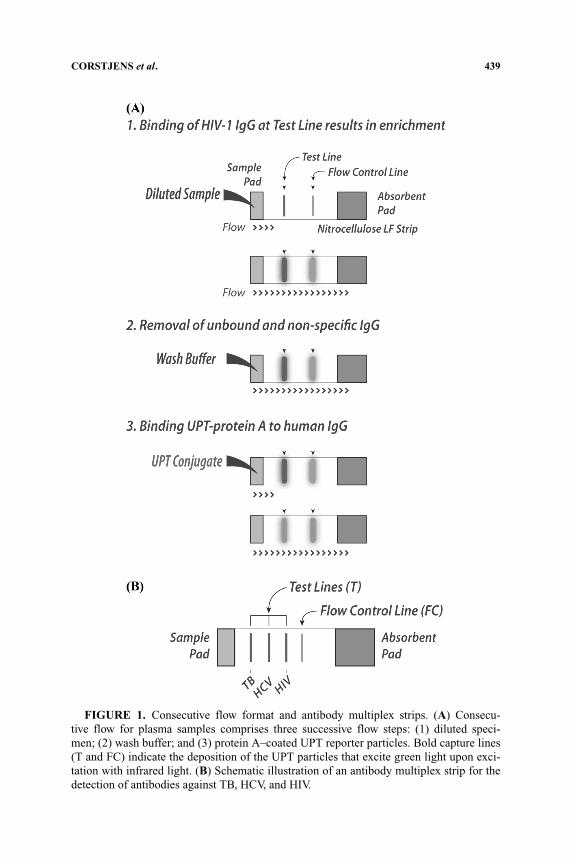
FIGURE 1 shows an illustration of UPT–CF designed for detection of humanantibodies against HIV. Antibodies to HIV-1 and -2 are captured at the testline which consists of HIV-specific antigens and the remaining human IgGsbind downstream to an anti-human IgG flow-control line. Nitrocellulose sheetsfor CF with proprietary HIV and HCV test lines were provided by OraSureTechnologies. A third test line with a proprietary TB antigen mixture (Courtesyof H.J. Houthoff and G.J. van Dam) was added to the CF strips in-house usinga Camag Linomat IV striper.9 Nitrocellulose sheets with only a TB-specifictest line were prepared in-house in a format similar to the sheets with theHIV-specific test line. Nitrocellulose sheets were assembled into LF strips asdescribed earlier.10 The assays described here used plasma, but can be easily
CORSTJENS et al. 439
FIGURE 1. Consecutive flow format and antibody multiplex strips. (A) Consecu-tive flow for plasma samples comprises three successive flow steps: (1) diluted speci-men; (2) wash buffer; and (3) protein A–coated UPT reporter particles. Bold capture lines(T and FC) indicate the deposition of the UPT particles that excite green light upon exci-tation with infrared light. (B) Schematic illustration of an antibody multiplex strip for thedetection of antibodies against TB, HCV, and HIV.
440 ANNALS OF THE NEW YORK ACADEMY OF SCIENCES
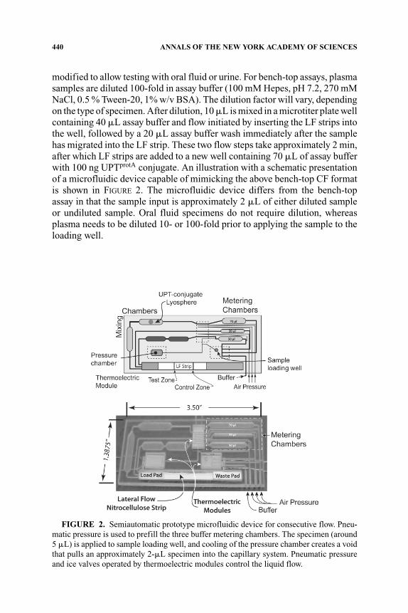
modified to allow testing with oral fluid or urine. For bench-top assays, plasmasamples are diluted 100-fold in assay buffer (100 mM Hepes, pH 7.2, 270 mMNaCl, 0.5 % Tween-20, 1% w/v BSA). The dilution factor will vary, dependingon the type of specimen. After dilution, 10 �L is mixed in a microtiter plate wellcontaining 40 �L assay buffer and flow initiated by inserting the LF strips intothe well, followed by a 20 �L assay buffer wash immediately after the samplehas migrated into the LF strip. These two flow steps take approximately 2 min,after which LF strips are added to a new well containing 70 �L of assay bufferwith 100 ng UPTprotA conjugate. An illustration with a schematic presentationof a microfluidic device capable of mimicking the above bench-top CF formatis shown in FIGURE 2. The microfluidic device differs from the bench-topassay in that the sample input is approximately 2 �L of either diluted sampleor undiluted sample. Oral fluid specimens do not require dilution, whereasplasma needs to be diluted 10- or 100-fold prior to applying the sample to theloading well.
FIGURE 2. Semiautomatic prototype microfluidic device for consecutive flow. Pneu-matic pressure is used to prefill the three buffer metering chambers. The specimen (around5 �L) is applied to sample loading well, and cooling of the pressure chamber creates a voidthat pulls an approximately 2-�L specimen into the capillary system. Pneumatic pressureand ice valves operated by thermoelectric modules control the liquid flow.

CORSTJENS et al. 441
HIV/TB Multiplexing
A collection of 300 banked plasma specimens (provided by H.J. Houthoffand G.J. van Dam) were analyzed with UPT–CF. The specimens consisted of100 healthy control samples (HIV–/TB–), 100 samples from HIV-negative butTB-infected patients (HIV–/TB+), and 100 samples from patients infectedwith HIV and TB (HIV+/TB+). The UPT assay threshold for plasma sam-ples as determined from an earlier study was verified by analyzing sampleson LF strips prepared with an HIV-specific test line only. In this experiment,samples generating a ratio signal (test signal divided by flow-control signal)>0.04 were considered antibody-reactive. A ratio value rather than an actualUPT signal value (measured in relative fluorescent units, RFU) is preferred asthis provides a convenient method to normalize test results; however, normal-ization is not a requirement. TB infection status was then tested on LF stripscarrying a TB-specific test line only. The resulting clinical parameters regard-ing TB testing are presented in TABLE 1 together with lab-based enzyme-linkedimmunosorbent assay (ELISA) results that were provided with the specimens.From these results we concluded that the accuracy of the rapid UPT assayis superior to the lab-based ELISA assay; it is important to note that bothassays used the same antigen mixture to capture TB-specific antibodies, sothat the increased performance is related to the difference in assay platform(ELISA vs. LF) as well as the difference in reporter technology (fluorescencevs. UPT).
Compared with testing for other infectious diseases, the actual specificityand sensitivity value for TB is relatively low, which is a known problem.11
Attempts to develop a low-complexity, rapid, immunologic-based TB assaywith sufficient accuracy have failed so far. In developing countries TB diagno-sis is especially problematic because children and HIV-compromised patientsare low responders, which increases the number of false negatives. TABLE 1
TABLE 1. UPT consecutive flow analysis of plasma specimen from 200 TB-diagnosed patients(50% being HIV compromised) and 100 healthy controls
Mixed(n = 300) Without HIV+/TB+ Without HIV–/TB+
HIV–/TB– (n = 100) (n = 200) (n = 200)HIV–/TB+ (n = 100) HIV–/TB– (n = 100) HIV+/TB+ (n = 100)HIV+/TB+ (n = 100) HIV–/TB+ (n = 100) HIV–/TB– (n = 100)
TB/HIV Statusa,b
Assay ELISAb UPT ELISAb UPT ELISAb UPT
Specificity 96.0 95.0 96.0 95.0 96.0 95.0Sensitivity 52.5 62.5 66.0 74.0 39.0 51.0Accuracy 50.4 59.4 63.4 70.3 37.4 48.5
aPlasma specimens of 200TB- diagnosed patients (TB+) and 100 healthy controls (HIV–/TB–)were analyzed. Half (n = 100) of the TB-diagnosed patients were also diagnosed as HIV+.
bPlasma specimens and corresponding ELISA data were provided by H.J. Houthoff and G.J. vanDam.

442 ANNALS OF THE NEW YORK ACADEMY OF SCIENCES
indicates that the UPT assay sensitivity improves from 62.5% to 74.0% whenthe HIV-compromised patient group (HIV+/TB+ samples) is omitted. This isalso evident when examining the ELISA data.
Analysis of selected samples on multiplex LF strips with TB and HIV testlines did not affect the qualitative result of the assay (results not shown).A detailed study is ongoing to examine the potential effect on signal valueof “preceding capture line interference,” including differences in the loca-tion/distance of the capture line from the sample application pad.
HIV/HCV Multiplexing
The UPT–CF format was also used for the detection of HCV infection.Analysis of three seroconversion panels demonstrated excellent performanceof the UPT assay compared to an EIA (TABLE 2), which demonstrates theapplicability as a model in developing UPT–CF multiplex assays. To furtherexplore the extent of potential “preceding capture line interference,” a “dilutioncheckerboard matrix” of a high-reactive HCV panel member mixed with a high-reactive HIV plasma sample was prepared in normal human plasma (NHP)and analyzed on multiplex LF strips. Each multiplex LF strip produced anHIV as well as HCV test signal, and a flow-control signal. HIV and HCV ratiosignals were calculated by dividing their individual test signal by the jointflow-control signal. In FIGURE 3 the ratio value is indicated on the y axis ofthree-dimensional histograms; panel A shows the results for HIV and panel Bshows the results for HCV. In both histograms the x- and z axis represents the
TABLE 2. Relative sensitivity of UPT consecutive flow for the analysis of HCV plasmaseroconversion panels
BBI Panel PHV905a BBI Panel PHV907a BBI Panel PHV914a
Daysb EIAc UPTd Daysb EIAc UPTd Daysb EIAc UPTd
0 0.0 0.02 0 0.0 0.02 0 0.0 0.014 0.0 0.01 4 0.0 0.02 5 0.0 0.027 0.0 0.03 7 0.0 0.02 9 0.0 0.01
11 0.3 0.02 13 0.1 0.09 12 0.0 0.0414 0.7 0.06 18 0.7 0.24 16 0.2 0.1618 0.7 0.07 21 1.5 0.42 19 0.3 0.2121 2.5 0.10 164 5.0 1.48 24 3.2 0.3725 >5.0 1.10 30 >4.7 0.7128 >5.0 2.00 33 >4.7 0.95
aDetailed panel information at www.seracare.com/bbidx/hcv panels.htm.bDays after first bleed.cBBI-provided data; test values ≥ 1 were considered positive (indicated in bold).dUPT ratio values above > 0.04 were considered positive (indicated in bold).

CORSTJENS et al. 443
FIGURE 3. Result of a multiplex analysis; the simultaneous detection of antibodiesagainst HIV and HCV in single plasma specimens. A dilution series of plasma speci-men from an HIV-compromised patient and an HCV-compromised patient were mixed inequal amounts. In the histograms “0” indicates the highest amount of HIV and/or HCV-compromised plasma specimen; the indicated dilutions were made in NHP. NHP is alsoused as the (no signal) control sample. The dilution of HIV plasma is indicated on the xaxis, the dilution of HCV plasma (z axis) (A) the HIV ratio signal, (B) the HCV ratio signal.All data points are the average value of three individual experiments.
test matrix with the HIV dilution on the x axis and the HCV dilution on thez axis. The experiment was performed in triplicate and the average signal ispresented in the histograms. For purpose of comparison the maximum ratiovalue was normalized to 1.
The actual ratio value as determined for undiluted HIV and HCV sampleswas 0.96 and 6.0, respectively (in FIGURE 3 these values were normalized to 1).In patients infected with both viruses, HIV and HCV ratio signals expectedlyare different because HIV and HCV test lines use different capture antigensto detect their respective antibodies; the amount of antibodies specific againstHIV and HCV is variable, as is the binding affinity of the applied antigen–antibody pairs. In the experiments performed here the HCV test line was closerto the sample application pad than the HIV test line (FIG. 1). As a consequence,the HCV test line signals are higher in comparison to assays where the sameHCV test line is localized at the (further downstream) position of the HIV testline. In theory this implies an assay cut-off threshold value that is dependent onthe distance of the test line from the sample application pad. For the experimentdescribed here, the actual test line ratio signal obtained on multiplex LF stripswith NHP (x5, z5 in FIG. 3) generated approximately the same value for HCVand HIV (respectively 0.032 and 0.027 [n = 3]) and the maintained cut-offthreshold value was > 0.04.
444 ANNALS OF THE NEW YORK ACADEMY OF SCIENCES
DISCUSSION
We describe a modified lateral flow format ( CF) for antibody detection andexplore its potential for multiplexing. The CF format was originally developedto detect human antibodies against HIV-1 and -2. Multiplexing is achieved byproviding the HIV-specific LF strips with additional capture lines specific forantibodies against other pathogens. Similar to the HIV-specific test line, theadditional capture lines comprise pathogen-specific antigens. The different testlines are placed transversely across the LF strip such that the tested specimenwill sequentially pass individual test lines. Antibodies present in the specimencan bind to these antigens. This procedure demands careful development andoptimization of the various capture lines in order to avoid nonspecific bindingand to prevent undesired cross-reactivity of the antibodies with preceding testlines. Furthermore, when applying an antibody-generic reporter, a reporterflow completely disconnected from the antibody flow is desired. CF appliesan initial flow of specimen, followed by a wash step and a final flow withantibody-generic UPT reporter particles. The initial flow allows a free flow ofthe antibodies present in the specimen, so that the antibodies against variouspathogens can enrich in the spatially separated capture zones. A wash stepthen removes non-specific-bound antibodies from the LF strip, and in the finalflow UPTprotA reporters bind to the antibodies in the pathogen-specific capturezones.
The results presented in this article indicate that multiple antibody test linesare feasible. Multiplex analysis of combined specimens with high loads ofantibodies against HIV and HCV did not show relevant interference. Furtherstudies are necessary to evaluate the maximum number of test lines that canbe applied to a LF strip without disturbing the UPT reporter particle flow. Inthis context the effect of high signals in the test lines closest to the sample padneeds to be carefully analyzed. But as most specimens will only show antibodyreactivity for a limited number of pathogens, disturbance of the reporter flowto further downstream test lines as a consequence of multiple coinciding hightest signals may only be a theoretical problem.
In earlier studies using LF strips with a single test line and a flow-controlline, ratio calculations of the test (T) divided by the flow control (FC) were ap-plied. This normalization allows for interassay comparison of results obtainedwith different LF strips. Increasing the number of test lines will probably af-fect the normalization algorithm. When the number of test lines is increased,differences in distance and signal may become more evident. In the multiplexassays presented here for TB and HCV these effects were minor and did notaffect the outcome of the multiplex tests. Note also that qualitative assays donot necessarily demand a ratio calculation (see, e.g., Mokkapati et al.7).
The CF format described here allows multiplex detection of various antibod-ies from a single specimen with an antibody-generic UPT reporter. As such,

CORSTJENS et al. 445
the CF format suits its objective and was therefore selected as the antibodytest module in a (modular) microfluidic device that permits simultaneous de-tection of viral and bacterial antigens, nucleic acids, and antibodies to thesepathogens. The bench-top CF format was therefore converted to a microfluidicmodule and a first semiautomated prototype is presented.
ACKNOWLEDGMENT
Shang Li and Geraldine Guillon are acknowledged for providing nitrocellu-lose striped with HIV and HCV antigens and the UPT reporters. Hendrik-JanHouthoff and Govert J. van Dam are acknowledged for providing the TB anti-gen mixture and 200 plasma samples from TB-diagnosed patients togetherwith 100 healthy control samples. Claudia J. de Dood and Dieuwke Kornelisare acknowledged for technical assistance. Part of this work was supported byNIH Grant UO1-DE-017855.
REFERENCES
1. ROCKSTROH, J.K. & U. SPENGLER. 2004. HIV and hepatitis C virus co-infection.Lancet Infect. Dis. 4: 437–444.
2. VERNET, G. 2004. Molecular diagnostics in virology. J. Clin. Virol. 31: 239–247.3. MALAMUD, D. et al. 2005. Point detection of pathogens in oral samples. Adv. Dent.
Res. 18: 12–16.4. LI, S. et al. 2002. Preparation, characterization and fabrication of uniform coated
Y2O2S:Er3+ up-converting phosphor particles for biological detection applica-tions. Proc. SPIE-Int. Soc. Opt. Eng. 4809: 100–109.
5. CORSTJENS, P.L.A.M. et al. 2005. Infrared up-converting phosphors for bioassays.IEE Proc. Nanobiotechnol. 152: 64–72.
6. ZARLING, D.A. et al. 1997. Up-converting reporters for biological and other assaysusing laser excitation techniques. US Patent 5: 674–698.
7. MOKKAPATI, V.K. et al. 2007. Evaluation of UPlink-RSV: a prototype rapid antigentest for detection of respiratory syncytial virus infection. Ann. N.Y. Acad. Sci.This volume.
8. CHEN, Z. et al. 2005. A disposable microfluidic point-of-care device for detectionof HIV: a new up-converting phosphor technology application. Presented at the9th International Conference on Miniaturized Systems for Chemistry and LifeSciences (�TAS).: 791–793.
9. NIEDBALA, R.S. et al. 2001. Detection of analytes by immunoassay using up-converting phosphor technology. Anal. Biochem. 293: 22–30.
10. CORSTJENS, P.L.A.M. et al. 2001. Use of up-converting phosphor reporters inlateral-flow assays to detect specific nucleic acid sequences: a rapid, sensitiveDNA test to identify human papillomavirus type 16. Infection 47: 1885–1893.
11. CHARLES, M. & J.W. PAPE. 2006. Turberculosis and HIV: implications in thedeveloping world. Curr. HIV/AIDS Rep. 3: 139–144.