rapid changes in plasma membrane protein - plant physiology
TRANSCRIPT

Plant Physiol. (1988) 86, 505-5090032-0889/88/86/0505/05/$01 .00/0
Rapid Changes in Plasma Membrane Protein Phosphorylationduring Initiation of Cell Wall Digestion'
Received for publication July 8, 1987 and in revised form October 31, 1987
DAVID P. BLOWERS, WENDY F. Boss*, AND ANTHONY J. TREWAVASDepartment of Botany, University of Edinburgh, Mayfield Road, Edinburgh, EH9 3JH, UK (D.P.B.,A.J.T.), and Department of Botany, Box 7612, North Carolina State University, Raleigh, NC 27695-7612(W.F.B.)
ABSTRACT
Plasma membrane vesicles from wild carrot cells grown in suspensionculture were isolated by aqueous two-phase partitioning, and ATP-de-pendent phosphorylation was measured with [y-32PJATP in the presenceand absence of calcium. Treatment of the carrot cells with the cell walldigestion enzymes, driselase, in a sorbitol osmoticum for 1.5 min alteredthe protein phosphorylation pattern compared to that of cells treated withsorbitol alone. Driselase treatment resulted in decreased phosphorylationof a band ofM, 80,000 which showed almost complete calcium dependencein the osmoticum treated cells; decreased phosphorylation of a band ofMr 15,000 which showed little calcium activation, and appearance of anew band of calcium-dependent phosphorylation at M, 22,000. Theseeffects appeared not to be due to nonspecific protease activity and neitherin vivo nor in vitro exposure to driselase caused a significant loss of Coom-assie blue-staining bands on the gels of the isolated plasma membranes.However, protein phosphorylation was decreased. Adding driselase to thein vitro reaction mixture caused a general decrease in the membraneprotein phosphorylation either in the presence or absence ofcalcium whichdid not mimic the in vivo response. Cells labeled in vivo with inorganic32P also showed a response to the Driselase treatment. An enzymicallyactive driselase preparation was required for the observed responses.
The plasma membrane of plant cells is a potential site for thetransduction of signals from the external environment to the cellinterior. The role of the plant plasma membrane in signal trans-duction has been under scrutiny for some time.The regulatory role of protein phosphorylation in plants is
becoming increasingly apparent (for review see Ref. 13), andcalcium-dependent protein kinases could provide a means ofsignal transduction across the plasma membrane. A calcium andcalmodulin-activated, autophosphorylating protein kinase of M,18,000 has been characterized in the plasma membranes isolatedfrom pea shoots (2, 3). Thus, there is the potential for the plantplasma membrane to respond to a stimulus and alter cellularmetabolism.To determine whether changes in protein phosphorylation oc-
cur in the plasma membranes of suspension culture cells in re-sponse to the cell wall digestion enzymes used to isolate proto-plasts, studies of protein phosphorylation of the carrot suspension
' Supported in part by funds from the Agricultural and Food ResearchCouncil to D. P. B. and in part by the National Science Foundationgrant DCB-8502813 to W. F. B. This is Paper No. 11165 of the JournalSeries of the North Carolina Agricultural Research Service, Raleigh, NC27695-7601.
culture cells were undertaken. These cells are normally plas-molysed in a solution of 0.4 molal sorbitol during cell wall diges-tion; therefore, the sorbitol osmoticum was used as a control.Dramatic and rapid changes were seen in the calcium-dependentin vitro phosphorylation patterns of plasma membranes isolatedfrom the cells exposed to Driselase compared to the sorbitolcontrols. Rapid changes in protein labeled in vivo with inorganic32p also were observed.
MATERIALS AND METHODS
Chemicals. [y-32P]ATP (specific activity 185 TBq/mmol) and32P (acid free, 370 MBq/ml) were obtained from Amersham In-ternational plc (Amersham, Bucks., UK). Other chemicals wereobtained from Sigma London Chemical Co (Poole, Dorset, UK).Driselase was obtained from Plenum Scientific Co.Treatment of Cells and Isolation of Plasma Membrane. Wild
carrot cells were maintained by weekly serial transfer into fusioninducing medium as previously described (4). For in vivo labelingwith 32p, the cells were grown on phosphate-free medium and 6to 10 MBq 32P were added for 15 h prior to harvesting. For allexperiments, the cells were harvested 4 d after transfer by fil-tration on filter paper, washed once with approximately 10 mlof water, and placed in the designated solutions at a concentra-tion of approximately 0.4 g fresh weight per 20 ml of solution.Incubations were performed at room temperature in 0.4 molalsorbitol in 1 mM MES (pH 4.8) with or without 2% (w/v) dri-selase. Stock driselase was prepared at 4% and the supernatantfrom a 1,000g centrifugation collected. At indicated times thecells or protoplasts were collected by centrifugation and washedtwice by resuspension and centrifugation in approximately S mlof 0.45 molal sorbitol 1 mM MES (pH 6.0).The washed cells and protoplasts were homogenized in a ground
glass homogenizer in a medium consisting of 8% sucrose, 95 mMLiCl, 10 mm KCl, 1 mm EDTA, 1 mM EGTA, 0.1 mM MgCl2,and 50 mm Tris (pH 7.5). The homogenate was centrifuged at1,000g for 4 min and the 1,000g supe-matant centrifuged at 40,000gfor 1 h. The 40,000g pellet was resuspended in 0.5 ml of waterand the plasma membrane-rich fraction isolated by aqueous twophase partitioning (1) using 6.3% PEG (approximate mol wt3,350) and dextran (average mol wt 500,000) as the polymers aspreviously described for the wild carrot cells (16) except that10% water of hydration was assumed for the dextran. The upperphase was collected, diluted with approximately 30 ml of ho-mogenizing buffer without the lithium or sucrose, and centri-fuged at 40,000g for 1 h.The pelleted plasma membrane-rich fraction (50-80 gg pro-
tein) was resuspended in approximately 50 ,l of water and usedfor in vitro phosphorylation as previously described (9) or forother treatments as described in the text. Protein was determined
505
Dow
nloaded from https://academ
ic.oup.com/plphys/article/86/2/505/6082921 by guest on 19 January 2022
Plant Physiol. Vol. 86. 1988
according to Lowry (10) with BSA as standard.Gel Electrophoresis. SDS-PAGE was performed using 12%
polyacrylamide gels 20 x 16 x 0.1 cm as previously described(8). Approximately equal amounts of protein were added to eachlane.
Protein Kinase Assays. Protein kinase assays for the isolatedplasma membrane fraction were carried out using TCA precip-itation onto cellulose discs as previously described (9).
RESULTS
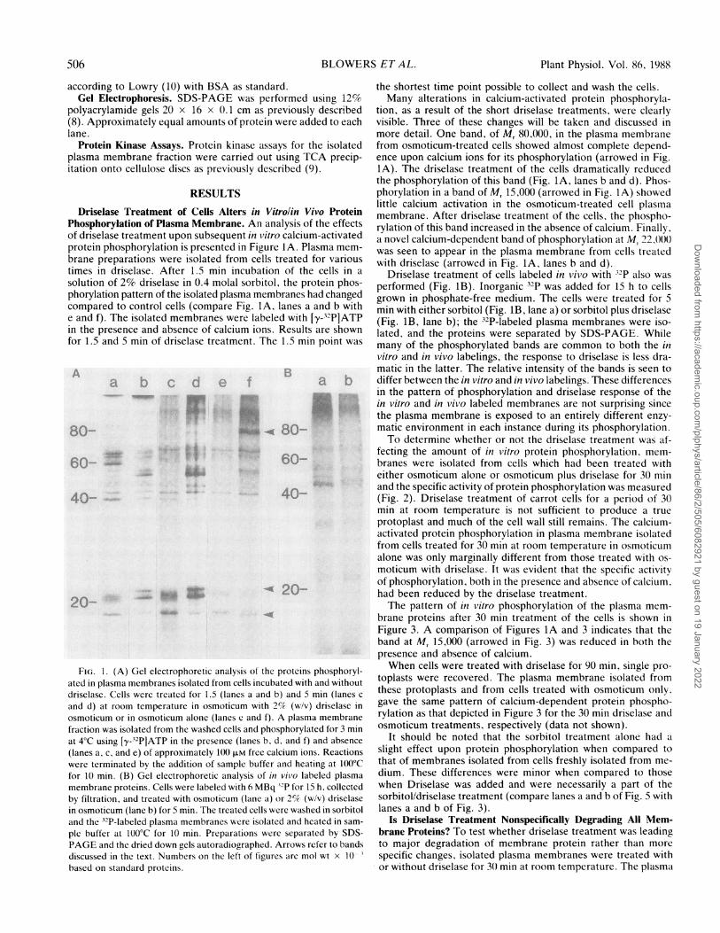
Driselase Treatment of Cells Alters in Vitrolin Vivo ProteinPhosphorylation of Plasma Membrane. An analysis of the effectsof driselase treatment upon subsequent in vitro calcium-activatedprotein phosphorylation is presented in Figure IA. Plasma mem-brane preparations were isolated from cells treated for varioustimes in driselase. After 1.5 min incubation of the cells in asolution of 2% driselase in 0.4 molal sorbitol, the protein phos-phorylation pattern of the isolated plasma membranes had changedcompared to control cells (compare Fig. IA, lanes a and b withe and f). The isolated membranes were labeled with [y-12PIATPin the presence and absence of calcium ions. Results are shownfor 1.5 and 5 min of driselase treatment. The 1.5 min point was
40;-)
....,-
FIG. 1. (A) Gel electrophoretic analysis of the proteins phosphoryl-ated in plasma membranes isolated from cells incubated with and withoutdriselase. Cells were treated for 1.5 (lanes a and b) and 5 min (lanes c
and d) at roorn temperature in osmoticum with 2C/c (w/v) driselase in
osmoticum or in osmoticum alone (lanes e and f). A plasma membranefraction was isolated from the washed cells and phosphorylated for 3 minat 4°C using [y-32PIATP in the presence (lanes b, d, and f) and absence
(lanes a, c, and e) of approximately 100 ,UM free calcium ions. Reactionswere terminated by the addition of sample buffer and heating at 100°Cfor 10 min. (B) Gel electrophoretic analysis of in vivo labeled plasmamembrane proteins. Cells were labeled with 6 MBq 32P for 15 h, collectedby filtration, and treated with osmoticum (lane a) or 2% (w/v) driselasein osmoticum (lane b) for 5 min. The treated cells were washed in sorbitoland the 32P-labeled plasma membranes were isolated and heated in sam-
ple buffer at 100°C for 10 min. Preparations were separated by SDS-PAGE and the dried down gels autoradiographed. Arrows refer to bandsdiscussed in the text. Numbers on the left of figures are mol wt x 10-3based on standard proteins.
the shortest time point possible to collect and wash the cells.Many alterations in calcium-activated protein phosphoryla-
tion, as a result of the short driselase treatments, were clearlyvisible. Three of these changes will be taken and discussed inmore detail. One band, of Mr 80,000, in the plasma membranefrom osmoticum-treated cells showed almost complete depend-ence upon calcium ions for its phosphorylation (arrowed in Fig.1A). The driselase treatment of the cells dramatically reducedthe phosphorylation of this band (Fig. IA, lanes b and d). Phos-phorylation in a band of M, 15,000 (arrowed in Fig. IA) showedlittle calcium activation in the osmoticum-treated cell plasmamembrane. After driselase treatment of the cells, the phospho-rylation of this band increased in the absence of calcium. Finally,a novel calcium-dependent band of phosphorylation at M, 22.000was seen to appear in the plasma membrane from cells treatedwith driselase (arrowed in Fig. IA, lanes b and d).
Driselase treatment of cells labeled in vivo with 32P also wasperformed (Fig. IB). Inorganic 32P was added for 15 h to cellsgrown in phosphate-free medium. The cells were treated for 5min with either sorbitol (Fig. iB, lane a) or sorbitol plus driselase(Fig. 1B, lane b); the 32P-labeled plasma membranes were iso-lated, and the proteins were separated by SDS-PAGE. Whilemany of the phosphorylated bands are common to both the invitro and in vivo labelings, the response to driselase is less dra-matic in the latter. The relative intensity of the bands is seen todiffer between the in vitro and in vivo labelings. These differencesin the pattern of phosphorylation and driselase response of thein vitro and in vivo labeled membranes are not surprising sincethe plasma membrane is exposed to an entirely different enzy-matic environment in each instance during its phosphorylation.To determine whether or not the driselase treatment was af-
fecting the amount of in vitro protein phosphorylation, mem-branes were isolated from cells which had been treated witheither osmoticum alone or osmoticum plus driselase for 30 minand the specific activity of protein phosphorylation was measured(Fig. 2). Driselase treatment of carrot cells for a period of 30min at room temperature is not sufficient to produce a trueprotoplast and much of the cell wall still remains. The calcium-activated protein phosphorylation in plasma membrane isolatedfrom cells treated for 30 min at room temperature in osmoticumalone was only marginally different from those treated with os-moticum with driselase. It was evident that the specific activityof phosphorylation, both in the presence and absence of calcium.had been reduced by the driselase treatment.The pattern of in vitro phosphorylation of the plasma mem-
brane proteins after 30 min treatment of the cells is shown inFigure 3. A comparison of Figures 1A and 3 indicates that theband at Mr 15,000 (arrowed in Fig. 3) was reduced in both thepresence and absence of calcium.When cells were treated with driselase for 90 min, single pro-
toplasts were recovered. The plasma membrane isolated fromthese protoplasts and from cells treated with osmoticum only.gave the same pattern of calcium-dependent protein phospho-rylation as that depicted in Figure 3 for the 30 min driselase andosmoticum treatments. respectively (data not shown).
It should be noted that the sorbitol treatment alone had aslight effect upon protein phosphorylation when compared tothat of membranes isolated from cells freshly isolated from me-dium. These differences were minor when compared to thosewhen Driselase was added and were necessarily a part of thesorbitol/driselase treatment (compare lanes a and b of Fig. 5 withlanes a and b of Fig. 3).
Is Driselase Treatment Nonspecifically Degrading All Mem-brane Proteins? To test whether driselase treatment was leadingto major degradation of membrane protein rather than morespecific changes, isolated plasma membranes were treated withor without driselase for 3() min at room temperature. The plasma
506 BLOWERS ET A L.
1. 11
itk'lo-.
.-W -a J
Dow
nloaded from https://academ
ic.oup.com/plphys/article/86/2/505/6082921 by guest on 19 January 2022

RAPID CHANGES IN PLASMA MEMBRANE PROTEIN PHOSPHORYLATION
CV,o 0-2 -0
VI
.; .CIo oa.I000 ao 0CL _
~0c w4 -
seconds
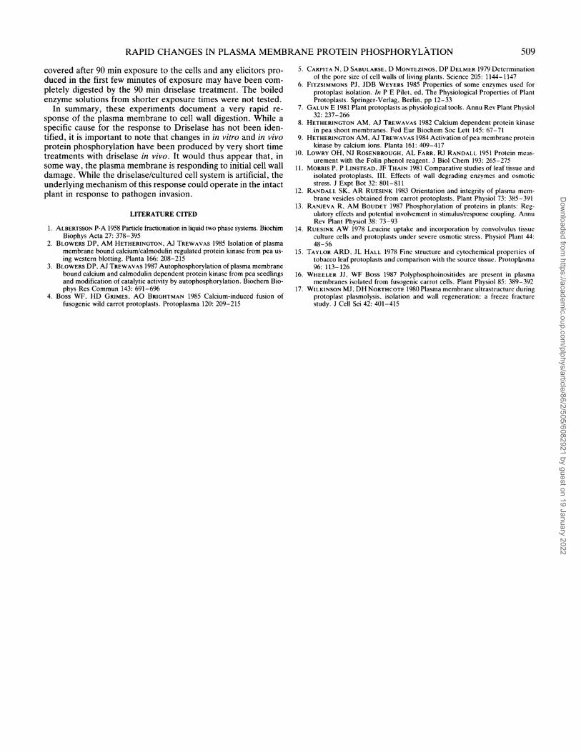
FIG. 2. Decrease in specific activity of in vitro plasma membranephosphorylation by driselase treatment of cells. Cells were treated for30 min at room temperature in osmoticum with (circles) or without(triangles) 2% (w/v) driselase. A plasma membrane fraction was thenisolated from the washed cells and used in an in vitro phosphorylationassay. Plasma membranes was incubated with [y-32P]ATP in the presence(closed symbols) or absence (open symbols) of aproximately 100 JiM freecalcium ions. Aliquots were removed at the times indicated and incor-porated phosphate estimated.
a b c d
8- ~
60- 4
40-
20-
FIG. 3. Gel electrophoretic analysis of the proteins phosphorylatedin plasma membranes isolated from cells incubated 30 min with osmo-ticum in the presence and absence of driselase. Cells were treated for30 min at room temperature in osmoticum with (lanes c and d) or without(lanes a and b) 2% (w/v) driselase. In vitro phosphorylation was as perFigure IA. Lanes b and d were plus 100 ,UM free calcium and a and cwere in the absence of added calcium. Arrows refer to bands discussedin the text. Numbers on left are mol wt x 10-3 based on standardproteins.
membrane was washed through a sorbitol cushion prior to anal-ysis of the proteins by SDS-PAGE. The effects of in vitro dri-selase treatment on the Coomassie blue-stained proteins werecompared to that of the in vivo treatment (Fig. 4). Only twodifferences were readily visible for the in vivo treatment-no-tably the loss of staining of two proteins at Mr 20,000 (arrowedin Fig. 4, lanes a and b). Driselase treatment in vitro (lanes cand d) leads to similar changes. A single protein of M, 15,000appears after in vitro driselase treatment of membranes (arrowedin Fig. 4, lane c). Presumably this band represents a degradationproduct or a driselase component which has co-isolated with thewashed membranes.The above in vitro driselase treatment completely abolished
all protein kinase activity in the resultant washed membranefractions. In addition, it was found that simply including driselasein the phosphorylation assay mixture led to decreased phospho-rylation as indicated in Figure 5. Driselase alone contained nomeasurable protein kinase activity (data not shown). Taken intoto, these results indicate that neither major degradation ofplasma membrane proteins nor phosphorylation of driselasecomponents contributed to the changes in protein phosphoryl-ation seen after in vivo driselase treatment.The reduced phosphorylation when driselase was added to the
in vitro reaction mixture was similar but not identical to theresponse to in vivo driselase treatment (e.g., compare Fig. 5,lanes c and d, to Fig. 1A, lanes a and b). Phosphorylation of theband of Mr 80,000 in the presence of calcium was reduced butnot eliminated (arrowed in Fig. 5). This band was characteristicof the membranes isolated from the control cells and was reduced
a b c d
40 X
20-
FIG. 4. A comparison of the protein content of plasma membraneafter in vivo and in vitro treatment with driselase. For in vivo treatment(lanes a and b) cells were incubated for 30 min at room temperature inthe presence (lane a) and absence (lane b) of 2% (w/v) Driselase. Aplasma membrane preparation was then isolated from the washed cells,added to sample buffer and separated by SDS-PAGE. The in vitro treat-ment consisted of a 30 min incubation at room temperature of plasmamembrane isolated from untreated cells in the presence (lane c) andabsence (lane d) of 2% (w/v) driselase in the osmoticum buffer withoutsorbitol. Membranes were then spun through a neutral pH sorbitol cush-ion at 11,500g, added to sample buffer heated at 100°C for 10 min andseparated by SDS-PAGE. The resultant polyacrylamide gels were stainedwith Coomassie blue, dried down and photographed. Arrows relate tobands discussed in the text. Numbers on left are mol wt x 10-1 basedon standard proteins
5()7
Dow
nloaded from https://academ
ic.oup.com/plphys/article/86/2/505/6082921 by guest on 19 January 2022

Plant Physiol. Vol. 86, 1988
a b c d
80- 460-
~ 4 0-
20-4WAM~ ~
FIG. 5. Demonstration of the lack of phosphorylation of driselase
proteins by the isolated plasma membrane fraction. A plasma membrane
fraction was isolated from untreated cells and phosphorylated by incu-
baiton in [y-32P]ATP in the presence (lanes c and d) and absence (lanes
a and b) of 2 % (w/v) driselase in the assay mix. Additionally, labelings
were performed in the absence (lanes a and c) and presence (lanes
and d) of approximately 100,UM free calcium ions. Reactions were ter-
minated after 3 min at4°C by the addition of sample buffer and heating
at 100°C for 10 min. Preparations were then separated by SDS-PAGE
and the dried down gels autoradiographed. Arrows relate to bands dis-
cussed in the text. Numbers on left are mol wt x 10-3
based on standard
proteins.
to a greater extent with an in vivo driselase treatment of only
1.5 min. The phosphorylation of the Mr 15,000 protein in the
presence of calcium was reduced by the in vitro driselase treat-
ment. Thus, direct interaction of driselase with the membranes
was capable of altering protein phosphorylation. It is important
to note, however, that long term in vitro treatment led to com-
plete loss of protein kinase activity, while even after 90 min
exposure to driselase in vivo, the distinct pattern of protein phos-
phorylation was evident both with and without calcium present.
What Alters Protein Phosphorylation? Incubation of cells in
osmoticum containing a boiled driselase supernatant had no ef-
fect on protein phosphorylation compared to the sorbitol con-
trols. This indicated that an active enzymatic component was
required for the response. Furthermore, incubation of cells with
a boiled 'once used' driselase mixture had no effect on the protein
phosphorylation pattern of the isolated plasma membranes. Thus,
any cell wall fragments remaining in the solution after the normal
90 min cell wall digestion period were not acting as elicitors for
this response.
An initial attempt was made to identify the component(s) in
the driselase which might cause such changes in protein phos-
phorylation. Since driselase is a crude mixture of fungal enzymes,
several hydrolytic enzymes were tested to see if they would give
a similar response. Cells were treated with either ribonuclease
(1,ug/ml), trypsin (1 Ag/ml), or pectinase (2 mg/ml) for 1.5 mmn.
None of the treatments gave detectable changes in the protein
phosphorylation pattern compared to the sorbitol controls. While
longer (30 min) treatment with trypsin decreased protein phos-
phorylation, the response was general and the pattern of phos-
phorylation did not vary either with or without calcium added
to the in vitro reaction mixture. In further attempts to eliminate
the effects of nonspecific proteases present in the driselase, cells
were treated for 5 min with driselase containing 1.0% BSA. Theaddition of BSA did not inhibit the driselase effect on proteinphosphorylation (data not shown).
If extracellular calcium was acting as a second messenger dur-ing the response to driselase in vivo, then removing extracellularcalcium with the calcium chelator, EGTA, should decrease theresponse and increasing intracellular calcium with the ionophore,A23187, should mimic the response. Neither EGTA (2.5 mM)with driselase, nor A23187 (10 /LM) in the absence of driselaseaffected the pattern of protein phosphorylation of the isolatedmembranes. Thus, the in vivo status of calcium appeared not tobe critical for the driselase-induced changes in the plasma mem-brane.
DISCUSSION
While differences in the structure (15, 17) and function (11,14) of the plasma membranes of protoplasts and the source tissue-have been reported, early biochemical changes in the plasmamembranes of cells during cell wall digestion have not beendescribed previously. The results presented in this paper indicatethat the plasma membranes of the wild carrot cells grown insuspension culture are very sensitive to exposure to cell walldigestion enzymes. Within 1.5 min after treatment of the cellswith the enzymes, the pattern of ATP-dependent protein phos-phorylation of the isolated membranes changed. The most dra-matic changes were evident when calcium was added to the invitro phosphorylation mixture. It is assumed that these representaltered/novel phosphorylation sites induced by the driselasetreatment. These changes in the plasma membrane persisted andwere evident in the fully isolated protoplasts (90 min treatmentof the cells with driselase).
It has been suggested that both the proteases contaminatingthe cell wall digestion enzymes and the osmotic stress imposedduring wall digestion could contibute to altering the plasma mem-brane of protoplasts (for review see Refs. 6 and 7). The responsesdescribed in this paper were rapid and did not result from osmoticstress alone. The responses were dependent on a heat-sensitivecomponent of the driselase; however, they were probably notdue to a nonspecific protease effect since neither in vivo nor invitro treatment with driselase resulted in major loss of Coomassieblue-staining bands. Treatment with a protease such as trypsinwould have caused major losses of membrane proteins (12). Inaddition, the driselase response could not be elicited by 1.5 minor 30 min treatment with trypsin in vivo and adding 1.0% BSAto the driselase to decrease nonspecific protease activity had noeffect.
Carpita et al. (5) have shown that 3.8 to 4.0 nm pores exist inthe cell walls of tissue culture cells and that these pores wouldallow large molecules to reach the plasma membrane. If someof the enzymes (e.g, glycosidases, phosphatases) in the driselasemixture reached the plasma membrane of the cells during the invivo treatments, then they could render them more or less sus-ceptable to calcium-dependent phosphorylation in vitro.
If the rapid response to driselase reported here was merelydue to a direct enzymatic alteration of the membrane by drise-lase, then the in vitro treatment with driselase should have mim-icked the in vivo treatment. Although some similaritieswereevident, increased calcium-dependent protein phosphorylationexemplified by the band of M, 22,000 was only seen with the invivo treatments. This suggested that the increased calcium-de-pendent phosphorylation either required cytosolic or cytoskeletalcomponents which were lost during membrane isolation or thatthe stimulus was not the enzymes per se but rather componentsof the wall released as result of the enzyme treatment.
Small saccharides released from the cell wall did not appearto be involved since the response was not elicited with a'onceused' boiled enzyme solution. This solution, however, was re-
508 BLOWERS ET AL.
Dow
nloaded from https://academ
ic.oup.com/plphys/article/86/2/505/6082921 by guest on 19 January 2022
RAPID CHANGES IN PLASMA MEMBRANE PROTEIN PHOSPHORYLATION
covered after 90 min exposure to the cells and any elicitors pro-duced in the first few minutes of exposure may have been com-pletely digested by the 90 min driselase treatment. The boiledenzyme solutions from shorter exposure times were not tested.
In summary, these experiments document a very rapid re-sponse of the plasma membrane to cell wall digestion. While aspecific cause for the response to Driselase has not been iden-tified, it is important to note that changes in in vitro and in vivoprotein phosphorylation have been produced by very short timetreatments with driselase in vivo. It would thus appear that, insome way, the plasma membrane is responding to initial cell walldamage. While the driselase/cultured cell system is artificial, theunderlying mechanism of this response could operate in the intactplant in response to pathogen invasion.
LITERATURE CITED
1. ALBERTSSON P-A 1958 Particle fractionation in liquid two phase systems. BiochimBiophys Acta 27: 378-395
2. BLOWERS DP, AM HETHERINGTON, AJ TREWAVAS 1985 Isolation of plasmamembrane bound calcium/calmodulin regulated protein kinase from pea us-ing western blotting. Planta 166: 208-215
3. BLOWERS DP, AJ TREWAVAS 1987 Autophosphorylation of plasma membranebound calcium and calmodulin dependent protein kinase from pea seedlingsand modification of catalytic activity by autophosphorylation. Biochem Bio-phys Res Commun 143: 691-696
4. Boss WF, HD GRIMES, AO BRIGHTMAN 1985 Calcium-induced fusion offusogenic wild carrot protoplasts. Protoplasma 120: 209-215
5. CARPITA N, D SABULARSE, D MONTEZINOS, DP DELMER 1979 Determinationof the pore size of cell walls of living plants. Science 205: 1144-1147
6. FITZSIMMONS PJ, JDB WEYERS 1985 Properties of some enzymes used forprotoplast isolation. In P E Pilet, ed, The Physiological Properties of PlantProtoplasts. Springer-Verlag, Berlin, pp 12-33
7. GALUN E 1981 Plant protoplasts as physiological tools. Annu Rev Plant Physiol32: 237-266
8. HETHERINGTON AM, AJ TREWAVAS 1982 Calcium dependent protein kinasein pea shoot membranes. Fed Eur Biochem Soc Lett 145: 67-71
9. HETHERINGTON AM, AJ TREWAVAS 1984 Activation of pea membrane proteinkinase by calcium ions. Planta 161: 409-417
10. LOWRY OH, NJ ROSENBROUGH, AL FARR, RJ RANDALL 1951 Protein meas-urement with the Folin phenol reagent. J Biol Chem 193: 265-275
1 1. MORRIS P, P LINSTEAD, JF THAIN 1981 Comparative studies of leaf tissue andisolated protoplasts. III. Effects of wall degrading enzymes and osmoticstress. J Expt Bot 32: 801-811
12. RANDALL SK, AR RUESINK 1983 Orientation and integrity of plasma mem-brane vesicles obtained from carrot protoplasts. Plant Physiol 73: 385-391
13. RANJEVA R, AM BOUDET 1987 Phosphorylation of proteins in plants: Reg-ulatory effects and potential involvement in stimulus/response coupling. AnnuRev Plant Physiol 38: 73-93
14. RUESINK AW 1978 Leucine uptake and incorporation by convolvulus tissueculture cells and protoplasts under severe osmotic stress. Physiol Plant 44:48-56
15. TAYLOR ARD. JL HALL 1978 Fine structure and cytochemical properties oftobacco leaf protoplasts and comparison with the source tissue. Protoplasma96: 113-126
16. WHEELER JJ, WF Boss 1987 Polyphosphoinositides are present in plasmamembranes isolated from fusogenic carrot cells. Plant Physiol 85: 389-392
17. WILKINSON MJ, DH NORTHCOTE 1980 Plasma membrane ultrastructure duringprotoplast plasmolysis, isolation and wall regeneration: a freeze fracturestudy. J Cell Sci 42: 401-415
509
Dow
nloaded from https://academ
ic.oup.com/plphys/article/86/2/505/6082921 by guest on 19 January 2022