rapid diagnosis of mycobacterium tuberculosis infection … · rapid diagnosis of mycobacterium...
TRANSCRIPT

Rapid diagnosis of Mycobacterium tuberculosis infectionin children using interferon-gamma release assays(IGRAs)
Shahla Riazi, M.D.,1,2 Barbara Zeligs,1,2 Henry Yeager, M.D.,3 Stephen M. Peters, Ph.D.,4,5
German A. Benavides, M.D.,1 Onorina Di Mita, M.D.,1 Joseph A. Bellanti, M.D.1,2,5
ABSTRACT
Diagnosis of tuberculosis (TB) in children by the tuberculin skin test (TST) poses a diagnostic challenge for physicians dueto its low specificity and cross-reactivity with nontuberculous mycobacteria and bacille Calmette-Guerin (BCG). Althoughinterferon-gamma release assays (IGRAs) have been shown as novel TST alternatives for diagnosis of latent TB infection (LTBI)in adults, their effectiveness is less clear in children. The present study examined QuantiFERON-TB Gold (QFT-G) responsesand IFN-gamma production capacity of TST-positive children, younger children �5 years. A total of 517 children of whom 434were TST positive ranging in age from 1 month to 18 years were evaluated by the QFT-G. Of the 517 children, 434 (84%) wereTST positive, 25 (5.8%) of whom were found to be QFT-G positive and 25 (5.4%) with an indeterminate response. Of the 517children, 355 (68.7%) were previously BCG immunized and 310/355 (87.3%) were TST positive including 18/27 (66.7%)QFT-positive children. Adequate IFN-gamma production by purified TB peptides or mitogen was observed in 92.8% ofchildren, 29.6% of whom were �5 years. This study shows that the QFT-G assay is useful for diagnosis of LTBI. The findingof 5.8% positive QFT-G in 434 TST-positive children underscores the superior specificity of the QFT-G than the TST and itsgreater cost effectiveness in preventing unnecessary and potentially toxic treatment in children. The study suggests that themajority of positive TST in children represent false-positive reactions and supports the use of IGRAs for diagnosis of LTBI inchildren, including those �5 years of age.
(Allergy Asthma Proc 33:217–226, 2012; doi: 10.2500/aap.2012.33.3574)
The tuberculin skin test (TST) was until recently theonly means of diagnosing latent tuberculosis (TB)
infection (LTBI). Recent advances in genomics and im-munology have led to the development of two newinterferon-� (IFN-�) release assays (IGRAs) for the di-agnosis of TB that utilize highly specific peptidesfor Mycobacterium tuberculosis absent from bacilleCalmette-Guerin (BCG) vaccine and most nontubercu-lous mycobacteria.1–13 The QuantiFERON-TB Gold(QFT-G) and QuantiFERON-TB Gold In-Tube (QFT-GIT) (Cellestis; Carnegie, Australia) assay11 measuresIFN-� by an enzyme-linked immunosorbent assay(ELISA) after stimulation by two antigens, i.e., the
early secretory antigenic target-6 (ESAT-6) and the cul-ture filtrate protein-10 (CFP-10) peptides. The enzyme-linked immunospot (ELISpot) (T-SPOT.TB; Oxford Im-munotec, Oxford, UK) enumerates IFN-�-secreting Tcells.9,14–16 Several studies performed primarily inadults indicate that both tests are more specific thanthe TST and are not confounded by previous BCGvaccination or by infection with most nontuberculousbacteria.9,17–31 Powell et al.30 recommend that with un-usual IFN-� measurements, not only should the QFT-Gand QFT-GIT be repeated with another blood sampleto avoid erroneous diagnosis of M. tuberculosis infec-tion and interpreted with caution if they recur but alsoencourage more meticulous recognition of these un-usual IFN-� measurements that may be a predictivemarker of inaccurate results. In the study by Pareek etal.,24 the use of screening by QFT-GIT in the UK forlatent TB in immigrant populations was found to becost-effective, thereby preventing substantial numbersof future cases of active TB.
In their recommendations for use of the test, theCenters for Disease Control and Prevention (CDC) hassuggested the need for additional studies in youngchildren, especially those �5 years, both to establishthe validity of the IGRAs and to make comparisons ofthese tests with the TST.12,13 Targeted TST testing iscurrently recommended by the American Academy of
From the 1International Center for Interdisciplinary Studies of Immunology andDepartments of 2Pediatrics, 3Medicine, 4Pathology, and 5Microbiology-Immunology,Georgetown University Medical Center, Washington, DCPresented as a poster at the Annual 2011 Meeting of the American College of Allergy,Asthma and Immunology (ACAAI) in Boston, MASupported by research grants from National Institutes of Health (RO3AI060856),the Potts Foundation (AWD4285501), and the MedStar Research Institute(FY2007IRGA-04)The authors have no conflicts of interest to declare pertaining to this articleAddress correspondence and reprint requests to Joseph A Bellanti, M.D., InternationalCenter for Interdisciplinary Studies of Immunology, Department of Pediatrics,Georgetown University, 4000 Reservoir Road NW, Building D, Room 158, Washing-ton, DC 20057E-mail address: [email protected] © 2012, OceanSide Publications, Inc., U.S.A.
Allergy and Asthma Proceedings 217

Pediatrics (AAP) Red Book Committee for the diagno-sis of TB for children at high risk of TB infection or withother TB-predisposing medical conditions, e.g., HIVinfection. The availability of these additional datawould also assist in the formulation of new recommen-dations for TB testing in the pediatric age group by theAAP Red Book Committee.
The primary purpose of the present study was toexamine the in vitro QFT-G responses of children atrisk to TB in children in the United States and tocompare age-related variations in IFN-� productionwith QFT-G responses in the younger age groups. Asecondary goal of this study is to alert the allergist-immunologist to the importance and usefulness of theIGRAs in the optimal management of patients whopresent with suspected TB infection that may be con-founded by an ambiguous positive TST.
MATERIALS AND METHODS
Study DesignThe study was approved by the Georgetown Univer-
sity Institutional Review Board under protocol 2003-080. Over a 7-year period from 2004 to 2011, 517healthy children from the United States were recruitedin the study. The study participants ranged in age from1 month to 18 years and were referred mainly bypediatricians from the private and public health clinicsin the greater Washington, DC, area who provide carefor U.S. and foreign-born children. All children wereenrolled in the study primarily because of an ambigu-ous diagnostically interpretable TST after their parentsor guardians were informed of the availability of a newin vitro QFT-G test that could improve the diagnosticaccuracy of the currently employed TST and after theyhad provided informed consent (Fig. 1). Detailed in-formation was obtained by questionnaire on demo-graphic and TB risk factors, including country of birth,travel, history of exposure to patients with active TB,and any previous history and date of BCG immuni-zation. This group included a relatively larger mi-nority population of Asians and Latinos, the major-ity of which had received BCG in countries whereactive TB infection is quite prevalent and where theuse of BCG immunization of children is the norm.
The majority of these children had their TST per-formed as part of international adoption policy andwere being referred because of the uncertainty ofinterpretation of their positive TST responses, whichcould have resulted from their previous BCG immu-nization or TB infection. All children were healthywith no underlying conditions and who were notreceiving any antiinflammatory or immunosuppres-sive therapy that might have affected the interpreta-tion of test results.
The basis for inclusion of relatively younger childrenin the study was to permit the evaluation of the effectsof the stage of maturation of the immune system on theQFT-G test results because of the known lowered im-mune responsiveness of the small infant. One of themajor goals of the study was to determine whetherthere were any age-related variations in IFN-� produc-tion, particularly in children �5 years of age that mightpreclude the future use of the QTB-G assay for thediagnosis of active TB or LTBI.
Written parental informed consent was obtained,and a verbal assent was obtained from older children(aged 12 years or more).
TST ProceduresThe TST procedures used methods currently de-
scribed in the classification and interpretation guide-lines for TST reactions recommended by the Divisionof Tuberculosis Elimination, CDC,31 and the AAPCommittee on Infectious Diseases.33,36,37 All TSTprocedures were performed by the referring health-care providers and were not repeated during thestudy.
QuantiFERON-TB GoldThe QFT-G (Cellestis Ltd., Carnegie, Victoria, Aus-
tralia) assay was used for the study of all children andwas performed according to the manufacturer’s in-structions.11 The test kit is supplied with peptides sim-ulating ESAT-6 and CFP-10 proteins, mitogen (phyto-hemagglutinin), and nil control, which are used tostimulate T cells in whole heparinized blood samplesduring incubation. The test uses a two-stage assaysystem; the first stage consists of incubation of whole
Figure 1. Chart showing the TST reactivity of totalstudy group in comparison with their BCG immuni-zation status.
218 May–June 2012, Vol. 33, No. 3
blood with antigens, mitogen, and nil control, and thesecond involves measurement of IFN-� production byELISA in harvested plasma. Briefly, �5 ml of bloodwas obtained from each subject and placed in a 10-mlheparinized BD Vacutainer tube. After evenly mixingthe blood by gentle rotation of the tube, a 1.0-ml ali-quot was dispensed into individual wells for test anti-gens, mitogen, and nil control. After the addition ofthree drops (125 �l) of each antigen, mitogen, or nilcontrol into each respective blood-containing well, theplate was covered, mixed thoroughly, and incubated ina humidified 37°C incubator for 16–24 hours. Afterincubation, plasma was removed from each well andthe amount of IFN-� measured by ELISA. The methodused for interpretation of QFT-G test results consistedof an algorithm requiring the satisfactory reactivity ofappropriate peptide and mitogen controls as shown inTable 1.
The information obtained from the QFT-G was notused by the investigators with an intent-to-treat pur-pose but may have been used in this way by thereferring healthcare providers after receiving the testresults.
Statistical AnalysisData were analyzed by one-way analysis of vari-
ance or Kruskal-Wallis analysis of variance on ranksfollowed by a Holm-Sidak’s multiple-comparisonstest (when data were not normally distributed orvariance was different between groups) using Sig-maStat software (Chicago, IL). The unpaired t testwas used to determine statistical differences be-tween two groups.
RESULTS
Characteristics of ChildrenThe age and gender distribution of children is shown
in Table 2 and Fig. 2. Among the 517 children, 211weremale and 306 female who ranged in age from 1 monthto 18 years and stratified into the following five sub-groups: 1 month to 1 11⁄12 years (61), 2–4 11⁄12 years(110), 5–9 11⁄12 years (125), 10–14 11⁄12 years (122), and
15–18 years (99). Of the 517 children, 434 were TSTpositive and 33 TST negative (Fig. 1). A total of 355children had received the BCG vaccine; of these, 310(87.3%) were TST positive and 26 (7.3%) TST negative.Shown in Table 3 is the ethnic distribution of the pop-ulation in comparison with the reported ethnic distri-bution on the greater Washington, DC, area. The ethnicdistribution of the study population was slightlyhigher in the Asian and Latino populations and lowerin the African-American population.
Results of the QFT-G Assay Performed inChildren
Shown in Table 4 is a summary comparison of theresults of the QFT-G determinations performed in chil-dren with their clinical and x-ray findings. Only onechild had a previous history of active TB diagnosed bychest x-ray and sputum culture after which he hadbeen treated for a 6-month period with anti-TB medi-cations. Of the 517 children studied, 27 (5.2%) showedpositive QFT-G responses, and 37 (7.15%) childrenshowed indeterminate results. When the data from the27 QFT-G-positive children were analyzed more
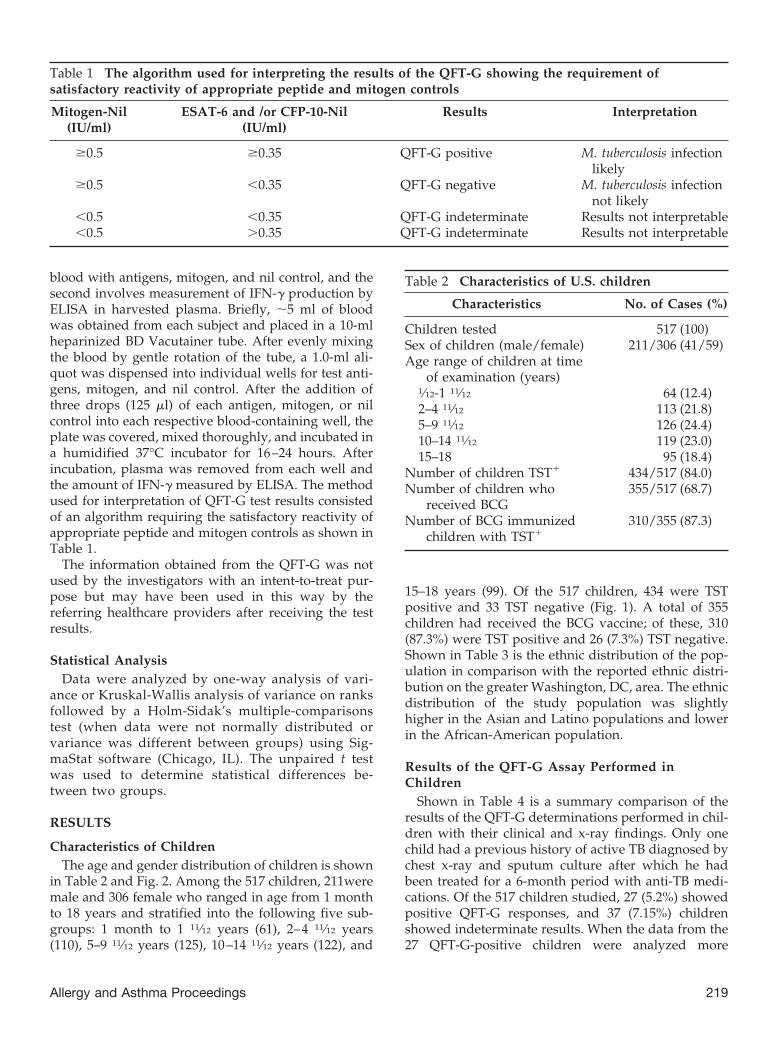
Table 1 The algorithm used for interpreting the results of the QFT-G showing the requirement ofsatisfactory reactivity of appropriate peptide and mitogen controls
Mitogen-Nil(IU/ml)
ESAT-6 and /or CFP-10-Nil(IU/ml)
Results Interpretation
�0.5 �0.35 QFT-G positive M. tuberculosis infectionlikely
�0.5 �0.35 QFT-G negative M. tuberculosis infectionnot likely
�0.5 �0.35 QFT-G indeterminate Results not interpretable�0.5 �0.35 QFT-G indeterminate Results not interpretable
Table 2 Characteristics of U.S. children
Characteristics No. of Cases (%)
Children tested 517 (100)Sex of children (male/female) 211/306 (41/59)Age range of children at time
of examination (years)1⁄12-1 11⁄12 64 (12.4)2–4 11⁄12 113 (21.8)5–9 11⁄12 126 (24.4)10–14 11⁄12 119 (23.0)15–18 95 (18.4)
Number of children TST� 434/517 (84.0)Number of children who
received BCG355/517 (68.7)
Number of BCG immunizedchildren with TST�
310/355 (87.3)
Allergy and Asthma Proceedings 219

closely with respect to BCG immunization and TBexposure history, several comparisons could be made(Table 5). The ages of the 27 QFT-G-positive childrenranged from 9 months to 18 years. It can be seen that 20of the 27 subjects had received previous BCG immuni-zation, 2 gave a history of previous exposure to TB, and1 had positive chest x-ray.
Results of Studies of Age-Related Capacity ofIFN-� Production
Because one of the major goals of this research wasto study variations in age-related immune capacityof the child to determine the validity of the use of theQFT-G assay in the younger age group particularlyin children �5 years of age, the capacity of IFN-�
Figure 2. Age and gender distribution of children.
Table 3 Ethnic distribution of the population
Ethnicity No. of participants % % Ethnic Distribution in Greater Washington,DC, Area
Whites* 301 58.2 51.7Asians 99 19.1 8.4Latinos# 77 14.9 11.6African-American 40 7.7 26.3Total 517 100 100
*This compartment includes persons born in the Middle East (traditional countries known as Middle East and ex-USSRcountries in Asia). Pakistan and Afghanistan are included in Asia. Maghreb (north of Africa) countries are not includedbecause there were no participants from this region.#Latinos includes Hispanics and Latinos from Brazil.
Table 4 Comparison of the clinical and QFT findings in 517 U.S. children
No.Children
Agerange(year)
Clinical Hx QFT-G Test Results
Hx ofBCG
vaccine
Hx Rxfor TB
TBExp
TST� 27 QFT-G� 37Indeterminate
QFT-G
BCG� BCG� CG� BCG�
TB�
ExpTB�
ExpTB�
ExpTB�
Exp
517 1⁄12-18 355 1 15 434 2 18 0 7 20 17
Exp, Exposure; Hx, history; Rx, treated.
220 May–June 2012, Vol. 33, No. 3

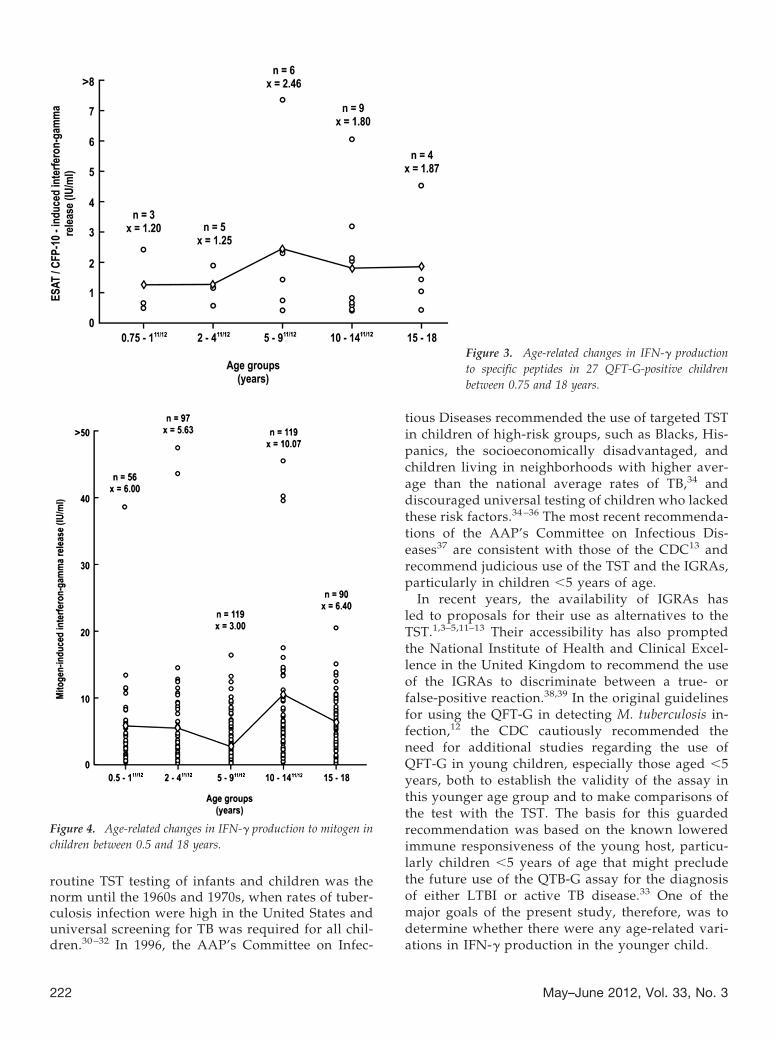
production was measured in various age groups ofchildren in response to specific TB peptides as wellas to mitogen. The results of these studies are shownin Figs. 3 and 4, respectively.
The IFN-� production in response to specific pep-tides in the 27 QFT-G-positive children could beevaluated only in children in the 9 months to 18years range, the group where the only positiveQFT-G responses were observed (Fig. 3). Within thisage range, no statistically significant differences inmean values were observed between the varioussubgroups that comprised this age range (p � 0.776).In contrast, IFN-� production in response to mitogencould be measured in all 481 subjects within the agerange of 6 months to 18 years (Fig. 4). In this agerange, adequate IFN-� responses were seen, and theonly statistically significant difference in mean val-ues among the various subgroups was seen betweenthe 5–9 11⁄12 year and the 10 –14 11⁄12 year groups (p �0.05). In children �5 years of age, comparable mean
values of IFN-� production in response to mitogenwas found, i.e., 5.8 IU/ml.
Of particular importance was the finding of ade-quate IFN-� responses even in the youngest agegroups �5 years with mean values of 5.9 IU/ml inthe 0.5–2 11⁄12 years group and 5.6 IU/ml in the2– 4 11⁄12 years group.
DISCUSSIONThe use of the traditional TST for diagnosis of TB
poses a perplexing challenge for the healthcare pro-vider entrusted to the care of children due to its lowspecificity and its cross-reactivity with nontubercu-lous mycobacteria or previous BCG immunization.13
The ambiguity of TST findings is of particular con-cern to the allergist-immunologist who often in-cludes TB infection in the differential diagnosisof children suspected to have allergic disease possi-bly complicated by TB. Despite its shortcomings,
Table 5 Comparison of the BCG immunization status and TB exposure history in the 27 QFT-G-positivechildren
Subject Age (years) Clinical Hx
Hx of BCG vaccine Hx TB Rx TB Exp TST� X�ray�
1 5 � � � � �2 5 � � � � �3 6 � � � � �4 17 � � � � �5 13 6⁄12 � � � � �6 11 6⁄12 � � � � �7 15 � � � � �8 2 � � � � �9 4 3⁄12 � � � ND �
10 9⁄12 � � � ND ND11 1 � � � � �12 2 5⁄12 � � � � �13 1 � � � � �14 4 5⁄12 � � � � ND15 10 2⁄12 � � � � �16 12 3⁄12 � � � � �17 8 1⁄12 � � � � �18 3 3⁄12 � � � � �19 8 � � � � �20 14 4⁄12 � � � � ND21 15 6⁄12 � � � � �22 17 � � � � �23 12 6⁄12 � � � � ND24 10 4⁄12 � � � � �25 7 5⁄12 � � � � ND26 16 � � � � �27 12 � � � � �
Exp, Exposure; Hx, history; ND, not done; Rx, treated.
Allergy and Asthma Proceedings 221
routine TST testing of infants and children was thenorm until the 1960s and 1970s, when rates of tuber-culosis infection were high in the United States anduniversal screening for TB was required for all chil-dren.30 –32 In 1996, the AAP’s Committee on Infec-
tious Diseases recommended the use of targeted TSTin children of high-risk groups, such as Blacks, His-panics, the socioeconomically disadvantaged, andchildren living in neighborhoods with higher aver-age than the national average rates of TB,34 anddiscouraged universal testing of children who lackedthese risk factors.34 –36 The most recent recommenda-tions of the AAP’s Committee on Infectious Dis-eases37 are consistent with those of the CDC13 andrecommend judicious use of the TST and the IGRAs,particularly in children �5 years of age.
In recent years, the availability of IGRAs hasled to proposals for their use as alternatives to theTST.1,3–5,11–13 Their accessibility has also promptedthe National Institute of Health and Clinical Excel-lence in the United Kingdom to recommend the useof the IGRAs to discriminate between a true- orfalse-positive reaction.38,39 In the original guidelinesfor using the QFT-G in detecting M. tuberculosis in-fection,12 the CDC cautiously recommended theneed for additional studies regarding the use ofQFT-G in young children, especially those aged �5years, both to establish the validity of the assay inthis younger age group and to make comparisons ofthe test with the TST. The basis for this guardedrecommendation was based on the known loweredimmune responsiveness of the young host, particu-larly children �5 years of age that might precludethe future use of the QTB-G assay for the diagnosisof either LTBI or active TB disease.33 One of themajor goals of the present study, therefore, was todetermine whether there were any age-related vari-ations in IFN-� production in the younger child.
Figure 3. Age-related changes in IFN-� productionto specific peptides in 27 QFT-G-positive childrenbetween 0.75 and 18 years.
Figure 4. Age-related changes in IFN-� production to mitogen inchildren between 0.5 and 18 years.
222 May–June 2012, Vol. 33, No. 3
The results of the present study confirm and ex-tend published observations supporting the validityof QFT IFN release assays in two groups of children.In a group of 517 U.S. children referred by healthcareproviders and pediatricians, 434 (84%) were TSTpositive. Of these 434 TST-positive children, 25(5.8%) were found to be QFT-G positive and wouldbe considered LTBI; because the vast majority ofTST-positive reactive children, i.e. 434 (84%), ap-peared not to be infected with TB, they would nothave required antituberculous treatment. In the ab-sence of the availability of the IGRAs, therefore,these children would have been treated unnecessar-ily for LTBI. Of the 517 children, 355 were previouslyimmunized with BCG; of these 355 children, 310(87.3%) were TST positive, which included 18/27(66.7%) children who were QFT positive. Therefore,of the 27 QFT-G-positive responses, 7 were seen innon-BCG-immunized children and 20 in childrenwho had also received the BCG vaccine. The simul-taneous occurrence in the 18 TST-positive childrenwho had received BCG could have contributed to afurther diagnostic dilemma of whether their skinreactivity was due either to previous immunizationor TB infection, a problem that was resolved by theuse of the QFT-G. The finding that only 18 TST-positive children were actually TB infected andwould require antituberculous therapy obviated theunnecessary use of antituberculous medication inthe remaining 292 children with TST-positive re-sponses that were related to previous BCG immuni-zation. Moreover, the unnecessary use of TB medi-cation resulting from a lack of specificity of TST mayhave resulted in an increased risk of significant drugtoxicity that was also avoided by the use of theQFT-G.
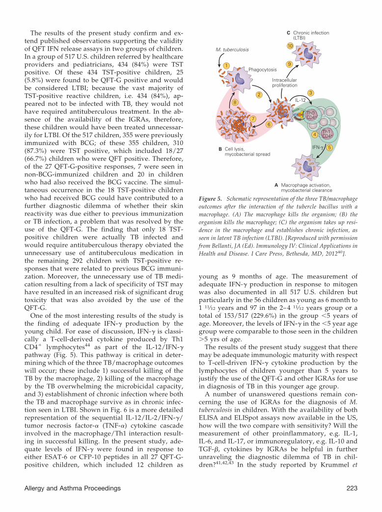
One of the most interesting results of the study isthe finding of adequate IFN-� production by theyoung child. For ease of discussion, IFN-� is classi-cally a T-cell-derived cytokine produced by Th1CD4� lymphocytes44 as part of the IL-12/IFN-�pathway (Fig. 5). This pathway is critical in deter-mining which of the three TB/macrophage outcomeswill occur; these include 1) successful killing of theTB by the macrophage, 2) killing of the macrophageby the TB overwhelming the microbicidal capacity,and 3) establishment of chronic infection where boththe TB and macrophage survive as in chronic infec-tion seen in LTBI. Shown in Fig. 6 is a more detailedrepresentation of the sequential IL-12/IL-2/IFN-�/tumor necrosis factor-� (TNF-�) cytokine cascadeinvolved in the macrophage/Th1 interaction result-ing in successful killing. In the present study, ade-quate levels of IFN-� were found in response toeither ESAT-6 or CFP-10 peptides in all 27 QFT-G-positive children, which included 12 children as
young as 9 months of age. The measurement ofadequate IFN-� production in response to mitogenwas also documented in all 517 U.S. children butparticularly in the 56 children as young as 6 month to1 11⁄12 years and 97 in the 2– 4 11⁄12 years group or atotal of 153/517 (229.6%) in the group �5 years ofage. Moreover, the levels of IFN-� in the �5 year agegroup were comparable to those seen in the children�5 yrs of age.
The results of the present study suggest that theremay be adequate immunologic maturity with respectto T-cell-driven IFN-� cytokine production by thelymphocytes of children younger than 5 years tojustify the use of the QFT-G and other IGRAs for usein diagnosis of TB in this younger age group.
A number of unanswered questions remain con-cerning the use of IGRAs for the diagnosis of M.tuberculosis in children. With the availability of bothELISA and ELISpot assays now available in the US,how will the two compare with sensitivity? Will themeasurement of other proinflammatory, e.g. IL-1,IL-6, and IL-17, or immunoregulatory, e.g. IL-10 andTGF-�, cytokines by IGRAs be helpful in furtherunraveling the diagnostic dilemma of TB in chil-dren?41,42,43 In the study reported by Krummel et
Figure 5. Schematic representation of the three TB/macrophageoutcomes after the interaction of the tubercle bacillus with amacrophage. (A) The macrophage kills the organism; (B) theorganism kills the macrophage; (C) the organism takes up resi-dence in the macrophage and establishes chronic infection, asseen in latent TB infection (LTBI). [Reproduced with permissionfrom Bellanti, JA (Ed). Immunology IV: Clinical Applications inHealth and Disease. I Care Press, Bethesda, MD, 201240].
Allergy and Asthma Proceedings 223

al.,41 a combination of a M. tuberculosis-specific IFN-�ELISpot with a M. tuberculosis-specific IL-2 ELISpotwas shown to significantly improve the identifica-tion of individuals with the highest risk of recent M.tuberculosis infection and offers a promising methodthat should be explored to target the treatment ofLTBI. Another promising diagnostic tool addressingthe need for additional assays to improve diagnosisof M. tuberculosis infection in children was reportedby Ruhwald et al.43 This study evaluated the perfor-mance of an IFN-�-inducible protein 10 (IP-10)- andIL-2-based test for the diagnosis of M. tuberculosisinfection in recently exposed children from Nigeria.After stimulation of lymphocytes in whole bloodpreparations with M. tuberculosis-specific antigensand mitogen, the high-risk children expressed sig-nificantly higher levels predominantly of IP-10 andlesser elevations of IL-2 than low-risk groups. Thesefindings suggest that IP-10 and possibly IL-2 couldbe alternative or adjunct markers to IFN-� in thediagnosis infection with M. tuberculosis.
Another practical application of IGRA tests is theiruse in screening patients before the use of corticoste-roids and the recently developed biologic responsemodifiers, e.g. anti-TNF-� agents known to exacerbateTB infection.44 The observation that anti-TNF-� ther-apy is associated with reactivation of LTBI has led to
clinical guidelines that recommend screening patientsfor previous evidence of TB infection before commenc-ing anti-TNF-� treatment.40 Of particular concern forthe allergist-immunologist is the known modifying ef-fect of corticosteroids commonly used in the treatmentof allergic disease on the TST, resulting in misleadingfalse-negative results, thus exposing undiagnosed pa-tients to the risk of TB reactivation when treated withthese agents.
In conclusion, the results of the present study arein keeping with those of other studies and show thatthe QFT-G and QFT-GIT offer distinct advantagesover the TST. They also provide new evidence thatthe young infant �5 years of age has sufficient im-munologic capacity for IFN-� production to allowthe test to be performed in younger populations. Itwould seem prudent to follow the National Instituteof Health and Clinical Excellence guidelines38,39 andperform the TST as an initial screening tool and carryout IFN-� testing in patients with positive TST re-sults.
REFERENCES1. Stout JE, and Menzies D. Predicting tuberculosis: does the IGRA
tell the tale? Am J Respir Crit Care Med 177:1055–1057, 2008.
Figure 6. Schematic representation of the IL-12/IFN-� pathway illustrating various activation pathways associated with the killingof intracellular bacterial organisms such as M. tuberculosis (TB) and Salmonella. After the uptake of the TB by the macrophage (1)into a phagosome (2), the macrophage is stimulated to produce IL-12 (3). The binding of IL-12 to its IL-12R on a Th CD4� cell (4)induces the synthesis of IL-2 (5), and after autocrine binding of IL-2 to its receptor IL-2R (6) leads to the synthesis of IFN-� (7). Thebinding of IFN-� to its receptor IFN�R (8) activates both the signal transducer and activator of transcription 1 (STAT1) and nuclearfactor (NF)-�B pathways (9) to kill the TB and to further enhance IL-12 production; the IFN- �-activated macrophage (10) and Toll-likereceptor 4 (TLR4) activation by the TB (11) induce TNF-� production that, after binding to its receptor (12), leads to furtherenhancement of intracellular killing of the TB (13). [Reproduced with permission from Bellanti, JA (Ed). Immunology IV: ClinicalApplications in Health and Disease. I Care Press, Bethesda, MD, 201240].
224 May–June 2012, Vol. 33, No. 3
2. Starke JR. Interferon-gamma release assays for diagnosis oftuberculosis infection in children. Pediatr Infect Dis J 25:941–942, 2006.
3. Rangaka MX, Wilkinson KA, Glynn JR, et al. Predictive value ofinterferon-� release assays for incident active tuberculosis: asystematic review and meta-analysis. Lancet Infect Dis 12:45–55, 2012.
4. Pavic I, Topic RZ, Raos M, et al. Interferon-� release assay forthe diagnosis of latent tuberculosis in children younger than 5years of age. Pediatr Infect Dis J 30:866–870, 2011.
5. Machingaidze S, Wiysonge CS, Gonzalez-Angulo Y, et al. Theutility of an interferon gamma release assay for diagnosis oflatent tuberculosis infection and disease in children: a system-atic review and meta-analysis. Pediatr Infect Dis J 30:694–700,2011.
6. Ling DI, Zwerling AA, Steingart KR, et al. Immune-based di-agnostics for TB in children: what is the evidence? PaediatrRespir Rev 12:9–15, 2011.
7. Zhang S, Shao L, Mo L, et al. Evaluation of gamma interferonrelease assays using Mycobacterium tuberculosis antigens for di-agnosis of latent and active tuberculosis in Mycobacterium bovisBCG-vaccinated populations. Clin Vaccine Immunol 17:1985–1990, 2010.
8. Lalvani A, and Pareek M. Interferon gamma release assays:principles and practice. Enferm Infecc Microbiol Clin 28:245–252, 2010.
9. Connell TG, Ritz N, Paxton GA, et al. A three-way comparisonof tuberculin skin testing, QuantiFERON-TB gold and T-SPOT.TB in children. PLoS One 3:e2624, 2008.
10. Dewan PK, Grinsdale J, Liska S, et al. Feasibility, acceptability,and cost of tuberculosis testing by whole-blood interferon-gamma assay. BMC Infect Dis 6:47, 2006.
11. Cellestis Web page, QuantiFERON-TB Gold In-Tube Test Manu-facturer’s instructions. Available at: www.cellestis.com; accessedJune 11, 2007.
12. Mazurek GH, Jereb J, Lobue P, et al. Guidelines for using theQuantiFERON-TB Gold test for detecting Mycobacterium tuber-culosis infection, United States. MMWR Recomm Rep. 2005 Dec16;54(RR-15):49–55. Erratum in: MMWR Morb Mortal WklyRep. 54(50):1288, December 23, 2005.
13. Mazurek GH, Jereb J, Vernon A, et al. IGRA Expert Committee;Centers for Disease Control and Prevention (CDC). Updatedguidelines for using interferon gamma release assays to detectMycobacterium tuberculosis infection: United States, 2010.MMWR Recomm Rep. 59(RR-5):1–25, 2010.
14. T-SPOT.TB T-Spot.TB is a type of ELISPOT assay, manufac-tured by Oxford Immunotec Ltd. in the UK, that counts thenumber of effector T cells that produce gamma interferon stim-ulated by proteins produced by the bacteria that cause tuber-culosis. www.oxfordimmunotec.com.
15. Cruz AT, Geltemeyer AM, Starke JR, et al. Comparing thetuberculin skin test and T-SPOT. TB blood test in children.Pediatrics 127:e31–e8, 2011.
16. Diel R, Loddenkemper R, Meywald-Walter K, et al. Compara-tive performance of tuberculin skin test, QuantiFERON-TB-Gold In Tube, and T-Spot. TB test in contact investigations fortuberculosis. Chest 135:1010–1018, 2009.
17. Bua A, Molicotti P, Delogu G, et al. QuantiFERON TB Gold: anew method for latent tuberculosis infection. New Microbiol30:477–480, 2007.
18. Diel R, Loddenkemper R, Meywald-Walter K, et al. Predictivevalue of a whole blood IFN-� assay for the development ofactive tuberculosis disease after recent infection with Mycobac-terium tuberculosis. Am J Respir Crit Care Med 177:1164–1170,2008.
19. Mori T, Sakatani M, Yamagishi F, et al. Specific detection oftuberculosis infection: an interferon-gamma-based assay us-
ing new antigens. Am J Respir Crit Care Med 170:59 – 64,2004.
20. Kang YA, Lee HW, Yoon HI, et al. Discrepancy between thetuberculin skin test and the whole-blood interferon gammaassay for the diagnosis of latent tuberculosis infection in anintermediate tuberculosis-burden country. JAMA 293:2756–2761, 2005.
21. Brock I, Munk ME, Kok-Jensen A, et al. Performance of wholeblood IFN-� test for tuberculosis diagnosis based on PPD or thespecific antigens ESAT-6 and CFP-10. Int J Tuberc Lung Dis5:462–467, 2001.
22. Brock I, Weldingh K, Lillebaek T, et al. Comparison of tuber-culin skin test and new specific blood test in tuberculosis con-tacts. Am J Respir Crit Care Med 170:65–69, 2004.
23. Diel R, Ernst M, Döscher G, et al. Avoiding the effect of BCGvaccination in detecting Mycobacterium tuberculosis infectionwith a blood test. Eur Respir J28:6–23, 2006.
24. Pareek M, Watson JP, Ormerod LP, et al. Screening of immi-grants in the UK for imported latent tuberculosis: a multicentrecohort study and cost-effectiveness analysis. Lancet Infect Dis11:435–444, 2011.
25. Wang SH, Powell DA, Nagaraja HN, et al. Evaluation of amodified interferon-gamma release assay for the diagnosis oflatent tuberculosis infection in adult and paediatric populationsthat enables delayed processing. Scand J Infect Dis 42:845–850,2010.
26. Lienhardt C, Fielding K, Hane AA, et al. Evaluation of theprognostic value of IFN-gamma release assay and tuberculinskin test in household contacts of infectious tuberculosis casesin Senegal. PLoS One 5:e10508, 2010.
27. Dyrhol-Riise AM, Gran G, Wentzel-Larsen T, et al. Diagnosisand follow-up of treatment of latent tuberculosis; the utilityof the QuantiFERON-TB Gold in-tube assay in outpatientsfrom a tuberculosis low-endemic country. BMC Infect Dis10:57, 2010.
28. Machado A Jr., Emodi K, Takenami I, et al. Analysis ofdiscordance between the tuberculin skin test and the inter-feron-gamma release assay. Int J Tuberc Lung Dis 13:446 –453, 2009.
29. Nienhaus A, Schablon A, and Diel R. Interferon-gamma releaseassay for the diagnosis of latent TB infection: analysis of dis-cordant results, when compared to the tuberculin skin test.PLoS One 3:e2665, 2008.
30. Powell RD III, Whitworth WC, Bernardo J, et al. Unusual inter-feron gamma measurements with QuanitFERON-TB Gold andQuantiFERON-TB In-Tube tests. PLoS ONE 6:1–6, 2011.
31. Centers for Disease Control. Tuberculosis in the UnitedStates: 1987. Atlanta, GA: Centers for Disease Control, USDepartment of Health and Human Services publication(CDC) 89-8322, 1989.
32. Diagnostic Standards and Classification of Tuberculosis inAdults and Children. This official statement of the AmericanThoracic Society and the Centers for Disease Control andPrevention was adopted by the ATS Board of Directors, July1999. This statement was endorsed by the Council of theInfectious Disease Society of America, September 1999. Am JRespir Crit Care Med 161(4 Pt 1):1376 –1395, 2000.
33. American Academy of Pediatrics, Committee on Infectious Dis-eases. Update on tuberculosis skin testing of children. Pediatrics97:282–284, 1996.
34. Starke JR, Jacobs RF, and Jereb J. Resurgence of tuberculosis inchildren. J Pediatr 120:839–855, 1992.
35. Cantwell MF, Snider DE Jr, Cauthen GM, et al. Epidemiology oftuberculosis in the United States, 1985 through 1992. JAMA272:535–539, 1994.
36. American Academy of Pediatrics, Committee on Infectious Dis-eases. Tuberculosis. In Report of the Committee on Infectious
Allergy and Asthma Proceedings 225

Diseases, 1991. 22nd ed. Elk Grove Village, IL: American Acad-emy of Pediatrics, 492–493, 1991.
37. American Academy of Pediatrics. Tuberculosis. In Red book:2009 report of the Committee on Infectious Disease, 28th ed.Pickering LK, Baker CJ, Kimberlin DW, and Long SS, (Eds).Elk Grove Village, IL: American Academy of Pediatrics, 680 –701, 2009.
38. NICE Guidelines. CG33 Tuberculosis: Full Guideline. 22 March 2006.39. Taylor RE, Cant AJ, and Clark JE. Potential effect of NICE
tuberculosis guidelines on paediatric tuberculosis screening.Arch Dis Child 93:200–203, 2008.
40. Holland SM, Freeman AF, and Bellanti JA. Immunity to bacte-ria. In Immunology IV. Clinical applications in health and dis-ease. Bellanti JA (Ed). Bethesda: I Care Press, 429–457, 2012.
41. Krummel B, Strassburg A, Ernst M, et al. Potential role for IL-2ELISpot in differentiating recent and remote infection in tuber-culosis contact tracing. PLoS One 5:e11670, 2010.
42. Lighter J, Rigaud M, Huie M, et al. Chemokine IP-10: an adjunctmarker for latent tuberculosis infection in children. Int J TubercLung Dis 1:731–736, 2009.
43. Ruhwald M, Petersen J, Kofoed K, et al. Improving T-cell assaysfor the diagnosis of latent TB infection: potential of a diagnostictest based on IP-10. PLoS One 3:e2858, 2008.
44. Saag KG, Teng GG, Patkar NM, et al. American College ofRheumatology. America College of Rheumatology 2008 recom-mendations for the use of nonbiologic and biologic disease-modifying antirheumatic drugs in rheumatoid arthritis. Arthri-tis Rheum 59:762–784, 2008. e
226 May–June 2012, Vol. 33, No. 3