rare disease - bmj case reports - bmj...
TRANSCRIPT
CASE REPORT
Two typical cases of pseudoankylosis of the jaw:same treatment, different outcomeFabiana Allevi,1 Valeria Battista,1 Laura Moneghini,2 Federico Biglioli1
1Department of Maxilllo FacialSurgery, San Paolo Hospital,University of Milan, Milan, Italy2Department of SurgicalPathology, San Paolo Hospital,University of Milan, Milan, Italy
Correspondence toDr Fabiana Allevi,[email protected]
Accepted 19 July 2015
To cite: Allevi F, Battista V,Moneghini L, et al. BMJCase Rep Published online:[please include Day MonthYear] doi:10.1136/bcr-2015-210099
SUMMARYPseudoankylosis of the temporomandibular joint is arare, extra-articular form of ankylosis of the jaw. It ischaracterised by limited mandibular movement caused byan extrinsic condition of the joint leading to fusionbetween the coronoid process and temporal, zygomaticor maxillary bone. Pseudoankylosis is less frequent thanthe intracapsular form. Extracapsular ankylosis can becongenital or acquired; approximately 70% of cases areassociated with trauma. A CT scan is usually requestedto achieve a diagnosis. CT can detect bony fusion, thusdifferentiating pseudoankylosis from true ankylosis. Oncesymptomatic bone ankylosis is diagnosed, surgery withpostoperative physiotherapy is the recommendedtreatment. The ankylotic bone is removed together withthe coronoid process and the mouth is forced openunder general anaesthesia. Two cases of post-traumaticpseudoankylosis of the jaw treated with bilateralcoronoidectomy and postoperative physiotherapy aredescribed.
BACKGROUNDPseudoankylosis of the temporomandibular joint(TMJ) is a rare condition that causes inability toopen the mouth. It is characterised by mandibularhypomobility caused by an extrinsic condition ofthe joint that results in fusion between the coron-oid process and temporal,1 zygomatic2 3 or maxil-lary bone.4 5
Pseudoankylosis of the TMJ is less frequent thanintracapsular ankylosis. Exact figures for the inci-dence of ankyloses and pseudoankyloses of theTMJ in the general population are not available inthe literature. It is even more difficult to comparethe incidence of ankyloses and pseudoankylosesgiven the lack of common definitions in most arti-cles.6 Nevertheless, it is known that extracapsularankylosis can be congenital or acquired and compli-cates up to 0.6% of undiagnosed zygomatic frac-tures.7 Approximately 88% of cases ofpseudoankylosis are associated with a history oftrauma.8 Traumas, with or without fracture, includ-ing surgery, can induce the formation of fibrous/scar adhesion in the temporal and massetermuscles.9 Diagnosis is based on clinical and radio-logical evaluations.Pseudoankylosis is clinically characterised by
limited mandibular movement with no pain.Orthopantomography can identify the presence ofelongated coronoids, condyle tumours and TMJankylosis. However, a CT scan is usually requestedas it can detect bony fusion, thus differentiatingpseudoankylosis from true ankylosis. Because CT
scans do not provide enough information aboutsoft tissue disorders, MRI may be indicated whenfibrous tissue formation is suspected.10 Once diag-nosed, symptomatic bone ankylosis can be treatedwith surgery (usually by coronoidectomy) or con-servatively (with forced mouth opening).10–12
A case of coronoid-zygomatic arch ankylosis anda case of coronoid-temporal bone ankylosis arereported.
CASE PRESENTATIONCase AA 32-year-old woman was referred to our unit forlimited jaw movement. The patient had beeninvolved in a road trauma accident at 22 years ofage, resulting in an ascending aortic rupture, frac-tures of the left forearm, humerus, scapula andright fibula, right pneumothorax, and spleen andliver contusions. She underwent multiple surgicalprocedures while she was in a drug-induced coma.In addition, she had displaced the left angle andright condylar and coronoid processes of the man-dible, which were treated with open reduction andinternal fixation with one plate on the angle andrigid intermaxillary fixation through arch bars for23 days. No physiotherapy was possible during thecoma.Ten years later, the patient requested a consult-
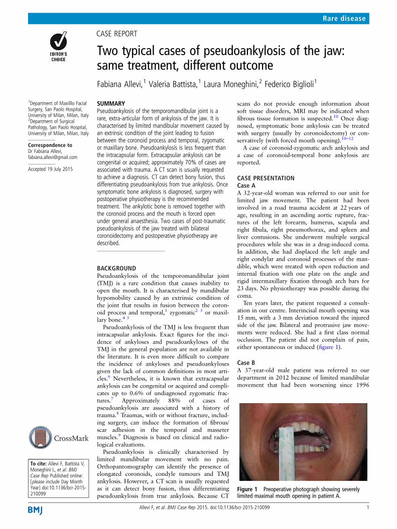
ation in our centre. Interincisal mouth opening was15 mm, with a 3 mm deviation toward the injuredside of the jaw. Bilateral and protrusive jaw move-ments were reduced. She had a first class normalocclusion. The patient did not complain of pain,either spontaneous or induced (figure 1).
Case BA 37-year-old male patient was referred to ourdepartment in 2012 because of limited mandibularmovement that had been worsening since 1996
Figure 1 Preoperative photograph showing severelylimited maximal mouth opening in patient A.
Allevi F, et al. BMJ Case Rep 2015. doi:10.1136/bcr-2015-210099 1
Rare disease
after a craniofacial trauma. After the event, he demonstratedoedema of the upper and middle third of the right side of hisface. No facial fractures were identified. Because of the pain dueto the trauma, the patient had limited ability to open his mouthimmediately after the traumatic event.
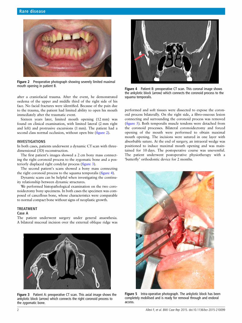
Sixteen years later, limited mouth opening (12 mm) wasfound on clinical examination, with limited lateral (2 mm rightand left) and protrusive excursions (1 mm). The patient had asecond class normal occlusion, without open bite (figure 2).
INVESTIGATIONSIn both cases, patients underwent a dynamic CT scan with three-dimensional (3D) reconstruction.
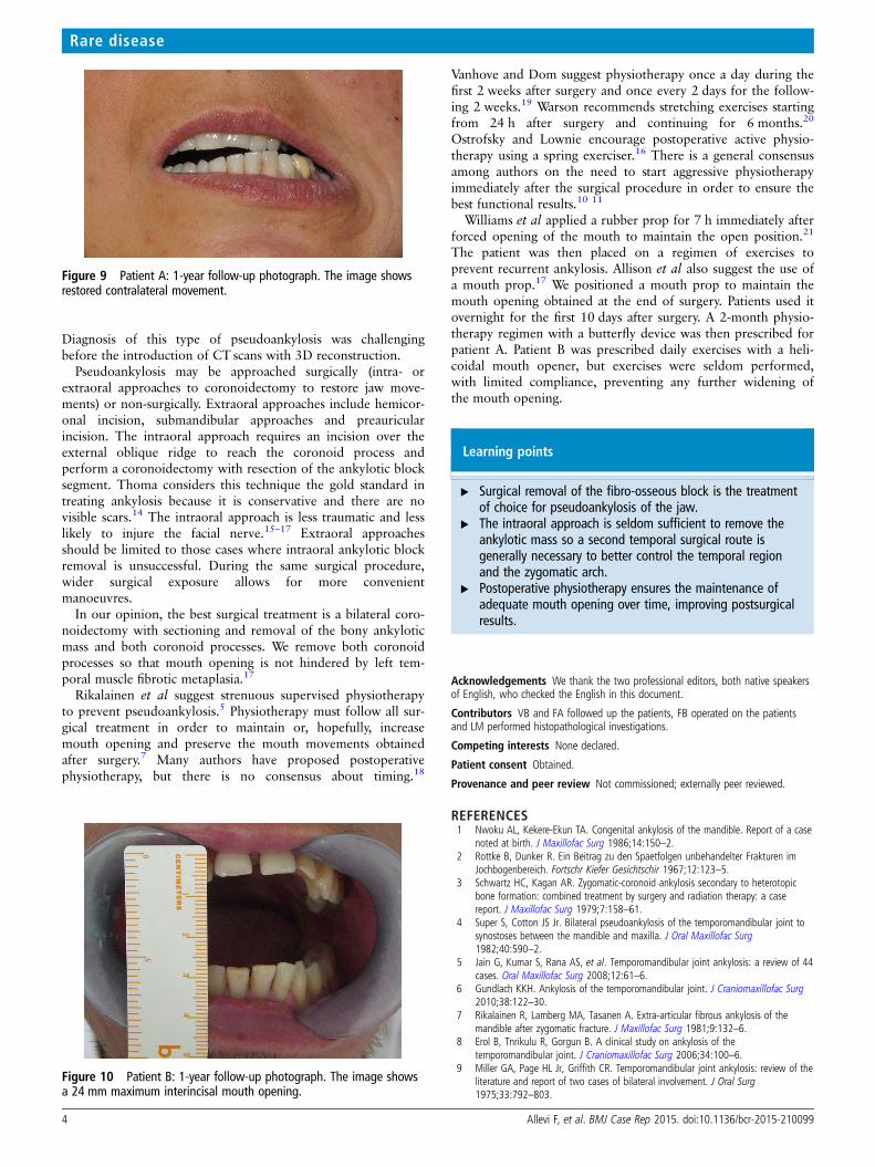
The first patient’s images showed a 2 cm bony mass connect-ing the right coronoid process to the zygomatic bone and a pos-teriorly displaced right condylar process (figure 3).
The second patient’s scans showed a bony mass connectingthe right coronoid process to the squama temporalis (figure 4).
Dynamic scans can be helpful when investigating the continu-ity relationship between dynamic structures.
We performed histopathological examination on the two coro-noidectomy bony specimens. In both cases the specimen was com-posed of cancellous bone, whose characteristics were comparableto normal compact bone without signs of neoplastic growth.
TREATMENTCase AThe patient underwent surgery under general anaesthesia.A bilateral mucosal incision over the external oblique ridge was
performed and soft tissues were dissected to expose the coron-oid process bilaterally. On the right side, a fibro-osseous lesionconnecting and surrounding the coronoid process was removed(figure 5). Both temporalis muscle tendons were detached fromthe coronoid processes. Bilateral coronoidectomy and forcedopening of the mouth were performed to obtain maximalmouth opening. The incisions were sutured in one layer withabsorbable suture. At the end of surgery, an intraoral wedge waspositioned to induce maximal mouth opening and was main-tained for 10 days. The postoperative course was uneventful.The patient underwent postoperative physiotherapy with a‘butterfly’ orthodontic device for 2 months.
Figure 2 Preoperative photograph showing severely limited maximalmouth opening in patient B.
Figure 3 Patient A: preoperative CT scan. This axial image shows theankylotic block (arrow) which connects the right coronoid process tothe zygomatic bone.
Figure 4 Patient B: preoperative CT scan. This coronal image showsthe ankylotic block (arrow) which connects the coronoid process to thesquama temporalis.
Figure 5 Intra-operative photograph. The ankylotic block has beencompletely mobilised and is ready for removal through and endoralaccess.
2 Allevi F, et al. BMJ Case Rep 2015. doi:10.1136/bcr-2015-210099
Rare disease

Case BThe patient underwent surgery under general anaesthesia. Anintraoral incision over the external right oblique ridge was per-formed. Another coronal incision was performed to expose thecoronoid process and the bony block. A right zygomatic osteot-omy (between the coronoid process and the squama temporalis)was performed to excise the ankylotic mass (figure 6). A leftintraoral coronoidectomy was performed through mucosal accessover the oblique external ridge. At the end of surgery, the mouthwas forced open to a 46 mm interincisal distance (figure 7). The
intraoral incisions were sutured with one-layered relapsablesuture, while the cutaneous incision was sutured with a two-layered relapsable–non-relapsable suture. An intraoral wedge wasplaced to maintain mouth opening for the first week aftersurgery. The postoperative course was uneventful.
The patient underwent postoperative physiotherapy with ahelicoidal mouth opener for 3 months.
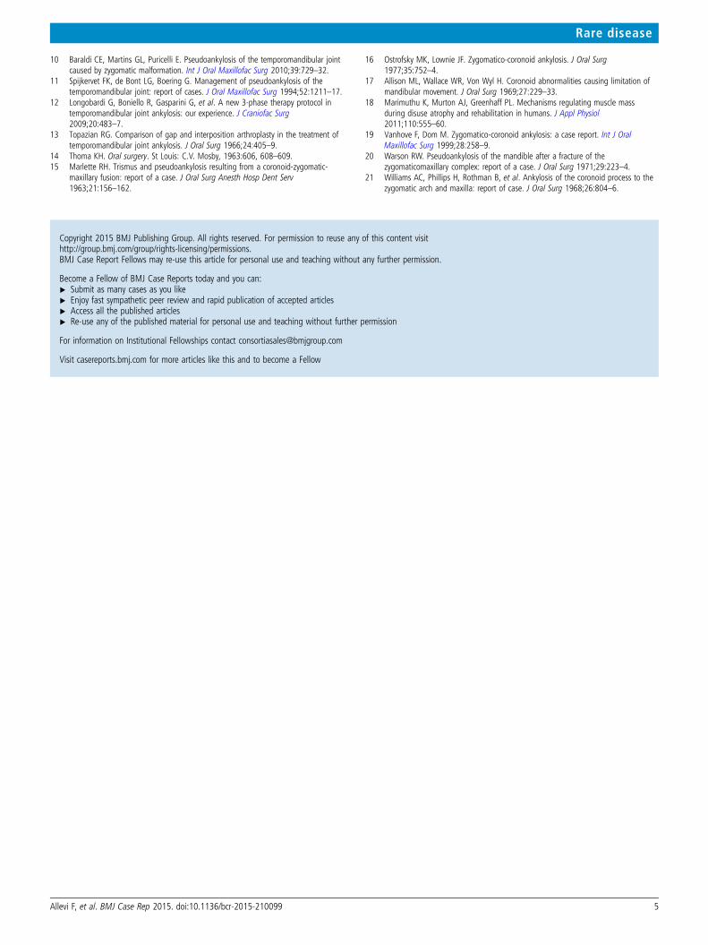
OUTCOME AND FOLLOW-UPCase ATwelve months after surgery, a 3D CT scan and orthopantomo-graphy confirmed the absence of relapse. Physical examinationshowed a 32 mm interincisal mouth opening with no lateraldeviation. Protrusive and lateral movements were restored(10 mm right and left, 7 mm of protrusion) (figures 8 and 9).
Case BSix months after surgery, physical examination showed a 34 mminterincisal mouth opening. Protrusive and lateral movementswere restored (9 mm right and left, 6 mm of protrusion). Thepatient was instructed to exercise five times a day with themouth opener to maintain a wide interincisal distance. Lowpatient compliance coupled with a lack of regular mouth exer-cise explained the reduction in the interincisal opening (24 mm)12 months after surgery (figure 10). A postoperative CT scanand orthopantomography showed no relapse of ankylosis.
DISCUSSIONThere are different types of false ankylosis. For example, thecoronoid process may fuse with the temporal bone, zygomaticarch or maxillary bone. Multiple causes of extracapsular anky-losis have been reported in the literature, including the develop-ment of intracapsular ankylosis anteriorly joining the sigmoidnotch to the zygomatic arch and skull base, trauma, infectionand iatrogenic causes.13
Tissue between bones can be fibrous3 or osseous, with identi-cal signs and symptoms but different pathogeneses.Haematomas caused by trauma or arising during correctivesurgery may develop in fibrous tissue. This tissue binds the cor-onoid bones to the bony structure surrounding the infratem-poral fossa. The temporalis and masseteric muscles are liable tocontusion. We assume that development of ankylosis is agradual process. Initially, fibrous tissue restricts mandibularmovement, and is soon replaced by fibro-osseous tissue.7 Theankylosis can be treated early with forced mouth opening.3–5 7
Figure 8 Patient A: 1-year follow-up photograph. The image shows a32 mm maximum interincisal mouth opening with no lateral deviation.
Figure 6 Histopathological examination showed that the specimenwas composed of cancellous bone, whose characteristics werecomparable to normal compact bone without signs of neoplasticgrowth.
Figure 7 Patient B: intraoperative photograph. The photographdepicts a 46 mm forced maximal interincisal opening at the end ofsurgery.
Allevi F, et al. BMJ Case Rep 2015. doi:10.1136/bcr-2015-210099 3
Rare disease
Diagnosis of this type of pseudoankylosis was challengingbefore the introduction of CT scans with 3D reconstruction.
Pseudoankylosis may be approached surgically (intra- orextraoral approaches to coronoidectomy to restore jaw move-ments) or non-surgically. Extraoral approaches include hemicor-onal incision, submandibular approaches and preauricularincision. The intraoral approach requires an incision over theexternal oblique ridge to reach the coronoid process andperform a coronoidectomy with resection of the ankylotic blocksegment. Thoma considers this technique the gold standard intreating ankylosis because it is conservative and there are novisible scars.14 The intraoral approach is less traumatic and lesslikely to injure the facial nerve.15–17 Extraoral approachesshould be limited to those cases where intraoral ankylotic blockremoval is unsuccessful. During the same surgical procedure,wider surgical exposure allows for more convenientmanoeuvres.
In our opinion, the best surgical treatment is a bilateral coro-noidectomy with sectioning and removal of the bony ankyloticmass and both coronoid processes. We remove both coronoidprocesses so that mouth opening is not hindered by left tem-poral muscle fibrotic metaplasia.17
Rikalainen et al suggest strenuous supervised physiotherapyto prevent pseudoankylosis.5 Physiotherapy must follow all sur-gical treatment in order to maintain or, hopefully, increasemouth opening and preserve the mouth movements obtainedafter surgery.7 Many authors have proposed postoperativephysiotherapy, but there is no consensus about timing.18
Vanhove and Dom suggest physiotherapy once a day during thefirst 2 weeks after surgery and once every 2 days for the follow-ing 2 weeks.19 Warson recommends stretching exercises startingfrom 24 h after surgery and continuing for 6 months.20
Ostrofsky and Lownie encourage postoperative active physio-therapy using a spring exerciser.16 There is a general consensusamong authors on the need to start aggressive physiotherapyimmediately after the surgical procedure in order to ensure thebest functional results.10 11
Williams et al applied a rubber prop for 7 h immediately afterforced opening of the mouth to maintain the open position.21
The patient was then placed on a regimen of exercises toprevent recurrent ankylosis. Allison et al also suggest the use ofa mouth prop.17 We positioned a mouth prop to maintain themouth opening obtained at the end of surgery. Patients used itovernight for the first 10 days after surgery. A 2-month physio-therapy regimen with a butterfly device was then prescribed forpatient A. Patient B was prescribed daily exercises with a heli-coidal mouth opener, but exercises were seldom performed,with limited compliance, preventing any further widening ofthe mouth opening.
Learning points
▸ Surgical removal of the fibro-osseous block is the treatmentof choice for pseudoankylosis of the jaw.
▸ The intraoral approach is seldom sufficient to remove theankylotic mass so a second temporal surgical route isgenerally necessary to better control the temporal regionand the zygomatic arch.
▸ Postoperative physiotherapy ensures the maintenance ofadequate mouth opening over time, improving postsurgicalresults.
Acknowledgements We thank the two professional editors, both native speakersof English, who checked the English in this document.
Contributors VB and FA followed up the patients, FB operated on the patientsand LM performed histopathological investigations.
Competing interests None declared.
Patient consent Obtained.
Provenance and peer review Not commissioned; externally peer reviewed.
REFERENCES1 Nwoku AL, Kekere-Ekun TA. Congenital ankylosis of the mandible. Report of a case
noted at birth. J Maxillofac Surg 1986;14:150–2.2 Rottke B, Dunker R. Ein Beitrag zu den Spaetfolgen unbehandelter Frakturen im
Jochbogenbereich. Fortschr Kiefer Gesichtschir 1967;12:123–5.3 Schwartz HC, Kagan AR. Zygomatic-coronoid ankylosis secondary to heterotopic
bone formation: combined treatment by surgery and radiation therapy: a casereport. J Maxillofac Surg 1979;7:158–61.
4 Super S, Cotton JS Jr. Bilateral pseudoankylosis of the temporomandibular joint tosynostoses between the mandible and maxilla. J Oral Maxillofac Surg1982;40:590–2.
5 Jain G, Kumar S, Rana AS, et al. Temporomandibular joint ankylosis: a review of 44cases. Oral Maxillofac Surg 2008;12:61–6.
6 Gundlach KKH. Ankylosis of the temporomandibular joint. J Craniomaxillofac Surg2010;38:122–30.
7 Rikalainen R, Lamberg MA, Tasanen A. Extra-articular fibrous ankylosis of themandible after zygomatic fracture. J Maxillofac Surg 1981;9:132–6.
8 Erol B, Tnrikulu R, Gorgun B. A clinical study on ankylosis of thetemporomandibular joint. J Craniomaxillofac Surg 2006;34:100–6.
9 Miller GA, Page HL Jr, Griffith CR. Temporomandibular joint ankylosis: review of theliterature and report of two cases of bilateral involvement. J Oral Surg1975;33:792–803.
Figure 9 Patient A: 1-year follow-up photograph. The image showsrestored contralateral movement.
Figure 10 Patient B: 1-year follow-up photograph. The image showsa 24 mm maximum interincisal mouth opening.
4 Allevi F, et al. BMJ Case Rep 2015. doi:10.1136/bcr-2015-210099
Rare disease
10 Baraldi CE, Martins GL, Puricelli E. Pseudoankylosis of the temporomandibular jointcaused by zygomatic malformation. Int J Oral Maxillofac Surg 2010;39:729–32.
11 Spijkervet FK, de Bont LG, Boering G. Management of pseudoankylosis of thetemporomandibular joint: report of cases. J Oral Maxillofac Surg 1994;52:1211–17.
12 Longobardi G, Boniello R, Gasparini G, et al. A new 3-phase therapy protocol intemporomandibular joint ankylosis: our experience. J Craniofac Surg2009;20:483–7.
13 Topazian RG. Comparison of gap and interposition arthroplasty in the treatment oftemporomandibular joint ankylosis. J Oral Surg 1966;24:405–9.
14 Thoma KH. Oral surgery. St Louis: C.V. Mosby, 1963:606, 608–609.15 Marlette RH. Trismus and pseudoankylosis resulting from a coronoid-zygomatic-
maxillary fusion: report of a case. J Oral Surg Anesth Hosp Dent Serv1963;21:156–162.
16 Ostrofsky MK, Lownie JF. Zygomatico-coronoid ankylosis. J Oral Surg1977;35:752–4.
17 Allison ML, Wallace WR, Von Wyl H. Coronoid abnormalities causing limitation ofmandibular movement. J Oral Surg 1969;27:229–33.
18 Marimuthu K, Murton AJ, Greenhaff PL. Mechanisms regulating muscle massduring disuse atrophy and rehabilitation in humans. J Appl Physiol2011;110:555–60.
19 Vanhove F, Dom M. Zygomatico-coronoid ankylosis: a case report. Int J OralMaxillofac Surg 1999;28:258–9.
20 Warson RW. Pseudoankylosis of the mandible after a fracture of thezygomaticomaxillary complex: report of a case. J Oral Surg 1971;29:223–4.
21 Williams AC, Phillips H, Rothman B, et al. Ankylosis of the coronoid process to thezygomatic arch and maxilla: report of case. J Oral Surg 1968;26:804–6.
Copyright 2015 BMJ Publishing Group. All rights reserved. For permission to reuse any of this content visithttp://group.bmj.com/group/rights-licensing/permissions.BMJ Case Report Fellows may re-use this article for personal use and teaching without any further permission.
Become a Fellow of BMJ Case Reports today and you can:▸ Submit as many cases as you like▸ Enjoy fast sympathetic peer review and rapid publication of accepted articles▸ Access all the published articles▸ Re-use any of the published material for personal use and teaching without further permission
For information on Institutional Fellowships contact [email protected]
Visit casereports.bmj.com for more articles like this and to become a Fellow
Allevi F, et al. BMJ Case Rep 2015. doi:10.1136/bcr-2015-210099 5
Rare disease