rationale, study design, and analysis plan of the lung
TRANSCRIPT

HAL Id: hal-01919201https://hal.archives-ouvertes.fr/hal-01919201
Submitted on 12 Nov 2018
HAL is a multi-disciplinary open accessarchive for the deposit and dissemination of sci-entific research documents, whether they are pub-lished or not. The documents may come fromteaching and research institutions in France orabroad, or from public or private research centers.
L’archive ouverte pluridisciplinaire HAL, estdestinée au dépôt et à la diffusion de documentsscientifiques de niveau recherche, publiés ou non,émanant des établissements d’enseignement et derecherche français ou étrangers, des laboratoirespublics ou privés.
Rationale, study design and analysis plan of the lungimaging morphology for ventilator settings in acuterespiratory distress syndrome study (LIVE study):
Study protocol for a randomised controlled trialMatthieu Jabaudon, Thomas Godet, Emmanuel Futier, Jean-Etienne Bazin,
Vincent Sapin, Laurence Roszyk, Bruno Pereira, Jean-Michel Constantin
To cite this version:Matthieu Jabaudon, Thomas Godet, Emmanuel Futier, Jean-Etienne Bazin, Vincent Sapin, et al..Rationale, study design and analysis plan of the lung imaging morphology for ventilator settingsin acute respiratory distress syndrome study (LIVE study): Study protocol for a randomised con-trolled trial. Anaesthesia Critical Care & Pain Medicine, Elsevier Masson, 2017, 36 (5), pp.301 - 306.�10.1016/j.accpm.2017.02.006�. �hal-01919201�

Accepted Manuscript
Title: Rationale, study design, and analysis plan of the LungImaging morphology for Ventilator settings in acuterespiratory distress syndrome study (LIVE study): studyprotocol for a randomised controlled trial
Authors: Matthieu Jabaudon Thomas Godet Emmanuel FutierJean-Etienne Bazin Vincent Sapin Laurence Roszyk BrunoPereira Jean-Michel Constantin, for AZUREA group
PII: S2352-5568(17)30030-9DOI: http://dx.doi.org/doi:10.1016/j.accpm.2017.02.006Reference: ACCPM 242
To appear in:
Received date: 2-2-2017Revised date: 5-2-2017Accepted date: 6-2-2017
Please cite this article as: Matthieu JabaudonThomas GodetEmmanuel FutierJean-Etienne BazinVincent SapinLaurence RoszykBruno PereiraJean-Michel ConstantinforAZUREA group, Rationale, study design, and analysis plan of the LungImaging morphology for Ventilator settings in acute respiratory distress syndromestudy (LIVE study): study protocol for a randomised controlled trial (2017),http://dx.doi.org/10.1016/j.accpm.2017.02.006
This is a PDF file of an unedited manuscript that has been accepted for publication.As a service to our customers we are providing this early version of the manuscript.The manuscript will undergo copyediting, typesetting, and review of the resulting proofbefore it is published in its final form. Please note that during the production processerrors may be discovered which could affect the content, and all legal disclaimers thatapply to the journal pertain.

Page 1 of 20
Accep
ted
Man
uscr
ipt
Rationale, study design, and analysis plan of the Lung Imaging morphology for Ventilator
settings in acute respiratory distress syndrome study (LIVE study): study protocol for a
randomised controlled trial.
Matthieu Jabaudon1,2
, Thomas Godet1,2
, Emmanuel Futier1,2
, Jean-Etienne Bazin1, Vincent
Sapin2,3
, Laurence Roszyk2,3
, Bruno Pereira4, Jean-Michel Constantin
1,2 for AZUREA group.
1Department of Perioperative Medicine, University Hospital of Clermont-Ferrand, Clermont-
Ferrand, France
2 Université Clermont Auvergne, CNRS, Inserm, GReD, F-63000 Clermont-Ferrand, France
3Department of Medical Biochemistry and Molecular Biology, University Hospital of
Clermont-Ferrand, Clermont-Ferrand, France
4 Biostatistics Unit, Department of Clinical Research and Innovation (DRCI), University
Hospital of Clermont-Ferrand, Clermont-Ferrand, France
Corresponding author: Pr JM Constantin, Department of Perioperative Medicine, University
Hospital of Clermont-Ferrand. [email protected]
Disclosure of interest
The authors declare that they have no conflicts of interest concerning this article.
Abstract: Different acute respiratory distress syndrome (ARDS) phenotypes may explain
controversial results in clinical trials. Lung-morphology is one of the ARDS-phenotypes and
physiological studies suggest different responses in terms of positive-end-expiratory-pressure
(PEEP) and recruitment-manoeuvres (RM) according to loss of aeration. To evaluate whether
Page 2 of 20
Accep
ted
Man
uscr
ipt
tailored ventilator regimens may impact ARDS outcomes, our group has designed a
randomised-clinical-trial of ventilator settings according to lung morphology in moderate-to-
severe ARDS (LIVE study).
Method: Patients will be enrolled within the first 12 hours of ARDS onset. In both groups,
volume-controlled ventilation with low tidal-volumes (Vt) will be used to target a plateau
pressure ≤ 30 cmH2O. In the control group, the PEEP level and inspired fraction of oxygen
(FiO2) will be set using the ARDSNet table; a Vt of 6 mL/kg of predicted body weight
(PBW) will be set, and prone position (PP) will be applied. In the intervention arm, the
ventilator will be set according to lung morphology (focal/non-focal) that will be assessed
according to CT-scan ± chest x-ray + lung echography. For focal ARDS patients, a Vt of
8 mL/kg PBW will be used along with low PEEP and PP. For non-focal ARDS patients, a Vt
of 6 mL/kg PBW will be used with RM and PEEP to reach a plateau pressure ≤ 30 cmH2O.
The primary outcome is all-cause 90-day mortality, and the secondary outcomes are: in-
hospital mortality, mortality at day 28, 60, 180 and 365; ventilator-free days at day 30, quality
of life at one year; ventilator-associated pneumonia rate; barotrauma; ICU and hospital length
of stay. This RCT is registered on Clinicaltrials.gov under identifier NCT02149589.

Page 3 of 20
Accep
ted
Man
uscr
ipt
Rationale, study design, and analysis plan for the Lung Imaging morphology for Ventilator
settings in acute respiratory distress syndrome study (LIVE study): study protocol for a
randomised controlled trial.
Introduction
Acute respiratory distress syndrome (ARDS) is a common problem in critically ill patients,
with a prevalence higher than 10% of intensive care unit (ICU) admissions [1]. ARDS is
associated with high in-hospital mortality (around 40%) and reduced quality of life among
survivors [1,2]. Optimal ventilator management for patients with ARDS remains uncertain.
Lower tidal volume (Vt) ventilation appears to be beneficial [3], but the optimal setting of Vt
in a given patient remains uncertain and challenging [4]. Optimal management of positive
end-expiratory pressure (PEEP) remains unclear. Higher levels of PEEP have only shown
equivocal benefits on outcomes in clinical trials [5,6]. Considering a prone position (PP), data
published in the last ten years remain controversial on the benefit of applying PP to all
patients with ARDS [7].
One hypothesis that may explain such controversial results is that behind the Berlin definition
[8], different patients with distinct forms (or phenotypes) of ARDS may exist. Numerous
studies have been published in this field, from the response to mechanical ventilation to the
concept of ARDS phenotypes [9-12]. Recent data suggest that lung morphology may be one
of the ARDS phenotypes [13,14].
Morphological characterization of CT-scan lung attenuation has contributed to the recognition
of subgroups of ARDS patients with distinct therapeutic responses (e.g., to PEEP, recruitment
manoeuvres (RM) …) [15] [16]. Non-focal ARDS, as defined by diffuse lung aeration loss, is
usually associated with significant lung recruitability, whereas focal ARDS is characterized
Page 4 of 20
Accep
ted
Man
uscr
ipt
by predominant aeration loss in lower lobes and dependent lung regions, with low
recruitability. Therefore, high PEEP levels and RMs seem more suitable for patients with
non-focal ARDS and may rather generate hyperinflation and haemodynamic instability in
patients with focal ARDS. Notwithstanding these physiological studies, whether or not such
ventilator setting strategies may influence the clinical outcomes of ARDS patients has never
been explored. To further explore the impact of tailored ventilation based on lung
morphology, we designed a randomised clinical trial of ventilator settings according to lung
morphology in moderate-to-severe ARDS. This paper describes the study protocol and
planned analyses for this clinical trial, registered on ClinicalTrials.gov under number
NCT02149589.
Methods
Objectives
Our primary objective is to determine if ventilator settings determined according to lung
morphology (focal or non-focal ARDS; the LIVE strategy) decrease the 90-day mortality rate
in patients with moderate-to-severe ARDS compared with conventional management
(ARDSNet strategy) [17]. The secondary objectives are to evaluate the effects of the LIVE
strategy, compared to the ARDSNet strategy, on the following outcomes: in-hospital
mortality, mortality at day 28, day 60, day 180 and day 365, ventilator-free days at day 30,
quality of life at one year, ventilator-associated pneumonia rate, barotrauma (pneumothorax,
pneumomediastinum), ICU and hospital lengths of stay.
Ethics and communication
Ethical details are in the main manuscript.
Page 5 of 20
Accep
ted
Man
uscr
ipt
The study was approved by an independent ethics committee (Comité de Protection des
Personnes Sud-Est VI, Clermont Ferrand, France; number: AU1099) and registered by the
French competent authority (Agence nationale de sécurité du médicament (ANSM); number:
2013-A01756-39).
Study design
Live is an investigator-initiated, patient-blinded, randomised, stratified, controlled,
multicentre trial with allocation and intention-to-treat analysis. Patients with ARDS will be
treated according to their lung morphology (LIVE strategy), as compared to an ARDSNet
strategy.
Study population
Inclusion criteria
- Age > 18 years
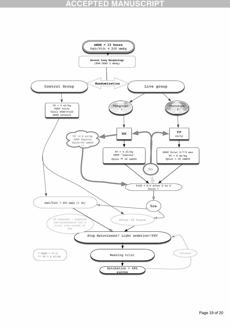
- Onset of ARDS < 12 hours
- PaO2/FiO2 ≤ 200 mmHg with PEEP ≥ 5 cmH2O
Non-inclusion criteria
- Mechanical ventilation for more than 7 consecutive days in the last 30 days.
- Previous history of ARDS in the last month
- Intracranial hypertension
- Morbid obesity with body mass index > 40 kg/m2
- Chronic respiratory diseases requiring long-term oxygen therapy
- Allogeneic bone marrow transplantation
- Metastatic cancer
Page 6 of 20
Accep
ted
Man
uscr
ipt
- Burn patients
- Liver cirrhosis with basal Child and Pugh of C
- Bronchopleural fistula
- Moribund patient
- Pregnancy
- Patient already enrolled in another interventional study
Sample size
For this study, 2 × 210 patients are needed to detect a hazard ratio of 1.45 in the censored
primary outcome at a two-sided α level of 0.05 and a statistical power of 90%, assuming a
33% survival in the control group according to Papazian et al. [18] (i.e., a difference between
33% and 20% in between-group mortality rates at 90 days).
Screening
Patients will be recruited from 21 clinical sites in France with experience in the identification
and management of ARDS. A full list of the participating institutions is displayed in Table 1.
The resulting study population is expected to be representative of the French adult acute care
hospital population. Study coordinators at each site will visit the ICUs at least daily to identify
potential candidates for enrolment. Screening logs will be maintained at each site and sent to
the study coordinator every month. Once a patient is deemed eligible for the study, the
designated substitute decision maker will be approached by a study investigator to give
informed consent. Due to the short window of inclusion, less than 12 hours after ARDS onset,
an emergency inclusion procedure will be possible. In this case, inclusion will be validated by
both a local investigator and an independent physician from outside the ICU.
Page 7 of 20
Accep
ted
Man
uscr
ipt
All patients with inclusion criteria and without non-inclusion criteria will be eligible for
inclusion in the study. After sedation and paralysation by neuromuscular blockers, a blood gas
analysis will be required at baseline. Lung morphology will be assessed by CT-scan. If the
physician considers the patient non-transportable to the department of radiology, a chest x-ray
± lung echography could be used. The local investigator in charge of patient inclusion will
define lung morphology, focal or non-focal [19]. A second analysis including 1 radiologist
and two intensivists, blinded from patient history and randomisation allocation arm, will be
performed after the end of trial inclusions for post-hoc analysis.
Randomisation
Patients will be randomised in a 1:1 ratio through centralised computer randomization
(www.tenalea.com) to the LIVE strategy (interventional group) or the ARDSNet strategy
(control group) stratified by investigator centre, lung morphology, and duration of mechanical
ventilation before ARDS onset (> 48 hours or < 48 hours). The random allocation will be
done with a dynamic balanced randomization, a method balancing treatment allocations both
within strata and across the trial as a whole. The method keeps a running tally on total
treatment allocation numbers at all stratification levels. When a patient accrues a hierarchical
decision rule is applied, and the allocation is deterministic if certain pre-defined limits are
exceeded, and random otherwise [20].
Interventions
In both randomisation groups, patients will be paralysed (with cis-atracurium) and sedated. In
both arms, tidal volume (VT) will be set according to predicted body weight (PBW).
Predicted body weight should be calculated for all patients according to the formula:
- Men: PBW (kg) = 50 + 2.3 ((height [cm] x 0.394) - 60)
Page 8 of 20
Accep
ted
Man
uscr
ipt
- Women: PBW (kg) = 45.5 + 2.3 ((height [cm] x 0.394) - 60)
The oxygenation target will be the same for both groups, with SpO2 > 88% or PaO2
> 55 mmHg.
Investigators will be encouraged to follow ICU guidelines for ventilator associated
pneumonia, sedation, nutrition, and the surviving sepsis campaign, but no one guideline will
be mandatory in terms of the global management of ICU patients.
Control group
In the control arm, the ventilator strategy will be the ARDSNet strategy, with VT= 6 ml/kg of
PBW, PEEP according to FiO2 (Table 2) and an early prone position (PP) as soon as possible
after randomisation. A maximal inspiratory plateau pressure (Pplat) of 30 cmH2O will be
targeted. In case of higher Pplat, PEEP will be decreased to keep Pplat < 30 cmH2O.
Interventional group
For the LIVE strategy groups, ventilators will be set according to lung morphology.
In patients with focal ARDS, VT will be 8 ml/kg of PBW, PEEP will be set minimally
between 5 to 10 cmH2O according to oxygenation targets, and PP will be required in the first
two hours after randomisation for a duration of 16 hours.
In patients with non-focal ARDS, VT will be set at 6 ml/kg of PBW and PEEP will be
increased to reach a Pplat of 30 cmH2O [5]. Immediately afterwards, a RM will be performed.
After the RM, PEEP will be increased to reach a Pplat of 30 cmH2O. If oxygenation improves
and reaches targeted levels, the PEEP level will remain the same until the patient is switched
to pressure support ventilation (PSV). If SaO2 or PaO2 decreases after initial improvement,
the VT should be decreased to 5.5 ml/kg of PBW and PEEP increased to reach a Pplat of 30

Page 9 of 20
Accep
ted
Man
uscr
ipt
cmH20 after a new RM. This process can be performed until VT = 4 ml/kg of PBW. RMs
should be repeated, if necessary, to maintain a steady SpO2.
In both arms, as soon as PaO2/FiO2 > 200 mmHg for 4 hours with FiO2 < 0.6, or 48 hours
after inclusion, neuromuscular blockers will be discontinued, the level of sedation targeted to
a Richmond Agitation-Sedation Scale (RASS) of 0/-1, and the ventilator mode switched to
PSV. In case of failure of PSV to maintain adequate gas exchange (PaO2/FiO2 > 200 mmHg
for 4 hours with FiO2 < 0.6), sedation should be increased, neuromuscular blockers should be
restarted if they have been used for less than 48 hours, and the ventilator set back to volume-
controlled ventilation, according to the allocation arm. If neuromuscular blockers have
already been used for 48 hours, they should be used again only as rescue therapy. When
PaO2/FiO2 is above 200 mmHg for 4 hours with FiO2 < 0.6, the same procedure should be
used again. All study interventions are summarised in Figure 1.
Blood Samples
Blood samples will be obtained at baseline (after randomisation and before initiation of study
interventions), then on days 1, 2, 3, 4 and 7 after inclusion. All blood samples will be stored
after centrifugation at -80°C until further analysis. Biomarkers of interest are listed in Table 3.
Statistical methods
Statistical analysis will be conducted on an intention-to-treat (ITT) basis. The time-to-event
curves will be estimated with the use of the Kaplan-Meier method, particularly for the
primary outcome. An unadjusted log-rank test will be considered for the primary analysis.
Then, adjusted analysis will be performed using marginal Cox proportional hazard regression,
(1) to take into account adjustment on possible confounding covariates selected according to
clinical relevance (age, SAPS II score, Baseline PaO2/FiO2, SOFA score at inclusion,), and
(2) to consider within- and between-centre variability (as a random effect). Results will be

Page 10 of 20
Accep
ted
Man
uscr
ipt
expressed as hazard-ratios with 95% confidence intervals. The chi-square test (or Fisher’s
exact test as appropriate) will be used for secondary categorical outcomes. Continuous
variables will be compared with the use of the unpaired t test or the Mann–Whitney U test
when appropriate. The Shapiro-Wilk test will be used to assess normality, and the Fisher-
Snedecor test to assess homoscedasticity. Adjusted analyses will be performed using the same
adjustment variables as described previously in the regression model (linear for quantitative
dependent outcomes and logistic for dichotomous variables). If the frequency of missing data
is > 5%, an additional analysis will be performed using the multiple imputation method.
Subgroups analyses are planned, according to clinical relevance. A particular attention will be
paid to analysis among Berlin classification and focal and non-focal ARDS. Before the sub-
group analysis, the interaction sub-group x randomized group will be studied in the regression
models previously described.
Longitudinal analysis using mixed models will be used to study fixed effect groups, time-
points evaluation and their interaction, taking into account between and within subject
variability. Imputation approaches developed by Verbeke and Molenberghs will be privileged.
A learning curve analysis will be performed to evaluate if an improvement in terms of
primary outcome is observed over time. As proposed by JA Cook et al. (Clinical Trials 2004),
this effect will be analysed using Bayesian hierarchical models, useful for adjusting trial
results for the existence of a learning curve effect. In the same way, a comparison between
centres familiar with RMs and other centres will be performed.
The analysis of concordance between radiologists and clinicians concerning specification of
focal and non-focal ARDS will be performed using the kappa concordance coefficient (noted
k). Results will be expressed as k, 95% confidence intervals and accuracy rates and will be
compared to values proposed in certain recommendations such as Partik et al. (2002). A
Page 11 of 20
Accep
ted
Man
uscr
ipt
modified per-protocol analysis will be proposed according to this concordance study,
considering focal and non-focal ARDS proposed by the radiologist.
All analyses will be conducted with Stata statistical software, version 13 (StataCorp LP,
College Station, TX, USA). A two-sided P value of less than 0.05 will be considered to
indicate statistical significance.
Role of the data safety and monitoring board (DSMB)
Safety oversight will be under the direction of an independent data safety and monitoring
board (DSMB). All serious adverse events will be reported to the site Institutional Review
Board within 24 h of the research team learning about the event. The medical coordinating
centre will prepare summaries of all reports and provide them to the DSMB at least every 6
months. The DSMB will convene by teleconferencing or in person at 25%, 50% and 75% of
enrolment to review adverse events or earlier if so needed.

Page 12 of 20
Accep
ted
Man
uscr
ipt
Discussion
During the last 50 years of intense research on ARDS, few interventions have shown their
efficiency in decreasing mortality. Apart from lower tidal volume ventilation (2 studies)
[21,22], prone position (one study) [23] and neuromuscular blockers (one study) [24], all
other interventions have been associated with negative results [5,17,25]. One hypothesis that
may explain this situation is that different ARDS phenotypes require different interventions.
Lung morphology is one ARDS phenotype, among others, and several studies highlighted
distinct responses to ventilator settings in patients with focal versus non-focal ARDS.
However, the effects of such a strategy on important patient outcomes remain to be
established. Therefore, evidence from well-designed and conducted trials is essential to
answer this question.
Here, we present the study protocol and data analysis plans for a new study of mechanical
ventilation settings in ARDS. This study is, to our knowledge, the first prospective RCT of
personalized ventilator settings in ARDS patients, which can be considered both as a strength
and a weakness, due to the exploratory design of such an intervention. If our study finds that a
strategy of alveolar recruitment plus PEEP titration for non-focal ARDS, and of prone
position plus low PEEP and higher tidal volume ventilation for focal ARDS is beneficial, this
will represent a valuable improvement for the management of patients with ARDS. In case of
negative results, we should analyse why moving from physiological evidence to clinical
evidence may change results and beliefs.
The assessment of lung morphology will be crucial in the LIVE trial. In expert studies,
misclassifications or non-agreement between experts occurred in less than 5% of cases [15].
Most investigators may not be so familiar with lung morphology assessment, and CT-scan

Page 13 of 20
Accep
ted
Man
uscr
ipt
will not be mandatory. As a result, predicting the degree of misclassification is impossible a
priori. When we designed the study, we decided not to require an expert assessment of lung
morphology at inclusion, in order to better represent real life conditions. A post-hoc analysis
will be done with an expert classification of lung morphology, and according to plasma
sRAGE levels. Indeed, plasma sRAGE is well correlated with lung morphology and has been
proposed as a surrogate for lung morphology in ARDS [13].
More broadly, a “negative” result remains a crucial result by providing important information
to the critical care community and suggests a shift of focus to more fruitful therapeutic
interventions. A lack of efficacy in the primary outcome may be offset by new findings in the
analysis of secondary outcomes, which could guide future research. In addition, the
physiological and biochemical data generated during the exploration of mechanistic outcomes
should lead to important insights into the reasons behind the negative result and generation of
important new knowledge. Finally, this study will allow, for the first time, the prospective
evaluation of ARDS phenotypes, and their related endotypes based on biomarker
measurements [12], their relationships with ARDS phenotypes of lung morphology, and
perhaps more importantly, their additional values to better understand the response to
personalized mechanical ventilation in patients with ARDS.

Page 14 of 20
Accep
ted
Man
uscr
ipt
Acknowledgements
The authors thank Sandrine Thibault, Dominique Morand and Lucile Barao for their helpful
collaboration in the steering committee.
References:
[1] Bellani G, Laffey JG, Pham T, Fan E, Brochard L, Esteban A, et al. Epidemiology,
Patterns of Care, and Mortality for Patients With Acute Respiratory Distress
Syndrome in Intensive Care Units in 50 Countries. Jama 2016;315:788–13.
doi:10.1001/jama.2016.0291.
[2] Herridge MS, Tansey CM, Matté A, Tomlinson G, Diaz-Granados N, Cooper A, et al.
Functional disability 5 years after acute respiratory distress syndrome. N Engl J Med
2011;364:1293–304. doi:10.1056/NEJMoa1011802.
[3] Needham DM, Yang T, Dinglas VD, Mendez-Tellez PA, Shanholtz C, Sevransky JE,
et al. Timing of Low Tidal Volume Ventilation and Intensive Care Unit Mortality in
Acute Respiratory Distress Syndrome. A Prospective Cohort Study. Am J Respir Crit
Care Med 2015;191:177–85. doi:10.1164/rccm.201409-1598OC.
[4] Protti A, Cressoni M, Santini A, Langer T, Mietto C, Febres D, et al. Lung Stress and
Strain During Mechanical Ventilation: Any Safe Threshold? Am J Respir Crit Care
Med 2011. doi:10.1164/rccm.201010-1757OC.
[5] Mercat A, Richard J-CM, Vielle B, Jaber S, Osman D, Diehl J-L, et al. Positive end-
expiratory pressure setting in adults with acute lung injury and acute respiratory
distress syndrome: a randomized controlled trial. Jama 2008;299:646–55.
doi:10.1001/jama.299.6.646.
[6] Brower RG, Lanken PN, MacIntyre N, Matthay MA, Morris A, Ancukiewicz M, et
al. Higher versus lower positive end-expiratory pressures in patients with the acute
respiratory distress syndrome. N Engl J Med 2004;351:327–36.
doi:10.1056/NEJMoa032193.
[7] Bloomfield R, Noble DW, Sudlow A. Prone position for acute respiratory failure in
adults. Chichester, UK: John Wiley & Sons, Ltd; 1996.
doi:10.1002/14651858.CD008095.pub2.
[8] The ARDS Definition Task Force. Acute Respiratory Distress SyndromeThe Berlin
DefinitionThe Berlin Definition of ARDS. Jama 2012;307:1–2533.
doi:10.1001/jama.2012.5669.
[9] Gattinoni L, Caironi P, Cressoni M, Chiumello D, Ranieri VM, Quintel M, et al.
Lung recruitment in patients with the acute respiratory distress syndrome. N Engl J
Med 2006;354:1775–86. doi:10.1056/NEJMoa052052.
[10] Rouby J, Puybasset L, Nieszkowska A, Lu Q. Acute respiratory distress syndrome:
lessons from computed tomography of the whole lung. Crit Care Med 2003;31:S285.
[11] Calfee CS, Janz DR, Bernard GR, May AK, Kangelaris KN, Matthay MA, et al.
Distinct Molecular Phenotypes of Direct Versus Indirect ARDS in Single and Multi-
Center Studies. Chest 2014. doi:10.1378/chest.14-2454.
[12] MD DCSC, PhD PKD, MD PPEP, MD PBTT, MD PLBW, MD PMAM, et al.
ArticlesSubphenotypes in acute respiratory distress syndrome: latent class analysis of
data from two randomised controlled trials. The Lancet Respiratory 2014:1–10.

Page 15 of 20
Accep
ted
Man
uscr
ipt
doi:10.1016/S2213-2600(14)70097-9.
[13] MD SM, Md MJ, PhD SJM, PhD CP-BM, PhD J-YLM, PhD J-JRM, et al. Elevated
Plasma Levels of sRAGE Are Associated With Nonfocal CT-Based Lung Imaging in
Patients With ARDS. Chest 2016;150:998–1007. doi:10.1016/j.chest.2016.03.016.
[14] Jabaudon M, Blondonnet R, Lutz J, Roszyk L, Bouvier D, Guérin R, et al. Net
alveolar fluid clearance is associated with lung morphology phenotypes in acute
respiratory distress syndrome. Anaesthesia Critical Care & Pain Medicine 2016:1–6.
doi:10.1016/j.accpm.2015.11.006.
[15] Constantin J-M, Grasso S, Chanques G, Aufort S, Futier E, Sebbane M, et al. Lung
morphology predicts response to recruitment maneuver in patients with acute
respiratory distress syndrome. Crit Care Med 2010;38:1108–17.
doi:10.1097/CCM.0b013e3181d451ec.
[16] Puybasset L, Gusman P, Muller J, Cluzel P, Coriat P, Rouby J. Regional distribution
of gas and tissue in acute respiratory distress syndrome. III. Consequences for the
effects of positive end-expiratory pressure. Intensive Care Medicine 2000;26:1215–
27.
[17] Brower R, Lanken P, MacIntyre N, Matthay M, Morris A, Ancukiewicz M, et al.
Higher versus lower positive end-expiratory pressures in patients with the acute
respiratory distress syndrome. N Engl J Med 2004;351:327.
[18] Papazian L, Forel J, Gacouin A, Penot-Ragon C, Perrin G, Loundou A, et al.
Neuromuscular blockers in early acute respiratory distress syndrome. New England
Journal of Medicine 2010;363:1107–16.
[19] Mrozek S, Jabaudon M, Jaber S, Paugam-Burtz C, Lefrant J-Y, Rouby J-J, et al.
Elevated Plasma Levels of sRAGE are Associated with Non-Focal CT-Based Lung
Imaging in ARDS patients.: A Prospective Multicenter Study. Chest 2016:1–31.
doi:10.1016/j.chest.2016.03.016.
[20] Signorini DF, Leung O, Simes RJ, Beller E, Gebski VJ, Callaghan T. Dynamic
balanced randomization for clinical trials. Stat Med 1993;12:2343–50.
[21] Amato MB, Barbas CS, Medeiros DM, Magaldi RB, Schettino GP, Lorenzi-Filho G,
et al. Effect of a protective-ventilation strategy on mortality in the acute respiratory
distress syndrome. N Engl J Med 1998;338:347–54.
[22] de Campos T. Ventilation with lower tidal volumes as compared with traditional tidal
volumes for acute lung injury and the acute respiratory distress syndrome. The Acute
Respiratory Distress Syndrome Network. N Engl J Med 2000;342:1301–8.
[23] Guérin C, Reignier J, Richard J-C, Beuret P, Gacouin A, Boulain T, et al. Prone
Positioning in Severe Acute Respiratory Distress Syndrome. N Engl J Med
2013:130520110015000. doi:10.1056/NEJMoa1214103.
[24] Papazian L, Forel J-M, Gacouin A, Penot-Ragon C, Perrin G, Loundou A, et al.
Neuromuscular blockers in early acute respiratory distress syndrome. N Engl J Med
2010;363:1107–16. doi:10.1056/NEJMoa1005372.
[25] Taccone P, Pesenti A, Latini R, Polli F, Vagginelli F, Mietto C, et al. Prone
Positioning in Patients With Moderate and Severe Acute Respiratory Distress
Syndrome: A Randomized Controlled Trial. Jama 2009;302:1977.

Page 16 of 20
Accep
ted
Man
uscr
ipt
Table 1: Trial centres at study initiation.
CHU de Clermont-Ferrand - Service de Réanimation Adultes
Hospice Civils de Lyon - Service d'Anesthésie-Réanimation
CHU de Montpellier - Hôpital St Eloi - Service de Réanimation Chirurgicale DAR B
AP- Hôpitaux de Marseille - Service de Réanimation
CHU de Nîmes - Service de Réanimation Chirurgicale
CHU de Nantes - Service d'Anesthésie-Réanimation Chirurgicale
AP-Hôpitaux de Paris - Service d'Anesthésie-Réanimation
Centre Hospitalier Le Puy en Velay - Service de Réanimation
CHU de Nice - Service d'Anesthésie-Réanimation
CHU de Poitiers - Service d'Anesthésie-Réanimation
Centre Jean-Perrin Clermont-Ferrand - Service de Réanimation
CHU de Clermont-Ferrand - Service de Réanimation Médicale Polyvalente
CHU d'Amiens - Service de Réanimation Chirurgicale
CHU d'Angers - Service d'Anesthésie-Réanimation chirurgicale
CHU de Rennes - Service de Réanimation chirurgicale
CHU de Rouen - Service de Réanimation Chirurgicale
CHU de Dijon- Service de Réanimation médicale
CHU de Tours - Service de Réanimation chirurgicale
Centre Hospitalier de Cannes - Service de Réanimation
Centre Hospitalier de St Brieuc - Service de Réanimation
Centre Hospitalier du Mans - Service de Réanimation médico-chirurgicale

Page 17 of 20
Accep
ted
Man
uscr
ipt
Table 2: ARDSNet table of FiO2 and PEEP values to maintain SpO2 ≥ 88% or PaO2 ≥ 55 mmHg
with a Pplat ≤ 30 cmH2O.
FiO2 (%) PEP cmH2O
30 5
40 5
40 8
50 8
60 10
70 10
70 10
70 12
80 14
90 14
90 16
90 18
100 24

Page 18 of 20
Accep
ted
Man
uscr
ipt
Table 3: Biomarkers of interest
- Plasma soluble receptor for advanced glycation end-products (sRAGE, produced
through the cleavage of full-length RAGE by proteinases, a marker of lung
epithelial injury)
- Plasma angiopoietin-2 (a marker of lung endothelial injury)
- Plasma interleukin (IL)-8, bicarbonate, and tumour necrosis factor receptor
(TNFr)-1 (markers previously used in a three-variable model that accurately
distinguished a hyperinflammatory phenotype from a hypoinflammatory
phenotype in patients with ARDS (Famous et al. Am J Respir crit Care Med 2016,
DOI:10.1164/rccm.201603-0645OC))
- Endogenous-secretory receptor for advanced glycation end-products (esRAGE, a
RAGE isoform produced by alternative splicing)
- RAGE ligands: high mobility group box-1 protein (HMGB1), S100A12 and
advanced glycation end-products (AGEs)
Page 19 of 20
Accep
ted
Man
uscr
ipt

Page 20 of 20
Accep
ted
Man
uscr
ipt