re-entrant tachycardia using two bypass tracts and syndrome
TRANSCRIPT

British Heart journal, 1978, 40, 1127-1133
Re-entrant tachycardia using two bypass tracts andexcluding AV node in short PR interval, normal QRSsyndromeD. E. WARD, A. J. CAMM, AND R. A. J. SPURRELL
From the Department of Cardiology, St Bartholomew's Hospital, London
SUMMARY In patients with the short PR interval, normal QRS complex syndrome, paroxysmaltachycardias are usually the result of circus movement involving the AV node and a partial or completeAV nodal bypass. We report 2 patients with this syndrome who suffered distressing rapid paroxysmsof tachycardia but in whom there was evidence of a concealed direct VA connection. In both patients,tachycardia was initiated with critical AV prolongation distal to the His bundle, in response to pro-grammed atrial premature stimuli. The constancy of the timing of the atrial echo from the onset of theQRS complex in the presence of a varying HV interval is evidence for involvement of the ventriclesin the re-entry pathway. In addition, in both patients the appearance of left bundle-branch block duringtachycardia was associated with appropriate prolongation of tachycardia cycle length consistent withthe presence of a direct VA connection. The short AH interval during tachycardia and the absence ofcritical AH prolongation suggests the participation of a rapidly conducting pathway in the anterogradelimb of the tachycardia circuit.
The syndrome of short PR interval and normalQRS complex associated with paroxysmal rapidheart action was described by Lown et al. (1952).The advent of techniques of intracardiac recordingand stimulation has allowed analysis of the syndromein great detail. It is now generally accepted that theshort PR interval is the result of conduction in apartial or complete AV nodal bypass (Durrer et al.,1970; Castellanos et al., 1971; Mandel et al., 1971).Intracardiac recordings in patients with this syn-drome show a short AH interval as a result of con-duction in the accessory pathway. Alternative ex-planations of the short PR interval include enhancedAV nodal conduction and have been enumerated byCaracta et al. (1973). The mechanism of tachy-cardia in the short PR interval, normal QRS com-plex syndrome has not been clearly elucidated.Intracardiac electrophysiological studies (Mandelet al., 1971; Denes et al., 1974) suggest that the mostcommon mechanism is associated with block in therapidly conducting pathway and critical prolonga-tion of the AH interval allowing re-entry towardsthe atrium in the fast extranodal pathway. In the 2patientg described in this report a different mechan-ism was evident.
Received for publication 6 February 1978
Patients and methods
A 63-year-old man (case 1) was admitted to hospitalin March 1977 for investigation of paroxysmalpalpitation which he had experienced for 12 years.Acute attacks had failed to respond reliably to intra-venous propranolol or verapamil and in recentmonths he had required DC conversion to restoresinus rhythm. Oral therapy which had includedpropranolol and disopyramide reduced the fre-quency of attacks. Physical examination and routineinvestigations were normal. The surface electro-cardiogram showed a PR interval of 0 11 s duringsinus rhythm and a normal QRS complex.The second patient (case 2), a 26-year-old man
suffered rapid paroxysms of palpitation since theage of 12 years. Various medications includingdigoxin, procainamide, propranolol, and verapamilhad failed to control attacks. In the 2 years beforeinvestigation he had had several attacks requiringDC conversion to sinus rhythm. On other occasionsintravenous verapamil was administered and suc-cessfully terminated the tachycardia only once. Thepatient was admitted for investigation in June 1977.Physical examination and routine investigations werenormal. The surface electrocardiogram revealeda PR interval of 0 12 s and normal QRS complexes.
1127

D. E. Ward, A. J. Camm, and R. A. Jr. Spurrell
Both patients were studied in the postabsorptivenon-sedated state. All medications were stopped4 days before study. Four pacing electrodes wereintroduced into the right femoral vein under localanaesthetic and were positioned fluoroscopically inthe apex of the right ventricle, against the septalleaflet of the tricuspid valve and in the high rightatrium. In case 1 an additional electrode catheterwas introduced via the left basilic vein and posi-tioned in the coronary sinus. All intracardiac signalswere filtered between 50 and 700 Hz and recordedsimultaneously with surface leads I, aVF, and Vion an Elema Mingograph at 100 mm/s, withtime markings at 1-second intervals.
Stimulation was performed using a Devices 4270isolated stimulator with 1-5 ms square wave pulsesdelivered at approximately twice the diastolicthreshold.
DEFINITIONSThe following abbrevations, terms, and definitionsare used throughout the text-A1, H1, Vi the atrial, His bundle, and ventricular
electrogram during basic atrial pacing.A2, H2, V2 the atrial, His bundle, and ventricular
eiectrograms produced by prematureatrial stimulation.
AH the interval between the intrinsicdeflection of the atrial electrogram andthe intrinsic deflection of the Hisbundle electrogram on the His bundleelectrode.
600
0 Vl V2. (ms)
H1 H2
400
HV
VA'
Results
CASE 1
the interval between the intrinsicdeflection of the His bundle electro-gram and the earliest ventricularactivation in any lead.the interval between the earliestventricular activation in any lead andthe retrograde atrial electrogram in thehigh right atrial electrode.
At electrophysiological study during sinus rhythmthe AH interval was 60 ms with an HV interval of35 ms. During incremental atrial pacing the AHinterval increased from 60 ms at a cycle length of610 ms to 80 ms at a cycle length of 375 ms. Pro-grammed atrial premature stimuli were deliveredthroughout electrical diastole. Fig. la shows theanterograde conduction curve constructed byplotting the H1H2 and V1V2 intervals against thecoupling interval of the premature stimulus, A1A2.A1A2 values between 610 and 385 ms produced asmall prolongation of the H1H2 interval. As thecoupling interval was shortened from 385 ms to310 ms prolongation of the H2V2 interval occurredand left bundle-branch block appeared. Within thisrange of coupling intervals the A2H2 intervalincreased by only 10 ms to 90 ms.
Fig. 2a shows initiation of tachycardia at acoupling interval of 310 ms. The H2V2 intervalincreased to 85 ms at this coupling interval. At all
Case 1
200F
300
A A2V2(m s) 200
* A2H2
100
Echo zo
BCL= 620
_____ . 400 600A1A2(ms)
A&A A Aa A A 'a
I*a
Fig. 1 Antero-grade conductioncurves in cases Iand 2.
I;
1128
Tachycardia in short PR, normal QRS
Case 1
Si SI'I 310 AH RAE-- -,
DCSE
MCSE
PCSE )ai IjV- n v iI
V2 A'-< 70 A H V
HlH2-340, VlV2-385, A2H2- 90 H2V2 85
iA
VA'-i50 VA'4lio
aVF 'r,
'Normal'ba
LBBB
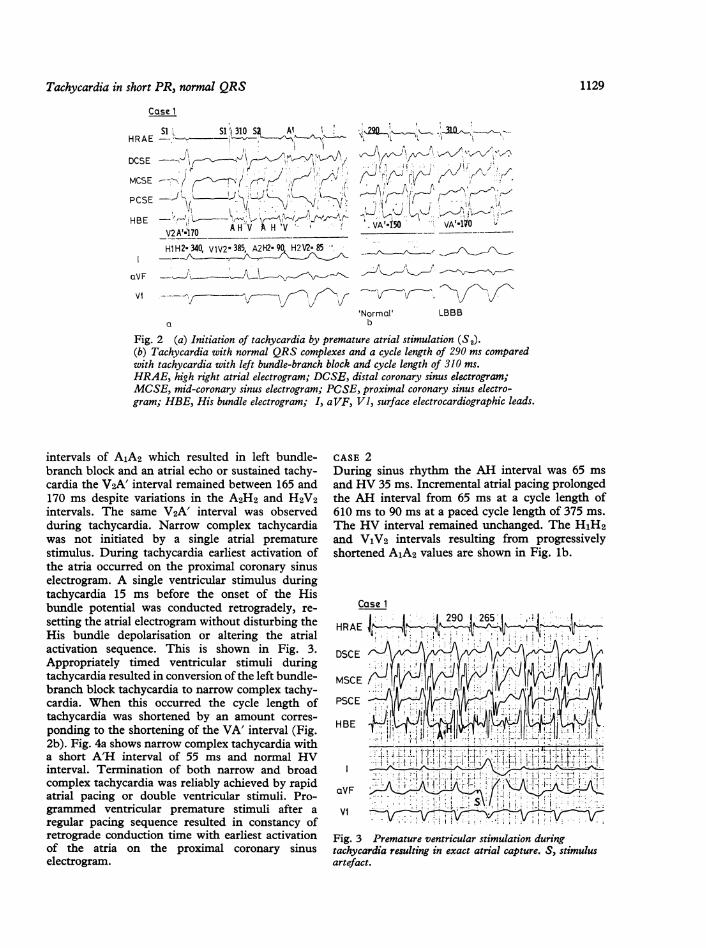
Fig. 2 (a) Initiation of tachycardia by premature atrial stimulation (S2).(b) Tachycardia with normal QRS complexes and a cycle length of 290 ms comparedwith tachycardia with left bundle-branch block and cycle length of 310 ms.HRAE, high right atrial electrogram; DCSE, distal coronary sinus electrogram;MCSE, mid-coronary sinus electrogram; PCSE, proximal coronary sinus electro-gram; HBE, His bundle electrogram; I, aVF, VI, surface electrocardiographic leads.
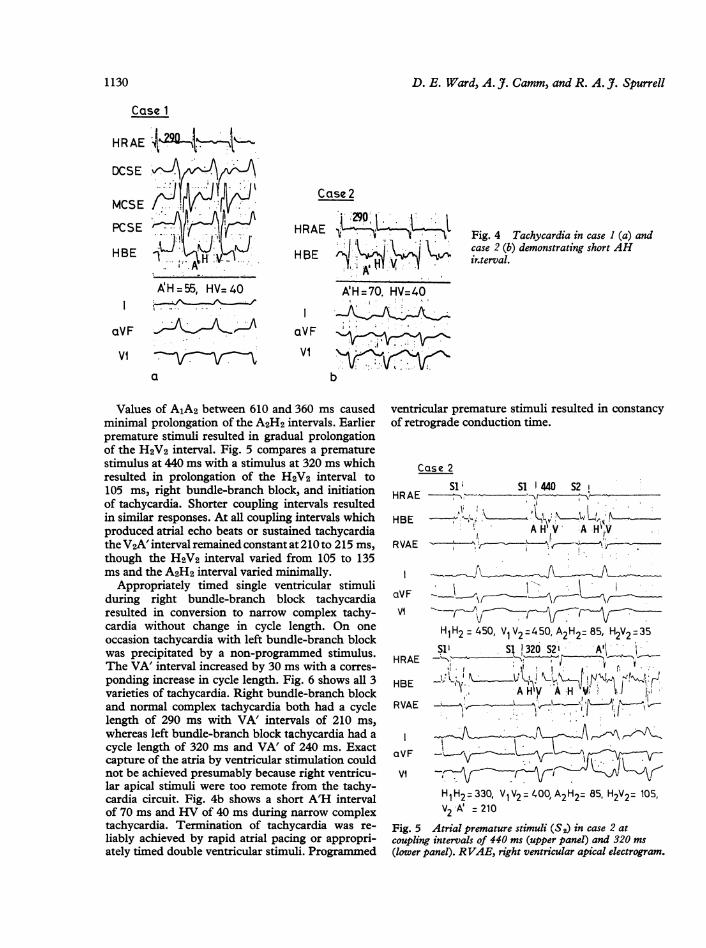
intervals of A1A2 which resulted in left bundle-branch block and an atrial echo or sustained tachy-cardia the V2A' interval remained between 165 and170 ms despite variations in the A2H2 and H2V2intervals. The same V2A' interval was observedduring tachycardia. Narrow complex tachycardiawas not initiated by a single atrial prematurestimulus. During tachycardia earliest activation ofthe atria occurred on the proximal coronary sinuselectrogram. A single ventricular stimulus duringtachycardia 15 ms before the onset of the Hisbundle potential was conducted retrogradely, re-setting the atrial electrogram without disturbing theHis bundle depolarisation or altering the atrialactivation sequence. This is shown in Fig. 3.Appropriately timed ventricular stimuli duringtachycardia resulted in conversion of the left bundle-branch block tachycardia to narrow complex tachy-cardia. When this occurred the cycle length oftachycardia was shortened by an amount corres-ponding to the shortening of the VA' interval (Fig.2b). Fig. 4a shows narrow complex tachycardia witha short A'H interval of 55 ms and normal HVinterval. Termination of both narrow and broadcomplex tachycardia was reliably achieved by rapidatrial pacing or double ventricular stimuli. Pro-grammed ventricular premature stimuli after aregular pacing sequence resulted in constancy ofretrograde conduction time with earliest activationof the atria on the proximal coronary sinuselectrogram.
CASE 2During sinus rhythm the AH interval was 65 msand HV 35 ms. Incremental atrial pacing prolongedthe AH interval from 65 ms at a cycle length of610 ms to 90 ms at a paced cycle length of 375 ms.The HV interval remained unchanged. The H1H2and V1V2 intervals resulting from progressivelyshortened A1A2 values are shown in Fig. lb.
Case 1
HRAE
DSCE \
MSCE (, (VJVYIJPSCE
II.aVF r
VI V
Fig. 3 Premature ventricular stimulation duringtachycardia resulting in exact atrial capture. S, stimulusartefact.
1129
D. E. Ward, A. 7. Camm, and R. A. J. Spurrell
Case 1
HRAE
DCSE
MCSE Y
PCSE
HBE 'J.
AH=55, HV=40
aVF
VIa
Case 2
HRAE
H E
aVF
VI
'iI!. N
A'H=70. HV=40
b
Fig. 4 Tachycardia in case I (a) andcase 2 (b) demonstrating short AHirnterval.
Values of A1A2 between 610 and 360 ms causedminimal prolongation of the A2H2 intervals. Earlierpremature stimuli resulted in gradual prolongationof the H2V2 interval. Fig. 5 compares a prematurestimulus at 440 ms with a stimulus at 320 ms whichresulted in prolongation of the H2V2 interval to105 ms, right bundle-branch block, and initiationof tachycardia. Shorter coupling intervals resultedin similar responses. At all coupling intervals whichproduced atrial echo beats or sustained tachycardiathe V2A' interval remained constant at 210 to 215 ms,though the H2V2 interval varied from 105 to 135ms and the A2H2 interval varied minimally.
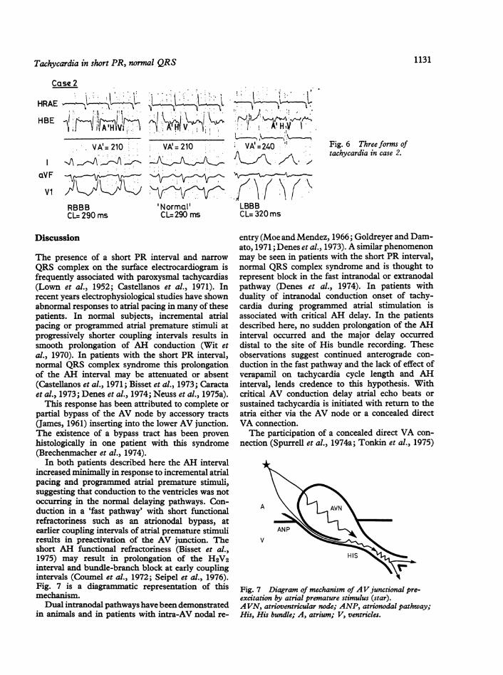
Appropriately timed single ventricular stimuliduring right bundle-branch block tachycardiaresulted in conversion to narrow complex tachy-cardia without change in cycle length. On oneoccasion tachycardia with left bundle-branch blockwas precipitated by a non-programmed stimulus.The VA' interval increased by 30 ms with a corres-ponding increase in cycle length. Fig. 6 shows all 3varieties of tachycardia. Right bundle-branch blockand normal complex tachycardia both had a cyclelength of 290 ms with VA' intervals of 210 ms,whereas left bundle-branch block tachycardia had a
cycle length of 320 ms and VA' of 240 ms. Exactcapture of the atria by ventricular stimulation couldnot be achieved presumably because right ventricu-lar apical stimuli were too remote from the tachy-cardia circuit. Fig. 4b shows a short A'H intervalof 70 ms and HV of 40 ms during narrow complextachycardia. Termination of tachycardia was re-
liably achieved by rapid atrial pacing or appropri-ately timed double ventricular stimuli. Programmed
ventricular premature stimuli resulted in constancyof retrograde conduction time.
Case 2S1i SI1 440 S2
HR AE tm>
HBE *-+'. ; _A H V A HV
RVAE E> ' , ' _
aVF J__
Hi H2 450, V1V2 4 50, A2H2= 85, H2V2 35
Si SI1 320 S21' A"HRAE -
HBE AiPY A e
RVAE -
aVF -
Hi H2 =330 V1V2 =400, A2H2= 85, H2V2= 105,V2 A' = 210
Fig. 5 Atrial premature stimuli (S2) in case 2 atcoupling intervals of 440 ms (upper panel) and 320 ms(lower panel). RVAE, right ventricular apical electrogram.
1130
Tachycardia in short PR, normal QRS 1131
Case 2
VA'= 210
RBBBCL=290ms
H v
VA' = 210
'Normal'CL= 290 ms
'I A'HV_VA'=240
fAv~
LBBBCL= 320ms
Fig. 6 Three forms oftachycardia in case 2.
Discussion
The presence of a short PR interval and narrow
QRS complex on the surface electrocardiogram isfrequently associated with paroxysmal tachycardias(Lown et al., 1952; Castellanos et al., 1971). Inrecent years electrophysiological studies have shownabnormal responses to atrial pacing in many of thesepatients. In normal subjects, incremental atrialpacing or programmed atrial premature stimuli atprogressively shorter coupling intervals results insmooth prolongation of AH conduction (Wit etal., 1970). In patients with the short PR interval,normal QRS complex syndrome this prolongationof the AH interval may be attenuated or absent(Castellanos et al., 1971; Bisset et al., 1973; Caractaet al., 1973; Denes et al., 1974; Neuss et al., 1975a).
This response has been attributed to complete or
partial bypass of the AV node by accessory tracts(James, 1961) inserting into the lower AV junction.The existence of a bypass tract has been proven
histologically in one patient with this syndrome(Brechenmacher et al., 1974).
In both patients described here the AH intervalincreased minimally in response to incremental atrialpacing and programmed atrial premature stimuli,suggesting that conduction to the ventricles was notoccurring in the normal delaying pathways. Con-duction in a 'fast pathway' with short functionalrefractoriness such as an atrionodal bypass, atearlier coupling intervals of atrial premature stimuliresults in preactivation of the AV junction. Theshort AH functional refractoriness (Bisset et al.,1975) may result in prolongation of the H2V2interval and bundle-branch block at early couplingintervals (Coumel et al., 1972; Seipel et al., 1976).Fig. 7 is a diagrammatic representation of thismechanism.Dual intranodal pathways have been demonstrated
in animals and in patients with intra-AV nodal re-
entry (Moe and Mendez, 1966; Goldreyer and Dam-ato, 1971; Denes et al., 1973). A similar phenomenonmay be seen in patients with the short PR interval,normal QRS complex syndrome and is thought torepresent block in the fast intranodal or extranodalpathway (Denes et al., 1974). In patients withduality of intranodal conduction onset of tachy-cardia during programmed atrial stimulation isassociated with critical AH delay. In the patientsdescribed here, no sudden prolongation of the AHinterval occurred and the major delay occurreddistal to the site of His bundle recording. Theseobservations suggest continued anterograde con-duction in the fast pathway and the lack of effect ofverapamil on tachycardia cycle length and AHinterval, lends credence to this hypothesis. Withcritical AV conduction delay atrial echo beats or
sustained tachycardia is initiated with return to theatria either via the AV node or a concealed directVA connection.The participation of a concealed direct VA con-
nection (Spurrell et al., 1974a; Tonkin et al., 1975)
A
ANPV
HIS
Fig. 7 Diagram of mechanism of AV junctional pre-excitation by atrial premature stimulus (star).AVN, atrioventricular node; ANP, atrionodal pathway;His, His bundle; A, atrium; V, ventricles.
HRAE
HBE
aVF
Vi
r

D. E. Ward, A. J. Camm, and R. A. 7. Spurrell
A
V /
Fig. 8 An atrial premature stimulus (star) may resultin anterograde conduction delay sufficient to allow re-
entry to the atrium (AE). Such re-entry may involve an
atrionodal pathway (ANP) or a direct ventriculoatrialpathway (VAP). Critical conduction delay above theHis bundle associated with a constant H-AE intervalsuggests re-entry within the AV node or via an atrionodalpathway. Delay below the site of the His bundle with a
constant V-AE interval suggests re-entry via a directVA pathway.
in the tachycardia circuit is suggested by the timingof the atrial echo beat. The constancy of the V2A'interval in the presence of a varying H2V2 intervalmakes it unlikely that the re-entrant pathway islocated entirely above the His bundle and impliesthe participation of the ventricles in the tachycardiacircuit. Intra-AV nodal re-entry or re-entry in an
atrionodal pathway results in nearly constant H2A'intervals. If, however, the H2V2 is constant no
conclusion can be drawn about the site of re-entry.Fig. 8 summarises these concepts.The appearance of atrial echoes with critical HV
conduction delay has been described previously(Lozano et al., 1973; Varghese et al., 1974; Neuss etal., 1975b). Both Lozano et al. (1973) and Vargheseet al. (1974) interpreted their findings as indicativeof a combination of intraventricular and intra AVnodal re-entry. However, a concealed, direct, VAconnection could not be excluded in their patients.The short AH in response to programmed atrialpremature beats and the lack of AH prolongationwith increasing prematurity of atrial stimulationsuggests the presence of a fast anterograde pathwayin the patient reported by Lozano et al. (1973).Neuss et al. (1975b) report a patient with similarfindings to the patients described here. They also
observed constancy of the V2A' interval in thepresence of an increasing H2V2 interval in responseto premature atrial stimuli delivered during sinusrhythm. This observation was regarded as proof ofinvolvement of the ventricles within the re-entrantcircuit. No comment was made conceming themechanism of critical conduction delay. Theabsence of atrial echo beats or tachycardia at latercoupling intervals of A2 is probably related tolimiting atrial refractoriness at the time of retrogradeactivation of the VA pathway (Pritchett et al., 1977),or alternatively to concealed anterograde invasion ofthe VA pathway during atrial stimulation (Zipes etal., 1974).
Additional evidence for an accessory concealedVA connection in our patients was provided by thefollowing observations.
(1) In both patients the appearance of leftbundle-branch block during tachycardias wasassociated with an increase in VA' conduction andtachycardia cycle length (Coumel and Attuel, 1974;Pritchett et al., 1976; Spurrell et al., 1974b).
(2) In case 1 coronary sinus recordings revealedearly activation of the low left atrium near theproximal coronary sinus during tachycardia. Thisdoes not prove that retrograde activation is via adirect AV pathway since retrograde conduction in anatrionodal pathway may produce similar eccentricatrial activation. However, a ventricular prematurestimulus, delivered 15 ms before the His bundlepotential, conducted retrogradely with exact atrialcapture and identical atrial activation sequence.Retrograde conduction in an atrionodal pathwaywould be impossible in these conditions and theretrograde limb of the re-entrant circuit, is there-fore, a direct VA pathway (Sellers et al., 1976).The coexistence of anomalous atrionodal and
atrioventricular pathways has been described pre-viously (Coumel et al., 1971, 1972; Neuss et al.,1975b; Zipes etal., 1974; Castellanos etal., 1975)andunusual re-entry mechanisms may result from thesecombinations. The mechanism of re-entry in ourpatients is dependent on an interaction between twofunctionally abnormal pathways. Conduction delayat infra-Hisian level occurs as a result of pre-activation of the AV junction and retrograde con-duction occurs when critical delay is sufficient toallow re-entry in the VA pathway and atrialmyocardium.
In the patients described by Lozano and hiscolleagues (1973) tachycardia was initiated in thepresence of a short AH interval which persistedduring sustained tachycardia. These observationssuggest the incorporation of a partial or completeAV nodal bypass tract in the anterograde limb of there-entrant circuit. Re-entrant circuits utilising an
1132

Tachycardia in short PR, normal QRS
anterograde fast pathway bypassing the AV nodeand a retrograde direct VA pathway may result inrapid, distressing tachycardias unresponsive totherapy directed towards the AV node itself, and inthese cases verapamil was not effective. Detailedelectrophysiological studies are required to definethe tachycardia mechanism in these patients.
References
Bisset, J. K., de Soyza, N., Kane, J. J., and Murphy, M. L.(1975). Altered refractory periods in patients with short PRintervals and normal QRS complex. American J3ournal ofCardiology, 35, 487-491.
Bisset, J. K., Thompson, A., de Soyza, N., and Murphy,M. L. (1973). Atrioventricular conduction in patients withshort PR intervals and normal QRS complexes. BritishHeartjournal, 35, 123-127.
Brechenmacher, C., Laham, J., Iris, L., Gerbaux, A., andLenegre, J. (1974). etude histologique des voies anormalesde conduction dans un syndrome de Wolff-Parkinson-White et dans un syndrome Lown-Ganong-Levine.Archives des Maladies du Coeur et des Vaisseaux, 67, 507-520.
Caracta, A. R., Damato, A. N., Gallagher, J. J., Josephson,M. E. I., Varghese, P. J., Lau, S. H., and Westura, E. F.(1973). Electrophysiological studies in the syndrome ofshort P-R interval, normal QRS complex. American Journalof Cardiology, 31, 245-253.
Castellanos, A., Agha, A. S., Befeler, B., and Myerburg, K.(1975). Double accessory pathways in Wolff-Parkinson-White syndrome. Circulation, 51, 1020-1025.
Castellanos, A., Castillo, C. A., Agha, A. S., and Tessler, M.(1971). His bundle electrograms in patients with short P-Rintervals, narrow QRS complexes and paroxysmal tachy-cardias. Circulation, 43, 667-678.
Coumel, P., and Attuel, P. (1974). Reciprocating tachycardiain overt and latent pre-excitation. European Journal ofCardiology, 1, 423-436.
Coumel, P., Wayneberger, M., Fabiato, A., Slama, R.,Aigueperse, J., and Bouvrain, Y. (1972). Wolff-Parkinson-White syndrome. Problems in evaluation of multipleaccessory pathways and surgical therapy. Circulation, 45,1216-1230.
Coumel, P., Wayneberger, M., Garnier, J. C., Slama, R., andBouvrain, Y. (1971). Syndrome de preexcitation ventricu-laire associant P-R court et onde delta, sans elargissment deQRS: (a propos de trois cas de syndrome de WPW acomplexes fins). Archives des Maladies du Coeur et desVaisseaux, 64, 1234-1255.
Denes, P., Wu, D., Dhingra, R. C., Chuquimia, R., andRosen, K. M. (1973). Demonstration of dual A-V nodalpathways in patients with paroxysmal supraventriculartachycardia. Circulation, 48, 549-555.
Denes, P., Wu, D., and Rosen, K. M. (1974). Demonstrationof dual A-V pathways in a patient with the Lown-Ganong-Levine syndrome. Chest, 65, 343-346.
Durrer, D., Schuilenburg, R. M., and Wellens, H. J. J. (1970).Pre-excitation revisited. American Journal of Cardiology, 25,690-697.
Goldreyer, B. N., and Damato, A. N. (1971). The essentialrole of atrioventricular conduction delay in the initiation ofparoxysmal supraventricular tachycardia. Circulation, 43,679-687.
James, T. N. (1961). The morphology of the human atrio-ventricular node, with remarks pertinent to its electro-physiology. American Heart Journal, 62, 756-771.
Lown, B., Ganong, W. F., and Levine, S. A. (1952). The
1133
syndrome of short P-R interval, normal QRS complex andparoxysmal rapid heart action. Circulation, 5, 693-706.
Lozano, J., Mandel, W. J., Hayakawa, H., Shine, K. I., andEber, L. M. (1973). Re-entrant tachycardia: participation ofthe distal A-V conduction system. Chest, 63, 23-34.
Mandel, W. J., Danzig, R., and Hayakawa, H. (1971). Lown-Ganong-Levine syndrome. Circulation, 44, 696-708.
Moe, G. K., and Mendez, C. (1966). The physiologic basis ofreciprocal rhythm. Progress in Cardiovascular Diseases, 8,461-482.
Neuss, H., Nowak, F., and Schlepper, M. (1975a). Analyseder A-V Uberleitung bei kurzer P-Q Zeit. In His-BundelElektrographie, p. 153. Ed. by L. Seipel, F. Loogen, and A.Both. Schattauer Verlag, Stuttgart.
Neuss, H., Schlepper, M., and Thormann, J. (1975b).Analysis of re-entry mechanisms in three patients withconcealed Wolff-Parkinson-White syndrome. Circulation,51, 75-81.
Pritchett, E. L. C., Gallagher, J. J., and Scheinman, M.(1977). Determinants of the echo zone in patients withreciprocating tachycardia and the Wolff-Parkinson-Whitesyndrome (abstract). American Journal of Cardiology, 39,319.
Pritchett, E. L. C., Tonkin, A. M., Dugan, F. A., Wallace,A. G., and Gallagher, J. J. (1976). Ventriculo-atrial con-duction time during reciprocating tachycardia with inter-mittent bundle branch block in Wolff-Parkinson-Whitesyndrome. British Heart Journal, 38, 1058-1064.
Seipel, L., Breithardt, G., and Both, A. (1976). Atrioventricu-lar (A-V) and ventriculoatrial (VA) conduction pattern inpatients with short PR interval and normal QRS. InCardiac Pacing-Diagnostic and Therapeutic Tools, p. 152.Ed. by B. Luderitz. Springer-Verlag, Berlin.
Sellers, T. D., Jr., Gallagher, J. J., Cope, G. D., Tonkin,A. M., and Wallace, A. G. (1976). Retrograde atrial pre-excitation following premature ventricular beats duringreciprocating tachycardia in the Wolff-Parkinson-Whitesyndrome. European Journal of Cardiology, 4, 283-294.
Spurrell, R. A. J., Krikler, D. M., and Sowton, E. (1974a).Concealed bypasses of the atrioventricular node in patientswith paroxysmal supraventricular tachycardia revealed byintracardiac electrical stimulation and Verapamil. AmericanJournal of Cardiology, 33, 590-595.
Spurrell, R. A. J., Krikler, D. M., and Sowton, E. (1974b).Retrograde invasion of the bundle branches producingaberration of the QRS complex during supraventriculartachycardia studied by programmed electrical stimulation.Circulation, 50, 487-495.
Tonkin, A. M., Gallagher, J. J., Svenson, R. H., Wallace,A. G., and Sealy, W. C. (1975). Anterograde block inaccessory pathways with retrograde conduction in recipro-cating tachycardia. European Journal of Cardiology, 3,143-152.
Varghese, P. J., Damato, A. N., Caracta, A. R., Gallagher,J. J., Josephson, M. E., and Lau, S. H. (1974). Intra-ventricular conduction delay as a determinant of atrial echobeats. Circulation, 49, 805-810.
Wit, A. L., Weiss, M. B., Berkowitz, W. D., Rosen, K.,Steiner, C., and Damato, A. N. (1970). Patterns of atrio-ventricular conduction in the human heart. CirculationResearch, 27, 345-359.
Zipes, D. P., DeJoseph, R. L., and Rothbaum, D. A. (1974).Unusual properties of accessory pathways. Circulation, 49,1200-1211.
Requests for reprints to Dr D. E. Ward, Departmentof Cardiology, St Bartholomew's Hospital, WestSmithfield, London EClA 7BE.