reactions between water and vitreous silica during irradiation
TRANSCRIPT
Journal of Nuclear Materials 430 (2012) 239–245
Contents lists available at SciVerse ScienceDirect
Journal of Nuclear Materials
journal homepage: www.elsevier .com/ locate / jnucmat
Reactions between water and vitreous silica during irradiation
Glenn K. Lockwood, Stephen H. Garofalini ⇑Interfacial Molecular Science Laboratory, Department of Materials Science and Engineering, Rutgers University, Piscataway, NJ 08854, United States
a r t i c l e i n f o
Article history:Received 23 April 2012Accepted 3 July 2012Available online 16 July 2012
0022-3115/$ - see front matter � 2012 Elsevier B.V. Ahttp://dx.doi.org/10.1016/j.jnucmat.2012.07.004
⇑ Corresponding author.E-mail address: [email protected] (S.H. Garofalini).
a b s t r a c t
Molecular dynamics simulations were conducted to determine the response of a vitreous silica surface incontact with water to radiation damage. The defects caused by radiation damage create channels thatpromote high H+ mobility and result in significantly higher concentration and deeper penetration of H+
in the silica subsurface. These subsurface H+ hop between acidic sites such as SiOHþ2 and Si–(OH)–Si untilsubsequent radiation ruptures siloxane bridges and forms subsurface non-bridging oxygens (NBOs);existing excess H+ readily bonds to these NBO sites to form SiOH. The high temperature caused by irra-diation also promotes the diffusion of molecular H2O into the subsurface, and although H2O does not pen-etrate as far as H+, it readily reacts with ruptured bridges to form 2SiOH. These SiOH sites are thermallystable and inhibit the reformation of bridges that would otherwise occur in the absence of water. In addi-tion to this reduction of self-healing, the presence of water during the self-irradiation of silica may causean increase in the glass’s proton conductivity.
� 2012 Elsevier B.V. All rights reserved.
1. Introduction
Vitrification is recognized as a key component to the immobili-zation and long-term storage of radioactive wastes, and borosili-cate-based glasses have become an international standard indeveloping waste glass compositions. These borosilicate glassesare particularly attractive because of their ability to be loaded toa high degree with a wide range of waste products, their relativeresistance to radiation damage, and the technological maturity ofglass processing [1,2]. However, given the very long lifetimes ofsome of the radionuclides involved (typically given to be a rangefrom 100,000 to 1,000,000 years for high-level waste [3]), somepercentage of waste forms must be expected to fail during thisservice life.
As a result of this, the predominant mechanism for radionucliderelease to the biosphere will be through dissolution of the wasteform into groundwater in waste repositories [4], and significantwork has been done in studying how radiation damage affects dis-solution and leaching rates in such waste glass materials. Unfortu-nately, the results from these experiments have often yieldedcontradictory results ranging from irradiation increasing dissolu-tion rates [5–7] to having negligible effects [8–11], underscoringthe need to explore how radiation damage affects the interactionsbetween water and glass at a more fundamental level.
Molecular dynamics (MD) simulations are well suited to study-ing the effects of radiation in vitreous silica [12] and have beenused for this purpose for over a decade [13]. As computationalpower has increased, the models used have also progressed to
ll rights reserved.
utilize much more realistic kinetic energies of recoil nuclei [14],to devise novel techniques to enable simulation of much larger sys-tems [15], and to increase compositional accuracy [15]. Theseadvancements have revealed a wealth of information on how sili-cate waste forms degrade under irradiation at the atomistic level;however, these simulations have never included water or consid-ered its effect.
Given the important role of water in the dissolution and degra-dation of glassy waste forms, including water in these molecularmodels of damage mechanisms is of critical importance to estab-lishing a comprehensive, fundamental understanding of howglassy waste forms will degrade. To this end, the authors recentlyconducted MD simulations that included hydroxyls and water inbulk vitreous silica and found that the presence of water acceler-ates the accumulation of residual structural damage in the formof non-bridging oxygen and larger voids in the glassy network [16].Based on this observed propensity for water to limit self-healingof radiation-damaged silica and increase the concentration of voidsunder irradiation, it is easy to envision radiation damage causingvoids and channels at the surface, providing a new pathway forthe accelerated diffusion of water into silica. This would then leadto further damage accumulation, ingress of water, and an overallaccelerated degradation of the silica network.
The interactions between water and small voids and channelsare also relevant beyond the scope of waste glasses. For example,abnormally high proton conduction has been observed in mesopor-ous silica [17–20], and evidence suggests that defects play a signif-icant role in H+ mobility in water–silica systems [21–25]. Given thefact that ballistic radiation creates large concentrations of defectsin silica, it is not unreasonable to consider how radiation damageaffects the mobility of H+ at the silica surface and into the bulk

Table 1Two-body potential parameters. The nr
O�H parameter is calculated as a function oftemperature and pressure as described elsewhere [26].
i–j Pair Arepij (fJ) nij (nm) nr
ij (nm) B12ij (J nm12) C6
ij (J nm6)
O–H 0.2283 2.4 f(T, P) 0 0O–O 0.0425 2.4 0.06100 4.0780 � 10�31 4.2260 � 10�24
H–H 0.0000 2.4 0.00000 0 0Si–O 0.2670 2.4 0.03730 3.5000 � 10�31 7.0000 � 10�24
Si–Si 0.0700 2.4 0.06400 0 0Si–H 0.5000 2.4 0.03500 5.6486 � 10�32 3.8000 � 10�24
Table 2Three-body potential parameters.
j–i–k Triplet kjik (fJ) cij (nm) cik (nm) r0ij (nm) r0
ik (nm) h0jik
O–Si–O 0.0150 0.28 0.28 0.30 0.30 109.5Si–O–Si 0.0010 0.20 0.20 0.28 0.28 109.5H–O–H 0.0300 0.13 0.13 0.16 0.16 100.0Si–O–H 0.0050 0.20 0.12 0.28 0.15 109.5
240 G.K. Lockwood, S.H. Garofalini / Journal of Nuclear Materials 430 (2012) 239–245
glass. Despite this, very little work has been done towards under-standing how radiation damage affects proton conductivity inwater–silica systems.
The study presented here explores the processes of H+ and H2Otransport into silica under irradiation more directly by modeling avitreous silica surface in contact with liquid water and simulatingthe effects of recoil nuclei in the silica subsurface. By simulatingthe self-irradiation of silica near a water–silica interface and usingan interatomic potential that allows water to dissociate, the effectsof both the water–silica reactions during the radiation damage pro-cess and the damaged region on accelerated water penetration anddegradation of the silica have been observed within a single simu-lated system.
2. Computational methods
2.1. Interatomic potential
A dissociative interatomic potential for water was used in thisstudy to model the interactions between all atoms. This modelbenefits from being able to reproduce the behavior of bulk water,bulk silica, and the water–silica interface in terms of structure,chemistry, transport processes, energetics, and other properties[21,23,26,27]. It also has been used previously to simulate the ef-fects of irradiation in silica [16] and to reproduce the characteristicrapid formation of damage followed by picosecond-scale healingproduced by other MD models [13,28]. Despite this wide range ofapplicability, it is a relatively simple potential comprised of two-and three-body interactions and takes the form:
U2�bodyij ðrijÞ ¼ Uq�q
ij þ Uqd�qdij þ Uq�qd
ij þ Uqd�qij þ Urep
ij þ Udispij
þ U12ij ð1Þ
where each energy term is a function of the interatomic spacing rij
and is given by:
Uq�qij ðrijÞ ¼
qiqj
rij� erfc
rij
b
� �ð2Þ
Uqd�qdij ðrijÞ ¼
qdi qd
j
rijerfð rij
2nijÞ � erfcðrij
bÞ ð3Þ
Uq�qd
ij ðrijÞ ¼qiq
dj
rijerf
rijffiffiffi2p
nij
!� erfc
rij
b
� �ð4Þ
Uqd�qij ðrijÞ ¼
qdi qj
rijerf
rijffiffiffi2p
nij
!� erfc
rij
b
� �ð5Þ
Urepij ðrijÞ ¼ Arep
ij
2nrij
rijerfcð rij
2nrij
Þ ð6Þ
Udispij ðrijÞ ¼ �
C6ij
r6ij
ð7Þ
U12ij ðrijÞ ¼ þ
B12ij
r12ij
ð8Þ
The parameters used in this study are listed in Table 1. The cal-culated interactions between pairs are limited to atoms separatedby a distance less than Rc = 1.0 nm, and as in past work, the long-range Coulomb interactions are determined by use of the Wolfsummation method [29] with a decay parameter of b = 0.446 nm.The point charges qi are partial charges and defined such thatqSi = +1.808, qO = �0.5qSi, and qH = +0.25qSi. The diffuse charges
are defined by the simple, empirical relationship qdi ¼ �0:25qi.
Eq. (8) is used to eliminate spurious attractive forces that might oc-cur at very short range during collisions rather than the ZBL [30]term often used. ZBL assumes full ionic charges and simple pointcharges in its empirical fit, whereas we use partial charges and in-clude diffuse charges in our potential so its incorporation would beproblematic.
The three-body component of the interatomic potential is a Stil-linger–Weber type function that serves to provide an energeticbias of certain j–i–k triplets towards certain angles, empiricallyequivalent to the effects introduced by partial bond covalency (asin SiO2) or lone pairs (as in H2O). It takes the form:
U3�bodyjik ðrij; rik; hjikÞ ¼ kjikexp
cij
rij � r0ij
þ cik
rik � r0ik
!ðcoshjik
� cosh0jikÞ
2 ð9Þ
and the parameters used here are listed in Table 2.
2.2. System assembly
The starting configuration for the systems simulated in thisstudy was comprised of a slab of amorphous silica(10.44 � 10.44 � 5 nm) in contact with a film of liquid water(about 2 nm) with about 5.5 nm of vacuum above, resulting in asystem size of 10.44 � 10.44 � 12.51 nm. The silica slab contained36,678 atoms and the water film contained 21,900 atoms.
To generate the silica slab, a simulation box of 10.50 nm on eachside was filled with 25,537 Si atoms and 51,074 O atoms placed atrandom, then simulated at 6000 K for 15 ps in the canonicalensemble (constant number of atoms, volume, and temperature)followed by 15 ps in the microcanonical ensemble (constant num-ber of atoms, volume, and energy). From this temperature, the sys-tem was quenched to a temperature of 300 K via intermediatesteps in a manner identical to previous work [21].
Once equilibrated to 300 K, the system was then simulated un-der constant temperature and pressure (or constant N–P–T, whereN = 76,611 atoms, T = 298 K, P = 1 atm) for 40 ps. The zero-straindimensions of the silica sample were assumed to be equal to theaverage dimensions taken over the final 20 ps of this 40 ps simula-tion; thus, after the 40 ps of N–P–T simulation, the final atomicconfiguration was rescaled in the x, y, and z dimensions to theseaverage dimensions of 10.44 nm in x, y, and z. All atoms abovez = 5.009 nm were then removed to maintain the large x–y surface
G.K. Lockwood, S.H. Garofalini / Journal of Nuclear Materials 430 (2012) 239–245 241
area but reduce the unnecessarily large size of the SiO2 sample. Theresulting system was an amorphous, stoichiometric SiO2 slabcontaining 36,678 atoms with dimensions xL = 10.44 nm andyL = 10.44 nm, and with a height of 5.009 nm in z.
The water film was generated by randomly inserting 7300 H2Omolecules between z = 5.3 nm and z = 8.3 nm such that no ran-domly inserted H2O molecule was within 0.2 nm of any previouslyinserted H2O molecule. Following this, the velocities of all atoms inthe resulting system were randomized to a Gaussian distributioncorresponding to T = 298.0 K. The bottom 0.5 nm of the systemwas frozen, and this system was simulated at 298.0 K for 20 ps un-der the canonical ensemble to allow the randomly placed water tocondense to liquid density and hydroxylate the SiO2 surface.
The final configuration of this 20 ps simulation, hereafter re-ferred to as the ‘‘starting configuration,’’ was then subjected tothe irradiation procedure detailed below.
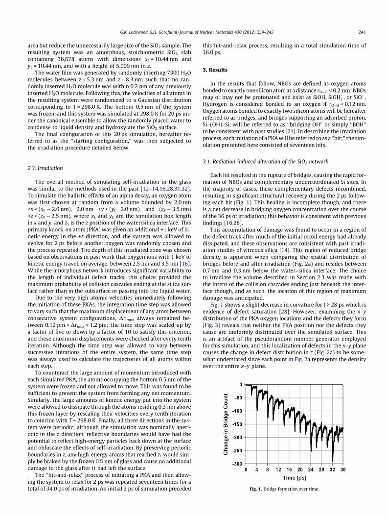
Fig. 1. Bridge formation over time.
2.3. Irradiation
The overall method of simulating self-irradiation in the glasswas similar to the methods used in the past [12–14,16,28,31,32].To simulate the ballistic effects of an alpha decay, an oxygen atomwas first chosen at random from a volume bounded by 2.0 nm<x < (xL � 2.0 nm), 2.0 nm <y < (yL 2.0 nm), and (zS � 3.5 nm)<z < (zS � 2.5 nm), where xL and yL are the simulation box lengthin x and y, and zS is the z position of the water/silica interface. Thisprimary knock-on atom (PKA) was given an additional +1 keV of ki-netic energy in the +z direction, and the system was allowed toevolve for 2 ps before another oxygen was randomly chosen andthe process repeated. The depth of this irradiated zone was chosenbased on observations in past work that oxygen ions with 1 keV ofkinetic energy travel, on average, between 2.5 nm and 3.5 nm [16].While the amorphous network introduces significant variability tothe length of individual defect tracks, this choice provided themaximum probability of collision cascades ending at the silica sur-face rather than in the subsurface or passing into the liquid water.
Due to the very high atomic velocities immediately followingthe initiation of these PKAs, the integration time step was allowedto vary such that the maximum displacement of any atom betweenconsecutive system configurations, Drmax, always remained be-tween 0.12 pm < Drmax < 1.2 pm; the time step was scaled up bya factor of five or down by a factor of 10 to satisfy this criterion,and these maximum displacements were checked after every tenthiteration. Although the time step was allowed to vary betweensuccessive iterations of the entire system, the same time stepwas always used to calculate the trajectories of all atoms withineach step.
To counteract the large amount of momentum introduced witheach simulated PKA, the atoms occupying the bottom 0.5 nm of thesystem were frozen and not allowed to move. This was found to besufficient to prevent the system from forming any net momentum.Similarly, the large amounts of kinetic energy put into the systemwere allowed to dissipate through the atoms residing 0.3 nm abovethis frozen layer by rescaling their velocities every tenth iterationto coincide with T = 298.0 K. Finally, all three directions in the sys-tem were periodic; although the simulation was nominally aperi-odic in the z direction, reflective boundaries would have had thepotential to reflect high-energy particles back down at the surfaceand obfuscate the effects of self-irradiation. By preserving periodicboundaries in z, any high-energy atoms that reached zL would sim-ply be braked by the frozen 0.5 nm of glass and cause no additionaldamage to the glass after it had left the surface.
The ‘‘hit-and-relax’’ process of initiating a PKA and then allow-ing the system to relax for 2 ps was repeated seventeen times for atotal of 34.0 ps of irradiation. An initial 2 ps of simulation preceded
this hit-and-relax process, resulting in a total simulation time of36.0 ps.
3. Results
In the results that follow, NBOs are defined as oxygen atomsbonded to exactly one silicon atom at a distance rSi–O < 0.2 nm; NBOsmay or may not be protonated and exist as SiOH, SiOHþ2 , or SiO�.Hydrogen is considered bonded to an oxygen if rO–H < 0.12 nm.Oxygen atoms bonded to exactly two silicon atoms will be hereafterreferred to as bridges, and bridges supporting an adsorbed proton,Si–(OH)–Si, will be referred to as ‘‘bridging OH’’ or simply ‘‘BOH’’to be consistent with past studies [21]. In describing the irradiationprocess, each initiation of a PKA will be referred to as a ‘‘hit;’’ the sim-ulation presented here consisted of seventeen hits.
3.1. Radiation-induced alteration of the SiO2 network
Each hit resulted in the rupture of bridges, causing the rapid for-mation of NBOs and complementary undercoordinated Si sites. Inthe majority of cases, these complementary defects recombined,resulting in significant structural recovery during the 2 ps follow-ing each hit (Fig. 1). This healing is incomplete though, and thereis a net decrease in bridging oxygen concentration over the courseof the 36 ps of irradiation; this behavior is consistent with previousfindings [16,28].
This accumulation of damage was found to occur in a region ofthe defect track after much of the initial recoil energy had alreadydissipated, and these observations are consistent with past irradi-ation studies of vitreous silica [14]. This region of reduced bridgedensity is apparent when comparing the spatial distribution ofbridges before and after irradiation (Fig. 2a) and resides between0.7 nm and 0.3 nm below the water–silica interface. The choiceto irradiate the volume described in Section 2.3 was made withthe intent of the collision cascades ending just beneath the inter-face though, and as such, the location of this region of maximumdamage was anticipated.
Fig. 1 shows a slight decrease in curvature for t > 28 ps which isevidence of defect saturation [28]. However, examining the x–ydistribution of the PKA oxygen locations and the defects they form(Fig. 3) reveals that neither the PKA position nor the defects theycause are uniformly distributed over the simulated surface. Thisis an artifact of the pseudorandom number generator employedfor this simulation, and this localization of defects in the x–y planecauses the change in defect distribution in z (Fig. 2a) to be some-what understated since each point in Fig. 2a represents the densityover the entire x–y plane.

Fig. 2. (a) Density of bridging oxygen as a function of distance below the silicasurface. (b) Side view of region showing interconnected void space less than 2.6 Åfrom any Si or O ions for pre-irradiated (yellow) and post-irradiated (blue) system,indicating more channel-like openings in latter. (c) Post-irradiated channel with redSi–O bonds included. Surface is at the top of (b) and (c) (water not shown). (Forinterpretation of the references to colour in this figure legend, the reader is referredto the web version of this article.)
Fig. 3. Distribution of PKAs and consequent net damage in the x–y plane. All PKAswere chosen randomly from the non-shaded region. Ruptured bridges shown reflectthe positions of bridges that ruptured following a decay event and did not re-forminto a bridging site during the 2 ps following that decay.
242 G.K. Lockwood, S.H. Garofalini / Journal of Nuclear Materials 430 (2012) 239–245
In addition to changes in bridge concentration, radiation dam-age in silica can manifest in the form of changes in the ring size dis-tribution [2]. Changes in this ring size distribution can occurindependently of the bridge concentration since ruptured bridgesreadily recombine with any adjacent complementary defect andnot necessarily their original neighbors. During the course of thisirradiation simulation, a total of 397 rings with four, five, six, or se-ven silica members (which comprise the majority of rings in unir-radiated bulk silica) were permanently destroyed. This destructionwas offset by the net formation of 93 rings having eight or moremembers (i.e. small voids) and 15 three-membered rings (strainedrings commonly found on silica surfaces). These changes in the ringsize distribution are very similar to the observations of others whohave utilized different interatomic potentials to model radiationdamage in silica [33].
The observed net destruction of bridging oxygens (Fig. 1) and theloss of bridging oxygen density near the interface (Fig. 2a) are indic-ative of a net increase in free volume, a loss of density, and the for-mation of new surface in the irradiated region [2]. Also observed isthe presence of interconnected void space, or ‘channels’, as shownin Fig. 2b and c that show side views of regions at and below thesurface. To examine the spatial distribution of this interconnectedvoid space in the pre-irradiated silica in comparison to the post-irradiated silica, a random space-filling procedure was carried outon the system configuration at the beginning of the irradiation sim-ulation and after the 36 ps of irradiation. Positions less than 2.6 Åfrom any Si or O atom were generated randomly, filling space thatenabled a view of interconnected channels into which water couldpenetrate. Fig. 2b shows that there are far fewer deep channels inthe pre-irradiated (yellow) silica in comparison to the post-irradi-ated (blue) glass. Fig. 2c includes the Si–O bonds in another regioncontaining a deep channel (�2 nm).
3.2. Water–silica reactions during irradiation
Water was found to readily react with the glassy network dur-ing the irradiation process as well, evidenced by a dramatic in-crease in SiOH concentration during irradiation. At the atomicconfiguration immediately preceding the first hit (t = 2.0 ps inFig. 1), the system contained 476 SiOH sites which correspondsto a surface density of 4.37 SiOH/nm2 and is consistent with exper-iment [34,35]. After seventeen hit-and-relax cycles (t = 36.0 ps),this concentration had increased to 977 SiOH.
These excess SiOH sites formed as bridges ruptured during theirradiation, producing a 3-coordinated � Siþ and Si–O� site andthen reacting with H2O (Fig. 4). The concentration of nonbridgingoxygen (NBO) defects over time (solid line in Fig. 5) is complemen-tary to the bridge concentration over time (Fig. 1); increases inNBO concentration mirror decreases in bridge concentration. How-ever, decomposing this NBO concentration into protonated (Si–OHand Si—OHþ2 ; shaded line in Fig. 5) and dry (Si–O�; dotted line)sites reveals that the spikes in NBO concentration are caused bythe formation of dry NBOs. These dry NBOs either heal back tobridging sites or form protonated NBOs very rapidly, and the net
Fig. 4. Adsorption–dissociation reaction mechanism between H2O and a rupturedbridge.
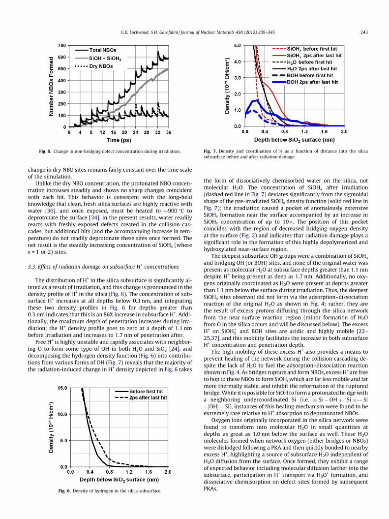
Fig. 5. Change in non-bridging defect concentration during irradiation. Fig. 7. Density and coordination of H as a function of distance into the silicasubsurface before and after radiation damage.
G.K. Lockwood, S.H. Garofalini / Journal of Nuclear Materials 430 (2012) 239–245 243
change in dry NBO sites remains fairly constant over the time scaleof the simulation.
Unlike the dry NBO concentration, the protonated NBO concen-tration increases steadily and shows no sharp changes coincidentwith each hit. This behavior is consistent with the long-heldknowledge that clean, fresh silica surfaces are highly reactive withwater [36], and once exposed, must be heated to �900 �C todeprotonate the surface [34]. In the present results, water readilyreacts with freshly exposed defects created in the collision cas-cades, but additional hits (and the accompanying increase in tem-perature) do not readily deprotonate these sites once formed. Thenet result is the steadily increasing concentration of SiOHx (wherex = 1 or 2) sites.
3.3. Effect of radiation damage on subsurface H+ concentrations
The distribution of H+ in the silica subsurface is significantly al-tered as a result of irradiation, and this change is pronounced in thedensity profile of H+ in the silica (Fig. 6). The concentration of sub-surface H+ increases at all depths below 0.3 nm, and integratingthese two density profiles in Fig. 6 for depths greater than0.3 nm indicates that this is an 86% increase in subsurface H+. Addi-tionally, the maximum depth of penetration increases during irra-diation; the H+ density profile goes to zero at a depth of 1.1 nmbefore irradiation and increases to 1.7 nm of penetration after.
Free H+ is highly unstable and rapidly associates with neighbor-ing O to form some type of OH in both H2O and SiO2 [24], anddecomposing the hydrogen density function (Fig. 6) into contribu-tions from various forms of OH (Fig. 7) reveals that the majority ofthe radiation-induced change in H+ density depicted in Fig. 6 takes
Fig. 6. Density of hydrogen in the silica subsurface.
the form of dissociatively chemisorbed water on the silica, notmolecular H2O. The concentration of SiOHx after irradiation(dashed red line in Fig. 7) deviates significantly from the sigmoidalshape of the pre-irradiated SiOHx density function (solid red line inFig. 7); the irradiation caused a pocket of anomalously extensiveSiOHx formation near the surface accompanied by an increase inSiOHx concentration of up to 10�. The position of this pocketcoincides with the region of decreased bridging oxygen densityat the surface (Fig. 2) and indicates that radiation damage plays asignificant role in the formation of this highly depolymerized andhydroxylated near-surface region.
The deepest subsurface OH groups were a combination of SiOHx
and bridging OH (or BOH) sites, and none of the original water waspresent as molecular H2O at subsurface depths greater than 1.1 nmdespite H+ being present as deep as 1.7 nm. Additionally, no oxy-gens originally coordinated as H2O were present at depths greaterthan 1.1 nm below the surface during irradiation. Thus, the deepestSiOHx sites observed did not form via the adsorption–dissociationreaction of the original H2O as shown in Fig. 4; rather, they arethe result of excess protons diffusing through the silica networkfrom the near-surface reaction region (minor formation of H2Ofrom O in the silica occurs and will be discussed below). The excessH+ on SiOHþ2 and BOH sites are acidic and highly mobile [22–25,37], and this mobility facilitates the increase in both subsurfaceH+ concentration and penetration depth.
The high mobility of these excess H+ also provides a means toprevent healing of the network during the collision cascading de-spite the lack of H2O to fuel the adsorption–dissociation reactionshown in Fig. 4. As bridges rupture and form NBOs, excess H+ are freeto hop to these NBOs to form SiOH, which are far less mobile and farmore thermally stable, and inhibit the reformation of the rupturedbridge. While it is possible for SiOH to form a protonated bridge witha neighboring undercoordinated Si (i.e. � Si� OHþ þSi �! Si�½OH� � Si), instances of this healing mechanism were found to beextremely rare relative to H+ adsorption to deprotonated NBOs.
Oxygen ions originally incorporated in the silica network werefound to transform into molecular H2O in small quantities atdepths as great as 1.0 nm below the surface as well. These H2Omolecules formed when network oxygen (either bridges or NBOs)were dislodged following a PKA and then quickly bonded to nearbyexcess H+, highlighting a source of subsurface H2O independent ofH2O diffusion from the surface. Once formed, they exhibit a rangeof expected behavior including molecular diffusion farther into thesubsurface, participation in H+ transport via H3O+ formation, anddissociative chemisorption on defect sites formed by subsequentPKAs.

244 G.K. Lockwood, S.H. Garofalini / Journal of Nuclear Materials 430 (2012) 239–245
3.4. Effect of elevated temperature
The average system temperature between hits was around515 K towards the end of the irradiation simulation despite the0.3 nm of atoms thermostated to 298.0 K. This temperature iswithin the range of conditions expected in waste repositories[38], and it is likely to have an effect on the kinetics of H2O diffu-sion into the silica surface and how the subsurface H+ concentra-tion changes during irradiation. To this end, the irradiationsimulation was repeated in the absence of irradiation; that is, thestarting configuration for the irradiation simulation (see Section2.2) was simulated for 36 ps within the canonical ensemble at573 K, which is the upper limit of expected relevant repositorytemperatures [38]. The periodic boundary in z was replaced by areflective boundary to prevent water vapor from interacting withthe frozen underside of the silica component of the system.
This high-temperature, unirradiated simulation showed a great-er subsurface concentration and a greater penetration depth ofmolecular H2O when compared to the irradiated system. This sug-gests that the additional ingress of molecular H2O observed duringirradiation is largely a thermal effect and will occur regardless ofwhether or not the defect tracks caused by the collision cascadesform channels to the silica surface. Thus, high repository tempera-tures make waste glasses susceptible to H2O-induced inhibition ofhealing by providing a means for the passivation reaction depictedin Fig. 4 to occur in the subsurface. The fact that this unirradiatedsimulation was carried out at a constant, high temperature ratherthan the gradual increase in temperature experienced in the irradi-ated system precludes the absolute values of H2O concentrationfrom being directly compared, but these observations still holdqualitative importance.
Unlike the H2O concentration, the SiOHx concentration in-creased only marginally in the absence of irradiation at 573 K.The SiOHx density profile retained its sigmoidal shape after the36 ps of non-irradiation simulation, and it lacks the pocket of in-creased SiOHx concentration evident in the irradiated system (redline in Fig. 7). The penetration depth remains approximately con-stant as well. Thus, the increase in subsurface SiOHx observed afterirradiation is not a thermal effect and arises as a result of radiationdamage to the silica network near the water–silica interface.
4. Discussion
The presence of liquid water during the self-irradiation of silicahas a definite adverse effect on the ability of silica to recover fromradiation damage, and the irradiation process is accompanied bythe ingress of a significant amount of H+. The majority of these pro-tons are bound in the form of SiOH, SiOHþ2 , or bridging OH, andthey exist far deeper into the subsurface after irradiation than insystems not exposed to self-irradiation. However, H2O moleculeswere unable to diffuse deep into the subsurface under the condi-tions simulated here, indicating that these deep protonated sitesform as the result of the hopping of protons along radiation-induced defect channels. Additionally, a small concentration ofdeep subsurface H2O forms as network oxygens are displaced fromthe network and capture excess protons.
These excess protons form near the water–silica interface as aresult of either radiolysis or dissociative chemisorption (Fig. 4)on bridges ruptured as the collision cascade reaches the interface.Although these excess protons cannot passivate both halves of aruptured bridge as depicted in Fig. 4, the high acidity of SiOHþ2and bridging OH sites allows these excess protons to rapidly moveto non-bridging defects (Si–O�) as they are formed by radiationdamage. Once adsorbed to such a non-bridging site to form SiOH,that SiOH is very stable, and the incidences of SiOH reforming abridge (whether it be a dry bridge or a protonated bridge) are very
rare; the net result is permanent, residual depolymerization of thesilica network and an opening of the network structure.
The 1 keV hits simulated here create defect regions with a largenumber of defect sites that are amenable to proton adsorption. Con-sequentially, protons rapidly diffuse along these defect tracks intothe subsurface by hopping between adjacent defects [21,24,25],and these excess subsurface protons will readily passivate neighbor-ing NBOs formed by subsequent collision cascades. The net result isthe inhibition of structural healing in the subsurface caused by H+;despite the fact that H2O molecules were not able to diffuse downthese defect tracks, their presence at the surface still caused in-creased damage accumulation away from the interface.
H+ diffusion into the subsurface is analogous to proton conduc-tion and suggests that radiation damage may increase the protonconductivity of silica near water–silica interfaces; similar phenom-enon has been observed in mesoporous silica [17–20]. This effectwould provide a means to directly measure the extent of the radio-lytic damage of glass surfaces by measuring changes in conductiv-ity. Additionally, radiation-induced enhanced proton conductivitymay enable, in a fashion not unlike so-called radioparagenesis[39], the synthesis of proton-conductive glasses via radiation-induced structural modifications.
The high temperatures associated with radioactive waste glasspromote the ingress of H2O into the glass subsurface, and the radia-tion damage promotes the formation of SiOH. Since radioactivewaste glasses are much more compositionally complicated thanpure silica, the results shown here are perhaps indicative of a ‘leastreactive’ case. In real waste glasses, the network modifier ions (alka-li, alkaline earth, and other low cation field strength species) that arepresent would be more mobile than Si and allow for a more opennetwork with modifier-rich channels as described in the ‘modifiedcontinuous random network model’ by Greaves [40], enabling en-hanced leaching and reactions with moisture [2]. Because the mobil-ity and passivating reactions of H2O is dissimilar to SiOHþ2 =BOH,modeling the true degradation of repository waste glasses (whetherit be via experiment or simulation) must account for both the hightemperatures and the radiation damage simultaneously to capturethe most realistic behavior. Experiments that irradiate silica glassunder dry conditions before exposure to water will show (1) greaterhealing during irradiation and therefore a lower subsurface SiOHx
and BOH concentration, and (2) lower concentrations of subsurfaceH2O after exposure unless elevated temperature is maintained dur-ing water exposure. Behavior of complex waste glasses under suchconditions would be more complicated because of the presence ofmodifier ions and multi-coordination species (such as B) in a mannerthat could only be conjectured at this point.
5. Conclusions
The presence of liquid water has a definite adverse effect on theability of silica to recover from radiation damage, and the irradia-tion process results in significant ingress of H+ into the silica sub-surface. The high temperatures accompanying the irradiation ofsilica glass promotes the diffusion of molecular H2O into the sub-surface where it can passivate ruptured bridges via an adsorp-tion–dissociation reaction, and the damage to the silica networkas the result of collision cascades provides a defect channel alongwhich rapid H+ transport occurs. Excess H+ in the deeper subsur-face occurs in the form of SiOHþ2 and Si–(OH)–Si, and the acidityof these sites allows them to rapidly react with any radiation-induced NBOs to form SiOH. Once formed, SiOH is very stableagainst both thermal deprotonation and reacting with undercoor-dinated Si to re-form bridges, causing an overall limited abilityfor vitreous silica to recover from radiation damage. Radiationdamage may also increase the proton conductivity of hydratedsilica via the formation of these acidic sites, suggesting that it
G.K. Lockwood, S.H. Garofalini / Journal of Nuclear Materials 430 (2012) 239–245 245
may be possible to quantify the radiolytic degradation of silica viaconductivity measurements, and it may be possible to increase theproton conductivity of hydrated silica by exposing it to ballisticradiation.
Acknowledgement
G.K.L. gratefully acknowledges support from the CorningFoundation.
References
[1] W.E. Lee, M.I. Ojovan, M.C. Stennett, N.C. Hyatt, Adv. Appl. Ceram. 105 (2006)3–12.
[2] W.J. Weber, R.C. Ewing, C.A. Angell, G.W. Arnold, A.N. Cormack, J.M. Delaye, D.L.Griscom, L.W. Hobbs, A. Navrotsky, D.L. Price, A.M. Stoneham, M.C. Weinberg, J.Mater. Res. 12 (1997) 1948–1978.
[3] I.S. Roxburgh, Geology of High Level Nuclear Waste Disposal, 1 ed., Chapmanand Hall, London, 1987.
[4] I.W. Donald, B.L. Metcalfe, R.N.J. Taylor, J. Mater. Sci. 32 (1997) 5851–5887.[5] J.C. Dran, M. Maurette, J.C. Petit, Science 209 (1980) 1518–1520.[6] L.R. Pederson, G.L. McVay, J. Am. Ceram. Soc. 66 (1983) 863–867.[7] W.J. Weber, Nucl. Instrum. Methods Phys. Res., Section B 32 (1988) 471–479.[8] J. Wald, F. Roberts, Commun. Am. Ceram. Soc. (1984) C-69–C-70.[9] D.M. Wellman, J.P. Icenhower, W.J. Weber, J. Nucl. Mater. 340 (2005) 149–162.
[10] S. Peuget, V. Broudic, C. Jégou, P. Frugier, D. Roudil, X. Deschanels, H. Rabiller,P.Y. Noel, J. Nucl. Mater. 362 (2007) 474–479.
[11] S. Peuget, J.-N. Cachia, C. Jegou, X. Deschanels, D. Roudil, V. Broudic, J.M.Delaye, J.-M. Bart, J. Nucl. Mater. 354 (2006) 1–13.
[12] J.M. Delaye, D. Ghaleb, Phys. Rev. B 61 (2000) 14481–14494.[13] J.-M. Delaye, V. Louis-Achille, D. Ghaleb, J. Non-Cryst. Sol. 210 (1997) 232–242.[14] J.-M. Delaye, D. Ghaleb, J. Nucl. Mater. 348 (2006) 243–255.[15] J.M. Delaye, D. Ghaleb, Phys. Rev. B 71 (2005) 224204.[16] G.K. Lockwood, S.H. Garofalini, J. Nucl. Mater. 400 (2010) 73–78.[17] M. Nogami, R. Nagao, C. Wong, J. Phys. Chem. 102 (1998) 5772–5775.[18] M. Nogami, Y. Abe, Phys. Rev. B 55 (1997) 12108–12112.
[19] M. Nogami, J. Sol–Gel Sci. Technol. 31 (2004) 359–364.[20] Y. Aoki, H. Habazaki, S. Nagata, A. Nakao, T. Kunitake, S. Yamaguchi, J. Am.
Chem. Soc. 133 (2011) 3471–3479.[21] G.K. Lockwood, S.H. Garofalini, J. Chem. Phys. 131 (2009) 074703.[22] Y. Ma, A.S. Foster, R.M. Nieminen, Reactions and clustering of water with silica
surface, J. Chem. Phys 122 (2005) 144709.[23] T.S. Mahadevan, S.H. Garofalini, J. Phys. Chem. C 112 (2008) 1507–1515.[24] H.A. Kurtz, S.P. Karna, IEEE Trans. Nucl. Sci. 46 (1999) 1574–1577.[25] K. Vanheusden, P.P. Korambath, H.A. Kurtz, S.P. Karna, D.M. Fleetwood, W.M.
Shedd, R.D. Pugh, IEEE Trans. Nucl. Sci. 46 (1999) 1562–1567.[26] T.S. Mahadevan, S.H. Garofalini, J. Phys. Chem. B 111 (2007) 8919–8927.[27] S.H. Garofalini, T.S. Mahadevan, S. Xu, G.W. Scherer, ChemPhysChem 9 (2008)
1997–2001.[28] A. Kubota, M.-J. Caturla, J. Stolken, B. Sadigh, S. Reyes, T. Diaz de la Rubia, J.F.
Latkowski, Nucl. Instrum. Methods Phys. Res. B 202 (2003) 88–92.[29] D. Wolf, P. Keblinski, S.R. Phillpot, J. Eggebrecht, J. Chem. Phys. 110 (1999)
8254–8282.[30] J.F. Ziegler, J.P. Biersack, U. Littmark, The Stopping and Range of Ions in Matter,
Pergamon, New York, 1985.[31] J.-M. Delaye, D. Ghaleb, Nucl. Instrum. Methods Phys. Res., Section B 153
(1999) 157–162.[32] A. Kubota, M.-J. Caturla, S.A. Payne, T. Diaz de la Rubia, J.F. Latkowski, J. Nucl.
Mater. 307–311 (2002) 891–894.[33] F. Mota, M.-J. Caturla, J.M. Perlado, J. Molla, A. Ibarra, J. Nucl. Mater. 386–388
(2009) 75–78.[34] R.K. Iler, The Chemistry of Silica, John Wiley and Sons, New York, 1979.[35] L.T. Zhuravlev, Langmuir 3 (1987) 316–318.[36] C.J. Brinker, R.K. Brow, D.R. Tallant, R.J. Kirkpatrick, J. Non-Cryst. Solids 120
(1990) 26–33.[37] L.J. Criscenti, J.D. Kubicki, S.L. Brantley, J. Phys. Chem. A 110 (2006) 198–
206.[38] Site characterization plan, Yucca mountain site, nevada research and
development area, Nevada DOE/RW-0199, in: Office of Civilian WasteManagement, U.S. Department of Energy, Washington, D.C., 1988, pp.8.3.5.9–51.
[39] C. Jiang, C. Stanek, N. Marks, K. Sickafus, B. Uberuaga, Philos. Mag. Lett. 90(2010) 435–446.
[40] G.N. Greaves, A. Fontaine, P. Lagarde, D. Raoux, S.J. Gurman, Nature 293 (1981)611–616.