(received december - journal of experimental...
TRANSCRIPT

232
STUDIES ON THE GROWTH OF TISSUES IN VITROVII. CARBOHYDRATE METABOLISM AND MITOSIS
BY C. M. POMERAT AND E. N. WILLMER
Physiological Laboratory, Cambridge
(Received 2* December 1938)
(With Sixteen Text-figures)
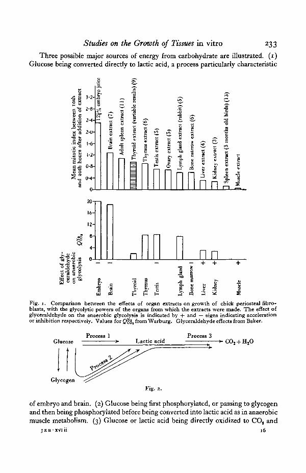
IN a previous paper of this series (Trowell & Willmer, 1939) an account was givenof the effects on the growth of chick periosteal fibroblasts of the addition of salineextracts of various organs from the growing and fully grown bird. Attention wascalled to the fact that those organs and tissues which yielded growth-promotingextracts were just those which have been shown by Warburg and his school(Warburg, 1930) to possess considerable powers of glycolysis. The parallelism isillustrated in Fig. 1. In the upper series are plotted the mean mitotic indices for thefibroblast cultures during the period from the tenth to the twentieth hour after theaddition of the tissue extracts, at which time the effects produced by such extractshave been shown to be most comparable. In the lower series, the anaerobic glyco-lytic activities, as measured by lactic acid production, of the corresponding tissuesare plotted. Comparison of these two sets of figures at once suggests that the organextracts may exert their effects by influencing the carbohydrate metabolism of thetissues to which they are applied. The only apparent exception is the thyroid, andthe effects of extracts of that organ on the growth of cultures were very variableand probably other factors were involved.
The work of Ashford & Holmes (1929), Needham & Nowinski (1937), Needham& Lehmann (1937 a, b) and Baker (1938) now emphasizes the correlation betweenhighly glycolysing tissues and the direct breakdown of glucose to lactic acid bysome mechanism which does not involve the intervention of the phosphorylatingsystems characteristic of the muscle cycle. Moreover, it is significant that tissueswhich show this property of direct glucose breakdown are again precisely thosewhose extracts are growth-stimulating (see Fig. 1). It is, of course, not yet knownwhy they should be growth-stimulating, but it seems conceivable that the extractsmay contain substances which affect the direct utilization of glucose by the tissues,and that this altered or increased carbohydrate metabolism may then affect thegrowth. Or possibly the extracts may contain substances which link up direct non-phosphorylating glucolysis with protein synthesis. In either case the carbohydratemetabolism would affect growth and the following working hypothesis would seemto be justifiable. It may be most easily appreciated by reference to the highlysimplified diagram in Fig. 2.
Studies on the Growth of Tissues in vitro 233
Three possible major sources of energy from carbohydrate are illustrated. (1)Glucose being converted directly to lactic acid, a process particularly characteristic
11HIfiio a
II
3-2-
2-8-
2-4-
2-0-
1-6-
1-2-
0-8-
(M-
ft
20
16-
12-
#£ | |
.5e .=m a ? I
£ 2
£ s
£QJ
a.
ti
"3£I
CO
' w *^ <&
•3 4J "3
3WJ
4-
.s o- n nn1
W CQ E- H E-
Fig. 1. Comparison between the effects of organ extracts on growth of chick periosteal fibro-blasts, with the glycolytic powers of the organs from which the extracts were made. The effect ofglyceraldehyde on the anaerobic glycolysis is indicated by + and — signs indicating accelerationor inhibition respectively. Values for Q&J, from Warburg. Glyceraldehyde effects from Baker.
Glucose
Glycogi
Process 1Lactic acid
Process 3
Fig. a.
of embryo and brain. (2) Glucose being first phosphorylated, or passing to glycogenand then being phosphorylated before being converted into lactic acid as in anaerobicmuscle metabolism. (3) Glucose or lactic acid being directly oxidized to CO, and
jEB-XVlii 16
234 C. M. POMERAT and E. N. WILLMER
H,0 as in most tissues under aerobic conditions. Process (3) necessitates a supplyof oxygen, while the other two are essentially anaerobic.
All or any of these processes might be involved in the phenomenon of growthby cell division. Experiments to be described in this paper, together with otheraccumulated data, make it probable that growing tissues make use primarily ofprocess (1), glucolysis. The evidence is as follows:
If, as has been suggested, the important process in growth is connected with thedirect breakdown of glucose (glucolysis) it should be possible to inhibit it by anymeans which inhibits glucolysis. Now it is known (Mendel, 1929; Needham &Lehmann, 193 7 a and b; Baker, 1938; Lehmann & Needham, 1938) that this processcan be specifically inhibited in tissues and tissue breis by the addition of glyceralde-hyde, and that this inhibition becomes almost complete at a concentration of aboutO-OO2 M glyceraldehyde. Growth also, if the assumption be correct, should beinhibited by the addition of glyceraldehyde in this concentration, assuming that thereagent can penetrate the cell walls of living healthy cells, or at least come into contactwith the reacting system, and that it is not toxic in any other way.
Cultures of chick periosteal fibroblasts were therefore grown in Carrel flasks,by the usual technique required for photographic recording, with a fluid phasecontaining embryo juice (10% in Tyrode solution). The periphery of the culturewas photographed every 6 min. from the twenty-fourth hour after planting. Thiswas carried on for from 4 to 6 hr., so that the normal growth rates of the culturescould be established. Individual variation between different cultures was found tobe such as to make this procedure advisable. Then the embryonic juice was removedand replaced by a fresh quantity (1 c.c.) containing the substance to be investigated,in this case glyceraldehyde. Different concentrations of the glyceraldehyde weretried. The results of a typical experiment are shown in Fig. 3. The curve at thebottom of the diagram shows the effect of adding 0-002 M glyceraldehyde which hadbeen previously boiled in alkaline solution and then neutralized. This processcaramelized the glyceraldehyde, and the absence of any effect upon the growthrate shows that the solution used was not toxic or in any way injurious because ofimpurities. The other curves show clearly that glyceraldehyde very strongly inhibitsgrowth, and the inhibition becomes maximal in the same concentration as thatnecessary to produce complete inhibition of glucolysis. Smaller concentrationswere proportionately less effective. The result might have been due to the conver-sion of the glyceraldehyde into lactic acid, but the effect of 0002 M lactic acid on thegrowth of cultures was found to be negligible. In other words the cessation of growthwas genuinely due to the glyceraldehyde itself or to some other process in whichglyceraldehyde is involved. The inhibition by fresh solutions was always completeand generally irreversible, but by older solutions it was less complete and was thenfound to be reversible as illustrated in Fig. 4. This is almost certainly connectedwith the fact that glyceraldehyde slowly, in the course of a few weeks, polymerizesinto an inactive substance. Under the influence of an effective dose all visibleactivity of the cells was brought to a standstill; the cells ceased to migrate and becamerounded off. Many of the cells in the culture whose behaviour is illustrated in Fig. 4

4
3
2
I
0
4
3
2
a '•oc3 oo
I
1 1
1 1
i i
0002 Af glyceraldehydeV (16 experiments)
0001 Af glyceraldehyde• (5 experiments)
00005 Af glyceraldehydef\ (2 experiments)
0002 Af caramelised glyceraldehyde(3 experiments)
0 2 4 6
Time in hours
10 12
Fig. 3. The effect of glyceraldehyde on the growth of chick periosteal fibroblasts. Glyceraldehydeadded at zero hour. There is 10 % embryo juice in the medium throughout the experiment.
•8 3
i 225
O002 Af glyceraldehyde10 % embryo j uice
_ 10% embryojuice
i 6 Y
15% embryo juice added afterwmhing with 1*5 <ui tyiodesolution
4 6 8 10 12 14 16 18 20 22 24 26 28Time in hours
Fig. 4. Recovery of a culture after inhibition by glyceraldehyde. This does not regularly occur,but the older the solution of glyceraldehyde the more easily does it take place.
16-2

C. M. POMERAT and E. N. WILLMER
were rounded off but recovered completely when the glyceraldehyde was removedand replaced by fresh embryo juice.
These results were definitely encouraging and lent support to the hypothesisconcerning the relation between growth and glucolysis, but further investigationsproduced results not so satisfactory to the hypothesis, although interesting in
8-a
ii2
is
0002 Af propyl aldehyde10 % embryo juice
(8 experiments)
8
Propyl aldehyde washed away andreplaced by 10% embryo juice
(4 experiments)
1 1 T~12 14 16 18 ~20~ 22 24
Time in hours
Propyl aldehyde
4r
0-002 M methyl glyoxal10 % embryo juice
(7 experiments)
0001 M methyl glyoxal10 % embryo juice
(2 experiments)
Time in hours Time in hoursMethyl glyoxal
0-002 M benialdehyda10 % embryo juice
(2 experiments)
4 2 O l
Time in hoursBenzaldehyde
Fig- 5-
themselves. They are not as yet fully explained. By boiling glyceraldehyde in acidsolution it is converted into methyl glyoxal and, as such, has no effect on glucolysis(Needham & Lehmann, 1937 b), nor indeed do any other aldehydes (Needham &Lehmann, 1937 b; Baker, 1938) which have so far been tried. On the other hand,the addition of 0-002 M concentrations of methyl glyoxal, propyl aldehyde, butylaldehyde and benzaldehyde to tissue cultures produced complete inhibition of

Studies on the Growth of Tissues in vitro 237
growth, migration and cellular activity (Fig. 5). The corresponding acids, pyruvic,propionic and benzoic and also glyceric and lactic, when neutralized with NaOHand applied in the same molar concentrations, produced no similar deleterious effects(Figs. 6, 7). The inhibition seems therefore to be a property of aldehydes as such.The reversibility of the effect has only been tested in the case of propyl aldehyde,
81-
7
6
sIso•a2 A
2
I
9
8
7
6
31 5o
S 4§
3
_ 10 % embryojuice
0'002 M glyceric acid10 % embryo joice
(2 experiments)
2 4 6 8Time in hoars
10 12 14 16
0002M benzoic acid10 % embryo juice
(2 experiments)
.growth rate in10% embryo
juice
2 4 6
Time in hours
10 12 14 16
Fig. 6. Glyceric and benzoic acids. The solutions were brought to pH 7-5 by means of NaOH.
but then the inhibition was found to be completely reversible even after it wasproduced by a fresh 0-002 M solution, so that it differs slightly in this way from theeffect of glyceraldehyde.
This property of aldehydes to inhibit growth and cell activity, though in itselfextremely interesting, is disturbing to the hypothesis when compared with the
2 3 8 C. M. POMERAT and E. N. WILLMER
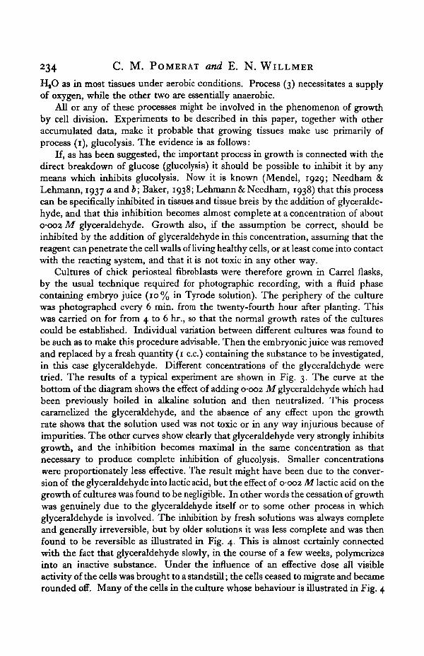
specificity of glyceraldehyde for the glucolytic process. So far it is unexplained, buta study of other aldehydes and their effects should prove illuminating. Perhaps twomechanisms are involved, as is suggested by the fact that recovery after propyl
3 3•oc
ii2
0-0O2 M 6odium pyruvate
10 % embryo juice
10 12 14 16Time in hours
18 20 22 24 26
0002 M sodium pyruvate0-002 Af glyceraldehyde10 % embryo juice
6 4 2 0 2 4 6 8 10 12 14 16 18 20
Time in hours
1oo
1
5
4
3
2
1
6
A / \- A / \/ \ / \-/ W \L V ^_ T
10% embryojuice
4 2 C
0-002 M glyceraldehyde10 % embryo juice
A/ \^ \s
2 4 6 8 lb
0-001 Af glyceraldehyde0001 M sodium pyruvate10% embryo juice
12 14 16 18 20
Time in hours
Fig. 7.
aldehyde is certain, but it is not so from glyceraldehyde at the same molar concen-tration. It is interesting to find that Boyland & Mawson (1938) have recentlyshown that certain aldehydes, notably citral, phloroglucinaldehyde and 3-4-dimeth-oxybenzaldehyde have been able to reduce the growth rate of Crocker Sarcoma180 in the living animal.

Studies on the Growth of Tissues in vitro 239
It may be significant that the concentration of 0-002 M for the glyceraldehydeis the same for growth inhibition as it is for glucolysis, but on this evidence aloneit is obviously impossible to link growth with glucolysis. Moreover, the blockingof embryonic glucolysis by glyceraldehyde is reversed by the addition of pyruvate.This does not appear to be the case with growth, for pyruvic acid has been foundto have no influence on growth of cultures either when applied alone, in conjunctionwith glyceraldehyde or after glyceraldehyde (Fig. 7). Finally, Sullmann (1938)concludes that the formation of hexose phosphate from glucose by lens tissue isinhibited by o-oi M glyceraldehyde, propyl aldehyde, benzaldehyde and formalde-hyde. This concentration is, of course, considerably higher than those used in thisinvestigation, but in growth experiments the reagents are allowed to act for aconsiderably longer time and so might eventually become effective. On the otherhand, the evidence adduced by Lehmann & Needham (1938) seems to rule out thepossibility of phosphorylation of glucose in the embryo, at any rate under anaerobicconditions; and further evidence now to be described points also to the sameconclusion.
The glucolytic process is affected, like the phosphorylating glycolysis, by theaddition of sodium fluoride and sodium iodoacetate, but the concentrations re-quired to produce inhibition are not the same for the two processes. Both substancescan be added to cultures, and an investigation of the concentrations necessary toinhibit growth has produced very suggestive results. At the same time it shouldbe remembered that these substances may also cause other effects in the cell, as,for example, the poisoning by iodoacetate of enzyme systems not related to themetabolism of glucose.
Sodium fluoride is found to inhibit phosphorylation in the muscle cycle at aconcentration of 0-005 M, but at this concentration a considerable amount of directglucose breakdown still occurs, namely about 45 % (Needham & Lehmann, 1937 b).This latter process is not stopped completely until a concentration of 0-02 M isreached. Fig. 8 shows the results of experiments in which sodium fluoride wasadded to cultures of periosteal fibrpblasts growing in vitro. It may be concludedfrom these results that mitoses although considerably reduced in number and aftera time suppressed by concentrations between 0-003 and o-oi M are not immediatelyknocked out until a concentration of 0-02 M is reached. Direct observation of thecultures confirms this. The cells are manifestly disorganized by a concentration of0-02 M almost at once, while at 0-007 M they remain healthy in appearance for aconsiderable time. There is thus a very striking parallel between the concentrationsof fluoride necessary to produce inhibition of growth and inhibition of directglucose breakdown. No such parallel exists between growth and phosphorylatingglycolysis, for mitoses still occur when this is presumably completely suppressed(0-007 M)- This result becomes even more significant when taken in conjunctionwith similar results obtained in experiments in which sodium iodoacetate took theplace of fluoride (Fig. 9). The results, though similar, show one striking and im-portant difference. Growth is checked when the concentration reaches 0-00005 M,the concentration at which non-phosphorylating glucolysis is checked. Ten times

0-0033 M(2 experiments)
0 2 4 6Time in hours
8 10 12
Fig. 8. The effect of different concentrations of sodium fluoride in io% embryo juiceon the growth rate of chick periostea! fibroblasts.
•B 3
o
10% embryojuice
0-00002 M 0-00005 M
B 4
.3 3
u
1
2 4 6 8 10 12
0-0002 M
- 10 7? embryojuice
8 10 12
Time in hours4 6 8 10 12
Time in hoursFig. 9. The effect of different concentrations of sodium iodoacetate in 10% embryo juice on the
growth rate of chick penosteal fibroblasts. Two experiments at each concentration.
Studies on the Growth of Tissues in vitro 241
this concentration is necessary to stop the phosphorylating mechanism. It shouldbe noted that these results are in marked contrast to those obtained by Ellis (1933)on segmenting eggs of Urechis caupo and of Strongylocentrotus purpuratus, in whichhe found that concentrations of o-oi M sodium iodoacetate and fluoride had noeffect on cleavage. The mechanism in these eggs must be very different.
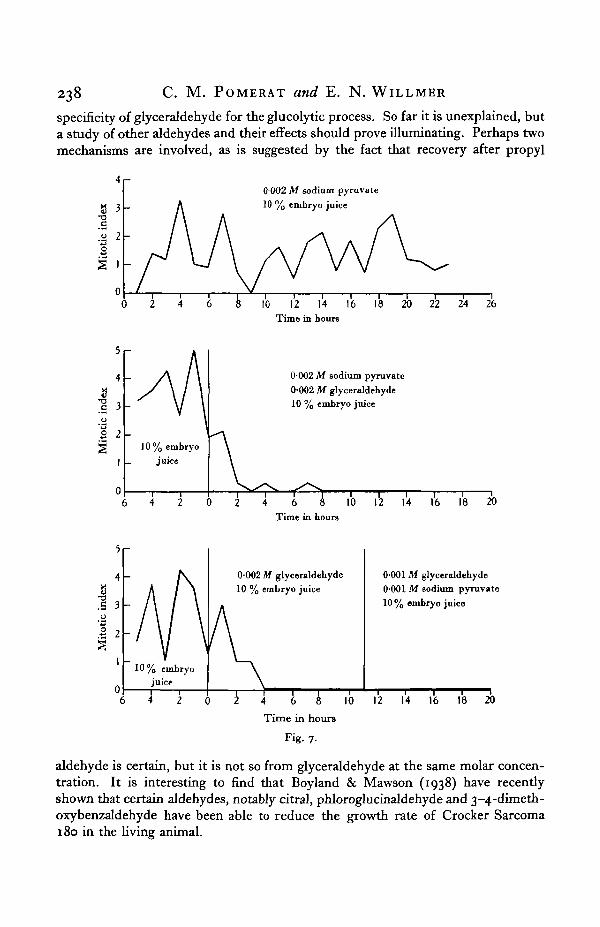
There is another substance which characteristically stops the process of phos-phorylation, namely, phlorizin. It can be effectively used for this purpose in con-centrations of 0-005 M. On applying it to cultures in this and even strongerconcentrations it produced no obvious decrease in the growth rate. The results areshown in Fig. 10. It is just possible that phlorizin does not reach the part of the cellwhere it can exert its effect, but, on the other hand, it acts on the kidney in vivo.
•o
'V10 % embryo juice
0 2 4Time in hours
10 12
Fig. 10. Effect of adding phlorizin in concentrations between o-oo8 M and 00033 Min 10% embryo juice (7 experiments).
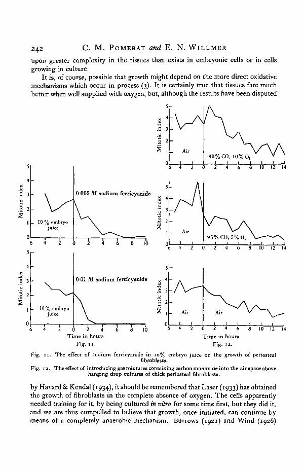
Lastly, it has been shown by Mendel (1937) that the addition of sodium ferri-cyanide to tissues, especially sarcoma, reduces their power of aerobic glycolysis.Anaerobic glycolysis is unaffected. The exact significance of this has not yet beenworked out in terms of direct glucose breakdown, but the two may be connected.The effects of ferricyanide on growth are precisely similar to its effects on aerobicglycolysis. The latter is inhibited to the extent of about 25 % by solutions of o-ooi Mand completely suppressed at o-oi M. Growth is quite definitely checked at0-005 M and is irreversibly stopped at o-oi M (Fig. 11).
Thus by the use of these inhibitors there is considerable evidence that growthof fibroblasts in cultures depends upon energy obtained from direct glucolysisrather than from phosphorylating glycolysis. Growth proceeds more or less inde-pendently of the latter, if indeed the latter process is occurring at all in these cultures;but, should the former be checked, then the growth suffers with it.
Referring back once again to the simplified diagram on page 233, it may be con-cluded that growth depends on process (1) rather than on process (2). The formeris probably the more fundamental method, as witnessed by its apparent dominancein the embryo. The second process which is clearly more elaborate may depend
242 C. M. POMERAT and E. N. WILLMER
upon greater complexity in the tissues than exists in embryonic cells or in cellsgrowing in culture.
It is, of course, possible that growth might depend on the more direct oxidativemechanisms which occur in process (3). It is certainly true that tissues fare muchbetter when well supplied with oxygen, but, although the results have been disputed
•a.5 3U
'•§ 2
_ Air90% CO, 10% 0,
1 1 1 1
0-002 M sodium ferricyanide
8 10 12 14
9 4XIc
".a 3
I 2_ Ai
8 10 12 14
- 10% embryojuice
0-01 M sodium ferricyanide
0 2 4Time in hours
Fig. 11.
8 10 0 2 4 6
Time in hoursFig. 12.
8 10 12 14
Fig. 11. The effect of sodium ferricyanide in 10% embryo juice on the growth of periostealfibroblasts.
Fig. 12. The effect of introducing gas mixtures containing carbon monoxide into the air space abovehanging drop cultures of chick periosteal fibroblasts.
by Havard & Kendal (1934), it should be remembered that Laser (1933) has obtainedthe growth of fibroblasts in the complete absence of oxygen. The cells apparentlyneeded training for it, by being cultured in vitro for some time first, but they did it,and we are thus compelled to believe that growth, once initiated, can continue bymeans of a completely anaerobic mechanism. Burrows (1921) and Wind (1926)

Studies on the Growth of Tissues in vitro 243
have also shown that very embryonic cells can display activity under anaerobiosis,but it is not clear from much of this work whether mitoses were occurring or not.In this connexion it is significant to note that Ephrussi et al. (1929), who examinedcultures after treatment with atmospheres containing only minimal quantities ofoxygen, found that mitoses were arrested in prophase, although the later stagescould, like the migration of the cells, go on almost unchecked. It is, however, notclear for how long the cells had been treated and the acidity produced in the mediumin the absence of oxidations might be sufficient to account for the results. On theother hand, sarcoma cells, which produce far more lactic acid, can survive anaero-biosis more easily than normal cells. The results from experiments on the effectsof anaerobiosis are therefore still somewhat equivocal.
It need hardly be pointed out that both processes (1) and (2) are anaerobic, and theevidence so far adduced seems to stress the importance of process (1). This becomesstill more significant when attempts, other than by altering the oxygen pressure,are made to vary the rate at which process (3) is occurring. There are several ways inwhich the respiratory exchanges of a tissue may be affected both favourably andadversely. Unfortunately few of the reagents are so specific that they affect respira-tion alone, but the addition of such substances to tissues growing in vitro bringsfurther evidence in favour of the independence of growth and respiration, for theyhave but little immediate effect upon the rate of growth. Before these experimentsare described it should be made quite clear that there is no question of denying theimportance of respiratory exchanges in the metabolism of tissues growing in vitro,which would be absurd, but the evidence shows that they are not the primarychanges concerned with the growth process. They rather, as in muscle, allow cata-bolites to be removed and may supply energy necessary for the continuous workingof the first process. That is to say that growth can go on at least for a time on gluco-lysis alone, and at a time when the respiration has been reduced to a minimum.
In the first place the action of carbon monoxide on the growth of cultures isinteresting. The actual part played by carbon monoxide in the chemistry of the cellis still somewhat obscure. It appears (Laser, 1937), in some tissues at least, to affectthe Pasteur reaction and increase the production of lactic acid rather than to decreasethe respiratory exchanges, although the cytochrome system is most probably putout of action. It is likely therefore that the increase in lactic acid is produced ratherby accumulation of this substance through faulty removal than by an increase insugar breakdown. It is interesting to note therefore that the introduction of amixture of 95 % carbon monoxide and 5 % oxygen into culture chambers of thetype described by Gill (1938) had no immediately deleterious effect upon the growthof the cells, which were growing on cover-slips in the chamber. The results withcarbon monoxide are illustrated in Fig. 12.
Similarly, the addition of cyanide to cultures produced striking results. Again,its action on cellular metabolism as a whole is complex and it may even act as astimulant to certain processes. Experiments, however, clearly show that, in thosecultures to which it was added, growth continued in concentrations of cyanide atwhich the respiration of the cells must have been profoundly altered (Fig. 13).

244 C. M. POMERAT and E. N. WILLMER
0-0005 M HCN still allowed considerable growth and even in a concentration of0-002 M mitoses were observed to be occurring as long as 10 hr. after the additionof the cyanide. That is to say the cells were definitely entering and completing theprocess of mitosis under these conditions.
0-002 M(2 experiments)
Time in hours
Fig. 13. The effect of different concentrations of HCN in 10% embryo juiceon the growth of chick periosteal fibroblastg.
The results with sodium azide were similar. This substance has actions verylike those of cyanide in inhibiting cellular respiration and oxidation of cytochrome(Keilin, 1936). Cytochrome oxidase is 80-95 % inhibited by o-ooi M NaN3. Theeffects of azide on growth are seen in Fig. 14 and it is clear that this concentrationstill allows of considerable growth. With both azide and cyanide the growth rateshows a progressive diminution, but undiminished activity would be no morelikely under these conditions than that a muscle should go on contracting anaero-bically for an indefinite period.

Studies on the Growth of Tissues in vitro 245
00001 a
2 4 £Time in hours
10 12 6 4 2 0 2 4 6 8 10 12Time in hours
Fig. 14. The effect of different concentrations of sodium azide in 10% embryo juiceon the growth of chick periosteal fibroblasts.
5f-
I'u•3O '
I2
10 % embryo juiceI I I
8 10 126 4 2 0 2 4 6Time in hours
Fig. 15. The effect of o-oi M sodium malonate on the growth of chick periosteal fibroblasts.
Sodium malonate has a specific action in inhibiting succinodehydrogenase andreducing respiration (Quastel, 1926; Gozsy & Szent-Gyorgi, 1934). Its effects ongrowth are negligible (Fig. 15).
In contrast with all the former substances, sodium fumarate stimulates respira-tion (Gozsy & Szent-Gyorgi, 1934; Stare & Baumann, 1936). But it too has no
246 C. M. POMERAT and E. N. WILLMER
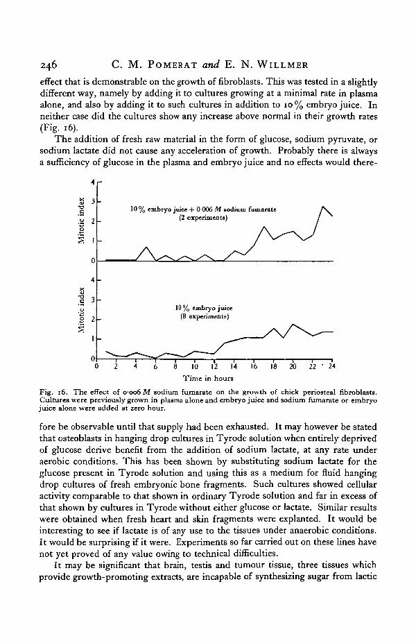
effect that is demonstrable on the growth of fibroblasts. This was tested in a slightlydifferent way, namely by adding it to cultures growing at a minimal rate in plasmaalone, and also by adding it to such cultures in addition to 10% embryo juice. Inneither case did the cultures show any increase above normal in their growth rates(Fig. 16).
The addition of fresh raw material in the form of glucose, sodium pyruvate, orsodium lactate did not cause any acceleration of growth. Probably there is alwaysa sufficiency of glucose in the plasma and embryo juice and no effects would there-
4 r
s 3-oc
.52 2S
10 % embryo juke + 0006 M sodium fumarate(2 experiment*)
10% embryo juice(8 experiments)
.s ->u
2 2
0 2 4 6 8 10 12 14
Time in hours
Fig. 16. The effect of C006 M sodium fumarate on the growth of chick periosteal fibroblasts.Cultures were previously grown in plasma alone and embryo juice and sodium fumarate or embryojuice alone were added at zero hour.
fore be observable until that supply had been exhausted. It may however be statedthat osteoblasts in hanging drop cultures in Tyrode solution when entirely deprivedof glucose derive benefit from the addition of sodium lactate, at any rate underaerobic conditions. This has been shown by substituting sodium lactate for theglucose present in Tyrode solution and using this as a medium for fluid hangingdrop cultures of fresh embryonic bone fragments. Such cultures showed cellularactivity comparable to that shown in ordinary Tyrode solution and far in excess ofthat shown by cultures in Tyrode without either glucose or lactate. Similar resultswere obtained when fresh heart and skin fragments were explanted. It would beinteresting to see if lactate is of any use to the tissues under anaerobic conditions.It would be surprising if it were. Experiments so far carried out on these lines havenot yet proved of any value owing to technical difficulties.
It may be significant that brain, testis and tumour tissue, three tissues whichprovide growth-promoting extracts, are incapable of synthesizing sugar from lactic
Studies on the Growth of Tissues in vitro 247
and pyruvic acids whereas other tissues like liver and kidney can do so (Benoy &Elliott, 1937). This might suggest that one of the actions of the extracts inpromoting growth may be in stimulating carbohydrate breakdown rather thansynthesis.
All these observations therefore provide evidence for the correctness of thehypothesis that the fundamental metabolic process concerned with growth is directglucose breakdown, rather than respiration, or phosphorylation as it occurs inanaerobic muscular metabolism. No single piece of evidence is in itself sufficient,but taken all together the strength of the case is apparent.
DISCUSSION
The significance of these findings may now be shortly discussed. In the firstplace Needham & Nowinski (1937) have shown fairly clearly that the embryo chickdoes not metabolize glycogen but relies on glucolysis or direct sugar breakdown.Secondly, growth can occur at extremely low oxygen pressures and, Laser (1933)believes, even under conditions of complete anaerobiosis. Moreover many workerson tissue culture have shown that glucose is necessary for tissue growth, and thatglycogen is of comparatively rare occurrence in the cells and, certainly, it is not anecessity that it should be there in detectable amounts before growth can occurfreely. Glucose has been shown to be used by tissues in vitro (Krontowski,1929; Krontowski & Bronstein, 1926), and it may have a pronounced proteinsparing action (Watchorn & Holmes, 1927; Litter et al., 1937). This effect is greaterin tissues in which mitoses are abundant than it is in tissues whose growth rates areless. The experiments described in this paper make it clear that respiratory poisonsdo not immediately put a stop to mitotic activity, nor does fumarate which increasesrespiratory activity increase the mitotic rate. Conditions which interfere with theprocess of phosphorylation do not at the same time affect growth. There wouldtherefore seem to be two remaining possibilities; either the requirements for growthenergy are so small or so little concerned with carbohydrates that gross disturbanceof the metabolism of the latter may occur without depriving the cells of their energysupplies, or else the essential process in the carbohydrate breakdown is neitherrespiration nor phosphorylation but glucolysis. Direct evidence for this is certainlyscanty. Besides the above-mentioned utilization of sugar by the embryo, and bytissue cultures, there is the evidence from the sources of active growth-promotingextracts, which always seem to be from glucolysing tissues, at least three of whichare unable to synthesize sugar from lactic or pyruvic acids. There is also the fact thatgrowth is inhibited by glyceraldehyde, though this is somewhat discounted by theinhibition caused by other aldehydes. And finally, there is the inhibition causedby appropriate doses of sodium fluoride and of sodium iodoacetate, which gainsconsiderable strength as evidence from the fact that the amount of inhibitornecessary to stop growth is in the one case larger and in the other smaller than theamount necessary to stop phosphorylation, in this way following the dosage requiredfor the inhibition of glucolysis.
248 C. M. POMERAT and E. N. WILLMER
It may be concluded therefore that the hypothesis that growth by cell divisionis intimately connected with direct glucose breakdown is not without foundation.One final point should be emphasized. There is nothing in the evidence presentedin this paper to suggest that the actual process of mitosis is the thing which isaffected by glucolysis, but rather is it clear that cells when glucolysing are capableof dividing and that the characteristic of actively growing tissues is their power ofglucolysis, upon which they rely for growth.
SUMMARY
1. Growth of chick periosteal fibroblasts is inhibited by 0-002 M solutions ofglyceraldehyde, benzaldehyde, butyl aldehyde, propyl aldehyde and methyl glyoxal.
2. The growth is not inhibited by sodium pyruvate, lactate, propionate,glycerate or benzoate.
3. Sodium pyruvate does not affect the growth under the conditions of theexperiments, nor does it alter the inhibition brought about by glyceraldehyde.
4. Growth is inhibited by sodium fluoride and sodium iodoacetate in theconcentrations in which these inhibit glucolysis. Growth inhibition does notcorrespond to inhibition of phosphorylation.
5. Growth is not inhibited by 0-008 M phlorizin.6. Growth is not immediately inhibited by an atmosphere containing 95 % CO
a n d 5 % 0 2 .7. Growth is not immediately inhibited by 0-002 M sodium azide, 0-002 M
HCN or by o-oi M sodium malonate. Azide and cyanide reduce the growth rateafter some hours.
8. Growth is not accelerated by the addition of 0-006 M sodium fumarate.9. Growth is inhibited by o-oi M sodium ferricyanide.10. The relationship between carbohydrate metabolism and growth by cell
division is discussed in the light of these results.
The expenses of this research have been in part defrayed by a grant from theBritish Empire Cancer Campaign, to whom the authors wish to express theirgratitude. The work was performed while C M . Pomerat was holding a RockefellerTravelling Fellowship.
The authors also wish to express their sincere thanks to Dr H. Lehmann forconstant advice and for providing many of the reagents in suitable form, and also toMrs Simon-Reuss for invaluable technical assistance.

Studies on the Growth of Tissues in vitro 249
REFERENCES
ASHFORD, C. A. & HOLMES, E. G. (1929). Biochem. J. 23, 748.BAKER, Z. (1938). Biochem. J. 32, 332.BENOY, M. P. & ELLIOTT, K. A. C. (1937). Biochem. J. 31, 1268.BOYLAND, E. & MAWSON, E. H. (1938). Biochem. J. 32, 1982.BURROWS, M. T. (1921). Proc. Soc. exp. Biol., N.Y., 18, 133.ELLIS, E. L. (1933). J. cell. comp. Pkytiol. 4, 127.EPHRUSSI, B., CHEVILLARD, L., MAYER, A. & PLANTEFOL, L. (1929). Aim. Pkysiol. Pkysicochim.
Biol. 5, 642.GILL, P. M. (1938). Biochem. J. 32, 179a.GOZSY, B. & SZENT-GYORGI, A. (1934). Hoppe-Seyl. Z. 224, 1.HAVARD, R. E. & KENDAL, L. P. (1934). Biochem. J. 28, 1121.REILIN, D. (1936). Proc. Toy. Soc. B, 121, 165.KHONTOWSKI, A- (1929). C.R. Soc. Biol., Paris, 102, 523.KRONTOWSKI, A. & BSONSTEIN, J. (1926). Arch. exp. Zellforsch. 3, 32.LASER, H. (1933). Biochem. Z. 264, 72.
(i937)- Biochem. J. 31, 1677.LEHMANN, H. & NEEDHAM, J. (1938). Enzymologia, 5, 95.LITTER, J., MARBLE, B. B. & SALTER, W. T. (1937). Amer. J. Cancer, 31, 268.MENDEL, B. (1929). Klin. Wtchr. 8, 169.
(i937)- Amer.J. Cancer, 30, 549.NEEDHAM, J. & LEHMANN, H. (1937 a). Biochem. J. 31, 1210.
(19376). Biochem. J. 31, 1913.NEEDHAM, J. & NOWINSKI, W. W. (1937). Biochem. J. 31, 1165.QUASTEL, J. H. (1926). Biochem. J. 20, 166.STARE, F. J. & BAUMANN, C. A. (1936). Proc. roy. Soc. B, 121, 338.SOLLMANN, H. (1938). Biochem. Z. 296, 325.TROWELL, O. A. & WILLMER, E. N. (1939). J. exp. Biol. 16, 60.WARBURG, O. (1930). The Metabolism of Tumours. London: Constable.WATCHORN, E. & HOLMES B. (1927). Biochem. J. 21, 1391.WIND, F. (1926). Biochem. Z. 179, 384.