recent applications of infrared thermography for … · inframation 2009 proceedings 2009-053...
TRANSCRIPT

InfraMation 2009 Proceedings 2009-053 Church
Recent Applications of Infrared Thermography for Animal Welfare and Veterinary Research: Everything from Chicks to Elephants John S. Church, Nigel J. Cook and Allan L. Schaefer Thompson Rivers University, Kamloops, British Columbia, Canada ABSTRACT Eye temperature, measured using infrared thermography (IRT), is a non-invasive tool for evaluating the stress response in cattle, bison and elk to surgical procedures. Infrared thermography has recently revealed that a rapid drop in eye temperature is likely a sympathetically mediated response via vasoconstriction that can be used to detect fear and/or pain related responses in animals to different handling procedures. Infrared thermography may be used in the future as an objective outcome based measure for the evaluation and assessment of animal welfare. Assessing the feather cover of laying hens by IRT, for example, has proven to be a fast, objective and accurate method that more precisely reflects either actual feather cover or areas of bare skin. Infrared thermography has historically been used primarily as a veterinary diagnostic tool in horses and can be expanded for use in other species as varied as elephants. In addition, IRT methodologies have been employed to detect medical conditions as diverse as naval infections in newborn chicks and mastitis in dairy cattle. Recent data has demonstrated that IRT scans of the orbital area in calves allows for the efficacious identification of animals at earlier stages of illness, often several days to over one week before clinical signs were manifest. In concurrent research, individual animal identification using radio frequency identification tags (RFID) and an IRT camera located at a water station have been employed in a remote monitoring system to automatically collect thermal data for the early detection of disease in feedlot cattle. INTRODUCTION Infrared thermography (IRT) has been used for many years in human and veterinary medicine. Thermal imaging has been used to study many different animals (see McCafferty 2007 for review). Infrared thermography can examine many different aspects of thermal physiology, diagnose injury and disease and is a useful technique for counting animal populations. The great advantage of IRT in animal research is that measurements can be made without touching the animal and can be made either at close range (<1 m) or at large distances (>1000 m). Infrared thermography has primarily been used as a diagnostic tool in veterinary science. A major application has been to diagnose injury and disease in horses (Figure 1) and there have been several useful studies detailing factors that impact normal temperature distributions and outline appropriate measurement protocols (see review by Eddy et al., 2001).

InfraMation 2009 Proceedings 2009-053 Church
Figure 1. The use of IRT to diagnose injury in horses. IRT pioneers Dr. Tracy Turner and Dr. Jim Waldsmith at the Veterinary Thermal Imaging Seminar, March 10-13, 2005 in San Luis Obispo, California, demonstrating the use of IRT in horses. A medical thermogram represents the surface temperature of skin, making thermography very useful for the detection of inflammation. Tendons and joints can show inflammatory changes up to two weeks before clinical lameness is apparent. Note the increased heat in the right front coronary band on the left image and the right front medial quarter on the right image. USE OF INFRARED THERMOGRAPHY IN ZOO ANIMALS Abnormal or asymmetrical temperature distributions have been used as indicators of underlying problems with blood circulation or inflammatory responses in numerous other species besides horses. When assessing a thermal image, asymmetry of 1oC or more is often significant and indicates possible pathology. In March, 2006 we were requested by the staff at the Valley Zoo in Edmonton, Alberta, Canada to undertake a thermographic examination of Lucy, a remarkable Asian elephant who has made her home at the Valley Zoo for 31 years (Figure 2). At an early age, Lucy was diagnosed with rheumatoid arthritis, but lately Lucy appeared to have some additional health challenges that were proving difficult to diagnose by her usual caregivers. The elephant appeared to be exhibiting symptoms of leg pain. The zoo curators had been noticing Lucy appeared to be reluctant to apply her full weight on one of her front legs, but her caregivers were unsure exactly what part of the leg was causing the problem. Irritation in her foot, her carpus or shoulder joint could all be causing Lucy to favor the leg. Lucy was brought inside the building for two hours prior to the examination to prevent solar loading. A thermal scan was performed and the left front leg was indeed determined to be the cause of the problem (Figure 3).
Figure 2. Veterinary thermographic examination of the Asian elephant Lucy. Infrared images of the bottom of Lucy’s feet were also taken to rule out any foreign object concerns such as a nail or sharp stick as possible causes of foot irritation. The main indication for thermography is to assess entire regions of the animal and determine if “heat” (inflammation) is present when it is not clinically apparent.
InfraMation 2009 Proceedings 2009-053 Church
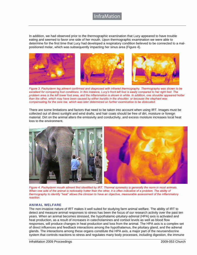
In addition, we had observed prior to the thermographic examination that Lucy appeared to have trouble eating and seemed to favor one side of her mouth. Upon thermographic examination we were able to determine for the first time that Lucy had developed a respiratory condition believed to be connected to a mal-positioned molar, which was subsequently impacting her sinus area (Figure 4).
Figure 3. Pachyderm leg ailment confirmed and diagnosed with infrared thermography. Thermography was shown to be excellent for comparing foot conditions. In this instance, Lucy’s front left foot is easily compared to her right foot. The problem area is the left lower foot area, and the inflammation is shown in white. In addition, one shoulder appeared hotter than the other, which may have been caused by either bursitis in the shoulder, or because the elephant was compensating for the sore toe, which was later determined on further examination to be dislocated. There are some limitations and factors that need to be taken into account when using IRT. Images must be collected out of direct sunlight and wind drafts, and hair coats should be free of dirt, moisture or foreign material. Dirt on the animal alters the emissivity and conductivity, and excess moisture increases local heat loss to the environment.
Figure 4. Pachyderm mouth ailment first identified by IRT. Thermal symmetry is generally the norm in most animals. When one side of the animal is noticeably hotter than the other, it is often indicative of a problem. The ability of thermography to identify “heat” allows the clinician to have an objective, measurable assessment of the inflammatory reaction. ANIMAL WELFARE The non-invasive nature of IRT makes it well suited for studying farm animal welfare. The ability of IRT to detect and measure animal responses to stress has been the focus of our research activity over the past ten years. When an animal becomes stressed, the hypothalamic-pituitary-adrenal (HPA) axis is activated and heat production, as a result of increases in catecholamines and cortisol levels as well as blood flow responses, will produce changes in heat production and loss from the animal. The HPA axis is a complex set of direct influences and feedback interactions among the hypothalamus, the pituitary gland, and the adrenal glands. The interactions among these organs constitute the HPA axis, a major part of the neuroendocrine system that controls reactions to stress and regulates many body processes, including digestion, the immune

InfraMation 2009 Proceedings 2009-053 Church
system, mood and emotions, sexuality, and energy storage and expenditure. This response can be detected using infrared cameras to collect real-time pictorial images, radiometric JPEGs, at a distance from the subject, usually with no need for contact or restraint. IRT has been used successfully to detect cattle that are pre-disposed to producing dark- firm-dry beef, pale-soft-exudative pork in swine, and to monitor transport stress in cattle. Cattle studies have used IRT to assess inflammation due to hot iron branding in cattle in order to determine the extent and duration of inflammation observed on branding sites, scrotal surface temperature as a measure of fertility in bulls and detecting hoof disorders and rises in body temperatures in cattle due to infection. My colleague Dr. Nigel Cook initially investigated the relationship between IRT and cortisol by measuring adrenocortical and metabolic activity in horses to determine HPA activity. Matched blood and saliva samples and IRT eye images were collected at set intervals before and after a challenge. The results showed a significant correlation between maximum eye temperature and both salivary and plasma cortisol suggesting that changes in eye temperature may be associated with activation of the HPA axis (Figure 5).
Figure 5. Endocrinologist Dr. Nigel Cook is shown in the picture on the left collecting saliva from the horses with cotton swabs for later analysis of the hormone cortisol via radioimmunoassay. The IRT image of the horses’ eye is on the right. Other studies have shown increases in eye temperature in response to velvet antler removal in elk and reindeer (Cook et al 2005; Cook et al 2006) (Figure 6), de-horning in bison and de-horning procedures in dairy calves (Stewart et al 2008) (Figure 7).
Figure 6. Infrared thermography was sensitive enough to pick up the %∆ change in eye temperature in Celsius before and after antler removal in young elk that received either lidocaine (Lido) or compression (Comp) anesthesia prior to having their antlers surgically removed, compared to animals that were in the organic treatment group (Org) who received no anesthesia prior to antler removal. A fourth control group that were simply restrained (Restr), and did not have their antlers removed showed the lowest %∆ change in eye temperature.

InfraMation 2009 Proceedings 2009-053 Church
EARLY DISEASE DETECTION Our research into early disease detection has demonstrated the diagnostic potential of IRT under controlled conditions for an induction model of bovine viral diarrhea (BVD). Considering up to 60% of the heat loss from an animal can occur in the infrared range, the observation that radiated heat loss can serve as an early indicator of fever conditions is very logical. Eye temperature, measured using IRT, was more effective at detecting bovine viral diarrhea as changes occurred as early as one day, compared to 5-6 days for other areas such as the nose, ear, body and hooves. Similarly, our research into bovine respiratory disease (BRD) has clearly demonstrated that infrared values were as much or even more efficient than clinical scores, core temperatures or hematology in identifying ill animals prior to the clinical appearance of BRD (Schaefer et al. 2007). Data collected from our research has demonstrated that infrared thermography scans of the orbital area in
Figure 7. The picture on the left is of a young bison calf being scanned with an infrared camera prior to dehorning. The picture on the right is an infrared image of a cattle eye that can be used to quantitatively measure stress during dehorning. The cross indicates the position of the maximum temperature within the area of the eye used for analysis, the medial posterior palpebral border of the lower eyelid and the lacrimal caruncle. This location has proven to be a very stable and useful spot to take temperature measurements in numerous studies. calves was efficacious as an early identifier of bovine respiratory disease onset (Figure 8). Such information would enable the earlier and more targeted treatment of affected animals thereby reducing animal suffering, improving the animal industry economics and reducing the likelihood of promoting antibiotic resistant
Figure 8. Retrospective facial infrared thermography images of the same animal during the onset of bovine respiratory disease. Clinical signs were positive (>3) on days 9-10. microbes. In concurrent research, individual animal identification using radio frequency identification tags (RFID) and an IRT camera located at a water station has been employed to automatically collect thermal data (Figure 9). This situation is impossible to achieve with more invasive measures such as hematology or core body temperatures. In such situations, the use of IRT for the non-invasive detection of early stages of disease in livestock is possible. One such strategy that we have tested is to incorporate an infrared scanning station
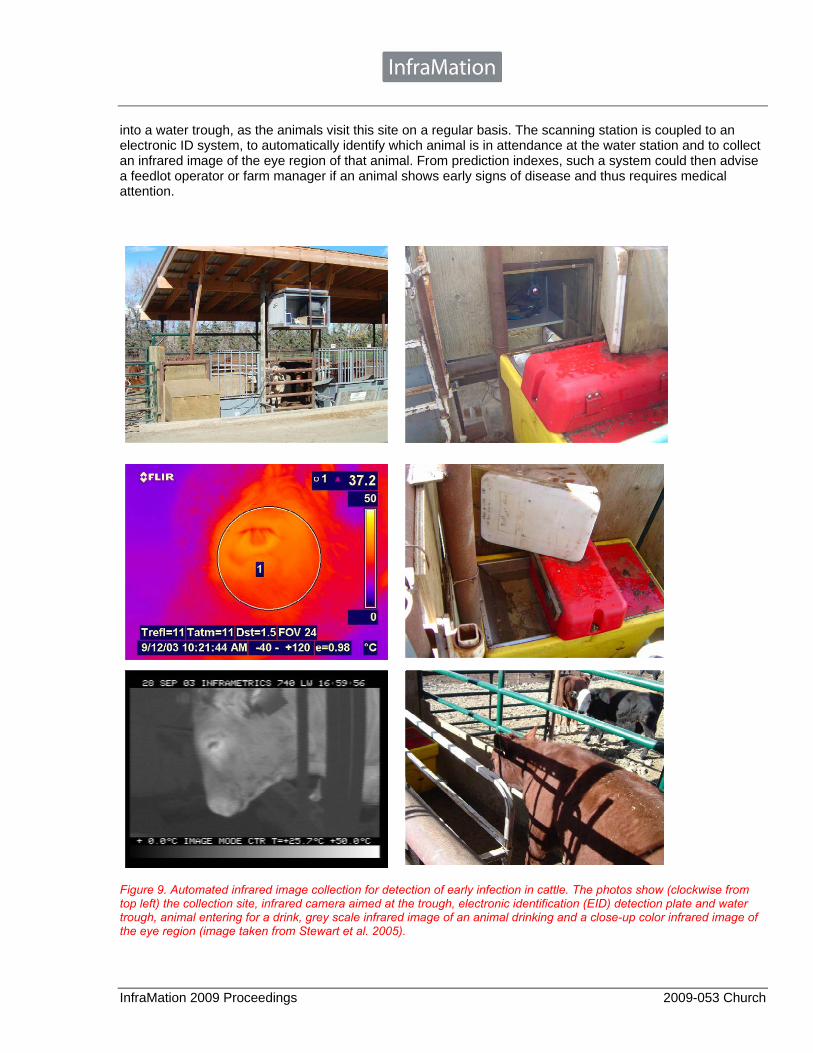
InfraMation 2009 Proceedings 2009-053 Church
into a water trough, as the animals visit this site on a regular basis. The scanning station is coupled to an electronic ID system, to automatically identify which animal is in attendance at the water station and to collect an infrared image of the eye region of that animal. From prediction indexes, such a system could then advise a feedlot operator or farm manager if an animal shows early signs of disease and thus requires medical attention.
Figure 9. Automated infrared image collection for detection of early infection in cattle. The photos show (clockwise from top left) the collection site, infrared camera aimed at the trough, electronic identification (EID) detection plate and water trough, animal entering for a drink, grey scale infrared image of an animal drinking and a close-up color infrared image of the eye region (image taken from Stewart et al. 2005).

InfraMation 2009 Proceedings 2009-053 Church
INFRARED THERMOGRAPHY IN THE DAIRY INDUSTRY Uses of IRT in the dairy industry thus far include early detection of estrus, mastitis and lameness (Stewart et al. 2005). Recent studies have focused on the use of infrared thermography to detect mastitis much earlier than previously possible. This is of considerable value since mastitis is a major welfare and economic concern for the dairy industry. The use of somatic cell counts (SCC) is currently the industry standard practice for detecting mastitis in milking cows. Changes in SCC are often found late into the time course of an udder infection and do not identify all classes of infection, subclinical infections, or those which take some time to display clinical signs. A reliable sign of an inflammatory response is the increase in temperature of the infected area. An alternative method for the early identification of mastitis would be to measure the radiated infrared temperature of the mammary gland, since it is the actual site of infection. In Figure 10 the background
Figure 10. Infrared thermographic image (grey scale) of the udder and hind quarters of a dairy cow. Using an endotoxin-induced mastitis model, Scott et al (2000) found that inflammation could be detected from temperature differences with IRT earlier than with somatic cell counts (SCC) or bovine serum albumin (BSA). BSA concentration peaked at 6 hours post-induction, whereas IRT temperature increases were evident within one hour post-induction. The middle image shows the udder pre-induction, while the image on the right shows the udder one hour post-induction with a temperature increase from 1-3 ºC. and legs of the cow are cooler and darker, while the udder is warmer and lighter. The skin temperature is affected by the flow of heat from the body core to the surface of the body, and by the flow of heat from the skin surface to the atmosphere. Such images can reveal very early signs of udder infections in lactating cows. INFRARED THERMOGRAPHY IN THE POULTRY INDUSTRY Feather losses in laying hens are predominantly due to age, wear, and feather pecking, a behavior that stems from a complex interaction of environmental, social, and genetic factors. The assessment of feather cover is an important factor for assessing bird welfare and, by extension, welfare-friendly production practices. Infrared thermography may be used in the future as an objective outcome-based measure for the evaluation and assessment of animal welfare. Currently, the most commonly used method is feather scoring, which is a visual assessment of the feather cover at several anatomical locations, followed by allocation of a subjective score. The subjectivity of feather scoring results in higher variation and lower repeatability than objective assessment on a continuous variable such as radiated temperature. In addition, feather scoring does not provide a measure of actual feather cover. Our research has recently demonstrated that infrared thermography is an objective, accurate, and repeatable method of assessing feather cover with obvious advantages over current subjective assessments (Cook et al. 2006). Each pixel in an infrared image has an associated temperature (°C); thus, an IRT image is effectively a temperature map of the object. Radiated temperature is more responsive to change than core body temperature and provides a sensitive measure of thermoregulatory fluctuations that occur in response to disease and stress. The data derived from infrared thermography has been shown to be a continuous variable that more accurately reflects either actual feather cover or areas of bare skin (Figure 11). Recently, Dr. Gaylene Fasenko at the University of Alberta has

InfraMation 2009 Proceedings 2009-053 Church
Figure 11. The loss of insulation resulting from poor feather cover in chickens is directly measurable as heat loss in the infrared spectrum and quantified by IRT. Real-time assessment of feather cover by IRT is achievable using the mean image temperature, although care should be taken to reduce or eliminate temperature information from extraneous sources. Quantitative measurements of feather cover, or bare skin, can be made from comparative analysis of areas of the image within specified temperature ranges. started to use infrared thermography to assess the health and viability of broiler embryos, chicks and turkey hatchings. The general purpose of the research is to evaluate infrared thermography (IRT) as a tool for early disease detection and diagnosis in production situations in which temperature is critical. The health of hatchlings is currently determined by a subjective visual examination. A more accurate early detection method for omphalitis and other hatchling health and production issues could result in more effective disease treatment and management. Infrared technology has been used with some success in other species and in an unpublished pilot study Dr. Fasenko has determined that the technology can detect navel temperature differences between chicks with healthy and unhealthy navels. POSSIBLE MEASUREMENT OF FEAR Understanding and identifying agricultural practices that can cause fear in animals is of importance from the perspective of both animal welfare and animal industry economics. In the past, the availability of tools to measure such responses has been lacking. As we have already discussed, the non-invasive use of infrared thermography has been used to study pain, stress and disease in animals. However, the differentiation of other factors such as fear (an immediate startle reflex) from factors such as distress or acute pain has received limited attention from scientists. The fear response of animals, for example, is thought to involve and operate principally through catecholamine and beta adrenergic systems whereas stress responses are typically thought to be more reflective of hypothalamic-pituitary-adrenal mechanisms (HPA axis). Recent studies have also shown that IRT is capable of detecting surface temperature changes in response not only to physical activity but also to fear. Particularly significant were the findings of Nakayama et al. (2005) which showed that changes in facial surface temperature patterns of Rhesus monkeys Macaca mulatta occurred in

InfraMation 2009 Proceedings 2009-053 Church
response to the threat of capture. Research was conducted to determine if eye temperature, measured by IRT, could non-invasively detect responses of cattle to various handling procedures (Stewart et al. 2008). The handling procedures studied were startling and human shouting and application of an electric prod commonly used on commercial farms to move cattle as well as a control treatment group. This groundbreaking research has shown that eye temperature, measured using IRT, can detect acute responses that may be due to the fear and/or pain associated with handling of cattle. Eye temperature dropped rapidly following all aversive treatments. This was the first time that eye temperature images were collected at very frequent intervals and a rapid drop in eye temperature was detected immediately following an electric prod, shouting and startling. It was suggested that this response might be due to sympathetically mediated vasoconstriction due to the rapidity of the response. It is important to note that IRT has shown promise as a non-invasive measure of sympathetic activity and while to date it has been validated during pain and fear responses its use may be extended to other situations where activity of the autonomic nervous system is changed, such as during pleasure or positive responses (e.g. provision of resources such as social contact, space or a comfortable resting areas). IRT responses to positive situations warrants further investigation for assessing emotional states in animals. FINAL CONCLUSIONS We have discovered that IRT offers advantages over other indicators of stress, pain and disease detection in animals due to the superior ability of IRT to non-invasively collect data, which minimizes the confounding factors that are often associated with other techniques. Eye temperature measures using IRT are especially promising, and are fast becoming an essential component in the development of a complementary index used to measure pain and stress in animals, and could eventually replace invasive procedures, such as the measurement of plasma catechloamines to measure autonomic nervous system responses for assessing animal welfare. The use of infrared thermography in both veterinary and animal science has advanced rapidly over the last five years and is currently providing more sensitive, detailed and immediate objective measurement of important variables such as acute pain. In addition, IRT is being used as an outcome based measure of animal welfare for variables such as feather cover. It is anticipated that IRT will have wider applications such as testing the efficacy of different analgesics and measuring animal emotions such as fear in the very near future. REFERENCES Cook, Nigel J.; Church, John S.; Schaefer, Allan L.; Webster, J. R.; Matthews, L. R. and Suttie, J. M.; Stress and pain assessment of velvet antler removal from Elk (Cervus elaphus Canadensis) and Reindeer (Rangifer tarandus); pp 13-25; Online Journal of Veterinary Research; Vol. 9; 2005 Cook, Nigel J.; Smykot, A. B.; Holm, D. E.; Fasenko, G. and Church, J. S.; “Assessing feather cover of laying hens by infrared thermography”; pp 274-279; Journal of Applied Poultry Research; Vol. 15; 2006 Cook, Nigel J.; Schaefer, Allan L. and Church, John S.; “Nutritional therapy modulates stress responses of elk (Cervus elaphus Canadensis) to removal of velvet antler”; pp 20-25; Online Journal of Veterinary Research; Vol. 10; 2006 Eddy, A. L.; Van Hoogmoed, L. M. and Snyder, J. R.; “The role of thermography in the management of equine lameness; pp 172-181; The Veterinary Journal; Vol. 163; 2001 McCafferty, Dominic J.; “The value of infrared thermography for research on mammals: previous applications and future directions; pp 207-223; Mammal Review; Vol. 37, No. 3; 2007 Nakayama, Katsura; Goto, Shunji; Kuraoka, Koji and Nakamura, Katsuki; “Decrease in nasal temperature of rhesus monkeys (Macaca mulatta) in negative emotional state”; pp 783-790; Physiology & Behavior; Vol. 84; 2005 Schaefer, A. L.; Cook, N. J.; Church, J. S.; Basarab, J.; Perry, B.; Miller, C. and Tong, A. W. K.; pp 376-384; Research in Veterinary Sciences; Vol. 83; 2007

InfraMation 2009 Proceedings 2009-053 Church
Stewart, M.; Webster, J. R.; Schaefer, A. L.; Cook, N. J. and Scott, S. L.; “Infrared thermography as a non-invasive tool to study animal welfare; pp 319-325; Animal Welfare; Vol. 14; 2005 Stewart, M.; Schaefer, A.L.; Haley, D. B.; Colyn, J.; Cook, N. J.; Stafford, K. J. and Webster, J. R.; “Infrared thermography as a non-invasive method for detecting fear-related responses of cattle to handling procedures; pp 387-393; Animal Welfare; Vol. 17; 2008 Stewart, M.; Schaefer, A. L. and Webster, J. R.; “Eye temperature and heart rate variability of calves disbudded with and without local anesthetic”; pp 789-797; Physiology & Behavior; Vol. 93; 2008 ACKNOWLEDGEMENTS The authors wish to thank the Alberta Farm Animal Care Organization, the Alberta Livestock Industry Development Fund, Agriculture and Agri-Food Canada, Animal Disease Research Institute and Thompson Rivers University for their support. We also wish to thank the following persons for their technical expertise without which this work could not have been done: B. Chabot, J. Colyn, D. Froehlich, L. Holt-Klimec, P. Lepage and S. Lohmann. The assistance of the Lacombe Research Centre beef unit staff is also gratefully acknowledged. ABOUT THE AUTHOR Dr. John S. Church, the lead author, is an experienced researcher and ranch manager. Dr. Church earned his PhD in Agriculture, Food and Nutritional Science from the University of Alberta in 1997, studying the effects of production practices on the behavior of ruminant animals. He holds an MSc in Biology from Dalhousie University in Nova Scotia (1993), and a BSc in Agriculture from the University of Alberta (1991). John was employed in the private sector for five years, as an animal behavior consultant to the oil and gas industry, working primarily with elk and bison, and as a manager of a large ranching operation in southern Alberta where he actively raised many diverse species such as bison, elk, white-tailed deer, reindeer and beef cattle. In 2008, Dr. Church accepted the new position as the B.C. Regional Innovation Chair in Cattle Industry Sustainability and Assistant Professor at Thompson Rivers University in Kamloops, British Columbia. The Chair leads a multidisciplinary research team dedicated to the exploration and invention of innovative practices and technologies such as IRT, leading to the sustainability and enhancement of the cattle industry, rangelands, and meat production and related products. The scope encompasses production of the grazing animals through to harvesting, raw and value added meat products, and the preservation of the natural resources on which these industries depend. Dr. John Church is a Level I Thermographer and has been involved in using the technology for the last 10 years.