recent insights from in vitro single-molecule studies into
TRANSCRIPT
Delft University of Technology
Recent insights from single-molecule studies into nucleosome structure and dynamics
Ordu, Orkide; Lusser, A.; Dekker, Nynke
DOI10.1007/s12551-016-0212-zPublication date2016Document VersionFinal published versionPublished inBiophysical Reviews
Citation (APA)Ordu, O., Lusser, A., & Dekker, N. (2016). Recent insights from single-molecule studies into nucleosomestructure and dynamics. Biophysical Reviews, 8(Supplement 1), 33-49. https://doi.org/10.1007/s12551-016-0212-z
Important noteTo cite this publication, please use the final published version (if applicable).Please check the document version above.
CopyrightOther than for strictly personal use, it is not permitted to download, forward or distribute the text or part of it, without the consentof the author(s) and/or copyright holder(s), unless the work is under an open content license such as Creative Commons.
Takedown policyPlease contact us and provide details if you believe this document breaches copyrights.We will remove access to the work immediately and investigate your claim.
This work is downloaded from Delft University of Technology.For technical reasons the number of authors shown on this cover page is limited to a maximum of 10.

REVIEW
Recent insights from in vitro single-molecule studiesinto nucleosome structure and dynamics
Orkide Ordu1& Alexandra Lusser2 & Nynke H. Dekker1
Received: 20 April 2016 /Accepted: 17 June 2016 /Published online: 18 October 2016# The Author(s) 2016. This article is published with open access at Springerlink.com
Abstract Eukaryotic DNA is tightly packed into a hierarchi-cally ordered structure called chromatin in order to fit into themicron-scaled nucleus. The basic unit of chromatin is the nu-cleosome, which consists of a short piece of DNA wrappedaround a core of eight histone proteins. In addition to their rolein packaging DNA, nucleosomes impact the regulation of es-sential nuclear processes such as replication, transcription, andrepair by controlling the accessibility of DNA. Thus, knowl-edge of this fundamental DNA–protein complex is crucial forunderstanding the mechanisms of gene control. While struc-tural and biochemical studies over the past few decades haveprovided key insights into both the molecular compositionand functional aspects of nucleosomes, these approaches nec-essarily average over large populations and times. In contrast,single-molecule methods are capable of revealing features ofsubpopulations and dynamic changes in the structure or func-tion of biomolecules, rendering them a powerful complemen-tary tool for probing mechanistic aspects of DNA–proteininteractions. In this review, we highlight how these single-molecule approaches have recently yielded new insights intonucleosomal and subnucleosomal structures and dynamics.
Keywords Single-molecule techniques . (Sub)nucleosomestructure and dynamics . Histones . Nucleosome remodeling .
Post-translational modifications . Transcriptional barrier
Introduction
The genome of eukaryotic organisms ranges from millions tohundreds of billions of base pairs for different species and canbe stretched accordingly to millimeters or meters (Kidwell2002; Merhej and Raoult 2012; Zimmer 2007). These lengthsare several orders of magnitude larger than the cell nucleus,with an average diameter of roughly 5 μm, and therefore thegenome must be tightly packed in order to fit into the nucleus.To achieve an appropriate compaction level, eukaryotic organ-isms organize their genome in hierarchical protein–DNA as-semblies termed chromatin that inevitably influence DNA ac-cessibility during key cellular processes. Hence, revealing thedetails of chromatin structure is essential for understanding theregulation of the genome. A major milestone was set aboutforty years ago, when chromatin was first reported to comprisea repeating unit of ∼200 bp of DNAwrapped around a core ofhistone proteins (Kornberg 1974; Olins and Olins 1974;Oudet et al. 1975). This basic component of chromatin,termed the nucleosome, has been a central subject of researchever since.
Bulk studies based on traditional biochemical approachesfrom molecular and structural biology have provided invalu-able insight into nucleosome structure and function (Kornberg1977; Kornberg and Lorch 1999; Li et al. 2007; McGhee andFelsenfeld 1980; Workman and Kingston 1998; Zentner andHenikoff 2013). However, thesemethods only reflect collectiveproperties of samples due to ensemble and time-averaging.Partial features, such as rare or transient events, and especially
This article is part of a Special Issue on ‘DNA supercoiling, proteininteractions and genetic function’ edited by Laura Finzi andWilma Olson
* Nynke H. [email protected]
1 Bionanoscience Department, Kavli Institute of Nanoscience,Delft University of Technology, Van der Maasweg 9,2629 HZ Delft, The Netherlands
2 Division of Molecular Biology, Biocenter, Medical University ofInnsbruck, Innrain 80-82, 6020 Innsbruck, Austria
Biophys Rev (2016) 8 (Suppl 1):S33–S49DOI 10.1007/s12551-016-0212-z

the intrinsic dynamics of usually heterogeneous biological sam-ples cannot be resolved.
When many identical biomolecules are present in a solu-tion, each one can occupy any one of a number of differentconformational states with similar energies. Assuming forsimplicity that each state corresponds to a distinct, visiblecolor, the whole solution will at any given time display a colorthat reflects the average of all the individual molecules, whichwill each occupy different states with their corresponding dis-tinct colors. Likewise, the observation of a single molecule ona long timescale, during which the molecule can convert be-tween all states with the corresponding colors, will also yieldan average color. The heterogeneity arising from both staticdifferences between single biomolecules and individual dy-namic changes in, for example, their structure, function orchemical modification remains therefore hidden.
Details of molecular properties can be revealed by directlystudying single molecules over time. However, the insightsthat can be obtained here strongly depend on the characteris-tics of the experimental system in use and the spatial andtemporal resolution of the applied technique. During the ob-servation within a certain time frame, the molecule might con-vert between barely different states at such high speed thatintermediates would be averaged out and obscured. Hence,since the 1990s the development of single-molecule studieshas not only included the interrogation of different biologicalsystems but also the continued improvement of their accuracyand sensitivity (Dulin et al. 2013; Eigen and Rigler 1994; Hell2009; Huang et al. 2009; Jares-Erijman and Jovin 2003; Jooet al. 2008; Moerner 2007; Neuman and Nagy 2008; Tinocoand Gonzalez 2011; Weiss 1999). Single-molecule techniqueshave become a highly suitable tool for investigating DNA–protein interactions at the molecular level (Duzdevich et al.2014; Heller et al. 2014; Monico et al. 2013). In the context ofDNA compaction into chromatin, they have also yielded new,complementary findings on the structure, function and espe-cially dynamics of chromatin and nucleosomes (Buning andvan Noort 2010; Choy and Lee 2012; Killian et al. 2012;Lavelle et al. 2011; Petesch and Lis 2012; Zlatanova andLeuba 2003).
In this review, we will introduce the most commonly usedsingle-molecule techniques in nucleosome research and pres-ent the recent insights they have provided into nucleosomaland subnucleosomal structure, function and dynamics withinthe last 3 years.
Single-molecule techniques in nucleosome research
Over the past two decades, time-resolved observation andmanipulation of single molecules have become very powerfulmeans to investigate biological systems. The major single-molecule techniques enable the researchers to either directly
visualize or influence individual molecules to reveal molecu-lar details of their structure, function and dynamics on thenanometer scale. Substantial technical advances in optical mi-croscopy and fluorescent probes have made fluorescence mi-croscopy and fluorescence spectroscopy routine methods fordirectly visualizing and observing single molecules over time(Giepmans et al. 2006; Moerner and Fromm 2003). The ma-nipulation of individual molecules using force spectroscopyhas become possible by the development of trapping methodswith different types of force transducers (Neuman et al. 2007).Among these techniques, atomic force microscopy (AFM) is aunique method that enables either the direct observation or themanipulation of single molecules by imaging or trapping, re-spectively (Butt et al. 2005; Hansma and Hoh 1994). Themostcommonly employed single-molecule techniques and their as-sociated specific experimental approaches are described in thefollowing subsections.
Fluorescence microscopy and fluorescence spectroscopy
Fluorescence microscopy essentially relies on the detection oflight emitted at a specific wavelength by specific molecules(fluorophores) that are fused to the biomolecule of interestfollowing their excitation at an initial, typically shorter, wave-length. The type and precise characteristics of thesefluorophores will ultimately determine the efficiency and ap-plicability of this technique to a specific study (Berlier et al.2003; Shaner et al. 2005). Fluorescent samples can be excitedeither in a large or a small area depending on the design of themicroscope. In wide-field microscopy, the sample is illumi-nated by a nearly collimated light beam, resulting in the si-multaneous excitation of numerous fluorophores at differentdepths of focus and therefore in considerable backgroundnoise (Funatsu et al. 1995; Schmidt et al. 1996). This out-of-focus fluorescence is substantially decreased in total internalreflection fluorescence microscopy (TIRFM), which restrictsthe illumination depth to about 100 nm via a highly localized,quickly decaying electromagnetic field (evanescent field) thatis generated at the sample surface (Axelrod 1981, 2001).Confocal microscopy reduces the excitation volume and al-most entirely eliminates out-of-focus light by using a focusedlaser beam and a spatial pinhole positioned just before thedetector (Minsky 1988; Nie et al. 1994). These excitationmethods are used in combination with different fluorescencespectroscopy techniques depending on the research question.
Förster or fluorescence resonance energy transfer (FRET)is a well-established method to study the structural dynamicsof single molecules. It is based on the non-radiative energytransfer between two fluorescent probes in very close proxim-ity (Förster 1948; Ha et al. 1996). An excited fluorophore(donor) can non-radiatively transfer its energy to another, suf-ficiently close fluorophore (acceptor) which then emits fluo-rescent light (Fig. 1a). By monitoring this energy transfer and
S34 Biophys Rev (2016) 8 (Suppl 1):S33–S49

its efficiency in real-time, the dynamics and distances of inter-and intramolecular interactions involved in conformationalchanges can be revealed on the scale of 1–10 nm. This methodwas first applied to nucleosomes for studying their structuraldynamics (Tomschik et al. 2005). The efficiency of FRETbetween a donor and an acceptor fluorophore each locatedon the nucleosomal DNA at ∼45 bp from its entry and exitsites showed fast dynamic changes in nucleosome structurebetween a long-lived, fully wrapped state (2–5 s) and atransient, substantially unwrapped conformation (100–200ms). This work represents the first attempt to directlyinvestigate the dynamic nature of nucleosomes, and theresults suggest a potential mechanism by which DNAaccessibility for DNA-binding proteins can be regulated.However, the observed dynamics was highly affected byfluorophore blinking, which influenced the results and wascorrected for in a later publication by the authors (Tomschiket al. 2009). Different FRET-based assays have subsequentlybeen developed and used, enabling more accurate, robust andreliable insights into nucleosome structure and dynamics(Blosser et al. 2009; Koopmans et al. 2007; North et al.2012; Simon et al. 2011).
Another frequently used single-molecule fluorescencetechnique is fluorescence correlation spectroscopy (FCS),which allows the study of the dynamics of individual mole-cules as they freely diffuse in solution (Elson and Magde
1974; Magde et al. 1972, 1974; Rigler et al. 1993). FCS isbased on the correlation analysis of fluctuations in the time-resolved fluorescence signal that arises from very few mol-ecules diffusing through the tiny excitation volume (∼10−3
pl) generated in a confocal microscope (Fig. 1b). Essentially,the fluorescence signal is compared to its replicas calculatedat different lag times to check their similarity and revealrepetitive patterns due to the underlying physical processes,such as free diffusion, chemical reaction or conformationalchanges. The resulting mathematical expression (the auto-correlation function) yields the characteristic parameters ofthese processes, such as diffusion constants, concentrations,hydrodynamic radii or reaction rates. FCS was first used toinvestigate the structural dynamics of nucleosomes in com-bination with FRET (Li et al. 2005). The un- andrewrapping rates of the nucleosome were initially deter-mined indirectly via FRET by trapping the open conforma-tion using a site-specific DNA-binding protein. FCS mea-surements were then performed on nucleosomes labeledwith either donor only or a donor–acceptor pair to directlyobserve conformational changes for a more reliable interpre-tation of the kinetics. The results obtained from both ap-proaches led to the conclusion that nucleosomal DNA un-wraps on a timescale of ∼250 ms and rewraps more rapidlywithin 10–50 ms. Subsequent efforts using this techniquehave provided additional insights into nucleosome structure
a
b
Fig. 1 Sketched examples of fluorescence spectroscopy techniques. aSingle-molecule fluorescence resonance energy transfer (smFRET). Adynamic molecule (orange/yellow) is labelled with a donor (green) andan acceptor (red) fluorophore. When the fluorophores are close togetherwithin 10 nm, the excited donor will transfer its energy to the acceptor forfluorescence emission. For distances of >10 nm, only the donor willfluoresce. The recorded time-resolved fluorescence signals of the donor(green) and acceptor (red) are used to calculate the efficiency of FRET. b
Fluorescence correlation spectroscopy. Very few molecules (black) dif-fusing through the tiny excitation volume generated in a confocal micro-scope are excited for fluorescence emission. The time-resolved fluores-cence signal is recorded and analyzed by autocorrelation, i.e. checking itssimilarity to its replicas shifted by different lag times (τ). The resultingautocorrelation function [G(τ)] yields the characteristic parameters of theunderlying process, such as the diffusion time (τD) at about half of theamplitude [G(0)/2]
Biophys Rev (2016) 8 (Suppl 1):S33–S49 S35

and dynamics (Bohm et al. 2011; Gansen et al. 2009;Koopmans et al. 2009; Poirier et al. 2009; Tims et al. 2011).
These studies convincingly demonstrate the great power ofthe most commonly used single-molecule fluorescence tech-niques to visualize DNA–protein interactions. When used incombination, they can yield complementary insights that al-low the researchers to draw more reliable conclusions.However, the data acquisition, analysis and interpretation influorescence microscopy studies must always take into ac-count the many factors related to the photophysics of thefluorophores (Ha and Tinnefeld 2012). This issue is entirelyeliminated in force spectroscopy techniques which rely on thedetection of light scattered by micron-sized massive particles.The most common approaches of these manipulation methodsare presented below.
Force spectroscopy
The basis of force spectroscopy techniques is the specificattachment of single molecules between a substrate and aforce transducer by interacting proteins or organic orengineered compounds. This tethering enables manipulationof the molecules by the application of forces and, in somecases, torques. The readouts provided by the force transduc-ers are tracked in real-time, allowing for the investigation ofprimarily mechanical properties of the sample. Dependingon the biological question of interest, different force trans-ducers are used together with distinct methods for trappingand monitoring them.
In optical tweezers (OT) dielectric micron-sized particlesare captured in the focus of an intense laser beam exerting aforce due to the light gradient (Ashkin 1992; Ashkin et al.1986; Smith et al. 1996). In biological applications, OT typi-cally involve a nucleic acid molecule tethered between anoptically trapped bead and a substrate, which can either bethe surface of the sample holder or another, fixed bead heldby a micropipette or even a second optical trap (Fig. 2a). Theunderlying mechanism is based on controlling the position ofthe trapped bead and, thereby, the molecule’s extension.Therefore, this type of trapping is referred to as an extensionclamp. Moving the focused laser beam with the trapped beadallows the manipulation of the molecule by inducing a con-comitant change in its extension, which concurrently affectsthe molecule’s tension that is related to the applied forcesranging between 0.1 and 100 pN. The bead’s position isrecorded indirectly by detecting the laser signal on aposition-sensitive device, which enables the simultaneousmeasurement of force and extension, the two key quantitiesof force spectroscopy. This methodwas first used in chromatinresearch to study the structure of native chromatin fibersextracted from chicken erythrocytes (Cui and Bustamante2000). The mechanical stretch–release manipulation revealeda reversible decondensation of the fibers at low forces (<6 pN),
which was attributed to internucleosomal interactions.Specifically, the fibers showed a pronounced transition be-tween condensation and decondensation at 5–6 pN underphysiological salt concentrations, indicating stronginternucleosomal interactions with energies comparable tothe thermal energy. Upon pulling at high forces (>20 pN), thefibers were observed to undergo irreversible changes in theirextension, which was explained by the possible eviction of thehistone proteins during this mechanical unfolding. The resultsof this study led to the first insights into the energy landscapeof chromatin structure and also suggested a considerable dy-namic nature due to thermal fluctuations. It was followed bymany other OT-based assays that shed more light on the struc-ture and dynamics of nucleosomes (Bennink et al. 2001; Bintuet al. 2012; Brower-Toland et al. 2005; Gemmen et al. 2005;Hall et al. 2009; Hodges et al. 2009; Jin et al. 2010; Mack et al.2012; Pope et al. 2005; Shundrovsky et al. 2006; Sirinakiset al. 2011; Zhang et al. 2006).
Another very common technique used for mechanical ma-nipulation of single molecules is magnetic tweezers (MT). Inthis method, magnetic beads are trapped by permanent orelectrical magnets that exert a force as a result of a magneticfield gradient (Amblard et al. 1996; Crick and Hughes 1950;Smith et al. 1992; Strick et al. 1996, 1998; Ziemann et al.1994). In the most common designs a nucleic acid moleculeis tethered between a magnetic bead and the surface of thesample holder at its two extremities and manipulated usingpermanent magnets (Fig. 2b). Vertical movement of the mag-nets results in a corresponding change of the applied force,ranging from 10−3 to 100 pN, and a concomitant change of thebead’s position, which is directly recorded by video micros-copy with a charge-coupled-device (CCD) camera. As theapplied force is the parameter that is precisely controlled inthis technique, the underlying mechanism is also referred to asforce clamp. However, besides forces, MT can also applytorques by rotating the magnets. MT were first used in chro-matin research to study the time- and force-dependence of theassembly and disassembly of chromatin fibers (Leuba et al.2003). Fibers were found to assemble only at forces up to 10pN, while assemblies at the higher forces within this rangewere observed to be reversible. These results revealed a strongdependency of chromatin assembly on the force applied to theDNA and illustrated the dynamic equilibrium of this process.Translated to a possible scenario in the cell, these experimentsallow conclusions to be drawn on the potential fate ofchromatin/nucleosomes under forces exerted by enzymes dur-ing DNA-templated processes. For example, the forces gener-ated by the E. coli RNA polymerase (RNAP) were shown tobe in the picoNewton range (Wang et al. 1998). SubsequentMT studies confirmed and further refined these results to amore detailed picture of the structure and dynamics of chro-matin and nucleosomes (Kruithof et al. 2009; Lia et al. 2006;Simon et al. 2011; Vlijm et al. 2012; Yan et al. 2007). The
S36 Biophys Rev (2016) 8 (Suppl 1):S33–S49

specific function of the nucleosome in higher-order folding ofchromatin based on inter-nucleosomal interactions has alsobeen assessed using single-molecule force and torque spec-troscopy, but it still remains elusive due to additional restric-tions on the electrostatics, topology and elasticity of the com-plex (Chien and van Noort 2009; Lavelle et al. 2010). WhileMT and OT have become the routine approaches for forcespectroscopy due to their simple yet robust principles, theyare, however, limited to mechanical manipulation of samplesand do not allow direct observation.
Atomic force microscopy
Atomic force microscropy (AFM), also called scanning forcemicroscopy (SFM), is a technique that is capable of either ob-serving or manipulating single molecules on the same instru-ment by imaging or force spectroscopy, respectively (Binniget al. 1986; Florin et al. 1994). Both principles are based on theuse of a cantilever as the force transducer. This cantilever iseither scanned over a sample to obtain a topographical imageby means of atomic interactions or tethered to one extremity ofan individual molecule for its mechanical manipulation. Itsinteraction with the sample involving forces ranging between10 and 104 pN leads to the bending of the cantilever, which istracked by the use of a laser beam directed on the cantilever and
reflected onto a position-sensitive device (Fig. 3). In this way,either the topology of a sample or the extension of a moleculecan be indirectly read out with near atomic resolution (≤1 nm)by controlling the position of the cantilever. Hence, like OT,AFM also operates primarily as an extension clamp in forcespectroscopy. In nucleosome research, this technique is mainlyused for imaging, as the applied forces are in the higher rangeof the molecular scale and the distinct structures of chromatin,such as mono- and polynucleosomes, or higher-order foldingsinto fibers are very suitable to study using this specific ap-proach. The first AFM study was performed on nucleosomearrays in order to directly observe and characterize their struc-tural details (Allen et al. 1993). This work convincinglyillustrated the applicability of AFM imaging for high-resolution studies on nucleosome structure and was followedup by many researchers investigating the dynamics, as well asthe role of nucleosomes in DNA accessibility (Bintu et al.2011; Dalal et al. 2007; Dimitriadis et al. 2010; Miyagi et al.2011; Shlyakhtenko et al. 2009; Yoda et al. 2000). Therefore,AFM represents another widely used technique in nucleosomeresearch in addition to fluorescence and force spectroscopy. Inthe following sections we highlight recent insights into(sub)nucleosomal structure and dynamics from studies usingmost of the specific single-molecule approaches presentedhere.
a b
Fig. 2 Overview of force spectroscopy techniques. aOptical tweezers. ADNA molecule (blue/green) is tethered between an optically trappeddielectric microsphere (violet) and either the glass coverslip (top) oranother bead fixed using a micropipette (middle) or a second opticaltrap (bottom). Moving the optical trap will change the tether’s extensionand tension related to the applied forces (F), ranging between 0.1 and 100pN (extension clamp). bMagnetic tweezers. A DNAmolecule is tetheredbetween the glass coverslip and a magnetic bead that is trapped using a
pair of cubic permanent magnets (red/blue) which accurately exert forcesranging between 10-3 and 100 pN due to themagnetic field gradient (forceclamp). Due to an induced horizontal magnetic moment (m0), the bead isalso torsionally trapped, which allows the application of torques byrotating the permanent magnets. Torque application leads tosupercoiling of the DNA molecule and the formation of plectonemes(circles of DNA). Non-magnetic reference beads adhered to the surfaceare used to correct for drift
Biophys Rev (2016) 8 (Suppl 1):S33–S49 S37

Nucleosome structure and dynamics
The nucleosome consists of 147 bp of DNA wrapped∼1.7 times in a left-handed superhelix around a discoidalprotein structure of ∼5 nm in height and ∼7 nm in diam-eter formed by eight histones (Davey et al. 2002; Lugeret al. 1997; Richmond et al. 1984) (Fig. 4). This histoneoctamer contains two copies of each of the so-called corehistones H2A, H2B, H3 and H4 that are assembled intofour heterodimers, i.e. two H2A/H2B and two H3–H4dimers, by short-range interactions between the centralα-helical histone-fold domains in a ‘handshake’ manner(Arents et al. 1991; Klug et al. 1980). The two H3–H4dimers join to form a tetramer through the four-helixbundles of the H3 histones centered on the pseudo-twofold symmetry (dyad) axis, while the two H2A/H2Bdimers attach to the tetramer via similar four-helix bun-dle interactions between the H2B and H4 histones. Eachcore histone further features a flexible N-terminal tail,while the histone H2A additionally exhibits a C-terminal tail. All of the histones are highly positively
charged, and as such they balance the negative chargeof the DNA. Hence, the histone octamer is only foundto be stable in the presence of DNA or at high saltconcentrations (∼2 M), and it dissociates into the (H3–H4)2 tetramer and the two H2A/H2B dimers at physio-logical conditions (Eickbush and Moudrianakis 1978).Likewise, the nucleosome is assembled in a stepwisemanner by the initial binding of the (H3–H4)2 tetramerto the DNA and the subsequent incorporation of theH2A/H2B dimers (Polo and Almouzni 2006). However,the nucleosome complex resulting from this well-definedassembly pathway is not static, but a highly dynamicentity. Its inter-dependent structural, mechanical, chemi-cal and functional properties are continuously altered bydifferent mechanisms, such as intrinsic dynamics, chem-ical modifications of the DNA and histones, ATP-dependent remodeling, as well as by forces and torquesexerted by genome processing enzymes. The concertedaction of all of these mechanisms makes it very difficultto study this complex system as a whole using single-molecule techniques. Such methods can, however,
a bFig. 3 Principles of atomic forcemicroscopy. A cantilever(orange) is used to exert atomicforces on the sample. Theirinteraction leads to distortions ofthe cantilever which is recordedusing a laser beam (red) that isreflected onto a position-sensitivedevice such as a quadrantphotodiode (blue). a Thecantilever can scan the sample toobtain a topographical image. bADNA molecule is tetheredbetween the glass coverslip andthe cantilever to exert forcesbetween 10 and 104 pN forforce spectroscopy
Fig. 4 Structure of thenucleosome. A total of 147 bp ofDNA (grey) are wrapped around adiscoidal protein structurecontaining two copies of the eightcore histones H2A (magenta),H2B (orange), H3 (green) and H4(blue) in a left-handed superhelix.a Top view. b Side view along thepseudo-twofold symmetry (dyad)axis. The images were createdfrom the structural data in theRCSB Protein Data Bank (PDB)with the identification code 1AOI(Luger et al. 1997) using thePyMOL Molecular GraphicsSystem, Version 1.7.2.1Schrödinger, LLC
S38 Biophys Rev (2016) 8 (Suppl 1):S33–S49

provide invaluable insights into the different individual mech-anisms and their impact on nucleosome structure, dynamicsand function (Choy and Lee 2012; Killian et al. 2012).
Intrinsic nucleosome dynamics
The observation of nucleosomes at the single-molecule levelhave revealed that their structure is intrinsically dynamic. Asmentioned above, about 30 bp at the entry and exit sites of thenucleosomal DNAwere reported to spontaneously unwrap andrewrap on a timescale between 10–300 ms (Koopmans et al.2007; Li et al. 2005; Miyagi et al. 2011) (Fig. 5a). However,until recently this phenomenon of DNA breathing was studiedeither indirectly with assays using DNA-binding proteins to trapthe open nucleosome conformation or directly using methodslimited in their specific time resolution for technical reasons.Very recently, the transient unwrapping and rewrapping of nu-cleosomal DNA ends has been identified on a timescale of 1–10 ms using a novel single-molecule technique combiningsingle-molecule FRET (smFRET) and FCS with stochastic dataanalysis based on maximum likelihood estimation (MLE) (Weiet al. 2015). This approach enables the study of the structuraldynamics of biomolecules on the sub-microsecond timescaleconsidering the photophysical properties of the fluorophores.By this means, this study provided the first direct evidencethat DNA breathing is a very fast process. In another recentwork, the first experimental evidence for a novel spontaneoustransition of nucleosome structure, called gaping, was reported
using smFRET (Ngo and Ha 2015). This phenomenon refers tothe transient opening of the two turns of nucleosomal DNAwithrespect to each other along the superhelical axis (Fig. 5b).Different labeling schemes were used to study this conforma-tional change associated with an estimated distance change of0.5–1 nm and a timescale of 1–10 min (Fig. 5c, d). However,due to technical limitations in terms of resolution owing to theuse of FRET and the labeling strategy based on the use of alinker, further high-resolution studies are needed to reveal thedetails of this phenomenon, including potential structural chang-es in the histone octamer. Further evidence supporting theserecent findings will certainly have strong implications for therole of the intrinsic structural dynamics of nucleosomes as amajor mechanism for regulating DNA accessibility in the con-text of genomic processes.
The sequence of the nucleosomal DNA
The structural, mechanical and functional properties of nucle-osomes have also been suggested to depend on the underlyingDNA sequence due to the influence of the latter on nucleo-some positioning (Widom 2001). This notion has provokedhigh interest in investigating nucleosomal DNA sequences toidentify weak and strong nucleosomes and has led to the de-velopment of different artificial sequences which have be-come widely used in in vitro studies (Trifonov and Nibhani2015). Recently, the influence of DNA sequence on nucleo-some structure was studied using a single-molecule approach
Fig. 5 Intrinsic nucleosome dynamics. a The nucleosomal DNA endstransiently wrap and unwrap from the histone octamer (breathing),indicated by the black arrow. b The two turns of nucleosomal DNAtransiently open with respect to each other along the superhelical axis(gaping), depicted by the red arrow. c Different labelling schemes toidentify the gaping transition which is best characterized by the FRETpair encircled in red. d Time-resolved fluorescence signals of the donor
(green) and acceptor (red) fluorophore from the best characterized FRETpair are recorded and yield the FRETefficiency, showing a slight increaseover several minutes due to gaping. All panels (a–d) are figures reprintedwith minor changes from Ngo and Ha (2015), Copyright (2015), usedunder a CCBY 4.0 license (http://creativecommons.org/licenses/by/4.0/).This figure is not included in the present article's Creative Commonslicense
Biophys Rev (2016) 8 (Suppl 1):S33–S49 S39

combining smFRET and OT (Ngo et al. 2015). This assayallowed the simultaneous manipulation and observation ofthe nucleosome to probe force-induced local conformationalchanges. Nucleosomes were found to disassemble by asym-metric and directional unwrapping under force, wherebythe relative stiffness of different regions of the nucleoso-mal DNA dictated the unwrapping direction, with a pref-erence for starting from the stiffer side. When the DNAexhibited similar flexibility on both sides, nucleosomesunwrapped stochastically from either side. Both ends fur-ther showed an interplay in which the opening of one endstabilized the other, indicating that even small differencesin DNA flexibility on either side could lead to an asym-metric stability of the nucleosome. These findings clearlydemonstrate the influence of local DNA flexibility, causedby its sequence composition, on nucleosome stability andDNA accessibility. In a more general context, they sug-gest a new mechanism contributing to the regulation ofDNA accessibility by the nucleosomal DNA sequence andits modifications. However, if and to what extentsequence-dependent effects on nucleosomal DNA dynam-ics play a role for in vivo processes awaits future investi-gation, which will likely be challenging, as many differentmechanisms cooperate to inf luence nucleosome(re)organization in the cell.
Post-translational modification of histones
One well-established mechanism that substantially influencesnucleosome structure and dynamics is the post-translationalmodification (PTM) of the unstructured histone tails protrud-ing from the nucleosome core complex at specific positions(Bowman and Poirier 2015). This form of dynamic chemicalalteration of the histones mediated by a great number of ded-icated enzymes has been shown to change the structural anddynamic properties of nucleosomes by affecting histone–DNA or histone–histone interactions. The best-studied chem-ical modifications include histone acetylation and phosphory-lation. Their effects on nucleosome structural dynamics werestudied recently by complementing different biochemical as-says with smFRET (Brehove et al. 2015). Phosphorylation oftyrosine 41 and threonine 45 of histone H3 located in thenucleosomal core near the DNA entry–exit sites was foundto enhance DNA accessibility by threefold, as did histoneacetylation of lysine 56 in the same region. Remarkably, si-multaneous phosphorylation and acetylation were observed toincrease DNA accessibility by an order of magnitude.Although DNA accessibility was tested indirectly by aprotein-binding assay, which does not allow direct quantifica-tion of the intrinsic dynamics of nucleosomes, the study stillclearly demonstrates the significant effect of PTMs on DNAunwrapping dynamics. In a broader context, these results sug-gest that particularly PTMs of the globular domains of the
histones have the ability to directly affect nucleosome stabilityby impacting histone–DNA interactions while modulating theability of the nucleosome to bind regulatory factors. A largeand increasing number of identified PTMs awaits furtherstudy at the single-molecule level to advance our understand-ing of nucleosome structure and dynamics (Arnaudo andGarcia 2013).
ATP-dependent remodeling
Complementary to the chemical modification of histones, an-other mechanism that affects the stability and dynamics ofnucleosomes is mediated by enzymes that actively reorganizenucleosome structure. These ATP-dependent chromatinremodelers catalyze changes in nucleosome position and com-position by inducing nucleosome sliding or (partial)disassembly/assembly of histones upon ATP-hydrolysis(Clapier and Cairns 2009). The nucleosome remodeling pro-cess by the ATP-dependent chromatin assembly and remodel-ing factor (ACF) that contains a catalytic subunit belonging tothe imitation switch (ISWI)-family and generates uniformlyspaced nucleosomal arrays was recently studied in moleculardetail using smFRET (Hwang et al. 2014). This approachallowed for the time-dependent observation of DNA translo-cation upon remodeling by ACF. Both the linker DNA and thehistone H4 tail were found to affect DNA translocation byincreasing the pause durations in the remodeling process.The catalytic and accessory subunit of ACF, Snfh2 andAcf1, respectively, were observed to cooperate in detectingthe linker DNAwith the help of the histone H4 tail. For shortlinker DNA lengths, Acf1 preferably bound to the N-terminalregion of the histone H4 tail, which resulted in autoinhibitionof the ATPase activity of Snfh2 while possibly increasingpause durations. With increasing linker DNA lengths, howev-er, Acf1 changed its binding preference towards the linkerDNA by releasing the histone H4 tail, which in turn relievedthe autorepression mechanism and resulted in activation of theSnf2h ATPase. These results indicate that DNA linker lengthand the histone H4 tail are important components of nucleo-some remodeling by enzymes of the ISWI-family and suggesta potential regulatory mechanism to direct nucleosome spac-ing. Many chromatin remodelers of different families havebeen identified, and the details of their remodeling mechanismstill need to be studied (Hota and Bartholomew 2011). Asdemonstrated by the results of this study, single-moleculetechniques are highly suitable for this purpose. The biggerchallenge seems to be the investigation of all mechanismsaffecting nucleosome structure and dynamics in combination.
Genome processing enzymes
In addition to the mechanisms presented above that specifi-cally target nucleosomes, non-specific external events
S40 Biophys Rev (2016) 8 (Suppl 1):S33–S49

mediated by other factors influence nucleosome structureand dynamics. Certain enzymes have to exert forces andtorques in order to perform their tasks in processing thegenome. In a recent study, the effect of force and torqueon nucleosome structure was investigated using an angularoptical trapping method called the optical torque wrench(OTW) (Sheinin et al. 2013). In addition to allowing themanipulation of biomolecules by means of force, this OT-based technique allows the application and measurement oftorque (La Porta and Wang 2004). Qualitatively, torque wasfound to only have a modest effect on nucleosome disas-sembly. The unwrapping of nucleosomes always followed adistinct two-step pattern, namely, a sudden release of nucle-osomal DNA at lower forces (<6 pN), attributed to the outerturns around the H2A/H2B dimers, and another release athigher forces (≥6 pN), assigned to the inner turn around the(H3–H4)2 tetramer. This interpretation and the details ofnucleosome unwrapping, however, might need to bereconsidered after the recent observation of asymmetricunwrapping reported above. Quantitatively, however, torquewas observed to significantly affect the disruption forces bystabilizing the outer turns and destabilizing the inner turn.Remarkably, the application of positive torque additionallyled to a striking loss of H2A/H2B dimers, whereas the (H3–H4)2 tetramer remains stably bound to the DNA. Thesefindings suggest a potential role of torque and supercoilingin regulating DNA-templated processes by facilitating theremoval of the H2A/H2B dimers. More recently, the effectof supercoiling on nucleosome structure was investigatedusing AFM and FCS (Elbel and Langowski 2015). The ar-chitecture of the nucleosomes was revealed by AFM imag-ing, while their stability was studied by measuring the dif-fusion constants upon salt-induced destabilization usingFCS. Nucleosome structure was found to be dependent onthe sign and density of the superhelical turns. Negativesupercoiling resulted in more compact and stable nucleo-somes that were resistant to changes in salt concentration.In contrast, nucleosomes reconstituted on either relaxed orpositively supercoiled DNA were observed to be more openand prone to salt-induced disassembly. Destabilization ofthese nucleosomes, leading to the enhanced eviction of theH2A/H2B dimers, was observed to start at ∼600–800 mMmonovalent salt concentration. The (H3–H4)2 tetramer, how-ever, was found to dissociate later at salt concentrations of>1000 mM. These results from the combined approach ofimaging and fluorescence spectroscopy clearly demonstratethe significant impact of DNA topology on nucleosomestructure and stability and further support the notion ofDNA supercoiling as a potential mechanism for regulatingthe genome by facilitating histone eviction. Investigatingstructural transitions in the histone octamer itself undertorque appears to be a logical next step to reveal more de-tails of the force- and torque-induced disassembly of
nucleosomes. The single-molecule approaches, however,need to take into account the biological relevance of allcomponents, such as the range of the applied forces andtorques, as well as the salt concentrations used. The regularobservation of distinct behavior for the H2A/H2B dimersand the (H3–H4)2 tetramer raises more questions about thearchitecture and dynamics of subnucleosomal structureswhich will be discussed in the next section.
Subnucleosomal structures and dynamics
Asmentioned above, nucleosomes have been observed to losetheir outer H2A/H2B dimers under force, torque or changes insalt concentration, while (H3–H4)2 tetramers remain bound tothe DNA. These findings indicate the existence of intermedi-ate nucleosome states, several of which have in fact beenreported in different studies (Andrews and Luger 2011;Lavelle and Prunell 2007; Luger et al. 2012; Zlatanova et al.2009). The assembly of nucleosomes happens in a stepwisemanner through the initial binding of the (H3–H4)2 tetramer tothe DNA, followed by the incorporation of the two H2A/H2Bdimers (Polo and Almouzni 2006). In the absence of DNA butunder otherwise physiological conditions, the histone octameritself dissociates into the (H3–H4)2 tetramer and H2A/H2Bdimers (Luger 2001). Specific proteins called histone chaper-ones exist to bind and stabilize histones and to control theirinteractions for the assembly or disassembly of nucleosomes(Gurard-Levin et al. 2014). These proteins can also alter thecomposition of nucleosomes by being involved in the replace-ment of core histones with histone variants which differ in theprotein sequence and can affect both histone–DNA and his-tone–histone interactions to specifically change nucleosomestructure and dynamics (Talbert and Henikoff 2010). In addi-tion, some histone chaperones contribute to the removal ofH2A/H2B dimers during transcription, such as the facilitateschromatin transcription (FACT) complex (Reinberg and Sims2006). Therefore, a reorganization of nucleosomes into sub-structures seems plausible, even crucial, in the context of chro-matin dynamics for regulating DNA-templated processes.Thus, investigating subnucleosomal particles can provide fur-ther insight into the structure and dynamics of fullnucleosomes.
As a whole, nucleosomes have been found to undergo aconformational transition upon positive torsional stress bychanging their ‘chirality’ from a left-handed to a right-handed DNA-wrapping into a reversed nucleosomal structurecalled reversome (Bancaud et al. 2007). Another nucleosomeconformation, termed the split nucleosome, was observed inthe form of partial splitting of the H2A/H2B dimers from the(H3–H4)2 tetramer while remaining bound to the DNA duringsalt-induced disassembly, with an eventual stepwise release ofthe H2A/H2B dimers and the (H3–H4)2 tetramer (Bohm et al.
Biophys Rev (2016) 8 (Suppl 1):S33–S49 S41

2011). Also, passage of the transcription enzyme RNAP IIthrough the nucleosome was found to produce a hexamericsubcomplex missing one H2A/H2B dimer, termed ahexasome (Bintu et al. 2011; Kireeva et al. 2002). Thesubnucleosomal structure that only contains the (H3–H4)2 tet-ramer is called a tetrasome (Alilat et al. 1999). Tetrasomeswere then also found to have the additional, remarkable fea-ture of intrinsically switching between a left-handed and aright-handed ‘chirality’. Thus, tetrasomes constitute an impor-tant subnucleosomal structure to study.
The nucleosome assembly protein 1 (NAP1)-mediated as-sembly of full nucleosomes or tetrasomes was recently inves-tigated in real-time using freely-orbiting magnetic tweezers(FOMT) and electro-magnetic torque tweezers (eMTT)(Vlijm et al. 2015a). These novel MT techniques enable thestudy of the dynamics and the impact of small, and well-con-trolled torques on biomolecules (Janssen et al. 2012; Lipfertet al. 2011). In this study, a bare double-stranded DNA(dsDNA) molecule was tethered between the glass coverslipand a magnetic bead that was trapped by a permanent magnetof cylindrical form, with the bead allowed to freely rotate on acircular trajectory (Fig. 6a). Upon injection of the histoneswith NAP1, nucleosome assembly occurred instantaneouslyin steps that were reflected both in the tether’s extension aswell as the rotation angle, corresponding to the tether’s twistand writhe (linking number). Nucleosome formation was al-so achieved by first assembling tetrasomes followed by theincorporation of the H2A/H2B dimers. Interaction with theDNAwas not observed for H2A/H2B dimers, whereas theyreadily bound to previously formed tetrasomes. This obser-vation again confirms the necessity of tetramer binding beforethe additional incorporation of H2A/H2B dimers. (H3–H4)2tetramers assembled instantaneously onto the DNA andremained stably bound for long times, indicating thattetrasomes are viable nucleosomal substructures (Fig. 6b).Remarkably, tetrasomes were further found to spontaneouslyswitch their ‘chirality’ between a preferred left-handed and aless frequently occurring right-handed DNAwrapping, whichis referred to as ‘handedness flipping’ (Fig. 6c). Thesestructural dynamics may explain the significantly delayedaccumulation of torque in the DNA tether containingtetrasomes in torque measurements (Fig. 6d, e). The conver-sion of tetrasomes from one chirality state to the other byapplying weak torques was suggested as the underlyingmechanism for this phenomenon. In contrast, this sort ofstructural dynamics and behavior was not observed fornucleosomes. On the whole, this study provides new in-sights into the structural dynamics of nucleosomes in thecontext of substructures, suggesting a potential mechanismto regulate supercoiling during DNA-templated processesby absorbing the generated torque. Very similar resultswere obtained in a more recent follow-up study with thehistone variant H3.3 (Vlijm et al. 2015b). Comparable
Fig. 6 Real-time assembly and structural dynamics of tetrasomes. a ADNA molecule is tethered between a glass coverslip and a magnetic beadtrapped by a cylindrical magnet, thereby allowing its free rotation. Theinjection of histones together with nucleosome assembly protein 1(NAP1) yields histone assembly reflected by a decrease in the tether’sextension (z, in μm) and the rotation angle (θ, in turns), which is relatedto the tether’s twist and writhe (linking number). b The extension and angletime traces show instantaneous changes in a stepwise manner upontetrasome assembly. c The angle time traces of assembled tetrasomesreveal frequent transitions between two distinct linking numberscorresponding to a structural change in their handedness. d Two pairs ofHelmholtz-coils are used to generate a horizontal magnetic field that isrotated by alternating the applied current to generate precisely controlledtorques. e The rotation–extension traces of DNA molecules containingtetrasomes (blue triangles) show smaller extensions and broadening com-pared to the traces obtained with bare DNA (black circles), indicatingassembled tethers and torque absorption, respectively. Torque absorptionis verified by the rotation–torque curves revealing an additional plateau forsmall torques (in pN∙nm) applied to tetrasomes (blue triangles) that isabsent with bare DNA (black circles). All panels (a–e) are figures reprintedor adapted from Vlijm et al. (2015a), Copyright (2015), with permissionfrom Elsevier. This figure is not included in the present article’s CreativeCommons license
S42 Biophys Rev (2016) 8 (Suppl 1):S33–S49
details of the nucleosome assembly and the structural dynam-ics of tetrasomes containing the histone variant H3.3 indicatethat the incorporation of this variant histone, which in the celloccurs upon histone loss in processes such as transcription,does not give rise to changes in nucleosomal structure anddynamics, but rather may affect other processes such as therecruitment of specific histone chaperones or remodelers.
The dynamics of (sub)nucleosomal structures were also in-vestigated using high-speed-AFM (HS-AFM) (Katan et al.2015). This novel technique enables the visualization of thestructure and dynamics of biomolecules at acquisition ratesof up to 10 Hz or higher (Ando et al. 2013). The histone–DNA complexes were either reconstituted by salt dialysis orassembled using NAP1 and deposited onto a mica substrate forincubation prior to imaging in liquid. Nucleosomes were foundto spontaneously disassemble in a fast process on a timescaleof 1 s, while tetrasomes underwent several different dynamicchanges, such as sliding, hopping between two stable positionsinvolving a change in the ‘handedness’ of the DNA-wrappingand disassembly with the concomitant formation of a DNA-loop that remains stable for minutes. In addition to illustratingthe suitability of HS-AFM for probing DNA–protein interac-tions, this study reveals the highly dynamic nature of(sub)nucleosomal structureswhichmay add an additional layerof flexibility in the accommodation and control of processessuch as transcription, replication and repair.
Overall, all nucleosomal (sub)structures and their proper-ties carry significant biological potential in the context of generegulation during essential cellular processes. There are stillmany questions left to be answered on their function whichcould also be explained in the context of other nucleosome-related mechanisms. Single-molecule techniques are a prom-ising tool to advance the research on this topic by the devel-opment and application of more complex assays, as describedin the next section.
The nucleosome as a barrier
As mentioned above, nucleosomes can act as dynamic me-chanical barriers to related DNA-binding proteins and DNA-processing enzymes. Many different processes that influencenucleosome structure, such as intrinsic dynamics and remod-eling involving histone chaperones, chromatin remodeling en-zymes as well as post-translational modifications, possiblyfacilitate the overcoming of the barrier. However, the exactmechanisms underlying genome processing through nucleo-somes that reveal the fate of colliding enzymes and histonesstill remain unclear. As the first step of gene expression andone of the crucial processes to maintain cell viability andfunction, transcription has become a topic of great interest inthe context of nucleosome research at the single-moleculelevel as well (Dangkulwanich et al. 2014; Teves et al. 2014).
Mimicking genome processing by unzipping dsDNA mole-cules containing single nucleosomes using OT revealed thelocations and features of histone–DNA interactions at ∼1-bpresolution (Hall et al. 2009). The 5-bp periodicity of thesestrong interactions within three broad regions indicates thatnucleosomes actually represent a considerable energy barrierto DNA-processing enzymes. This conclusion was further con-firmed by several direct studies of transcription through nucle-osomes using purified RNAP II in different assays based on thecommon single-molecule techniques (Bintu et al. 2011, 2012;Hodges et al. 2009; Jin et al. 2010). Nucleosomeswere found tohave a significant effect on the dynamics of RNAP II by locallyincreasing the density and duration of its pausing, as well as bydecreasing their actual (pause-free) velocity (Hodges et al.2009). The authors concluded that the changes in polymerasedynamics are governed by fluctuations in nucleosomeunwrapping, which would either deny or give the polymeraseaccess to nucleosomal DNA in the closed or open nucleosomeconformation, respectively. In addition to increasing the pausedensity and duration, nucleosomes were also observed to in-duce backtracking of polymerases. A successive RNAP was,however, found to release the preceding polymerase frombacktracking to restart and even continue with elongation at ahigher rate (Jin et al. 2010). This finding suggests that multipleRNAP II enzymes could cooperatively increase transcriptionefficiency. The nucleosomal barrier to transcription was furthershown to be highly controlled by specific structural elements ofthe nucleosome (Bintu et al. 2012). Elimination of the histonetails and destabilization of specific histone–DNA interactionsenabled transcription to overcome the nucleosomal barriermoreeasily. The greater efficiency of transcription observed forweakened histone–DNA interactions shows their essential rolein nucleosome stability, while, alternatively, the histone tailscould have a significant function in the recruitment and modeof action of specific remodelers. Some details of nucleosomalfate during transcription were revealed by simultaneous imag-ing of RNAP and nucleosomes at different stages of transcrip-tion using AFM (Bintu et al. 2011). While some nucleosomesdid not change their position upon transcription, others werefound upstream of their initial location, which was explainedby a DNA-looping mechanism for histone transfer. In addition,some of the transcribed nucleosomes showed a smaller sizedepending on the elongation rate, which was ascribed to theloss of one H2A/H2B dimer during transcription resulting inthe formation of a hexamer. These studies have convincinglyillustrated how transcription through nucleosomes both requiresand causes structural changes that may occur by RNAP-mediated changes in supercoiling and/or the action of the ac-cessory factors, such as histone chaperones, chromatinremodelers and other transcription factors.
Single-molecule research on the transcription of nucleo-some substrates is now moving towards more complex sys-tems involving additional factors. The effect of nucleosomes
Biophys Rev (2016) 8 (Suppl 1):S33–S49 S43
on the binding and dissociation of transcription factors (TF)was recently studied using fluorescence microscopy, includ-ing smFRET (Luo et al. 2014). In this study, dsDNA mole-cules containing a binding site for either the TF LexA or theTF Gal4 were used to reconstitute nucleosomes whichremained intact or were trapped in an open conformation uponTF binding. Monitoring the FRET efficiency allowed the TFbinding and dissociation rates to be determined. Nucleosomeswere not only found to decrease the binding rate of the TFs by∼500-fold, but also to significantly increase their dissociationrate by ∼1000-fold compared to bare dsDNA molecules.These results show that nucleosomes regulate TF access toDNA and propose a possible mechanism for facilitating TFexchange. This regulatory function of the nucleosome mayalso apply to other DNA-binding proteins.
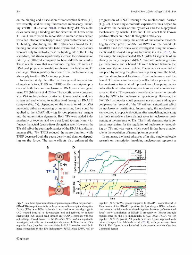
In another study, the effect of two general transcriptionelongation factors, TFIIS and TFIIF, on the transcription pro-cess of both bare and nucleosomal DNA was investigatedusing OT (Ishibashi et al. 2014). The specific assay compriseda dsDNA molecule directly attached to one bead at its down-stream end and tethered to another bead through an RNAP IIcomplex (Fig. 7a). Depending on the orientation of the DNAmolecule, either an opposing or an assisting force could beapplied to the RNAP, allowing for complementary insightsinto the transcription dynamics. Both TFs were added inde-pendently or together and were not found to significantly in-fluence the actual (pause-free) elongation rate. However, theTFs did affect the pausing dynamics of the RNAP in a distinctmanner (Fig. 7b). TFIIS reduced the pause duration, whileTFIIF decreased both the pause density and duration depend-ing on the force. The same effects also enhanced the
progression of RNAP through the nucleosomal barrier(Fig. 7c). These single-molecule experiments thus helped topin down the details on the dynamics and the biophysicalmechanisms by which TFIIS and TFIIF enact their knownpositive effects on RNAP II elongation efficiency.
In a very recent study, the effects of nucleosome remodel-ling by either yeast SWI/SNF or ISW1a on the bound TFGal4DBD and vice versa were investigated using the above-mentioned OT-based unzipping technique (Li et al. 2015). Inthis assay, the single-stranded DNA (ssDNA) segments of analready partially unzipped dsDNA molecule containing a sin-gle nucleosome and a bound TF were tethered between theglass coverslip and a microsphere. The molecules were furtherunzipped by moving the glass coverslip away from the bead,and the strengths and locations of the nucleosome and thebound TF were simultaneously reflected as peaks in theforce-extension traces at ∼1 bp resolution. Unzipping mole-cules after finalized remodeling reactions with either remodelerrevealed that a TF represents a considerable barrier to remod-eling by ISW1a for nucleosome repositioning. However, theSWI/SNF remodeler could generate nucleosome sliding ac-companied by removal of the TF without a significant effecton nucleosome positioning. Interestingly, the nucleosomeswere located in opposite directions after remodeling, indicatingthat both remodelers have distinct roles in nucleosome posi-tioning in the presence of TFs. This study demonstrates a po-tential mechanism for the regulation of nucleosome remodel-ing by TFs and vice versa, which could further have a majorrole in the regulation of transcription in general.
In summary, the latest advances of in vitro single-moleculeresearch on transcription involving nucleosomes represent a
Fig. 7 Real-time dynamics of transcription enzyme RNA polymerase II(RNAP II) elongation activity in the presence of transcription elongationfactors (TFs). a A DNA molecule is attached to an anti-digoxigenin(AD)-coated bead at its downstream end and tethered to anotherstreptavidin (SA)-coated bead through an RNAP II complex with twooptical traps. Two different TFs (TFIIS, blue; TFIIF, red) are injected toinvestigate their effect on transcription dynamics. b Time traces of theopposing force (in pN) to the transcribing RNAP II complex reveal facil-itated elongation by the TFs individually (TFIIS, blue; TFIIF, red) or
together (TFIIF/TFIIS, green) compared to RNAP II alone (black). cTime traces of the RNAP II position (in bp) along a DNA moleculecontaining an initially well-positioned single nucleosome (yellow-shadedband) show stimulation of RNAP II processivity (black) throughnucleosomes by the TFs individually (TFIIS, blue; TFIIF, red) ortogether (TFIIF/S, green). All panels (a–c) are figures reprinted withminor changes from Ishibashi et al. (2014), with permission fromPNAS. This figure is not included in the present article's CreativeCommons license
S44 Biophys Rev (2016) 8 (Suppl 1):S33–S49
successful next step towards understanding more details of thiscomplex process. The combination of multiple proteins andmechanisms in a crowded environment seems a promising ap-proach to elucidate additional details of this complex fundamen-tal process. The further development of this kind of assays isexpected to also finally explain the still striking feature of tran-scription through chromatin in vivo occurring at similar rates toin vitro transcription of bare DNA (Izban and Luse 1992).
Conclusions and future perspectives
As the basic packaging unit of chromatin, the nucleosomerepresents a fundamental DNA–protein complex whose studyis required for understanding the organization and regulationof the genome during essential cellular processes. In this con-text, nucleosomes are not static, but highly ordered and dy-namic entities. Their structure and dynamics are continuouslyaltered by different mechanisms involving spontaneous con-formational changes, the properties of the underlying DNA,post-translational modifications of the histones, ATP-dependent remodelers, external forces and torques, the incor-poration of histone variants, and interactions with histonechaperones and other related proteins.
In this review, we have introduced the most commonly usedsingle-molecule techniques in nucleosome research and pre-sented recent insights they have provided into nucleosomestructure, function and dynamics. Studies of individual
nucleosomes in a time-resolved manner have revealed tran-sient conformational states on fast timescales, such as breath-ing and possibly gaping. The structural and chemical proper-ties of the nucleosomal DNA are suggested to influence nucle-osome structure and dynamics. Post-translational modifica-tions of histones have been found to significantly affect theintrinsic dynamics and the stability of nucleosomes by alteringDNA–histone or histone–histone interactions. More assayshave been developed and extended by using othernucleosome-related components, such as histone chaperonesand remodeling enzymes, to study their underlying molecularmechanisms and impact on nucleosome (dis)assembly, archi-tecture and dynamics. Various subnucleosomal structures havebeen identified both in vivo and in vitro and are believed toplay an important role in the regulation of the genome duringnuclear processes. Some great insights into the function andfate of the nucleosome as a barrier during DNA-templatedprocesses have been provided by the incorporation of genomeprocessing machineries. Single-molecule studies in the contextof histone variants and replication could provide more insightsinto (sub)nucleosomal structure and dynamics and possiblyreveal new mechanisms to complement current knowledge.
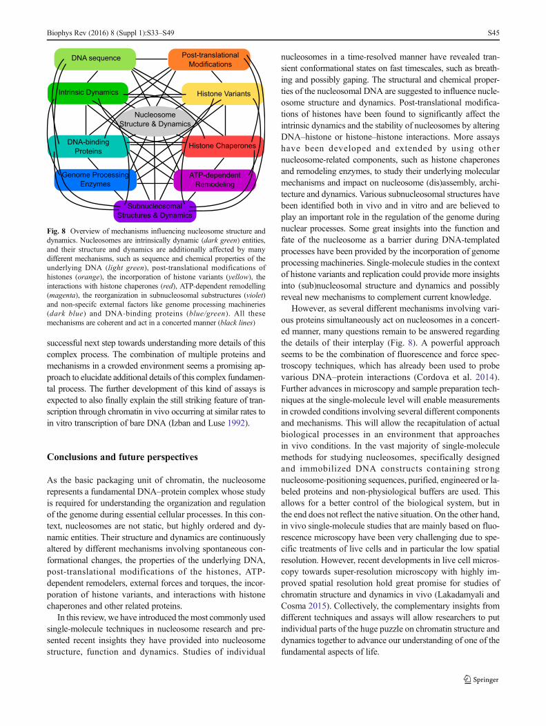
However, as several different mechanisms involving vari-ous proteins simultaneously act on nucleosomes in a concert-ed manner, many questions remain to be answered regardingthe details of their interplay (Fig. 8). A powerful approachseems to be the combination of fluorescence and force spec-troscopy techniques, which has already been used to probevarious DNA–protein interactions (Cordova et al. 2014).Further advances in microscopy and sample preparation tech-niques at the single-molecule level will enable measurementsin crowded conditions involving several different componentsand mechanisms. This will allow the recapitulation of actualbiological processes in an environment that approachesin vivo conditions. In the vast majority of single-moleculemethods for studying nucleosomes, specifically designedand immobilized DNA constructs containing strongnucleosome-positioning sequences, purified, engineered or la-beled proteins and non-physiological buffers are used. Thisallows for a better control of the biological system, but inthe end does not reflect the native situation. On the other hand,in vivo single-molecule studies that are mainly based on fluo-rescence microscopy have been very challenging due to spe-cific treatments of live cells and in particular the low spatialresolution. However, recent developments in live cell micros-copy towards super-resolution microscopy with highly im-proved spatial resolution hold great promise for studies ofchromatin structure and dynamics in vivo (Lakadamyali andCosma 2015). Collectively, the complementary insights fromdifferent techniques and assays will allow researchers to putindividual parts of the huge puzzle on chromatin structure anddynamics together to advance our understanding of one of thefundamental aspects of life.
Fig. 8 Overview of mechanisms influencing nucleosome structure anddynamics. Nucleosomes are intrinsically dynamic (dark green) entities,and their structure and dynamics are additionally affected by manydifferent mechanisms, such as sequence and chemical properties of theunderlying DNA (light green), post-translational modifications ofhistones (orange), the incorporation of histone variants (yellow), theinteractions with histone chaperones (red), ATP-dependent remodelling(magenta), the reorganization in subnucleosomal substructures (violet)and non-specifc external factors like genome processing machineries(dark blue) and DNA-binding proteins (blue/green). All thesemechanisms are coherent and act in a concerted manner (black lines)
Biophys Rev (2016) 8 (Suppl 1):S33–S49 S45
Acknowledgements The authors thank Dr. Belen Solano, Dr. Aysen L.Erdem and Artur Kaczmarczyk for their critical reading of and valuablecomments on the manuscript. The authors further acknowledge the manyefforts in the field of chromatin research and regret that it was not possibleto include more high-quality studies due to limitations in space.
Compliance with ethical standards
Funding Funding for this work was provided by the EuropeanResearch Council (ERC) via a Consolidator Grant DynGenome(No:312221) to N.H.D and the Austrian Science Fund (FWF) [STARTY275-B12] to A.L.
Conflict of interest Orkide Ordu declares that she does not have anyconflicts of interest. Alexandra Lusser declares that she does not have anyconflicts of interest. Nynke H. Dekker declares that she does not have anyconflicts of interest.
Ethical approval This article does not contain any studies with humanparticipants or animals performed by any of the authors.
Open Access This article is licensed under a Creative CommonsAttribution 4.0 International License, which permits use, duplication,adaptation, distribution and reproduction in any medium or format, aslong as you give appropriate credit to the original author(s) and thesource, provide a link to the Creative Commons license and indicate ifchanges were made.The images or other third party material in this article are included in thearticle’s Creative Commons license, unless indicated otherwise in thecredit line; if such material is not included in the article’s CreativeCommons license and the respective action is not permitted by statutoryregulation, users will need to obtain permission from the license holder toduplicate, adapt or reproduce the material.To view a copy of this license, visit http://creativecommons.org/licenses/by/4.0/
References
Alilat M, Sivolob A, Revet B, Prunell A (1999) Nucleosome dynamicsIV. Protein and DNA contributions in the chiral transition of thetetrasome, the histone (H3–H4)(2) tetramer-DNA particle. J MolBiol 291:815–841. doi:10.1006/jmbi.1999.2988
AllenMJ, DongXF, Oneill TE, Yau P, Kowalczykowski SC, Gatewood J,Balhorn R, Bradbury EM (1993) Atomic-force microscope mea-surements of nucleosome cores assembled along defined DNA-se-quences. Biochemistry 32:8390–8396. doi:10.1021/bi00084a002
Amblard F, Yurke B, Pargellis A, Leibler S (1996) A magnetic manipu-lator for studying local rheology and micromechanical properties ofbiological systems. Rev Sci Instrum 67:818–827. doi:10.1063/1.1146816
Ando T, Uchihashi T, Kodera N (2013) High-Speed AFM and applica-tions to biomolecular systems. Annu Rev Biophys 42:393–414.doi:10.1146/annurev-biophys-083012-130324
Andrews AJ, Luger K (2011) Nucleosome structure(s) and stability: var-iations on a theme. Annu Rev Biophys 40:99–117. doi:10.1146/annurev-biophys-042910-155329
Arents G, Burlingame RW, Wang BC, Love WE, Moudrianakis EN(1991) The nucleosomal core histone octamer at 3.1 Å resolution:tripartite protein assembly and a left-handed superhelix. Proc NatlAcad Sci USA 88:10148–10152
Arnaudo AM, Garcia BA (2013) Proteomic characterization of novelhistone post-translational modifications. Epigenetics Chromatin 6:7. doi:10.1186/1756-8935-6-24
Ashkin A (1992) Forces of a single-beam gradient laser trap on a dielec-tric sphere in the ray optics regime. Biophys J 61:569–582
Ashkin A, Dziedzic JM, Bjorkholm JE, Chu S (1986) Observation of asingle-beam gradient force optical trap for dielectric particles. OptLett 11:288–290. doi:10.1364/ol.11.000288
Axelrod D (1981) Cell-substrate contacts illuminated by total internalreflection fluorescence. J Cell Biol 89:141–145. doi:10.1083/jcb.89.1.141
Axelrod D (2001) Total internal reflection fluorescence microscopy incel l biology. Traff ic 2:764–774. doi :10.1034/j .1600-0854.2001.21104.x
Bancaud A, Wagner G, Cond e Silva N, Lavelle C, Wong H,Mozziconacci J, Barbi M, Sivolob A, Le Cam E, Mouawad L,Viovy JL, Victor JM, Prunell A (2007) Nucleosome chiral transitionunder positive torsional stress in single chromatin fibers. Mol Cell27:135–147. doi:10.1016/j.molcel.2007.05.037
Bennink ML, Leuba SH, Leno GH, Zlatanova J, de Grooth BG, Greve J(2001) Unfolding individual nucleosomes by stretching single chro-matin fibers with optical tweezers. Nat Struct Biol 8:606–610.doi:10.1038/89646
Berlier JE, Rothe A, Buller G, Bradford J, GrayDR, Filanoski BJ, TelfordWG, Yue S, Liu JX, Cheung CY, Chang W, Hirsch JD, BeechemJM, Haugland RP, Haugland RP (2003) Quantitative comparison oflong-wavelength Alexa Fluor dyes to Cy dyes: fluorescence of thedyes and their bioconjugates. J HistochemCytochem 51:1699–1712
Binnig G, Quate CF, Gerber C (1986) Atomic force microscope. PhysRev Lett 56:930–933. doi:10.1103/PhysRevLett.56.930
Bintu L, Kopaczynska M, Hodges C, Lubkowska L, Kashlev M,Bustamante C (2011) The elongation rate of RNA polymerase de-termines the fate of transcribed nucleosomes. Nat Struct Mol Biol18:1394–1399. doi:10.1038/nsmb.2164
Bintu L, Ishibashi T, DangkulwanichM, Wu YY, Lubkowska L, KashlevM, Bustamante C (2012) Nucleosomal elements that control thetopography of the barrier to transcription. Cell 151:738–749.doi:10.1016/j.cell.2012.10.009
Blosser TR, Yang JG, Stone MD, Narlikar GJ, Zhuang X (2009)Dynamics of nucleosome remodelling by individual ACF com-plexes. Nature 462:1022–1027. doi:10.1038/nature08627
Bohm V, Hieb AR, Andrews AJ, Gansen A, Rocker A, Toth K, Luger K,Langowski J (2011) Nucleosome accessibility governed by thedimer/tetramer interface. Nucleic Acids Res 39:3093–3102.doi:10.1093/nar/gkq1279
Bowman GD, Poirier MG (2015) Post-translational modifications of his-tones that influence nucleosome dynamics. Chem Rev 115:2274–2295. doi:10.1021/cr500350x
Brehove M, Wang T, North J, Luo Y, Dreher SJ, Shimko JC, Ottesen JJ,Luger K, Poirier MG (2015) Histone core phosphorylation regulatesDNA accessibility. J Biol Chem 290:22612–22621. doi:10.1074/jbc.M115.661363
Brower-Toland B, Wacker DA, Fulbright RM, Lis JT, Kraus WL, WangMD (2005) Specific contributions of histone tails and their acetyla-tion to the mechanical stability of nucleosomes. J Mol Biol 346:135–146. doi:10.1016/j.jmb.2004.11.056
Buning R, van Noort J (2010) Single-pair FRET experiments on nucleo-some conformational dynamics. Biochimie 92:1729–1740.doi:10.1016/j.biochi.2010.08.010
Butt HJ, Cappella B, Kappl M (2005) Force measurements with theatomic force microscope: technique, interpretation and applications.Surf Sci Rep 59:1–152. doi:10.1016/j.surfrep.2005.08.003
Chien FT, van Noort J (2009) 10 years of tension on chromatin: resultsfrom single molecule force spectroscopy. Curr Pharm Biotechnol10:474–485
Choy JS, Lee TH (2012) Structural dynamics of nucleosomes at single-molecule resolution. Trends Biochem Sci 37:425–435. doi:10.1016/j.tibs.2012.06.006
S46 Biophys Rev (2016) 8 (Suppl 1):S33–S49

Clapier CR, Cairns BR (2009) The biology of chromatin remodelingcomplexes. Annu Rev Biochem 78:273–304. doi:10.1146/annurev.biochem.77.062706.153223
Cordova JC, Das DK, Manning HW, Lang MJ (2014) Combining single-molecule manipulation and single-molecule detection. Curr OpinStruct Biol 28:142–148. doi:10.1016/j.sbi.2014.08.010
Crick FHC, Hughes AFW (1950) The physical properties of cyto-plasm: a study by means of the magnetic particle method. PartI. Experimental. Exp Cell Res 1:37–80. doi:10.1016/0014-4827(50)90048-6
Cui Y, Bustamante C (2000) Pulling a single chromatin fiber reveals theforces that maintain its higher-order structure. Proc Natl Acad SciUSA 97:127–132. doi:10.1073/pnas.97.1.127
Dalal Y, Wang H, Lindsay S, Henikoff S (2007) Tetrameric structure ofcentromeric nucleosomes in interphase Drosophila cells. PLoS Biol5:1798–1809. doi:10.1371/journal.pbio.0050218
Dangkulwanich M, Ishibashi T, Bintu L, Bustamante C (2014) Molecularmechanisms of transcription through single-molecule experiments.Chem Rev 114:3203–3223. doi:10.1021/cr400730x
Davey CA, Sargent DF, Luger K, Maeder AW, Richmond TJ (2002)Solvent mediated interactions in the structure of the nucleosomecore particle at 1.9 Å resolution. J Mol Biol 319:1097–1113.doi:10.1016/s0022-2836(02)00386-8
Dimitriadis EK, Weber C, Gill RK, Diekmann S, Dalal Y (2010)Tetrameric organization of vertebrate centromeric nucleosomes.Proc Natl Acad Sci USA 107:20317–20322. doi:10.1073/pnas.1009563107
Dulin D, Lipfert J, Moolman MC, Dekker NH (2013) Studying genomicprocesses at the single-molecule level: introducing the tools andapplications. Nat Rev Genet 14:9–22. doi:10.1038/nrg3316
Duzdevich D, Redding S, Greene EC (2014) DNA dynamics and single-molecule biology. Chem Rev 114:3072–3086. doi:10.1021/cr4004117
Eickbush TH, Moudrianakis EN (1978) Histone core complex—octamerassembled by two sets of protein-protein interactions. Biochemistry17:4955–4964. doi:10.1021/bi00616a016
Eigen M, Rigler R (1994) Sorting single molecules - application to diag-nostics and evolutionary biotechnology. Proc Natl Acad Sci USA91:5740–5747. doi:10.1073/pnas.91.13.5740
Elbel T, Langowski J (2015) The effect of DNA supercoiling on nucleo-some structure and stability. J Phys Condens Matter 27:064105.doi:10.1088/0953-8984/27/6/064105
Elson EL, Magde D (1974) Fluorescence correlation spectroscopy. I.Conceptual basis and theory. Biopolymers 13:1–27. doi:10.1002/bip.1974.360130102
Florin EL, Moy VT, Gaub HE (1994) Adhesion forces between individ-ual ligand–receptor pairs. Science 264:415–417. doi:10.1126/science.8153628
Förster T (1948) Intermolecular energy migration and fluorescence[Zwischenmolekulare Energiewanderung und Fluoreszenz]. AnnPhys 2:55–75
Funatsu T, Harada Y, Tokunaga M, Saito K, Yanagida T (1995) Imagingof single fluorescent molecules and individual ATP turnovers bysingle myosin molecules in aqueous solution. Nature 374:555–559. doi:10.1038/374555a0
Gansen A, Valeri A, Hauger F, Felekyan S, Kalinin S, Toth K, LangowskiJ, Seidel CA (2009) Nucleosome disassembly intermediates charac-terized by single-molecule FRET. Proc Natl Acad Sci USA 106:15308–15313. doi:10.1073/pnas.0903005106
Gemmen GJ, Sim R, Haushalter KA, Ke PC, Kadonaga JT, Smith DE(2005) Forced unraveling of nucleosomes assembled on heteroge-neous DNA using core, histones, NAP-1, and ACF. J Mol Biol 351:89–99. doi:10.1016/j.jmb.2005.05.058
Giepmans BNG, Adams SR, Ellisman MH, Tsien RY (2006) Review—The fluorescent toolbox for assessing protein location and function.Science 312:217–224. doi:10.1126/science.1124618
Gurard-Levin ZA, Quivy JP, Almouzni G (2014) Histone chaperones:assisting histone traffic and nucleosome dynamics. Annu RevBiochem 83:487–517. doi:10.1146/annurev-biochem-060713-035536
Ha T, Tinnefeld P (2012) Photophysics of fluorescent probes for single-molecule biophysics and super-resolution imaging. Ann Rev PhysChem 63:595–617. doi:10.1146/annurev-physchem-032210-103340
Ha T, Enderle T, Ogletree DF, Chemla DS, Selvin PR, Weiss S (1996)Probing the interaction between two single molecules: fluorescenceresonance energy transfer between a single donor and a single ac-ceptor. Proc Natl Acad Sci USA 93:6264–6268. doi:10.1073/pnas.93.13.6264
Hall MA, Shundrovsky A, Bai L, Fulbright RM, Lis JT, Wang MD(2009) High-resolution dynamic mapping of histone-DNA interac-tions in a nucleosome. Nat Struct Mol Biol 16:124–129.doi:10.1038/nsmb.1526
HansmaHG, Hoh JH (1994) Biomolecular imagingwith the atomic-forcemicroscope. Annu Rev Biophys Biomol Struct 23:115–139.doi:10.1146/annurev.bb.23.060194.000555
Hell SW (2009) Microscopy and its focal switch. Nat Methods 6:24–32.doi:10.1038/nmeth.1291
Heller I, Hoekstra TP, King GA, Peterman EJG, Wuite GJL (2014)Optical tweezers analysis of DNA–protein complexes. Chem Rev114:3087–3119. doi:10.1021/cr4003006
Hodges C, Bintu L, Lubkowska L, Kashlev M, Bustamante C (2009)Nucleosomal fluctuations govern the transcription dynamics ofRNA polymerase II. Science 325:626–628. doi:10.1126/science.1172926
Hota SK, Bartholomew B (2011) Diversity of operation in ATP-dependent chromatin remodelers. Biochim Biophys Acta 1809:476–487. doi:10.1016/j.bbagrm.2011.05.007
Huang B, Bates M, Zhuang XW (2009) Super-resolution fluorescencemicroscopy. Annu Rev Biochem 78:993–1016. doi:10.1146/annurev.biochem.77.061906.092014
Hwang WL, Deindl S, Harada BT, Zhuang X (2014) Histone H4 tailmediates allosteric regulation of nucleosome remodelling by linkerDNA. Nature 512:213–217. doi:10.1038/nature13380
Ishibashi T, Dangkulwanich M, Coello Y, Lionberger TA, Lubkowska L,Ponticelli AS, Kashlev M, Bustamante C (2014) Transcription fac-tors IIS and IIF enhance transcription efficiency by differentiallymodifying RNA polymerase pausing dynamics. Proc Natl AcadSci USA 111:3419–3424. doi:10.1073/pnas.1401611111
Izban MG, Luse DS (1992) Factor-stimulated rna polymerase II tran-scribes at physiological elongation rates on naked DNA but verypoorly on chromatin templates. J Biol Chem 267:13647–13655
Janssen XJA, Lipfert J, Jager T, Daudey R, Beekman J, Dekker NH(2012) Electromagnetic Torque Tweezers: a versatile approach formeasurement of single-molecule twist and torque. Nano Lett 12:3634–3639. doi:10.1021/nl301330h
Jares-Erijman EA, Jovin TM (2003) FRET imaging. Nat Biotechnol 21:1387–1395. doi:10.1038/nbt896
Jin J, Bai L, Johnson DS, Fulbright RM, Kireeva ML, Kashlev M, WangMD (2010) Synergistic action of RNA polymerases in overcomingthe nucleosomal barrier. Nat Struct Mol Biol 17:745–752.doi:10.1038/nsmb.1798
Joo C, Balci H, Ishitsuka Y, Buranachai C, Ha T (2008) Advances insingle-molecule fluorescence methods for molecular biology.Annu Rev Biochem 77:51–76. doi :10.1146/annurev.biochem.77.070606.101543
Katan AJ, VlijmR, Lusser A, Dekker C (2015) Dynamics of nucleosomalstructures measured by high-speed atomic force microscopy. Small11:976–984. doi:10.1002/smll.201401318
Kidwell MG (2002) Transposable elements and the evolution of genomesize in eukaryotes . Genetica 115:49–63. doi:10.1023/a:1016072014259
Biophys Rev (2016) 8 (Suppl 1):S33–S49 S47

Killian JL, Li M, Sheinin MY, Wang MD (2012) Recent advances insingle molecule studies of nucleosomes. Curr Opin Struct Biol 22:80–87. doi:10.1016/j.sbi.2011.11.003
Kireeva ML, Walter W, Tchernajenko V, Bondarenko V, Kashlev M,Studitsky VM (2002) Nucleosome remodeling induced by RNApolymerase II: Loss of the H2A/H2B dimer during transcription.Mol Cell 9:541–552. doi:10.1016/s1097-2765(02)00472-0
Klug A, Rhodes D, Smith J, Finch JT, Thomas JO (1980) A low resolu-tion structure for the histone core of the nucleosome. Nature 287:509–516
Koopmans WJA, Brehm A, Logie C, Schmidt T, van Noort J (2007)Single-pair FRET microscopy reveals mononucleosome dynamics.J Fluoresc 17:785–795. doi:10.1007/s10895-007-0218-9
Koopmans WJ, Buning R, Schmidt T, van Noort J (2009) spFRET usingalternating excitation and FCS reveals progressive DNAunwrapping in nucleosomes. Biophys J 97:195–204. doi:10.1016/j.bpj.2009.04.030
Kornberg RD (1974) Chromatin structure: a repeating unit of histonesand DNA. Science 184:868–871
Kornberg RD (1977) Structure of chromatin. Annu Rev Biochem 46:931–954. doi:10.1146/annurev.bi.46.070177.004435
Kornberg RD, Lorch YL (1999) Twenty-five years of the nucleosome,fundamental particle of the eukaryote chromosome. Cell 98:285–294. doi:10.1016/s0092-8674(00)81958-3
Kruithof M, Chien FT, Routh A, Logie C, Rhodes D, van Noort J (2009)Single-molecule force spectroscopy reveals a highly compliant he-lical folding for the 30-nm chromatin fiber. Nat Struct Mol Biol 16:534–540. doi:10.1038/nsmb.1590
La Porta A, Wang MD (2004) Optical Torque Wrench: angular trapping,rotation, and torque detection of quartz microparticles. Phys RevLett 92:190801. doi:10.1103/PhysRevLett.92.190801
Lakadamyali M, Cosma MP (2015) Advanced microscopy methods forvisualizing chromatin structure. FEBS Lett 589:3023–3030.doi:10.1016/j.febslet.2015.04.012
Lavelle C, Prunell A (2007) Chromatin polymorphism and the nucleo-some superfamily—a genealogy. Cell Cycle 6:2113–2119
Lavelle C, Victor JM, Zlatanova J (2010) Chromatin fiber dynamicsunder tension and torsion. Int J Mol Sci 11:1557–1579.doi:10.3390/ijms11041557
Lavelle C, Praly E, Bensimon D, Le Cam E, Croquette V (2011)Nucleosome-remodelling machines and other molecular motors ob-served at the single-molecule level. FEBS J 278:3596–3607.doi:10.1111/j.1742-4658.2011.08280.x
Leuba SH, Karymov MA, Tomschik M, Ramjit R, Smith P, Zlatanova J(2003) Assembly of single chromatin fibers depends on the tensionin the DNA molecule: magnetic tweezers study. Proc Natl Acad SciUSA 100:495–500. doi:10.1073/pnas.0136890100
Li B, Carey M, Workman JL (2007) The role of chromatin during tran-scription. Cell 128:707–719. doi:10.1016/j.cell.2007.01.015
Li G, Levitus M, Bustamante C, Widom J (2005) Rapid spontaneousaccessibility of nucleosomal DNA. Nat Struct Mol Biol 12:46–53.doi:10.1038/nsmb869
LiM, Hada A, Sen P, Olufemi L, Hall MA, Smith BY, Forth S,McKnightJN, Patel A, Bowman GD, Bartholomew B, Wang MD (2015)Dynamic regulation of transcription factors by nucleosome remod-eling. Elife 4. doi:10.7554/eLife.06249
Lia G, Praly E, Ferreira H, Stockdale C, Tse-Dinh YC, Dunlap D,Croquette V, Bensimon D, Owen-Hughes T (2006) Direct observa-tion of DNA distortion by the RSC complex. Mol Cell 21:417–425.doi:10.1016/j.molcel.2005.12.013
Lipfert J, Wiggin M, Kerssemakers JWJ, Pedaci F, Dekker NH (2011)Freely orbiting magnetic tweezers to directly monitor changes in thetwist of nucleic acids. Nat Commun 2:9. doi:10.1038/ncomms1450
Luger K (2001) Nucleosomes: structure and function. In: eLS. JohnWiley & Sons Ltd, Chichester. http://www.els.net [doi:10.1038/npg.els.0001155]
Luger K, Mäder AW, Richmond RK, Sargent DF, Richmond TJ (1997)Crystal structure of the nucleosome core particle at 2.8 Å resolution.Nature 389:251–260
Luger K, Dechassa ML, Tremethick DJ (2012) New insights into nucle-osome and chromatin structure: an ordered state or a disorderedaffair? Nat Rev Mol Cell Biol 13:436–447. doi:10.1038/nrm3382
Luo Y, North JA, Rose SD, Poirier MG (2014) Nucleosomes acceleratetranscription factor dissociation. Nucleic Acids Res 42:3017–3027.doi:10.1093/nar/gkt1319
Mack AH, Schlingman DJ, Ilagan RP, Regan L, Mochrie SGJ (2012)Kinetics and thermodynamics of phenotype: unwinding and rewind-ing the nucleosome. J Mol Biol 423:687–701. doi:10.1016/j.jmb.2012.08.021
Magde D, Webb WW, Elson E (1972) Thermodynamic fluctuations in areacting system—measurement by fluorescence correlation spec-troscopy. Phys Rev Lett 29:705. doi:10.1103/PhysRevLett.29.705
Magde D, Elson EL, Webb WW (1974) Fluorescence correlation spec-troscopy. II. Experimental realization. Biopolymers 13:29–61.doi:10.1002/bip.1974.360130103
McGhee JD, Felsenfeld G (1980) Nucleosome structure. Annu RevB i o c h em 4 9 : 1 1 1 5 – 11 5 6 . d o i : 1 0 . 1 1 4 6 / a n n u r e v .bi.49.070180.005343
Merhej V, Raoult D (2012) Rhizome of life, catastrophes, sequence ex-changes, gene creations, and giant viruses: how microbial genomicschallenges Darwin. Front Cell Infect Microbiol 2:17. doi:10.3389/fcimb.2012.00113
Minsky M (1988) Memoir on inventing the confocal scanning micro-scope. Scanning 10:128–138
Miyagi A, Ando T, Lyubchenko YL (2011) Dynamics of nucleosomesassessed with time-lapse high-speed atomic force microscopy.Biochemistry 50:7901–7908. doi:10.1021/bi200946z
Moerner WE (2007) New directions in single-molecule imaging andanalysis. Proc Natl Acad Sci USA 104:12596–12602. doi:10.1073/pnas.0610081104
Moerner WE, Fromm DP (2003) Methods of single-molecule fluores-cence spectroscopy and microscopy. Rev Sci Instrum 74:3597–3619. doi:10.1063/1.1589587
Monico C, Capitanio M, Belcastro G, Vanzi F, Pavone FS (2013) Opticalmethods to study protein–DNA interactions in vitro and in livingcells at the single-molecule level. Int J Mol Sci 14:3961–3992.doi:10.3390/ijms14023961
Neuman KC, Nagy A (2008) Single-molecule force spectroscopy: opticaltweezers, magnetic tweezers and atomic force microscopy. NatMethods 5:491–505. doi:10.1038/nmeth.1218
Neuman KC, Lionnet T, Allemand JF (2007) Single-molecule microma-nipulation techniques. Annu Rev Mater Res 37:33–67. doi:10.1146/annurev.matsci.37.052506.084336
Ngo TT, Ha T (2015) Nucleosomes undergo slow spontaneous gaping.Nucleic Acids Res 43:3964–3971. doi:10.1093/nar/gkv276
Ngo TT, Zhang Q, Zhou R, Yodh JG, Ha T (2015) Asymmetricunwrapping of nucleosomes under tension directed by DNA localflexibility. Cell 160:1135–1144. doi:10.1016/j.cell.2015.02.001
Nie SM, Chiu DT, Zare RN (1994) Probing individual molecules withconfocal fluorescence microscopy. Science 266:1018–1021.doi:10.1126/science.7973650
North JA, Shimko JC, Javaid S, Mooney AM, Shoffner MA, Rose SD,Bundschuh R, Fishel R, Ottesen JJ, Poirier MG (2012) Regulationof the nucleosome unwrapping rate controls DNA accessibility.Nucleic Acids Res 40:10215–10227. doi:10.1093/nar/gks747
Olins AL, Olins DE (1974) Spheroid chromatin units (ν bodies). Science183:330–332
Oudet P, Gross-Bellard M, Chambon P (1975) Electron microscopic andbiochemical evidence that chromatin structure is a repeating unit.Cell 4:281–300
S48 Biophys Rev (2016) 8 (Suppl 1):S33–S49

Petesch SJ, Lis JT (2012) Overcoming the nucleosome barrier duringtranscript elongation. Trends Genet 28:285–294. doi:10.1016/j.tig.2012.02.005
Poirier MG, Oh E, Tims HS, Widom J (2009) Dynamics and function ofcompact nucleosome arrays. Nat Struct Mol Biol 16:938–U959.doi:10.1038/nsmb.1650
Polo SE, Almouzni G (2006) Chromatin assembly: a basic recipe withvarious flavours. Curr Opin Genet Dev 16:104–111. doi:10.1016/j.gde.2006.02.011
Pope LH, BenninkML, van Leijenhorst-Groener KA, Nikova D, Greve J,Marko JF (2005) Single chromatin fiber stretching reveals physical-ly distinct populations of disassembly events. Biophys J 88:3572–3583. doi:10.1529/biophysj.104.053074
Reinberg D, Sims RJ (2006) De FACTo nucleosome dynamics. J BiolChem 281:23297–23301. doi:10.1074/jbc.R600007200
Richmond TJ, Finch JT, Rushton B, Rhodes D, KlugA (1984) Structure ofthe nucleosome core particle at 7 Å resolution. Nature 311:532–537
Rigler R, Mets U, Widengren J, Kask P (1993) Fluorescence correlationspectroscopy with high count rate and low-background—analysis oftranslational diffusion. Eur Biophys J Biophys 22:169–175
Schmidt T, Schutz GJ, Baumgartner W, Gruber HJ, Schindler H (1996)Imaging of single molecule diffusion. Proc Natl Acad Sci USA 93:2926–2929. doi:10.1073/pnas.93.7.2926
Shaner NC, Steinbach PA, Tsien RY (2005) A guide to choosing fluores-cent proteins. Nat Methods 2:905–909. doi:10.1038/nmeth819
Sheinin MY, Li M, Soltani M, Luger K, Wang MD (2013) Torque mod-ulates nucleosome stability and facilitates H2A/H2B dimer loss. NatCommun 4:8. doi:10.1038/ncomms3579
Shlyakhtenko LS, Lushnikov AY, Lyubchenko YL (2009) Dynamics ofnucleosomes revealed by time-lapse atomic force microscopy.Biochemistry 48:7842–7848. doi:10.1021/bi900977t
Shundrovsky A, Smith CL, Lis JT, Peterson CL,WangMD (2006) ProbingSWI/SNF remodeling of the nucleosome by unzipping single DNAmolecules. Nat Struct Mol Biol 13:549–554. doi:10.1038/nsmb1102
Simon M, North JA, Shimko JC, Forties RA, Ferdinand MB, ManoharM, Zhang M, Fishel R, Ottesen JJ, Poirier MG (2011) Histone foldmodifications control nucleosome unwrapping and disassembly.Proc Natl Acad Sci USA 108:12711–12716. doi:10.1073/pnas.1106264108
Sirinakis G, Clapier CR, Gao Y, Viswanathan R, Cairns BR, Zhang YL(2011) The RSC chromatin remodelling ATPase translocates DNAwith high force and small step size. EMBO J 30:2364–2372.doi:10.1038/emboj.2011.141
Smith SB, Finzi L, Bustamante C (1992) Direct mechanical measure-ments of the elasticity of single DNA-molecules by using magneticbeads. Science 258:1122–1126. doi:10.1126/science.1439819
Smith SB, CuiYJ, BustamanteC (1996)OverstretchingB-DNA: The elasticresponse of individual double-stranded and single-stranded DNAmol-ecules. Science 271:795–799. doi:10.1126/science.271.5250.795
Strick TR, Allemand JF, Bensimon D, Bensimon A, Croquette V (1996)The elasticity of a single supercoiled DNA molecule. Science 271:1835–1837. doi:10.1126/science.271.5257.1835
Strick TR, Allemand JF, Bensimon D, Croquette V (1998) Behavior ofsupercoiled DNA. Biophys J 74:2016–2028
Talbert PB, Henikoff S (2010) Histone variants—ancient wrap artists ofthe epigenome. Nat Rev Mol Cell Biol 11:264–275. doi:10.1038/nrm2861
Teves SS, Weber CM, Henikoff S (2014) Transcribing through the nucle-osome. Trends Biochem Sci 39:577–586. doi:10.1016/j.tibs.2014.10.004
Tims HS, Gurunathan K, Levitus M, Widom J (2011) Dynamics of nu-cleosome invasion by DNA binding proteins. J Mol Biol 411:430–448. doi:10.1016/j.jmb.2011.05.044
Tinoco I, Gonzalez RL (2011) Biological mechanisms, one molecule at atime. Genes Dev 25:1205–1231. doi:10.1101/gad.2050011
Tomschik M, Zheng HC, van Holde K, Zlatanova J, Leuba SH (2005)Fast, long-range, reversible conformational fluctuations in nucleo-somes revealed by single-pair fluorescence resonance energy trans-fer. Proc Natl Acad Sci USA 102:3278–3283. doi:10.1073/pnas.0500189102
Tomschik M, van Holde K, Zlatanova J (2009) Nucleosome dynamics asstudied by single-pair fluorescence resonance energy transfer: a re-evaluation. J Fluoresc 19:53–62. doi:10.1007/s10895-008-0379-1
Trifonov EN, Nibhani R (2015) Review fifteen years of search for strongnucleosomes. Biopolymers 103:432–437. doi:10.1002/bip.22604
Vlijm R, Smitshuijzen JSJ, Lusser A, Dekker C (2012) NAP1-assistednucleosome assembly on DNA measured in real time by single-molecule magnetic tweezers. PLoS One 7:11. doi:10.1371/journal.pone.0046306
Vlijm R, Lee M, Lipfert J, Lusser A, Dekker C, Dekker NH (2015a)Nucleosome assembly dynamics involve spontaneous fluctuationsin the handedness of tetrasomes. Cell Rep 10:216–225. doi:10.1016/j.celrep.2014.12.022
Vlijm R, Lee M, Ordu O, Boltengagen A, Lusser A, Dekker NH, DekkerC (2015b) Comparing the assembly and handedness dynamics of(H3.3-H4)2 tetrasomes to canonical tetrasomes. PLoS One 10:e0141267. doi:10.1371/journal.pone.0141267
Wang MD, Schnitzer MJ, Yin H, Landick R, Gelles J, Block SM (1998)Force and velocity measured for single molecules of RNA polymer-ase. Science 282:902–907. doi:10.1126/science.282.5390.902
Wei S, Falk SJ, Black BE, Lee TH (2015) A novel hybrid single moleculeapproach reveals spontaneous DNA motion in the nucleosome.Nucleic Acids Res 43:e111. doi:10.1093/nar/gkv549
Weiss S (1999) Fluorescence spectroscopy of single biomolecules.Science 283:1676–1683. doi:10.1126/science.283.5408.1676
Widom J (2001) Role of DNA sequence in nucleosome stability anddynamics. Q Rev Biophys 34:269–324
Workman JL, Kingston RE (1998) Alteration of nucleosome structure asa mechanism of transcriptional regulation. Annu Rev Biochem 67:545–579. doi:10.1146/annurev.biochem.67.1.545
Yan J, Maresca TJ, Skoko D, Adams CD, Xiao BT, Christensen MO,Heald R, Marko JF (2007) Micromanipulation studies of chromatinfibers in Xenopus egg extracts reveal ATP-dependent chromatinassembly dynamics. Mol Biol Cell 18:464–474. doi:10.1091/mbc.E06-09-0800
Yoda K, Ando S, Morishita S, Houmura K, Hashimoto K, Takeyasu K,Okazaki T (2000) Human centromere protein A (CENP-A) can re-place histone H3 in nucleosome reconstitution in vitro. Proc NatlAcad Sci USA 97:7266–7271. doi:10.1073/pnas.130189697
Zentner GE, Henikoff S (2013) Regulation of nucleosome dynamics byhistone modifications. Nat Struct Mol Biol 20:259–266.doi:10.1038/nsmb.2470
Zhang YL, Smith CL, Saha A, Grill SW, Mihardja S, Smith SB,Cairns BR, Peterson CL, Bustamantel C (2006) DNA transloca-tion and loop formation mechanism of chromatin remodeling bySWI/SNF and RSC. Mol Cell 24:559–568. doi:10.1016/j.molcel.2006.10.025
Ziemann F, Radler J, Sackmann E (1994) Local measurements of visco-elastic moduli of entangled actin networks using an oscillating mag-netic bead micro-rheometer. Biophys J 66:2210–2216
Zimmer C (2007) Evolution - Jurassic genome. Science 315:1358–1359.doi:10.1126/science.315.5817.1358
Zlatanova J, Leuba SH (2003) Chromatin fibers, one-at-a-time. J MolBiol 331:1–19. doi:10.1016/s0022-2836(03)00691-0
Zlatanova J, Bishop TC, Victor JM, Jackson V, van Holde K (2009) Thenucleosome family: Dynamic and growing. Structure 17:160–171.doi:10.1016/j.str.2008.12.016
Biophys Rev (2016) 8 (Suppl 1):S33–S49 S49