recent news - university of western ontario
TRANSCRIPT

Journal of Surgical Research 138, 141–150 (2007)
�-Catenin Signaling in Fibroproliferative Disease
Erin Bowley, B.Sc.,*,† David B. O’Gorman, M.Sc., Ph.D.,*,†,‡ andBing Siang Gan, M.D., Ph.D., F.R.C.S.C., F.A.C.S.*,†,‡,§,¶,1
*Cell and Molecular Biology Laboratory of the Hand & Upper Limb Centre; †Department of Surgery, ‡Department of Physiology,§Deparment of Pharmacology, and ¶Department of Medical Biophysics, University of Western Ontario, Lawson Health Research Institute,
London, Ontario, Canada
Submitted for publication June 19, 2006
doi:10.1016/j.jss.2006.07.026
Background. �-catenin has been historically recog-nized as both an intermediate in the “canonical Wntsignaling pathway” and as a component of functionaladherens junctions.
Materials and methods. Cellular accumulation of�-catenin levels can result in transactivation of genetranscription and cellular proliferation during normalcellular and disease development. Recent evidencehas identified �-catenin in an additional role as a com-ponent of cutaneous wound healing.
Results. This finding is in keeping with previous ob-servations that post-translational modifications of�-catenin that are associated with its cytoplasmic ac-cumulation are frequently observed in fibroprolifera-tive diseases with characteristics of dysregulatedwound healing. These diseases include hypertrophicscar formation, aggressive fibromatoses, Lederhosedisease, and Dupuytren’s contracture (DC).
Conclusions. While its precise roles in disease initi-ation and progression remain to be explored, this re-view highlights our current knowledge of �-cateninregulation and describes some potential upstream me-diators of �-catenin accumulation and signaling in fi-broproliferative disease. © 2007 Elsevier Inc. All rights reserved.
Key Words: Wnt/beta-catenin signaling; fibroprolif-erative disorders; Dupuytren’s contracture; woundhealing; hypertrophic scarring.
INTRODUCTION
�-catenin was once thought to have only two distinctfunctional roles within the cell. One was as an inter-mediary in the “canonical Wnt-pathway”, a critical sig-naling system in embryonic growth and development,
1 To whom correspondence and reprint requests should be ad-dressed at Hand and Upper Limb Centre, St. Joseph’s Health Care,268 Grosvenor Street, London, Ontario, Canada N6A 4L6. E-mail:
[email protected].141
and the second was as a structural protein that, inconjunction with E-cadherin, served in the functioningof cellular adherens junctions. However, it is now clearthat aberrant �-catenin signaling can occur in a num-ber of human diseases, such as certain types of cancerand fibroproliferative disorders, and that a large num-ber of signaling pathways use �-catenin to direct thetranscription of genes whose products are characteris-tic of those conditions. The purpose of this paper is toreview the known interactions of �-catenin and placethese in the context of newly discovered findings re-lated to a number of fibrotic conditions including hy-pertrophic scar formation, aggressive fibromatoses(desmoid tumors) and Dupuytren’s contracture (DC).To understand the central importance of this moleculein these areas, we will begin with an overview of thepathways known to regulate �-catenin bioavailability.
THE CANONICAL WNT/�-CATENIN PATHWAY
The canonical Wnt pathway (Fig. 1) is reliant on�-catenin as a central signaling molecule. This path-way is one of the most well-studied regulators of em-bryonic development [1], exerting remarkable controlover cellular proliferation, differentiation, invasionand adhesion [2, 3]. When the canonical Wnt pathwayis activated by the appropriate Wnt ligand, the Friz-zled (Fz) receptor and co-receptor LRP5/6 (lipoproteinreceptor-related proteins 5 or 6) [4–6] form a complexand trigger a cascade of signaling events through thekinase Dishevelled (Dvl), resulting in the phosphory-lation, and subsequent inactivation, of glycogen synthasekinase-3� (GSK-3�). GSK-3� activity is the key compo-nent of the so-called “destruction complex” that also in-cludes Axin, the product of the adenomatous polyposiscoli gene (APC), and casein kinase-1 (CK-I) [7–9]. WhenGSK-3� phosphorylates complexed �-catenin on serine
and threonine residues, primarily S33, S37, T41, and0022-4804/07 $32.00© 2007 Elsevier Inc. All rights reserved.

n c
142 JOURNAL OF SURGICAL RESEARCH: VOL. 138, NO. 1, MARCH 2007
S45, the hyper-phosphorylated �-catenin is recognizedby the F-box containing protein slimb/b-TrCP, a com-ponent of the E3 ubiquitin (Ub) ligase complex, and�-catenin is targeted for degradation via the 26S pro-teasome [10–16]. Alternatively, GSK-3� can be inacti-vated by phosphorylation of Serine 9 resulting in�-catenin release from the destruction complex and itsaccumulation in the cytoplasm as a stabilized pool. Ina poorly understood process, this cytoplasmic pool of�-catenin is then thought to translocate to the nucleusthrough binding to Tcf4 and BCL9/Pygopus while APCand Axin act to inhibit this process and retain the�-catenin in the cytoplasm [17]. Nuclear �-cateninfunctions as a transcriptional activator for members ofthe T-cell factor/lymphoid enhancer factor (Tcf/Lef)family of DNA binding proteins [18, 19] that activate asubset of genes in a cell-context specific manner.
A number of �-catenin-Tcf/Lef target genes havebeen identified [11] and many of these play an impor-tant role in cell cycle control, proliferation and cell fatedetermination [20, 21]. (A current online list of Wnt/�-catenin target genes with Tcf/Lef binding sites can befound at http://www.stanford.edu/�rnusse/pathways/targets.html). Both independently and in conjunctionwith Tcf/Lef, �-catenin can interact with nuclear recep-tors resulting in an array of cellular effects includingchanges in cellular adhesion, tissue morphogenesis,and tumor development. Whereas androgen receptorinteractions are perhaps the best characterized, nu-clear �-catenin has also been shown to affect the activ-
FIG. 1. The Wnt/�-catenin pathway. The two historically recognpathway and adherens junctions. Wnt signaling through Frizzled return inactivates GSK3�. GSK3� inactivation allows �-catenin to avtranslocate to the nucleus and transactivate the Tcf/Lef transcriptio
ities of receptors for retinoic acid, vitamin D, glucocor-
ticoid, progesterone, thyroid, estrogen, and peroxisomeproliferator-activator and these interactions are re-viewed in detail in [22].
The Wnt pathway has now become well recognizedas a molecular contributor to the development of manydisease states [23–25]. Dysregulation of the canonicalWnt signaling system is involved in greater than 90%of colorectal carcinomas [11] and evidence continues toaccumulate supporting the role of this pathway in amultitude of other malignant disorders [2, 26, 27].Since nuclear translocation of �-catenin and Tcf/Lefinteraction can induce transcription of cellular regula-tors of proliferation and differentiation such as CyclinD1 and c-myc [11], the Wnt/�-catenin pathway pro-vides a direct connection between extra-cellular sig-nals, gene transcription and cell cycle control. There-fore, aberrant behavior of this pathway as a result ofactivating mutations of �-catenin have the potential topromote both initiation and the progression of cancer[11, 25]. Not surprisingly, the critical serine/threonineresidues required for �-catenin phosphorylation andubiquitination, normally used as regulators of the canon-ical Wnt signaling pathway, are ‘hot spots’ in those can-cers involving �-catenin mutations. Clinically, strongnuclear or cytoplasmic �-catenin staining in colorectalcancer correlates with more invasive tumor growth, ahigher susceptibility of disease recurrence after sur-gery and a lower survival rate [28].
Similarly, the adenomatous polyposis coli (APC)gene product is a negative regulator of cytoplasmic
d intracellular sources of �-catenin are the canonical Wnt/�-catenintor/LRP5/6 results in phosphorylation of Dishevelled (Dvl), which in
the destruction complex and to accumulate within the cytoplasm,omplex. (Color version of figure is available online.)
izecepoid
�-catenin. Part of the Axin/APC/GSK-3� destruction

143BOWLEY ET AL.: �-CATENIN SIGNALING IN FIBROPROLIFERATIVE DISEASE
complex, APC is responsible for sequestering cytoplas-mic �-catenin and targeting it for ubiquitination anddegradation at the proteasome. Functioning in thisregard, APC is an important negative regulator of freecytoplasmic �-catenin, and inactivating mutations ofthe APC gene play an important role in cancer progres-sion. Monoallelic inactivating mutations in APC occurin patients with familial adenomatous polyposis (FAP),a condition that results in many adenomas in the colo-rectum [29]. As well, 85% of colorectal cancers demon-strate a mutation in the APC gene and it is impliedthat the remaining 15% are likely to contain mutationsin �-catenin [11, 19, 30, 31]. However, many cases ofhepatocellular carcinoma reveal that mutation and nu-clear staining of �-catenin correlates with less aggres-sive tumor growth and increased survival rates [32],while there is no correlation apparent with �-cateninnuclear staining and tumor type in gastric cancer [33].In summary, while there are obvious correlations be-tween dysregulation of the Wnt/�-catenin pathway andcolorectal disease, the relevance of altered �-cateninaccumulation in other malignancies is still not clearlydefined.
WNT-INDEPENDENT �-CATENIN SIGNALING
Although the canonical Wnt signaling pathway is amajor player in the regulation of stabilized cytoplasmicpools of �-catenin, several Wnt-independent signaling
FIG. 2. GSK-3� is a central mediator of Wnt/�-catenin and recepas the type 1 IGF receptor shown here, can signal through a variety ofactivity. GSK-3� is also a component of the Wnt/�-catenin pathwathreonine phosphorylation and subsequent cytoplasmic accumulatio
pathways have also been shown to converge at GSK-3�
to achieve this result (Fig. 2). These include growthfactor signaling pathways such as those used byinsulin-like growth factors (IGFs), platelet-derivedgrowth factors (PDGFs), and hepatocyte growth factor(HGF). These molecules induce tyrosine phosphoryla-tion of downstream targets through activation of theirrespective receptor tyrosine kinases and signalthrough a variety of intracellular intermediate mole-cules including phosphatidylinositol 3 (PI3)-kinase[34, 35]. PI3 kinase activity results, via phosphoinosi-tide dependent kinases 1 and 2 (PDK1/2), in the phos-phorylation of the serine 473 and threonine 308 resi-dues of Akt. Akt, also known as protein kinase B, is aserine/threonine kinase with an established role inregulating proliferation and apoptosis in cancer [36, 37],wound healing and fibrosis [38]. Among several tar-gets, Akt can phosphorylate GSK-3� at serine 9 result-ing in �-catenin accumulation and Tcf/Lef transcrip-tion complex activation [34, 39]. From a clinicalperspective, androgen-independent prostate cancer isfrequently associated with the silencing of PTEN(phosphatase and tensin homologue deleted on chro-mosome 10), a potent inhibitor of PI3 kinase-mediatedactivation of Akt. PTEN silencing can indirectly resultin inhibition of GSK-3� activity and subsequent nu-clear transactivation through interactions of nuclear�-catenin [40].
In another Wnt-independent �-catenin regulatory
tyrosine kinase signaling pathways. Receptor tyrosine kinases, suchermediate molecules including PI3 kinase and Akt to inhibit GSK-3�s shown, and thus acts as a central mediator of �-catenin serine/Color version of figure is available online.)
torinty an. (
pathway, integrin receptors connect the extra-cellular

144 JOURNAL OF SURGICAL RESEARCH: VOL. 138, NO. 1, MARCH 2007
matrix (ECM) to the actin cytoskeleton [41] at the cellmembrane. Stimulation by the appropriate ligand thenleads to integrin receptor clustering and recruitment ofactin filaments and signaling proteins [42] to the com-plex. In this way, ECM molecules can regulate cellularsurvival by signaling through integrin receptors andintegrin-linked kinase (ILK) to activate, among others,the PI3 kinase and Akt pathways [43] inducing�-catenin/Tcf/Lef-mediated transcription of cell cyclegenes such as cyclin D1 [44, 45].
�-CATENIN AND ADHERENS JUNCTIONS
Other than as a component of the Wnt-dependentand independent �-catenin pathways, �-catenin haslong been established as a component of adherensjunctions in association with cadherins. Cadherinsare a large family of calcium-dependent cell adhesionproteins that include epithelial cadherin (E-cadherin), neural cadherin (N-cadherin), placentalcadherin (P-cadherin), muscle cadherin (M-cadherin), and vascular endothelial cadherin (VE-cadherin, or CDH5). While N-cadherin andP-cadherin have also been shown to associate with�-catenin and �-catenin, E-cadherin is the best char-acterized cadherin component of adherens junctions[46]. �-catenin is an essential component of this molec-ular complex, linking transmembrane E-cadherin tothe actin cytoskeleton through �-catenin as well asother catenin molecules such as plakoglobin (or�-catenin) [47, 48]. Adherens junctions allow for cell-celladhesion, and when required, can disassemble to allowcell migration [3], a process important in embryologicaldevelopment [21], the wound healing response [49] andepithelial-mesenchymal transition [50, 51]. Associa-tion of the transmembrane cadherins with the intra-cellular cytoskeleton is essential for the regulation ofcell-cell adhesion, stability and contact [52]. Clinically,a loss of the E-cadherin/�-catenin adhesion complexcorrelates with poor prognosis in some malignancies[53], and therefore could potentially act as a modulatorof disease progression.
A variety of growth factors and ECM moleculeshave been demonstrated to exert effects on cytoplas-mic �-catenin accumulation by disrupting theE-cadherin/�-catenin/�-catenin complex. Pleiotro-phin has an effect on cell adhesion in part by signal-ing through receptor protein tyrosine phosphatases�/� to promote the disruption of adherens junctions[54]. HGF not only promotes adherens junction dis-ruption but is involved in E-cadherin endocytosis[55, 56] and �-catenin release [57]. Epidermalgrowth factors (EGFs) signaling through the EGFreceptor (EGFR), initiate changes in cell morphology[58] that are associated with EGFR co-localizationwith cadherin molecules at the cell membrane [58,
59]. This co-localization induces tyrosine phosphor-ylation of �-catenin and destabilization ofE-cadherin/�-catenin complexes in adherens junc-tions [59, 60], in turn resulting in cytoplasmic accu-mulation of tyrosine phosphorylated �-catenin. Gly-cosylation has a role in this process, as the additionof bisecting N-acetylglucosamine residues toE-cadherin combined with epidermal growth factorstimulation alters tyrosine phosphorylation of�-catenin [61]. Similarly, IGF-I and -II acting throughthe type 1 IGF receptor (IGFRI), can also induce disman-tling of the E-cadherin/�-catenin cellular scaffold (Fig. 3).
Phosphorylation events are clearly paramount in thestructural integrity of the cadherin-catenin complex.In general, when �-catenin or E-cadherin are serine/threonine phosphorylated the complex is stabilized [62,63]. However, when �-catenin is tyrosine phosphorylatedby an intracellular signaling event, the E-cadherin-�-catenin complex is generally disrupted and cell adhesionis lost [60, 64, 65]. It has also been demonstrated thatthe interaction between �-catenin and �-catenin is reg-ulated by the tyrosine kinases Fer/Fyn, which are inturn activated by the tyrosine kinase, Yes [66]. At leasttwo critical tyrosine residues on �-catenin are targetedby these kinases: tyrosine 142 by cMet (the HGF re-ceptor), Fyn and Fer, and tyrosine 654 by Src and theEGF receptor [67]. Tyrosine phosporylation can resultin release of �-catenin from E-cadherin, decreased cell-cell adhesion and increased cell migration and inva-siveness [64, 68].
The role of adherens junction-derived �-catenin inpromoting Tcf/Lef mediated gene transcription isstill an area of some debate [69]. The literature sug-gests that there are at least two molecular pools of�-catenin and that �-catenin associated withE-cadherin at adherens junctions is preferentiallybound to �-catenin and functionally distinct from the�-catenin that promotes Tcf/Lef transcription [70].Other studies have found evidence that suggeststyrosine-phosphorylated �-catenin is capable oftransactivating signal transduction. For instance,the tyrosine phosphatase SHP-1 has been shown todecrease �-catenin signaling [71] implying that ty-rosine phosphorylation of �-catenin enhances itstransactivation of the Tcf/Lef transcription complex.In some systems, transcriptional activity associatedwith tyrosine phosphorylated �-catenin appears de-pendent on the presence of growth factors including IGFand HGF [72, 73]. For example, IGF signaling has beenreported to promote disruption of adherens junctionswith associated nuclear translocation of �-catenin andsubsequent expression of �-catenin-Tcf/Lef target genesin vitro [72, 74]. It is still unclear, however, whether theadherens junctions-derived �-catenin itself promotescell proliferation and, if it does, this process may be
cell-type specific.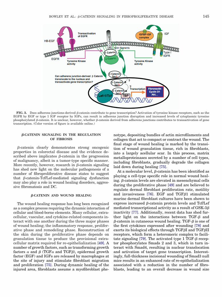
145BOWLEY ET AL.: �-CATENIN SIGNALING IN FIBROPROLIFERATIVE DISEASE
�-CATENIN SIGNALING IN THE REGULATIONOF FIBROSIS
�-catenin clearly demonstrates strong oncogenicproperties in colorectal disease and the evidence de-scribed above implicates �-catenin in the progressionof malignancy, albeit in a tumor-type specific manner.More recently, however, research in �-catenin signalinghas shed new light on the molecular pathogenesis of anumber of fibroproliferative disease states to suggestthat �-catenin-Tcf/Lef-mediated signaling dysfunctionmay also play a role in wound healing disorders, aggres-sive fibromatosis and DC.
�-CATENIN AND WOUND HEALING
The wound healing response has long been recognizedas a complex process requiring the dynamic interaction ofcellular and blood-borne elements. Many cellular, extra-cellular, vascular, and cytokine-related components in-teract with one another during the three major phasesof wound healing; the inflammatory response, prolifer-ative phase and remodeling phase. Reconstruction ofthe skin during the proliferative phase depends ongranulation tissue to produce the provisional extra-cellular matrix required for re-epithelialization [49]. Anumber of growth factors, such as transforming growthfactors � and � (TGF� and TGF�), epidermal growthfactor (EGF) and IGFs are released by macrophages atthe site of injury and stimulate fibroblast migrationand proliferation [75]. During dynamic healing of the
FIG. 3. Does adherens junctions-derived �-catenin contribute toEGFR by EGF or type 1 IGF receptor by IGFs, can result in adhephosphorylated �-catenin. It is unclear, however, whether �-catenintranscription. (Color version of figure is available online.)
injured area, fibroblasts assume a myofibroblast phe-
notype, depositing bundles of actin microfilaments andcollagen that act to compact or contract the wound. Thefinal stage of wound healing is marked by the transi-tion of wound granulation tissue, rich in fibroblasts,into a largely acellular scar. In this process, matrixmetalloproteinases secreted by a number of cell types,including fibroblasts, gradually degrade the collagenlaid down during healing [75].
At a molecular level, �-catenin has been identified asplaying a cell-type specific role in normal wound heal-ing. �-catenin levels are elevated in mesenchymal cellsduring the proliferative phase [49] and are believed toregulate dermal fibroblast proliferation rate, motilityand invasiveness [76]. EGF and TGF�1 stimulatedmurine dermal fibroblast cultures have been shown toexpress increased �-catenin protein levels and Tcf/Lefmediated transcriptional activity as a result of GSK-3�inactivity [77]. Additionally, recent data has shed fur-ther light on the interactions between TGF-� and�-catenin in cutaneous wound healing. TGF-� is one ofthe first cytokines expressed after wounding [78] andexerts its biological effects through TGF�I and TGF�IIreceptors, which form a heteromeric complex to facili-tate signaling [79]. The activated type 1 TGF-� recep-tor phosphorylates Smads 2 and 3, which in turn in-teract with Smad4, resulting in nuclear translocationand activation of target gene transcription. Interest-ingly, full-thickness incisional wounding of Smad3 nullmice results in an enhanced rate of re-epithelializationassociated with a reduction in the number of fibro-
e transcription? Activation of tyrosine kinase receptors, such as thes junction disruption and increased levels of cytoplasmic tyrosineived from adherens junctions contributes to transactivation of gene
genrender
blasts, leading to an overall decrease in wound size

146 JOURNAL OF SURGICAL RESEARCH: VOL. 138, NO. 1, MARCH 2007
[80–82]. Cheon et al. have recently demonstrated thatthis phenotype is dependent on �-catenin expression,because stabilized �-catenin expression reverses theSmad3 null effect on wound size. TGF �-mediated fi-broblast proliferation and hyperplastic wound forma-tion was also shown to be dependent on �-cateninexpression, demonstrating the central role of thismolecule in the healing process [83] and highlightingthe negative effects of �-catenin dysregulation. HGFsignaling, which can induce tyrosine phosphorylated�-catenin release from adherens junctions, has alsobeen shown to promote wound healing [84, 85], al-though a role of �-catenin signaling in this context hasnot been reported. In contrast to the fibroblast re-sponse, �-catenin inhibits migration of human epithe-lial cells in culture [86] and, as normal epithelial celldifferentiation and proliferation in wounded �-cateninnull mice demonstrates, it is not an essential compo-nent of the epithelium for wound healing [87].
When the wound healing response is dysregulated, avariety of epithelial and mesenchymal disorders canoccur. Fibroproliferative disorders, characterized byexcessive proliferation of mesenchymal cells, range inseverity from hypertrophic scars to neoplasms such asaggressive fibromatosis (desmoid tumors). These con-ditions display cellular and biochemical features thatare remarkably similar to those involved in woundhealing [49]. This similarity has given rise to thewidely accepted hypothesis that fibroproliferative dis-orders may be the result of an unchecked or exagger-ated wound healing response.
HYPERPLASTIC SCARRING AND �-CATENIN
Hyperplastic scars are composed of fibrous bundles inboth the deeper and upper dermis. Although the molec-ular cause of hyperplastic scarring is still unknown, cy-tological and gene expression studies comparing this dis-order to wound healing suggest that it is an exaggerated,prolonged healing response. Wounding of �-catenin over-expressing mice results in hyperplastic scar formation[76] giving rise to the speculation that �-catenin plays asignificant role in the pathomechanism driving hyper-plastic scarring. In wound healing, the elevated �-cateninlevels normally found in granulation tissue (fibroblasts)only during the proliferative phase are detectible in hy-perplastic scar tissue for more than 2 years after initialinjury [49]. Although �-catenin protein levels in hyper-plastic scars do not correlate with mRNA levels, they docorrelate with levels of inactive (phospho-serine 9) GSK-3�. This suggests that the increased �-catenin levelspresent during normal wound healing and in hyperplas-tic scars are a result of a post-transcriptional mechanisminvolving signaling systems that inhibit GSK-3� activity.Additionally, cell cultures derived from normal woundand hyperplastic scar patient excisions show �-catenin-
Tcf/Lef transcriptional activation [49]. Increased levels of�-catenin and activation of �-catenin-Tcf/Lef mediatedgene transcription in both the proliferative phase ofwound healing and hyperplastic wounds suggests�-catenin may play an important role in hyperplastic scarformation, as well as other fibrotic disorders.
�-CATENIN AND AGGRESSIVE FIBROMATOSIS
Aggressive fibromatoses or desmoid tumors areclonal lesions characterized by locally invasive andproliferative fibroblast-like spindle cells [88]. Evidencesuggests that desmoid tumors are neoplastic in nature,deriving from a single progenitor cell with a growthadvantage [89]. Desmoid tumors can occur as a spo-radic lesion or as part of familial adenomatous polyp-osis (FAP) caused by a germline mutation of the APCgene [90]. Recent studies focused on the molecular causeof desmoid tumors suggest that this disorder may be aresult of abnormal �-catenin signaling. Desmoid tumorsshow �-catenin stabilizing mutations in both the APCand �-catenin genes [91, 92] and demonstrate cytoplas-mic and nuclear accumulation of �-catenin as shown byimmunohistochemical (IH) analysis [91]. Additionally,�-catenin stabilization in desmoid tumors activatesTcf/Lef transcriptional gene expression [93] and simi-lar results are found in IH analysis of solitary fibroustumors, another type of spindle cell neoplasm [94].
Transgenic mouse models expressing a stabilizedform of �-catenin in mesenchymal cells develop symp-toms of aggressive fibromatosis as well as hyperplasticcutaneous wounds [76]. Fibroblasts derived from thesemice display increased proliferation, motility and inva-siveness when grafted into nude mice and primary cellcultures demonstrate Tcf-dependent transcriptionalactivation [76], consistent with the hypothesis thatnuclear �-catenin transactivation of target genes is aprimary component of this fibrosis.
Oligonucleotide array analysis of global gene expres-sion in desmoid tumors has identified insulin-growthfactor binding protein-6 (IGFBP6), an established in-hibitor of IGF-II signaling [95], as one of these targetgenes of �-catenin transactivation of the Tcf/Lef tran-scription complex. Unusually, IGFBP6 mRNA expres-sion is down-regulated by �-catenin-Tcf/Lef mediatedtranscription [96], implying that expression of theIGFBP6 gene might be inhibitory to the development ofthis disease. In association, the gene encoding A Disin-tegrin And Metalloprotease 12, ADAM12, has beenshown to be up regulated in aggressive fibromatoses [97].ADAM12 is a protease of IGFBPs including IGFBP-3 and-5. A combination of up-regulated ADAM12 and down-regulated IGFBP-6 levels is predicted to result in in-creased IGF signaling, a situation previously shown topromote adherens junction disruption, nuclear localiza-tion of �-catenin [72, 74] and fibroblast proliferation[98]. Overall, these data suggest that dysregulation of
the �-catenin signaling mechanisms and �-catenin-Tcf/
147BOWLEY ET AL.: �-CATENIN SIGNALING IN FIBROPROLIFERATIVE DISEASE
Lef mediated transcription are key factors in thepathogenesis of aggressive fibromatoses and implythat targeting �-catenin may have therapeutic utilityfor treatment of this disease.
�-CATENIN AND BENIGN SUPERFICIAL FIBROMATOSIS
Histologically and biochemically, benign fibroprolifera-tive disorders including DC, Peyronie’s disease (fibroma-tosis of the penis), Lederhose disease (plantar fibromato-sis), and frozen shoulder syndrome (FSS) share manysimilarities with wound granulation tissue, leading tothe view that these conditions represent a dysregu-lated, fibrotic wound healing responses in differentphysiological contexts.
DC is a superficial fibromatosis that affects the pal-mar fascia and displays an invasive phenotype. Theearliest stage of the disease is characterized by theappearance of a hyperproliferative nodule composed ofboth fibroblasts and activated myofibroblasts whichdeposit type 3 collagen in thick bundles throughout theaffected area [99, 100]. Eventually, this process givesrise to a scar-like contractile collagen rich cord, result-ing in permanent digit contracture [101]. Histologicalanalysis has shown that DC and wound granulationtissue share many biochemical characteristics includ-ing the appearance of collagen type 3 and TGF� [102,103]. TGF�1 stimulation of DC derived fibroblast cul-tures in vitro results in an increased myofibroblastphenotype as determined by the presence of a smoothmuscle actin [102]. Additionally, TGF� stimulates col-lagen production in cultured fibroblasts from DC andpatient matched control tissue [104]. Type 3 collagen isusually absent in normal adult palmar fascia but iscommonly found in DC tissue extracted from patients[103]. This biochemical change in palmar fascia com-position is similar to the changes that occur during theproliferative phase of wound healing suggesting thatDC may be a type of dysregulated wound healing pro-cess.
Although the molecular cause of DC still remainsto be elucidated, similarities in the clinical progres-sion of DC with the wound healing response and addi-tional accumulating molecular evidence suggests that�-catenin may be a component of the initiation andprogression of DC. IH and Western blot analysis of DCpatient tissue reveals increased cytoplasmic and nu-clear staining of tyrosine phosphorylated �-catenincompared to control tissue [105]. Unlike aggressive fi-bromatoses, however, there is no evidence of �-cateninmutations in disease or control samples [105]. Further-more, in vitro analysis of DC primary cells cultured in afibroblast populated collagen lattice (FPCL), a threedimensional collagen environment designed to mimicin vivo disease conditions, demonstrates increased lev-els of �-catenin compared to levels in control lattices
[106]. Interestingly, cellular levels of �-catenin are rap-idly and differentially regulated by tension in FPCLculture compared to two-dimensional tissue culture,implying that both extra-cellular matrix (ECM) inter-actions and mechano-tension are essential componentsof this disease [106]. These results suggest that�-catenin may be a key player in the development ofthis and related superficial fibromatoses and that al-terations in �-catenin accumulation in DC are poten-tially regulated by the ECM.
Cytoplasmic and nuclear accumulation of �-cateninaccumulation has been reported in Lederhose diseaseand, in common with other superficial fibromatoses, nomutations in either exon 3 of �-catenin or in APC weredetected. Whether the presence or absence of thesemutations directly affects the clinical characteristics ofthis and other superficial fibromatoses, which are typ-ically less invasive and proliferative than deep fibro-matoses, such as desmoid tumors that frequently con-tain such mutations, remains unclear at present [107].
Adhesive capsulitis of the joint capsule, or FSS, ischaracterized by painful and restricted shoulder mo-tion and affects an estimated 2% of adults. Based onimmunohistochemical studies and clinical correlation,it has been suggest that FSS may share a similarpatho-mechanism with that of DC [108, 109]. Histolog-ically, both conditions are characterized by the pres-ence of collagen-rich (types 1 and 3) nodules and bandsthat are populated by contractile fibroblasts and myo-fibroblasts. Furthermore, clinical studies suggest thatthese two connective tissue disorders are strongly as-sociated with one another. A recent study of patientswith FSS showed a marked increase in the incidence ofDD compared to the general population [110]. Prelim-inary observations from our laboratory indicate that�-catenin expression is up-regulated in FSS comparedto arthroscopy samples derived from subacromial de-compression for rotator cuff syndrome (unpublisheddata). As with DC and Lederhose disease, the role ofcytoplasmic �-catenin in this fibrotic disorder is pres-ently unknown.
Peyronie’s disease is a localized fibrotic lesion withinthe tunica albuginea of the penis [111, 112] that is verypoorly understood at a molecular level. While there areno reports of �-catenin accumulation in this fibrosis,recent microarray analysis of gene expression in Pey-ronie’s disease plaques compared normal tunica albu-ginea identified the mRNA for pleiotrophin as mark-edly up-regulated in the disease plaques [113]. Asdescribed previously, pleiotrophin regulates cell adhe-sion by signaling through receptor protein tyrosinephosphatases �/� [54], which can promote the disrup-tion of adherens junctions and release of cytoplasmic�-catenin. Whether this process is involved in thepathophysiology of this disease is at present not clear.
In conclusion, dysregulation of �-catenin processing
and cellular accumulation are likely to be important
148 JOURNAL OF SURGICAL RESEARCH: VOL. 138, NO. 1, MARCH 2007
components of the pathogenesis of a number of fibroticdisorders. Mutational analysis of the �-catenin andAPC genes in aggressive fibromatosis indicate that, asin cancers associated with familial adenomatous pol-yposis, somatic and germline mutations are very likelycontributing to disease progression. However, muta-tions in �-catenin and related biomolecular partnersare not evident in hypertrophic scarring and benignfibrotic conditions such as DC, Lederhose disease andFSS, disorders which display characteristics of an ex-aggerated wound healing response. Evidence suggeststhat �-catenin is dysregulated in these disordersthrough protein signaling pathways yet to be fully elu-cidated. Accumulating evidence supporting the impor-tance of �-catenin in a variety of signaling pathwayssuggests that this molecule plays a much larger role incellular processes than is currently appreciated.
ACKNOWLEDGMENTS
Work in the authors’ lab is supported by the Canadian Institutes ofHealth Research, the Lawson Health Research Institute Internal Re-search Fund, The Advanced Surgical Technologies Group at LHRI, theU.S. Plastic Surgery Education Foundation, the Canadian OrthopedicFoundation, and the Canadian Society for Surgery of the Hand.
REFERENCES
1. Korswagen HC. Canonical and non-canonical Wnt signalingpathways in Caenorhabditis elegans: Variations on a commonsignaling theme. Bioessays 2002;24:801.
2. Smalley MJ, Dale TC. Wnt signalling in mammalian develop-ment and cancer. Cancer Metastasis Rev 1999;18:215.
3. Nelson WJ, Nusse R. Convergence of Wnt, beta-catenin, andcadherin pathways. Science 2004;303:1483.
4. Wehrli M, Dougan ST, Caldwell K, et al. Arrow encodes anLDL-receptor-related protein essential for Wingless signal-ling. Nature 2000;407:527.
5. Tamai K, Semenov M, Kato Y, et al. LDL-receptor-relatedproteins in Wnt signal transduction. Nature 2000;407:530.
6. Pinson KI, Brennan J, Monkley S, Avery BJ, Skarnes WC. AnLDL-receptor-related protein mediates Wnt signalling in mice.Nature 2000;407:535.
7. Amit S, Hatzubai A, Birman Y, et al. Axin-mediated CKIphosphorylation of beta-catenin at Ser 45: A molecular switchfor the Wnt pathway. Genes Dev 2002;16:1066.
8. Dixelius J, Cross M, Matsumoto T, Sasaki T, Timpl R,Claesson-Welsh L. Endostatin regulates endothelial cell adhe-sion and cytoskeletal organization. Cancer Res 2002;62:1944.
9. Yanagawa S, Matsuda Y, Lee JS, et al. Casein kinase I phos-phorylates the Armadillo protein and induces its degradationin Drosophila. Embo J 2002;21:1733.
10. Aberle H, Bauer A, Stappert J, Kispert A, Kemler R. Beta-catenin is a target for the ubiquitin-proteasome pathway.Embo J 1997;16:3797.
11. Giles RH, van Es JH, Clevers H. Caught up in a Wnt storm:Wnt signaling in cancer. Biochim Biophys Acta 2003;1653:1.
12. Miller JR, Hocking AM, Brown JD, Moon RT. Mechanism andfunction of signal transduction by the Wnt/beta-catenin andWnt/Ca2� pathways. Oncogene 1999;18:7860.
13. Orford K, Crockett C, Jensen JP, Weissman AM, Byers SW.
Serine phosphorylation-regulated ubiquitination anddegradation of beta-catenin. J Biol Chem 1997;272:24735.
14. Hart M, Concordet JP, Lassot I, et al. The F-box proteinbeta-TrCP associates with phosphorylated beta-catenin andregulates its activity in the cell. Curr Biol 1999;9:207.
15. Latres E, Chiaur DS, Pagano M. The human F box proteinbeta-Trcp associates with the Cul1/Skp1 complex and regu-lates the stability of beta-catenin. Oncogene 1999;18:849.
16. Winston JT, Strack P, Beer-Romero P, Chu CY, Elledge SJ,Harper JW. The SCFbeta-TRCP-ubiquitin ligase complex as-sociates specifically with phosphorylated destruction motifs inIkappaBalpha and beta-catenin and stimulates IkappaBalphaubiquitination in vitro. Genes Dev 1999;13:270.
17. Krieghoff E, Behrens J, Mayr B. Nucleo-cytoplasmic distribu-tion of {beta}-catenin is regulated by retention. J Cell Sci2006;119:1453.
18. Behrens J, von Kries JP, Kuhl M, et al. Functional interactionof beta-catenin with the transcription factor LEF-1. Nature1996;382:638.
19. Morin PJ, Sparks AB, Korinek V, et al. Activation of beta-catenin-Tcf signaling in colon cancer by mutations in beta-catenin or APC. Science 1997;275:1787.
20. Molenaar M, van de Wetering M, Oosterwegel M, et al. XTcf-3transcription factor mediates beta-catenin-induced axis for-mation in Xenopus embryos. Cell 1996;86:391.
21. Cadigan KM, Nusse R. Wnt signaling: A common theme inanimal development. Genes Dev 1997;11:3286.
22. Mulholland DJ, Dedhar S, Coetzee GA, Nelson CC. Interactionof nuclear receptors with the Wnt/beta-catenin/Tcf signalingaxis: Wnt you like to know? Endocr Rev 2005;26:898.
23. Nusse R. Wnt signaling in disease and in development. CellRes 2005;15:28.
24. Logan CY, Nusse R. The Wnt signaling pathway in develop-ment and disease. Annu Rev Cell Dev Biol 2004;20:781.
25. Moon RT, Kohn AD, De Ferrari GV, Kaykas A. WNT andbeta-catenin signalling: Diseases and therapies. Nat RevGenet 2004;5:691.
26. Yardy GW, Brewster SF. Wnt signalling and prostate cancer.Prostate Cancer Prostatic Dis 2005;8:119.
27. Kolligs FT, Bommer G, Goke B. Wnt/beta-catenin/tcf signal-ing: A critical pathway in gastrointestinal tumorigenesis. Di-gestion 2002;66:131.
28. Maruyama K, Ochiai A, Akimoto S, et al. Cytoplasmic beta-catenin accumulation as a predictor of hematogenous metas-tasis in human colorectal cancer. Oncology 2000;59:302.
29. Groden J, Thliveris A, Samowitz W, et al. Identification andcharacterization of the familial adenomatous polyposis coligene. Cell 1991;66:589.
30. de La Costeqq A, Romagnolo B, Billuart P, et al. Somaticmutations of the beta-catenin gene are frequent in mouse andhuman hepatocellular carcinomas. Proc Natl Acad Sci USA1998;95:8847.
31. Kinzler KW, Vogelstein B. Lessons from hereditary colorectalcancer. Cell 1996;87:159.
32. Hsu HC, Jeng YM, Mao TL, Chu JS, Lai PL, Peng SY. Beta-catenin mutations are associated with a subset of low-stagehepatocellular carcinoma negative for hepatitis B virus andwith favorable prognosis. Am J Pathol 2000;157:763.
33. Grabsch H, Takeno S, Noguchi T, Hommel G, Gabbert HE,Mueller W. Different patterns of beta-catenin expression ingastric carcinomas: Relationship with clinicopathological pa-rameters and prognostic outcome. Histopathology 2001;39:141.
34. Desbois-Mouthon C, Cadoret A, Blivet-Van Eggelpoel MJ, et al.

149BOWLEY ET AL.: �-CATENIN SIGNALING IN FIBROPROLIFERATIVE DISEASE
Insulin and IGF-1 stimulate the beta-catenin pathway throughtwo signalling cascades involving GSK-3beta inhibition and Rasactivation. Oncogene 2001;20:252.
35. Chen EY, Mazure NM, Cooper JA, Giaccia AJ. Hypoxia activatesa platelet-derived growth factor receptor/phosphatidylinositol3-kinase/Akt pathway that results in glycogen synthase kinase-3inactivation. Cancer Res 2001;61:2429.
36. Cheng JQ, Lindsley CW, Cheng GZ, Yang H, Nicosia SV. TheAkt/PKB pathway: Molecular target for cancer drug discovery.Oncogene 2005;24:7482.
37. Chen YL, Law PY, Loh HH. Inhibition of PI3K/Akt signaling:An emerging paradigm for targeted cancer therapy. Curr MedChem Anticancer Agents 2005;5:575.
38. Jun JB, Kuechle M, Min J, et al. Scleroderma fibroblastsdemonstrate enhanced activation of Akt (protein kinase B) insitu. J Invest Dermatol 2005;124:298.
39. Cross DA, Alessi DR, Cohen P, Andjelkovich M, HemmingsBA. Inhibition of glycogen synthase kinase-3 by insulin medi-ated by protein kinase B. Nature 1995;378:785.
40. Mulholland DJ, Dedhar S, Wu H, Nelson CC. PTEN andGSK3beta: Key regulators of progression to androgen-independent prostate cancer. Oncogene 2006;25:329.
41. Brakebusch C, Fassler R. The integrin-actin connection, aneternal love affair. Embo J 2003;22:2324.
42. Hynes RO. Integrins: Bidirectional, allosteric signaling ma-chines. Cell 2002;110:673.
43. Nho RS, Xia H, Kahm J, Kleidon J, Diebold D, Henke CA. Roleof integrin-linked kinase in regulating phosphorylation of Aktand fibroblast survival in type I collagen matrices through abeta1 integrin viability signaling pathway. J Biol Chem 2005;280:26630.
44. D’Amico M, Hulit J, Amanatullah DF, et al. The integrin-linked kinase regulates the cyclin D1 gene through glycogensynthase kinase 3beta and cAMP-responsive element-bindingprotein-dependent pathways. J Biol Chem 2000;275:32649.
45. Tan C, Costello P, Sanghera J, et al. Inhibition of integrin linkedkinase (ILK) suppresses beta-catenin-Lef/Tcf-dependent tran-scription and expression of the E-cadherin repressor, snail, inAPC-/- human colon carcinoma cells. Oncogene 2001;20:133.
46. Wheelock MJ, Johnson KR. Cadherins as modulators of cellu-lar phenotype. Annu Rev Cell Dev Biol 2003;19:207.
47. Kintner C. Regulation of embryonic cell adhesion by the cad-herin cytoplasmic domain. Cell 1992;69:225.
48. Nagafuchi A, Takeichi M. Cell binding function of E-cadherinis regulated by the cytoplasmic domain. Embo J 1988;7:3679.
49. Cheon S, Poon R, Yu C, et al. Prolonged beta-catenin stabili-zation and tcf-dependent transcriptional activation in hyper-plastic cutaneous wounds. Lab Invest 2005;85:416.
50. Iwano M, Plieth D, Danoff TM, Xue C, Okada H, Neilson EG.Evidence that fibroblasts derive from epithelium during tissuefibrosis. J Clin Invest 2002;110:341.
51. Kalluri R, Neilson EG. Epithelial-mesenchymal transition andits implications for fibrosis. J Clin Invest 2003;112:1776.
52. Meniel V, Clarke AR. Wnt-cadherin connections in normal andneoplastic mammary epithelium. J Mammary Gland Biol Neo-plasia 2003;8:435.
53. Krishnadath KK, Tilanus HW, van Blankenstein M, et al.Reduced expression of the cadherin-catenin complex in oe-sophageal adenocarcinoma correlates with poor prognosis.J Pathol 1997;182:331.
54. Beltran PJ, Bixby JL. Receptor protein tyrosine phosphatasesas mediators of cellular adhesion. Front Biosci 2003;8:d87.
55. Potempa S, Ridley AJ. Activation of both MAP kinase and
phosphatidylinositide 3-kinase by Ras is required for hepato-cyte growth factor/scatter factor-induced adherens junctiondisassembly. Mol Biol Cell 1998;9:2185.
56. Kimura T, Sakisaka T, Baba T, Yamada T, Takai Y. Involve-ment of the Ras-Ras-activated Rab5 guanine nucleotide ex-change factor RIN2-Rab5 pathway in the HGF-induced endo-cytosis of E-cadherin. J Biol Chem 2006;281:10598.
57. Muller T, Bain G, Wang X, Papkoff J. Regulation of epithelialcell migration and tumor formation by beta-catenin signaling.Exp Cell Res 2002;280:119.
58. Fukuyama R, Shimizu N. Detection of epidermal growth factorreceptors and E-cadherins in the basolateral membrane ofA431 cells by laser scanning fluorescence microscopy. Jpn JCancer Res 1991;82:8.
59. Hoschuetzky H, Aberle H, Kemler R. Beta-catenin mediatesthe interaction of the cadherin-catenin complex with epider-mal growth factor receptor. J Cell Biol 1994;127:1375.
60. Shibamoto S, Hayakawa M, Takeuchi K, et al. Tyrosine phos-phorylation of beta-catenin and plakoglobin enhanced by he-patocyte growth factor and epidermal growth factor in humancarcinoma cells. Cell Adhes Commun 1994;1:295.
61. Kitada T, Miyoshi E, Noda K, et al. The addition of bisectingN-acetylglucosamine residues to E-cadherin down-regulatesthe tyrosine phosphorylation of beta-catenin. J Biol Chem2001;276:475.
62. Bek S, Kemler R. Protein kinase CKII regulates the interac-tion of beta-catenin with alpha-catenin and its protein stabil-ity. J Cell Sci 2002;115:4743.
63. Lickert H, Bauer A, Kemler R, Stappert J. Casein kinase IIphosphorylation of E-cadherin increases E-cadherin/beta-catenin interaction and strengthens cell-cell adhesion. J BiolChem 2000;275:5090.
64. Behrens J, Vakaet L, Friis R, et al. Loss of epithelial differen-tiation and gain of invasiveness correlates with tyrosine phos-phorylation of the E-cadherin/beta-catenin complex in cellstransformed with a temperature-sensitive v-SRC gene. J CellBiol 1993;120:757.
65. Taddei ML, Chiarugi P, Cirri P, et al. Beta-catenin interactswith low-molecular-weight protein tyrosine phosphatase lead-ing to cadherin-mediated cell-cell adhesion increase. CancerRes 2002;62:6489.
66. Piedra J, Miravet S, Castano J, et al. p120 Catenin-associatedFer and Fyn tyrosine kinases regulate beta-catenin Tyr-142phosphorylation and beta-catenin-alpha-catenin Interaction.Mol Cell Biol 2003;23:2287.
67. Lilien J, Balsamo J. The regulation of cadherin-mediated ad-hesion by tyrosine phosphorylation/dephosphorylation of beta-catenin. Curr Opin Cell Biol 2005;17:459.
68. Hulsken J, Birchmeier W, Behrens J. E-cadherin and APCcompete for the interaction with beta-catenin and the cytoskel-eton. J Cell Biol 1994;127:2061.
69. Gavard J, Mege RM. Once upon a time there was beta-cateninin cadherin-mediated signalling. Biol Cell 2005;97:921.
70. Gottardi CJ, Gumbiner BM. Distinct molecular forms of beta-catenin are targeted to adhesive or transcriptional complexes.J Cell Biol 2004;167:339.
71. Duchesne C, Charland S, Asselin C, Nahmias C, Rivard N.Negative regulation of beta-catenin signaling by tyrosinephosphatase SHP-1 in intestinal epithelial cells. J Biol Chem2003;278:14274.
72. Playford MP, Bicknell D, Bodmer WF, Macaulay VM. Insulin-like growth factor 1 regulates the location, stability, and tran-scriptional activity of beta-catenin. Proc Natl Acad Sci USA2000;97:12103.
73. Herynk MH, Tsan R, Radinsky R, Gallick GE. Activation of

150 JOURNAL OF SURGICAL RESEARCH: VOL. 138, NO. 1, MARCH 2007
c-Met in colorectal carcinoma cells leads to constitutiveassociation of tyrosine-phosphorylated beta-catenin. Clin ExpMetastasis 2003;20:291.
74. Morali OG, Delmas V, Moore R, Jeanney C, Thiery JP, Larue L.IGF-II induces rapid beta-catenin relocation to the nucleus dur-ing epithelium to mesenchyme transition. Oncogene 2001;20:4942.
75. Singer AJ, Clark RA. Cutaneous wound healing. N Engl J Med1999;341:738.
76. Cheon SS, Cheah AY, Turley S, et al. Beta-Catenin stabilizationdysregulates mesenchymal cell proliferation, motility, and inva-siveness and causes aggressive fibromatosis and hyperplasticcutaneous wounds. Proc Natl Acad Sci USA 2002;99:6973.
77. Cheon SS, Nadesan P, Poon R, Alman BA. Growth factorsregulate beta-catenin-mediated TCF-dependent transcrip-tional activation in fibroblasts during the proliferative phaseof wound healing. Exp Cell Res 2004;293:267.
78. Werner S, Grose R. Regulation of wound healing by growthfactors and cytokines. Physiol Rev 2003;83:835.
79. Annes JP, Munger JS, Rifkin DB. Making sense of latentTGFbeta activation. J Cell Sci 2003;116:217.
80. Flanders KC. Smad3 as a mediator of the fibrotic response. IntJ Exp Pathol 2004;85:47.
81. Falanga V, Schrayer D, Cha J, Butmarc J, Carson P, RobertsAB, Kim SJ. Full-thickness wounding of the mouse tail as amodel for delayed wound healing: Accelerated wound closurein Smad3 knock-out mice. Wound Repair Regen 2004;12:320.
82. Ashcroft GS, Yang X, Glick AB, et al. Mice lacking Smad3show accelerated wound healing and an impaired local inflam-matory response. Nat Cell Biol 1999;1:260.
83. Cheon SS, Wei Q, Gurung A, et al. Beta-catenin regulateswound size and mediates the effect of TGF-beta in cutaneoushealing. FASEB J 2006;20:692.
84. Nayeri F, Olsson H, Peterson C, Sundqvist T. Hepatocytegrowth factor; expression, concentration and biological activityin chronic leg ulcers. J Dermatol Sci 2005;37:75.
85. Nayeri F, Stromberg T, Larsson M, Brudin L, Soderstrom C,Forsberg P. Hepatocyte growth factor may accelerate healing inchronic leg ulcers: A pilot study. J Dermatolog Treat 2002;13:81.
86. Stojadinovic O, Brem H, Vouthounis C, et al. Molecular patho-genesis of chronic wounds: The role of beta-catenin and c-mycin the inhibition of epithelialization and wound healing. Am JPathol 2005;167:59.
87. Posthaus H, Williamson L, Baumann D, et al. Beta-Catenin isnot required for proliferation and differentiation of epidermalmouse keratinocytes. J Cell Sci 2002;115:4587.
88. Bhattacharya B, Dilworth HP, Iacobuzio-Donahue C, et al.Nuclear beta-catenin expression distinguishes deep fibroma-tosis from other benign and malignant fibroblastic and myofi-broblastic lesions. Am J Surg Pathol 2005;29:653.
89. Alman BA, Pajerski ME, Diaz-Cano S, Corboy K, Wolfe HJ.Aggressive fibromatosis (desmoid tumor) is a monoclonal dis-order. Diagn Mol Pathol 1997;6:98.
90. Kinzler KW, Nilbert MC, Su LK, et al. Identification of FAPlocus genes from chromosome 5q21. Science 1991;253:661.
91. Tejpar S, Nollet F, Li C, et al. Predominance of beta-cateninmutations and beta-catenin dysregulation in sporadic aggres-sive fibromatosis (desmoid tumor). Oncogene 1999;18:6615.
92. Alman BA, Li C, Pajerski ME, Diaz-Cano S, Wolfe HJ. In-creased beta-catenin protein and somatic APC mutations insporadic aggressive fibromatoses (desmoid tumors). Am J
Pathol 1997;151:329.93. Tejpar S, Li C, Yu C, et al. Tcf-3 expression and beta-cateninmediated transcriptional activation in aggressive fibromatosis(desmoid tumour). Br J Cancer 2001;85:98.
94. Rakheja D, Molberg KH, Roberts CA, Jaiswal VR. Immuno-histochemical expression of beta-catenin in solitary fibroustumors. Arch Pathol Lab Med 2005;129:776.
95. Bach LA. Insulin-like growth factor binding protein-6: the“forgotten” binding protein? Horm Metab Res 1999;31:226.
96. Denys H, Jadidizadeh A, Amini Nik S, et al. Identification ofIGFBP-6 as a significantly downregulated gene by beta-catenin in desmoid tumors. Oncogene 2004;23:654.
97. Skubitz KM, Skubitz AP. Gene expression in aggressive fibro-matosis. J Lab Clin Med 2004;143:89.
98. Grotendorst GR, Rahmanie H, Duncan MR. Combinatorialsignaling pathways determine fibroblast proliferation andmyofibroblast differentiation. FASEB J 2004;18:469.
99. Bisson MA, Mudera V, McGrouther DA, Grobbelaar AO. Thecontractile properties and responses to tensional loading of Du-puytren’s disease–derived fibroblasts are altered: A cause of thecontracture? Plast Reconstr Surg 2004;113:611; discussion 622.
100. Kloen P. New insights in the development of Dupuytren’scontracture: A review. Br J Plast Surg 1999;52:629.
101. Rayan GM. Palmar fascial complex anatomy and pathology inDupuytren’s disease. Hand Clin 1999;15:73.
102. Bisson MA, McGrouther DA, Mudera V, Grobbelaar AO. Thedifferent characteristics of Dupuytren’s disease fibroblasts de-rived from either nodule or cord: Expression of alpha-smoothmuscle actin and the response to stimulation by TGF-beta1.J Hand Surg [Br] 2003;28:351.
103. Brickley-Parsons D, Glimcher MJ, Smith RJ, Albin R, AdamsJP. Biochemical changes in the collagen of the palmar fascia inpatients with Dupuytren’s disease. J Bone Joint Surg Am1981;63:787.
104. Alioto RJ, Rosier RN, Burton RI, Puzas JE. Comparativeeffects of growth factors on fibroblasts of Dupuytren’s tissueand normal palmar fascia. J Hand Surg [Am] 1994;19:442.
105. Varallo VM, Gan BS, Seney S, et al. Beta-catenin expressionin Dupuytren’s disease: Potential role for cell-matrix interac-tions in modulating beta-catenin levels in vivo and in vitro.Oncogene 2003;22:3680.
106. Howard JC, Varallo VM, Ross DC, et al. Elevated levels ofbeta-catenin and fibronectin in three-dimensional collagencultures of Dupuytren’s disease cells are regulated by tensionin vitro. BMC Musculoskelet Disord 2003;4:16.
107. Montgomery E, Lee JH, Abraham SC, Wu TT. Superficialfibromatoses are genetically distinct from deep fibromatoses.Mod Pathol 2001;14:695.
108. Bunker TD, Anthony PP. The pathology of frozen shoulder. ADupuytren-like disease. J Bone Joint Surg Br 1995;77:677.
109. Bunker TD, Reilly J, Baird KS, Hamblen DL. Expression ofgrowth factors, cytokines and matrix metalloproteinases infrozen shoulder. J Bone Joint Surg [Br] 2000;82:768.
110. Smith SP, Devaraj VS, Bunker TD. The association betweenfrozen shoulder and Dupuytren’s disease. J Shoulder ElbowSurg 2001;10:149.
111. Chilton CP, Castle WM, Westwood CA, Pryor JP. Factorsassociated in the aetiology of peyronie’s disease. Br J Urol1982;54:748.
112. Hellstrom WJ, Bivalacqua TJ. Peyronie’s disease: Etiology,medical, and surgical therapy. J Androl 2000;21:347.
113. Qian A, Meals RA, Rajfer J, Gonzalez-Cadavid NF. Compari-son of gene expression profiles between Peyronie’s disease and
Dupuytren’s contracture. Urology 2004;64:399.