reconstruction of oropharyngeal · pdf filereconstruction of tongue base defects ... who...
TRANSCRIPT
Reconstruction of Tongue Base Defects
Michael Briscoe Jr., MD Faculty Advisor: Susan McCammon, MD The University of Texas Medical Branch
Department of Otolaryngology Grand Rounds Presentation
February 27, 2008

Outline
Introduction
Tongue base carcinoma
Surgical Anatomy
Surgical Resection
Reconstruction Options
Conclusion

Introduction
The oropharynx plays a key role in speech, swallowing, and host defenses.
Squamous cell cancers in this region can cause significant morbidity, and affects quality of life.
Reconstruction of these defects attempts to improve function and quality of life.
Epidemiology
Oropharyngeal carcinoma has an incidence of 11.9/100,000
30,000 new cases annually in the United States.
The tongue base is the number one site for oropharyngeal tumors, accounting for approximately half.
Epidemiology
2.5-3:1 male to female predominance
African American males account for most new cases
Etiology
Alcohol use
Cigarrette use
Betel nut use
Tongue base SCCa
Present at advanced stage
Base of tongue drains to levels II and III.
High incidence of nodal disease on presentation (60%)
Good locoregional control with multidisciplinary approach
TNM staging
WHO classification

Symptoms
Sore throat
Otalgia
Dysphagia
Weight loss
Neck mass

Oropharynx
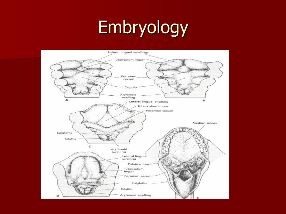
Oropharyngeal embryology – 4th week of life, the pharyngeal arches, clefts, and
pouches develop.
– Anterior tongue develops from 1st arch, while the posterior tongue develops from 3rd arch.
– The epiglottis is formed from the hypopharyngeal eminence, a condensation of the 3rd and 4th arch.
– Palatine tonsils and tonsillar fossa are formed from the 2nd pharyngeal pouch
– Secondary palate is formed around the ninth week by the fusion of the intermaxillary process, and the lateral maxillary processes.
Embryology

Oropharynx
Superior boundary – Superior border of soft palate
Inferior boundary – Superior surface of hyoid bone
Anterior boundary – V-shaped circumvallate papillae
– Anterior border of soft palate/uvula
– Palatoglossal arch (anterior tonsillar pillar)
Posterior boundary (pharyngeal wall)

Surgical anatomy
The oropharynx consists of four distinct sites
– Soft palate
– Tonsillar fossa/palatine tonsil
– Posterior pharyngeal wall
– Base of tongue

Oropharynx
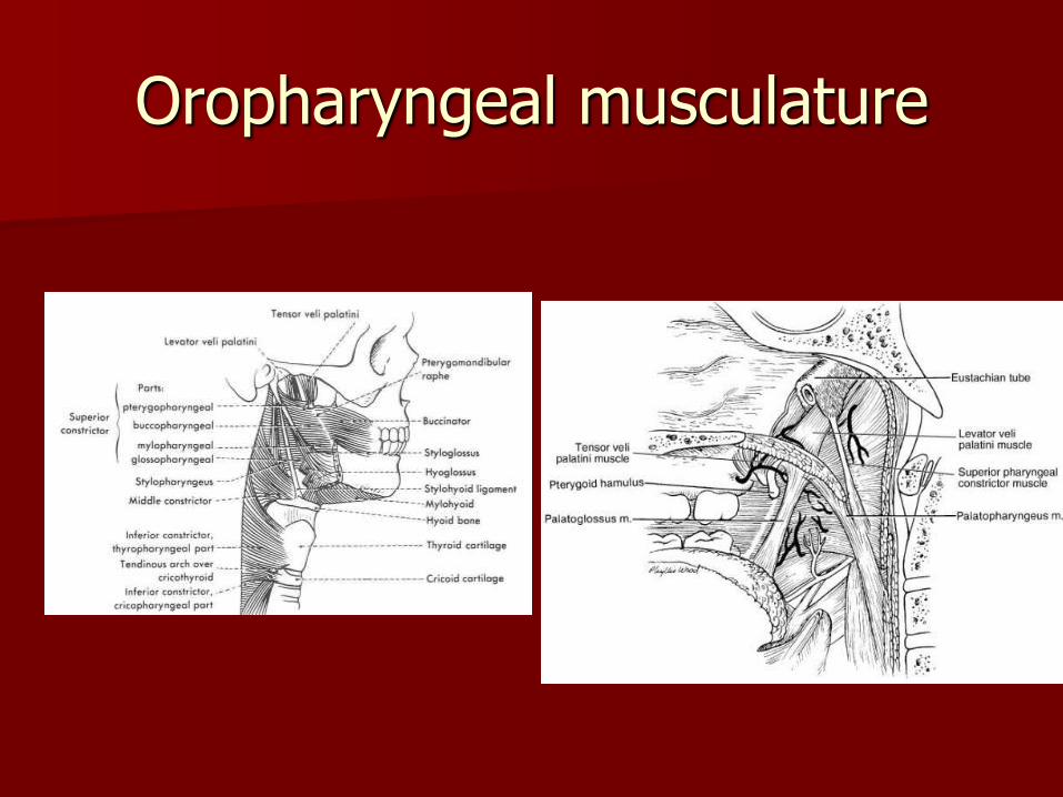
Oropharyngeal musculature

Base of tongue landmarks
The sulcus terminalis (V-shaped furrow on dorsal surface of tongue) divides anterior/posterior tongue
Foramen cecum – area where thyroid descends.
Taste papillae, mucus glands
Lingual tonsils
Base of tongue – blood supply
Lingual arteries supply the tongue
Enter the tongue base medial to the hyoglossal muscle
Septum linguae – near bloodless plain in the midline of tongue
Submandibular arteries provide important anastomosis to contralateral tongue
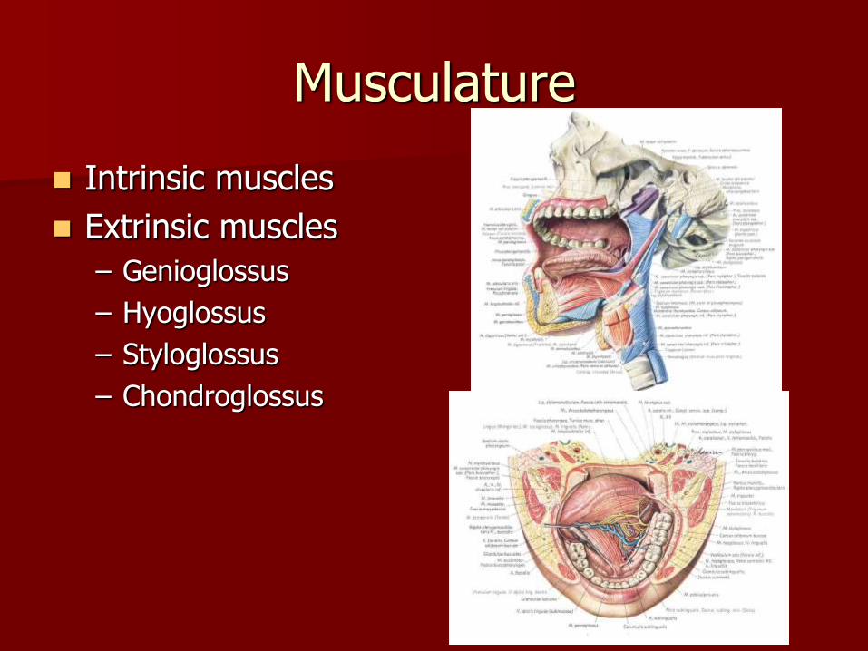
Musculature
Intrinsic muscles
Extrinsic muscles
– Genioglossus
– Hyoglossus
– Styloglossus
– Chondroglossus
Innervation
Base of tongue motor innervation by hypoglossal nerve
Damage to this nerve causes
– deviation to ipsilateral side
– Fasiculations
– atrophy
Taste from glossopharyngeal nerve
Oral Cavity
Oral cavity begins at the lips, and ends at the circumvillate papillae.
– It consists of the lips, alveolar ridge, anterior tongue, retromolar trigone, floor of mouth, buccal mucosa, and hard palate
Many tumors of the oropharynx extend into the oral cavity.
Approaches to the oropharynx require dissection through the oral cavity.
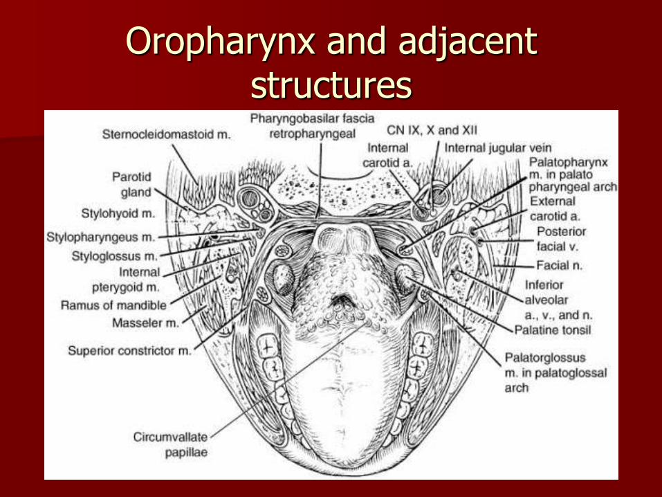
Oropharynx and adjacent structures
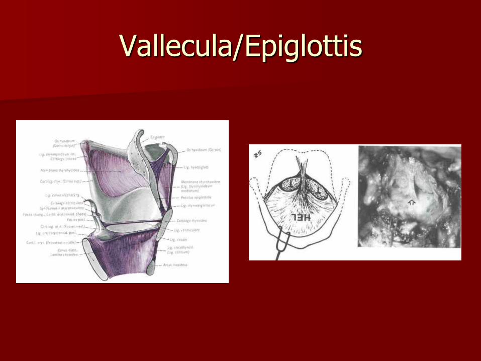
Vallecula and Epiglottis
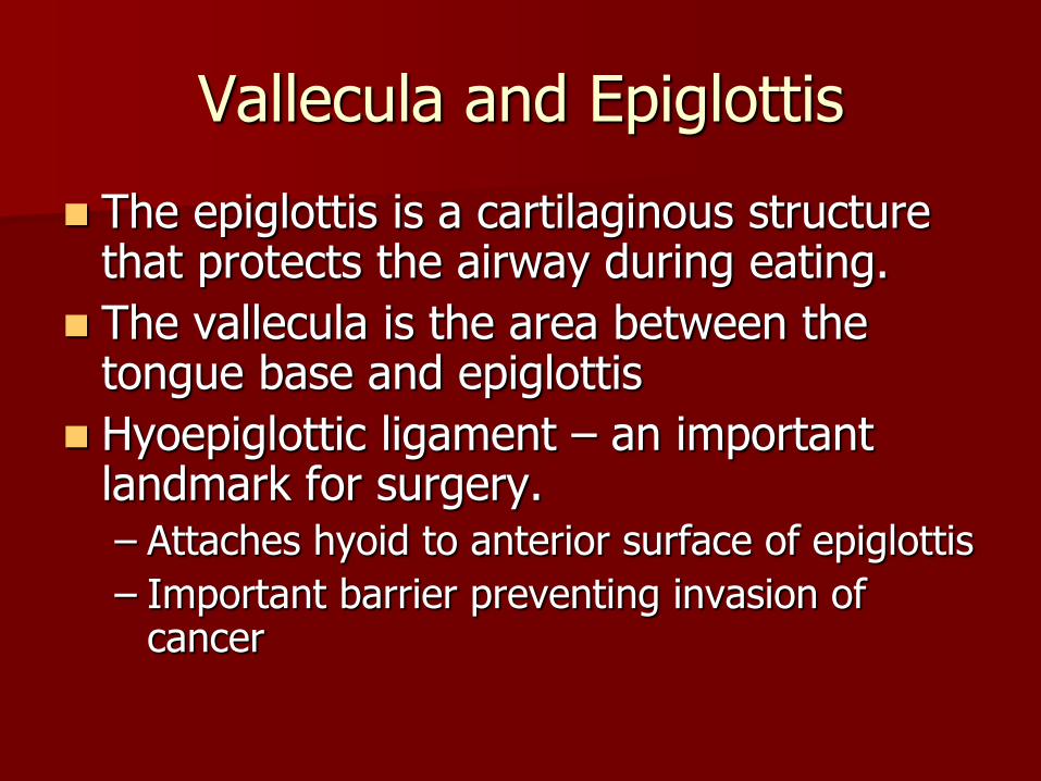
The epiglottis is a cartilaginous structure that protects the airway during eating.
The vallecula is the area between the tongue base and epiglottis
Hyoepiglottic ligament – an important landmark for surgery. – Attaches hyoid to anterior surface of epiglottis
– Important barrier preventing invasion of cancer
Vallecula/Epiglottis
Surgery of the tongue base
Intubation may be difficult.
Need wide exposure to ensure clear margins and to reconstruct defects.
Close proximity of mandible, vascular structures, nerves, and narrow introitus make resection challenging.
Surgical approaches
The base of tongue can be approached via the oral cavity or the neck.
Approaches through the oral cavity give wide exposure of the tongue base, but have significant morbidity associated with them
Approaches through the neck have decreased morbidity, but limited access.
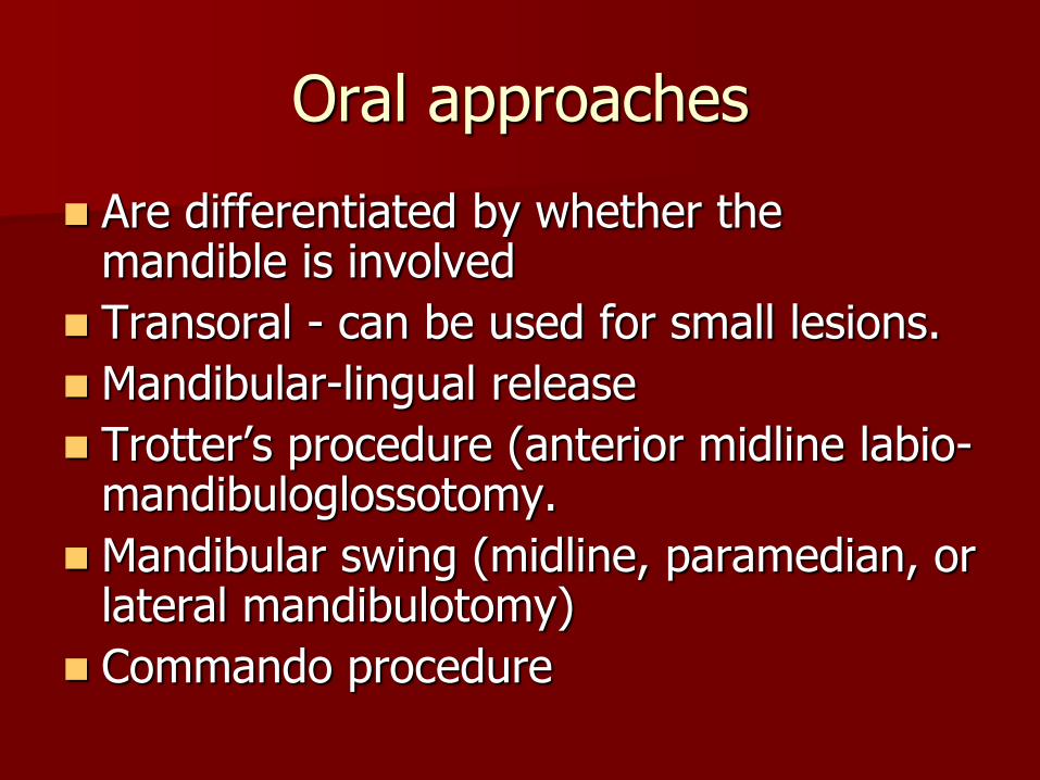
Oral approaches
Are differentiated by whether the mandible is involved
Transoral - can be used for small lesions.
Mandibular-lingual release
Trotter’s procedure (anterior midline labio-mandibuloglossotomy.
Mandibular swing (midline, paramedian, or lateral mandibulotomy)
Commando procedure
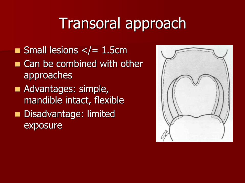
Transoral approach
Small lesions </= 1.5cm
Can be combined with other approaches
Advantages: simple, mandible intact, flexible
Disadvantage: limited exposure
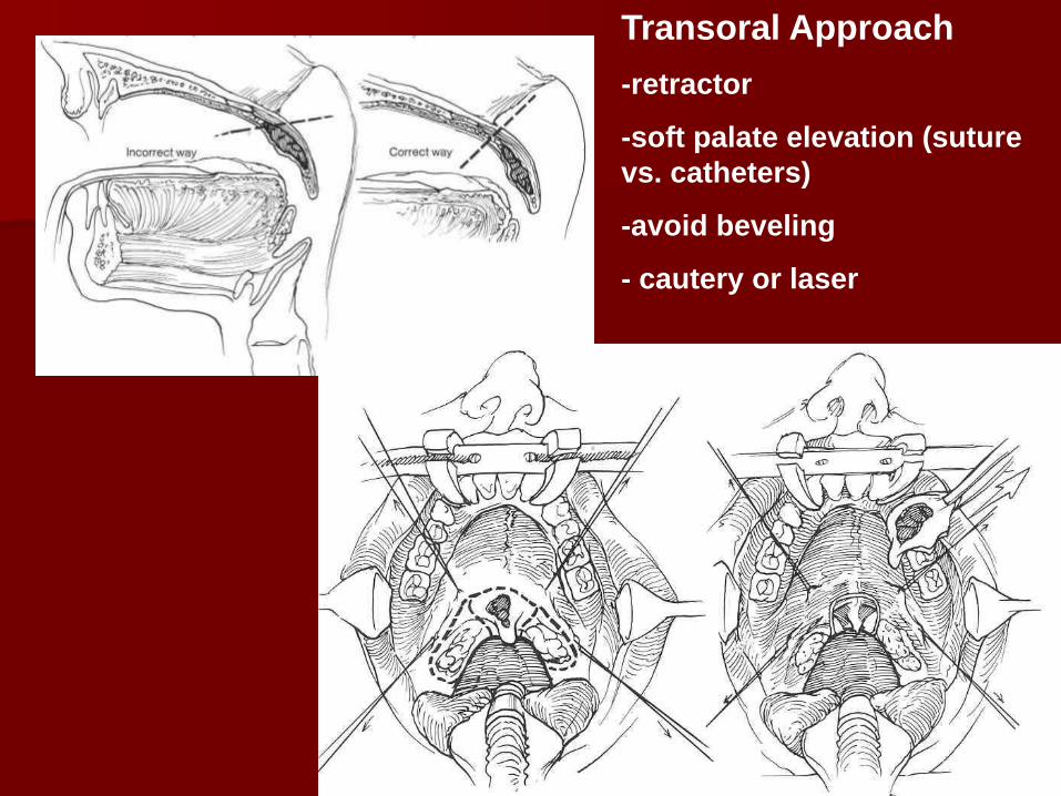
Transoral Approach
-retractor
-soft palate elevation (suture
vs. catheters)
-avoid beveling
- cautery or laser
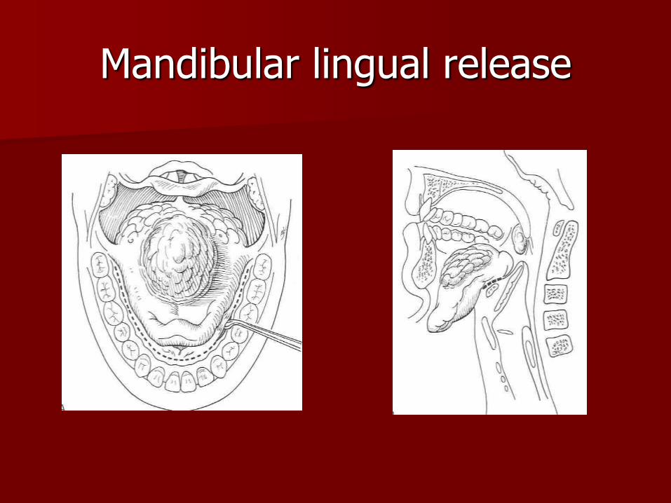
Mandibular lingual release

Mandibulotomy
Lip incision in midline (vs. visor flap) – Mark vermillion
border – Usually curve around
chin pad
Incision of vestibular mucosa with minimal elevation of periosteum (no more lateral than mental n.)
Shape plate and drill holes before osteotomy
Midline vs. paramedian vs. lateral osteotomy – Thin blade saw vs.
Gigli saw – Stairstep vs. notched
vs. straight
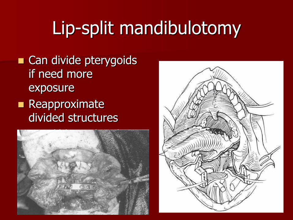
Lip-split mandibulotomy
Can divide pterygoids if need more exposure
Reapproximate divided structures
Mandible is plated.
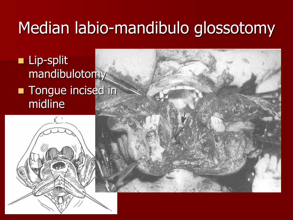
Median labio-mandibulo glossotomy
Lip-split mandibulotomy
Tongue incised in midline
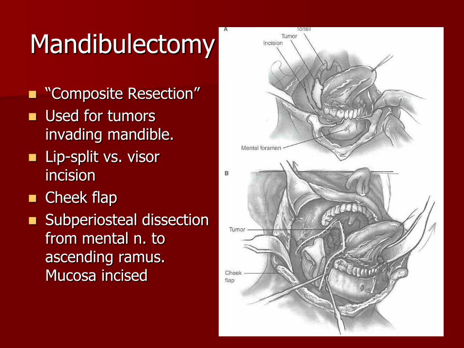
Mandibulectomy
“Composite Resection”
Used for tumors invading mandible.
Lip-split vs. visor incision
Cheek flap
Subperiosteal dissection from mental n. to ascending ramus. Mucosa incised

Neck approches
Anterior pharyngotomy
– Suprahyoid
– Subhyoid
– transhyoid
Laryngectomy
– Supraglottic
– Partial
– Total
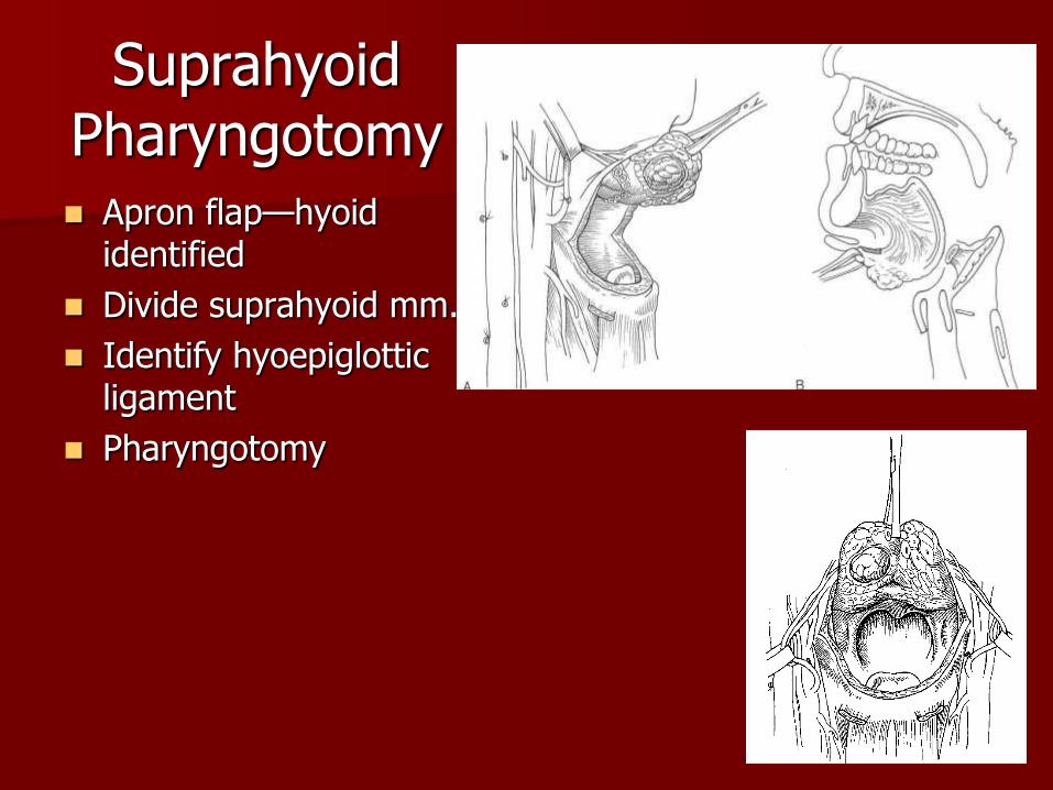
Suprahyoid Pharyngotomy Apron flap—hyoid
identified
Divide suprahyoid mm.
Identify hyoepiglottic ligament
Pharyngotomy
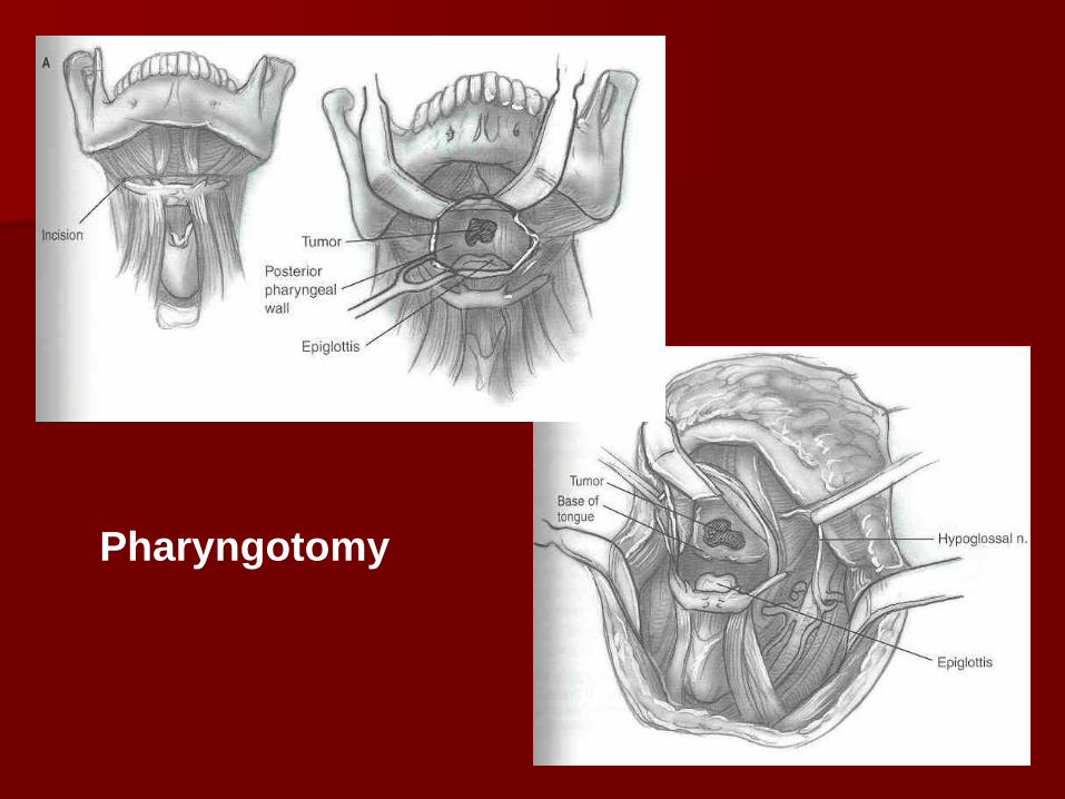
Pharyngotomy

Supra/Subhyoid supraglottic laryngotomy/ectomy
Used to excise tongue-base lesions which are adjacent to or invade the vallecula. The more extensive the tumor, the farther inferior the approach.
Approach is similar to suprahyoid pharyngotomy except: – Hyoepiglottic ligament is divided at its origin
– Dissection in underlying preepiglottic fat reveals lateral border of epiglottis
– Laryngotomy performed between epiglottis and false cords
At least one sup. Laryngeal neurovascular bundle is preserved.
Closure includes suspension of the hyoid/thyroid cartilage and partial closure of larynx, if indicated
Transthyroid supraglottic laryngotomy/ectomy
Oropharyngeal lesions which deeply invade the supraglottic larynx, but do not involve the true vocal cords or lower paraglottic space.
Can be combined with pull-through approach
Approach similar to supraglottic laryngectomy with transthyroid cartilage laryngotomy
Total laryngectomy is performed for patients with oropharyngeal lesions which involve the larynx. It should also be considered for patients with poor pulmonary reserve.
Reconstruction of defects
Goals of reconstruction are
– Maintenance of airway
– Physiologic swallowing function
– Maintenance of intelligible speech
Tongue base not involved with articulation, but if a significant portion of the tongue is removed, then articulation will be affected.
Base of tongue function
Tongue base is the most important structure of the oropharynx
Responsible for pharyngeal closure during the oral phase
Driving for force for the bolus in the pharyngeal phase
Need at least one hypoglossal and one lingual artery for mobility and survival of remaining tongue

Base of tongue
Reconstruction must
– restore bulk
– Recreate glossopharyngeal fold
– ensure continued mobility of tongue
Reconstruction
Ideal reconstruction prevents aspiration
Sensate tissue
– More physiologic swallowing
– Dynamic capability needed for articulation
Reconstructive Options
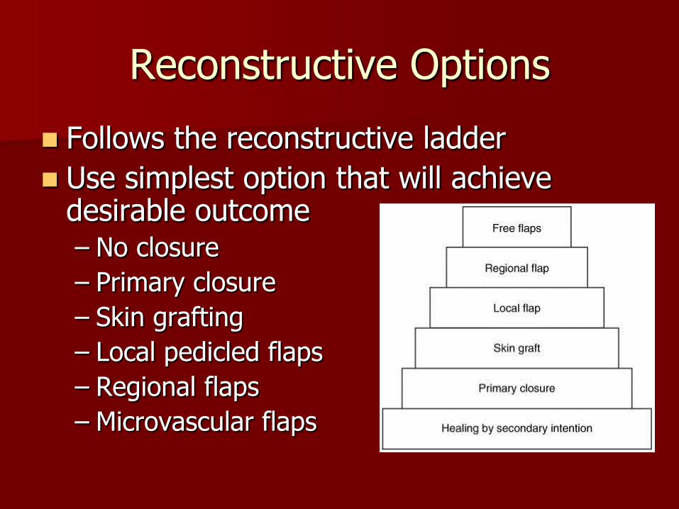
Follows the reconstructive ladder
Use simplest option that will achieve desirable outcome – No closure
– Primary closure
– Skin grafting
– Local pedicled flaps
– Regional flaps
– Microvascular flaps
Small defects
Defects up to 1/3 volume of the tongue base
Closed primarily
Split-thickness skin graft
Granulation
Minimal functional defecit
Large defects
Larger than 1/3 volume of base of tongue
Require reconstruction
Primary closure/skin grafting causes functional deficit
– Tongue tethering
– Pharyngeal stenosis
Local flaps
Have fallen out of favor
Limited amount of tissue
Inferior functional results
Not very useful for tongue defects
– Tongue flaps, divide tongue anteriorly and rotate posteriorly
– Limited tongue motion
Regional flaps
Advantages
– Abundant, well-vascularized tissue
– Single stage reconstruction
– Easy to harvest
Disadvantages
– Limited superior reach
– Bulk
– Tip necrosis

Regional flaps
Pectoralis major
Latissimus dorsi
Trapezius
Platysma
Sternocleidomastoid

Microvascular flaps
Overcome the deficiencies of regional flaps
Ability to provide sensory or motor innervation
Microvascular flaps
Fasciocutaneous
– Forearm
– Lateral thigh
– Lateral arm
Latissimus dorsi
Rectus abdominis
Radial Forearm
Workhorse flap
Lateral antebrachial cutaneous nerve can be used for sensation

Neurovascular pedicle
Up to 20 cm long
Vessel caliber 2 – 2.5 mm
Radial artery
Venae comitantes / cephalic vein
Lateral antebrachial cutaneous nerve (sensory) – Anastomose to lingual nerve
– Increased two point discrimination after inset
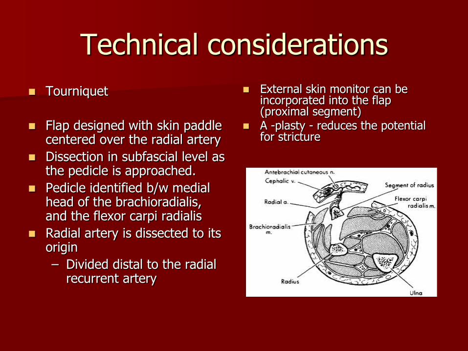
Technical considerations
Tourniquet
Flap designed with skin paddle centered over the radial artery
Dissection in subfascial level as the pedicle is approached.
Pedicle identified b/w medial head of the brachioradialis, and the flexor carpi radialis
Radial artery is dissected to its origin
– Divided distal to the radial recurrent artery
External skin monitor can be incorporated into the flap (proximal segment)
A -plasty - reduces the potential for stricture

Radial Forearm Flap
Morbidity
– Hand ischemia
– Fistula rates - 42% to 67% in early series
Subsequent series - 15% and 38%.
Creation of a controlled fistula or use of a salivary bypass stent can protect the suture line from salivary soilage and decrease the potential for fistulization.
– Stricture formation - 9% to 50%.
– Radial nerve injury
– Variable anesthesia over dorsum of hand.

Radial Forearm Flap
Preoperative considerations
– Allen test
Tests viability of palmar arch system
– No IVs / blood draws in donor arm.
– Skin graft (must preserve paratenon layer)
– Should not be used if defect extends below the thoracic inlet
Postoperative management
– Forearm and wrist immobilization w/volar splint
– 7-10 days
– Oral intake can generally begin within 7 to 10 days
2 weeks is best if the patient has been previously irradiated.
Lateral Arm Flap
Described by Song in 1982
Moderately thin fasciocutaneous flap
Donor site skin 6-8 cm (1/3 circumference of arm)
Thick skin from the upper arm
Tongue base
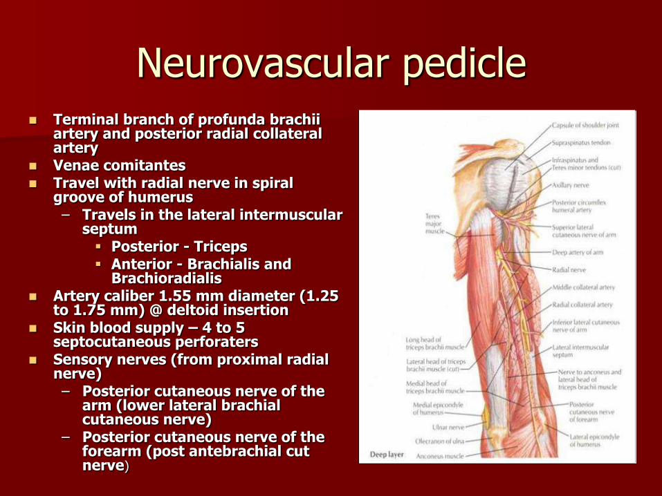
Neurovascular pedicle Terminal branch of profunda brachii
artery and posterior radial collateral artery
Venae comitantes Travel with radial nerve in spiral
groove of humerus – Travels in the lateral intermuscular
septum Posterior - Triceps Anterior - Brachialis and
Brachioradialis Artery caliber 1.55 mm diameter (1.25
to 1.75 mm) @ deltoid insertion Skin blood supply – 4 to 5
septocutaneous perforaters Sensory nerves (from proximal radial
nerve) – Posterior cutaneous nerve of the
arm (lower lateral brachial cutaneous nerve)
– Posterior cutaneous nerve of the forearm (post antebrachial cut nerve)
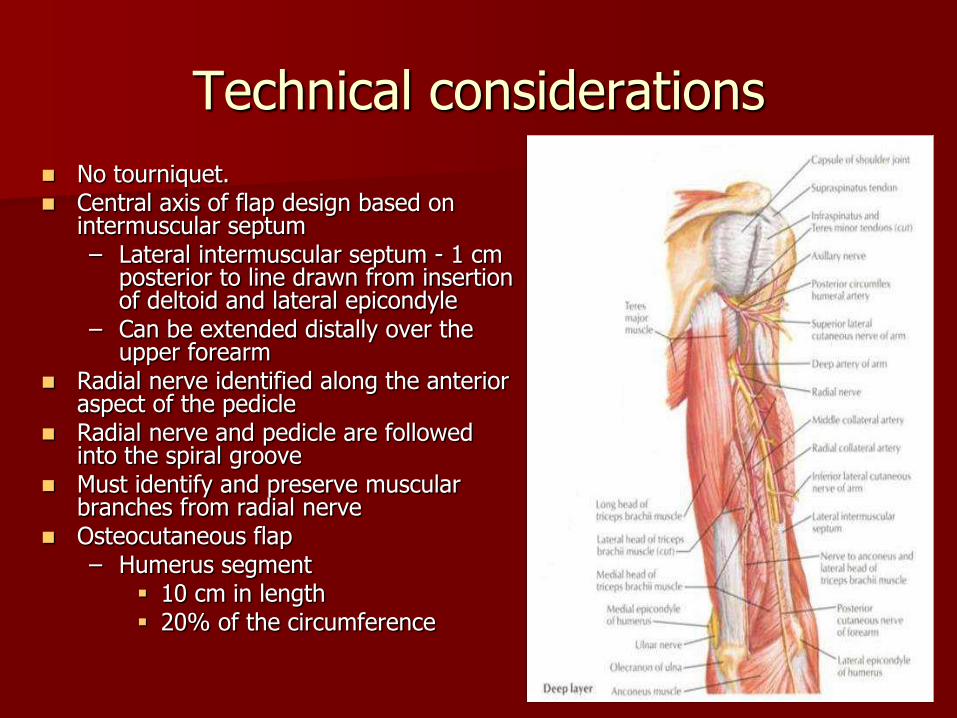
Technical considerations
No tourniquet. Central axis of flap design based on
intermuscular septum – Lateral intermuscular septum - 1 cm
posterior to line drawn from insertion of deltoid and lateral epicondyle
– Can be extended distally over the upper forearm
Radial nerve identified along the anterior aspect of the pedicle
Radial nerve and pedicle are followed into the spiral groove
Must identify and preserve muscular branches from radial nerve
Osteocutaneous flap – Humerus segment
10 cm in length 20% of the circumference
Lateral Arm Flap
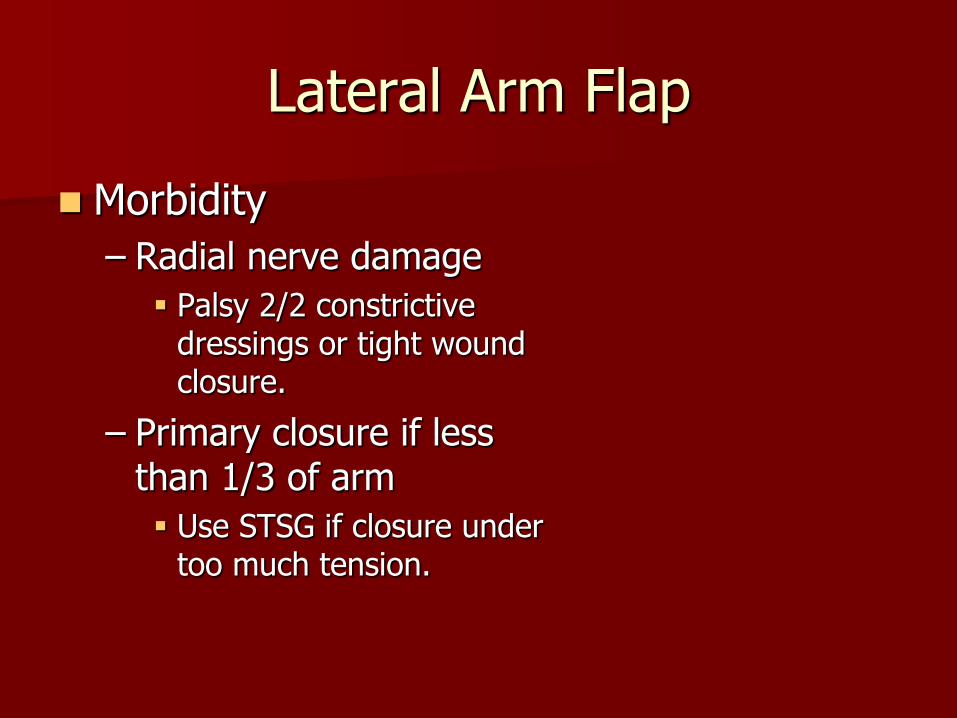
Morbidity
– Radial nerve damage
Palsy 2/2 constrictive dressings or tight wound closure.
– Primary closure if less than 1/3 of arm
Use STSG if closure under too much tension.
Lateral Arm Flap
Preoperative Considerations
– Easy scar camouflage
– Male patients may have less hair in this region when compared to forearm
Consider for intraoral reconstruction
– Flap becomes thinner more distally

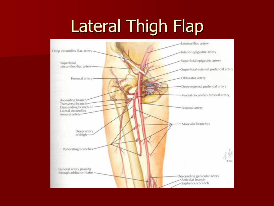
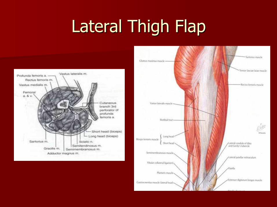
Lateral Thigh Flap
Described by Baek in 1983
Large surface area
Expendable tissue
Flap size up to 25 x 14 cm
Fasciocutaneous flap – thin to moderately thick
Intraoral and pharyngeal reconstruction
Reinnervated via lateral femoral cutaneous nerve
Neurovascular pedicle
Third perforator of profunda femoris
Travels w/in intermuscular septum
Pedicle 8 – 12 cm
Vessel caliber 2 – 4 mm
Lateral femoral cutaneous nerve of the thigh
– Anterosuperior entry into flap
– Does not travel with vascular pedicle
Terminal cutaneous branch of second or fourth perforators are the dominant arterial supply (rare)
– 4th perforator usually included in dissection to account for variations
– When 2nd perforator dominant – pedicle length limited by muscular branch vessels to preserve femoral blood supply.
Lateral Thigh Flap
Lateral Thigh Flap
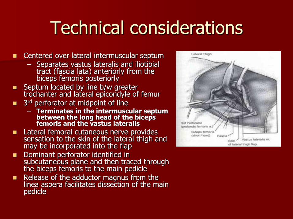
Technical considerations
Centered over lateral intermuscular septum – Separates vastus lateralis and iliotibial
tract (fascia lata) anteriorly from the biceps femoris posteriorly
Septum located by line b/w greater trochanter and lateral epicondyle of femur
3rd perforator at midpoint of line – Terminates in the intermuscular septum
between the long head of the biceps femoris and the vastus lateralis
Lateral femoral cutaneous nerve provides sensation to the skin of the lateral thigh and may be incorporated into the flap
Dominant perforator identified in subcutaneous plane and then traced through the biceps femoris to the main pedicle
Release of the adductor magnus from the linea aspera facilitates dissection of the main pedicle
Lateral Thigh Flap
Morbidity
– Atherosclerosis of profunda femoris and its branches
– Avoid in pts with h/o peripheral vascular disease
– Sciatic nerve injury
Lateral Thigh Flap
Preoperative Considerations
– Assess for PVD (palpate peripheral pulses)
– Not advised for use in obese individuals or in those with previous surgery or trauma to the thigh
Postoperative management
– Primary closure of donor site
– Early walking

Rectus abdominis
Easy to harvest
Long pedicle
Skin from abdomen and lower chest
Myocutaneous flap or muscle only flap
Not used for functional motor reconstruction
Total glossectomy defects

Neurovascular pedicle
Two dominant pedicles – Deep superior epigastric artery/vein – Deep inferior epigastric artery and
vein
Based on inferior epigastrics when used for h/n recon because of larger pedicle size
Inferior epigastric diameter – 3 to 4 mm
Reinnervated with any of the lower six intercostal nerves.
Pedicle may travel along lateral aspect of muscle before taking intramuscular route
Technical considerations
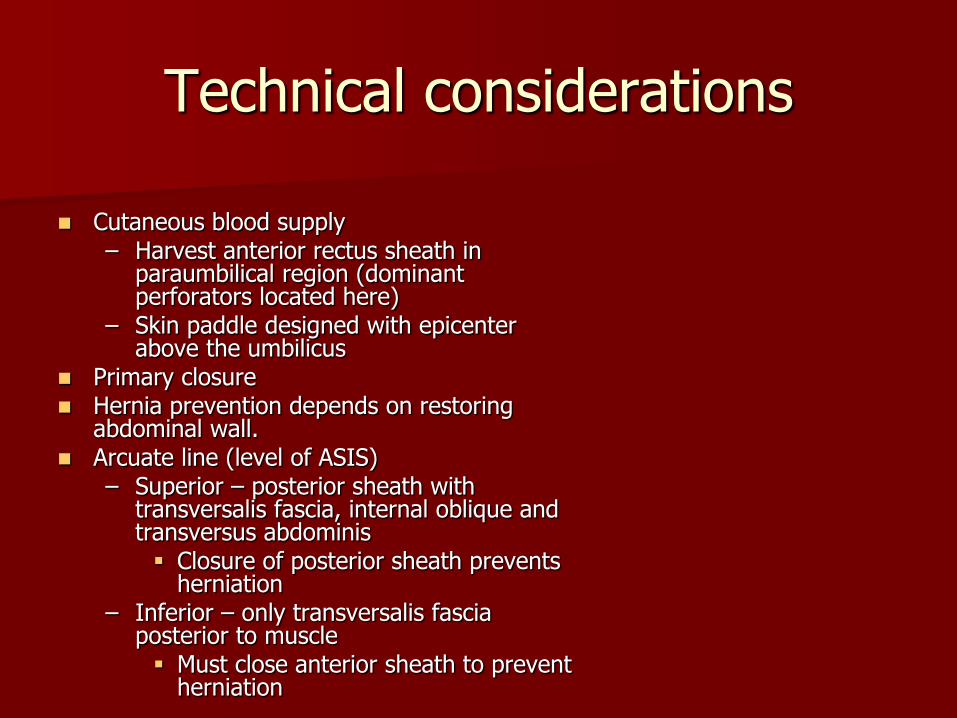
Cutaneous blood supply – Harvest anterior rectus sheath in
paraumbilical region (dominant perforators located here)
– Skin paddle designed with epicenter above the umbilicus
Primary closure Hernia prevention depends on restoring
abdominal wall. Arcuate line (level of ASIS)
– Superior – posterior sheath with transversalis fascia, internal oblique and transversus abdominis Closure of posterior sheath prevents
herniation – Inferior – only transversalis fascia
posterior to muscle Must close anterior sheath to prevent
herniation

Technical considerations 1. Dissect superiorly first 2. Dissect down to underlying muscle 3. Split fascia to the costal margin 4. Lateral and inferior portions of skin paddle
incised next 5. Small cuff of anterior rectus fascia preserved
medially and laterally, to preserve cutaneous perforators
6. Split fascia vertically down to the public region 7. Divide rectus superiorly and free from
posterior rectus sheath 8. Dissection below the arcuate line 9. Vascular pedicle identified below arcuate line
along the lateral deep aspect of the muscle. 10. Divide rectus inferiorly 11. Pedicle dissected inferiorly to origin off the
external iliac system
Rectus abdominis
Morbidity
– Abdominal weakness
– Hernia

Rectus abdominis
Preoperative Considerations – Prior abdominal
surgery
– Prior inguinal herniorrhapy may compromise pedicle dissection 2/2 scarring
– Hernia
– Diastasis recti
Postoperative management
– Ileus
– Avoid abdominal strain for 6 weeks.
Latissimus dorsi
Pedicle or free flap Free flaps
– Better flap positioning – Cutaneous portion can be centered over pedicle – Less risk of pedicle kinking
Musculocutaneous – Large volume defects of large cutaneous neck defects
STSG for final resurfacing Non-sensate Motor reconstruction possible Useful after total glossectomy

Neurovascular pedicle
Thoracodorsal artery Arise from subscapular vessels
off of third portion of axillary artery and vein
Vessel diameter at origin – 2.7 mm(1.5 to 4.0)
Vein diameter – 3.4 mm (1.5 to 4.5)
Pedicle length 9.3 cm (6 to 16.5) – Can be lengthened by sacrificing
branch to serratus anterior
Numerous variations – Most common: independent origin of
thoracodorsal vein/artery
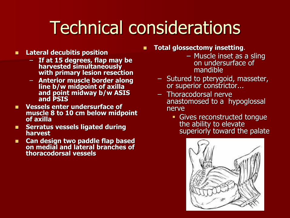
Technical considerations Lateral decubitis position
– If at 15 degrees, flap may be harvested simultaneously with primary lesion resection
– Anterior muscle border along line b/w midpoint of axilla and point midway b/w ASIS and PSIS
Vessels enter undersurface of muscle 8 to 10 cm below midpoint of axilla
Serratus vessels ligated during harvest
Can design two paddle flap based on medial and lateral branches of thoracodorsal vessels
Total glossectomy insetting.
– Muscle inset as a sling on undersurface of mandible
– Sutured to pterygoid, masseter, or superior constrictor...
– Thoracodorsal nerve anastomosed to a hypoglossal nerve Gives reconstructed tongue
the ability to elevate superiorly toward the palate

Latissimus dorsi
Morbidity
– Marginal flap necrosis
– Pedicled flaps pass b/w pec major and minor
Changes in arm position may occlude pedicle
Should immobilize arm in flexed position

Latissimus dorsi
Preoperative Considerations
– Relative contraindications - prior axillary LN dissection
– Preop angiography advocated to assess vessel patency
Postoperative management
– Suction drains
– High incidence of seroma
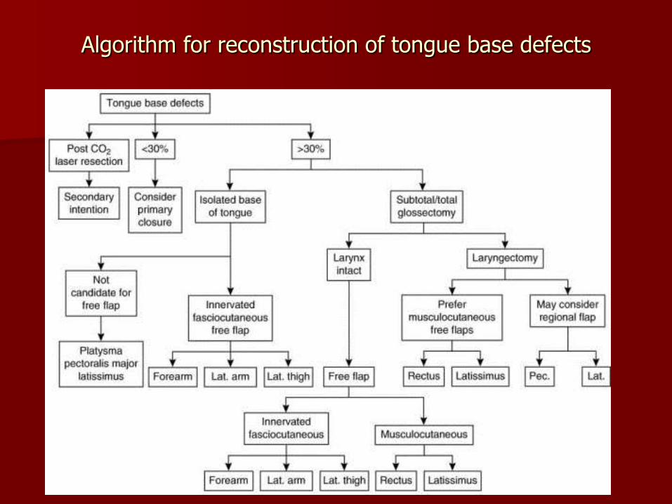
Algorithm for reconstruction of tongue base defects
Conclusion
The tongue base is a very important structure found in the oropharynx
Over half of all oropharyngeal SCCa involve the base of tongue
Resection of these cancers leave anatomic as well as functional defects.
Reconstruction of these defects tries to restore airway protection, swallowing, and speech functions.
Bibliography
Agrawal, A., et al. Resection of cancer of the tongue base and tonsil via the transhyoid approach. Laryngoscope 2000; 110(11):1802-1806.
Azizzadeh, B., et al. Long-term survival outcome in transhyoid resection of base of tongue squamous cell carcinoma. Archives of Otolaryngology—Head & Neck Surgery 2002; 128(9):1067-1070.
Amin, M.R., et al. Straight midline mandibulotomy revisited. Laryngoscope 1999; 109(9):1402-1405. Bailey, B.J., et al. Surgery of the Oral Cavity Year Book Medical Publishers, Inc., Chicago, IL c. 1989. Bailey, B.J., et al. Head & Neck Surgery—Otolaryngology Lippincott Williams & Wilkins, Philadelphia, PA, c. 2006. Dai, T.S., et al. Complications of mandibulotomy: midline versus paramidline. Otolaryngology—Head & Neck Surgery 2003;
128(1):137-141. Davidson J., et al. Mandibulotomy in the irradiated patient. Archives of Otolaryngology Head and Neck Surgery 1989;
115:497-499. Eisen M.D., et al. Morbidity after midline mandibulotomy and radiation therapy. American Journal of Otolaryngology 2000;
21:312-317. Ferner, H. Eduard Pernkopf Atlas of Topographical and Applied Human Anatomy, Volume I, Urban & Schwarzenberg, Inc.,
Baltimore, MA, c.1980. Lore, J.M., et al. An Atlas of Head and Neck Surgery W.B. Saunders Co., Philadelphia, PA, c.1988. Myers, E.N. Operative Otolaryngology Head and Neck Surgery W.B. Saunders Co., Philadelphia, PA, c. 1997. Nasri, S., et al. Transpharyngeal approach to base of tongue tumors: a comparative study. Laryngoscope 1996; 106(8):945-
950. Pan, W.L., et al. The anatomical basis for mandibulotomy: midline versus paramidline. Laryngology 2003; 113(2):377-380. Riddle, S.A., et al. Midline mandibular osteotomy: an analysis of functional outcomes. Laryngoscope 1997; 107(7):893-896. Sessions, D.G., et al. Atlas of Access & Reconstruction in Head & Neck Surgery. Mosby Year Book, Inc., St. Louis, MO.
C.1992. Shah, J.P., et al. Comparative elvaluation of fixation methods after mandibulotomy for oropharyngeal tumors. American
Journal of Surgery 1993; 166:431-434. Warwick R., Williams, P.L. Gray’s Anatomy W.B. Saunders, Co., Philadelphia, PA, c. 1973. Zeitels, S.M., et al. Surgical Management of Tumors of the Oropharynx American Academy of Otolaryngology—Head and Neck
Surgery Foundation, Inc. Custom Printing, Inc. Rochester, MI c.1997. Zeitels, S.M., et al. Suprahyoid pharyngotomy for oropharynx cancer including the tongue base. Archives of Otolaryngology
Head and Neck Surgery 1991; 117:757-760.