reconstruction of the flagellar apparatus and microtubular...
TRANSCRIPT

Protoplasma 121, 186--I98 (1984) PROTOPLASMA �9 by Springer-Verlag 1984
Reconstruction of the Flagellar Apparatus and Microtubular Cytoskeleton in Pyramimonas gelidicola ( Prasinophyceae, Chlorophyta)
G. I. McFADDEN * and R. WETHERBEE
School of Botany, University of Melbourne
Received September 5, 1983 Accepted November 9, 1983
Summary
The absolute configuration of the flagellar apparatus in Pyramimonas gelidicola MCFADDEN et al. has been determined and shows identity with P. obovata, indicating that they are closely related. Comparison with the flagellar apparatus of quadriflagellate zoospores from the more advanced Chlorophyeeae suggest that Pyramimonas may be a primitive ancestral form. The microtubular cytoskeleton has been examined in detail and is shown to be unusual in that it does not attach to the flagellar apparatus. CytoskeletaI microtubules are nucleated individually, and this is interpreted as an adaptation to the methods of mitosis and scale deployment. In view of the primitive nature of these processes, it is proposed that this type of cytoskeletal organization may represent a less advanced condition than that of the flagellar root MTOCs (microtubule organizing centers) observed in the Chlorophyceae.
Keywords: Flagellar apparatus; Microtubular cytoskeleton; Phylogeny; Prasinophyceae; Pyramimonas.
1. Introduction
Evidence accunmlated f rom comparat ive ultra- structure indicates that there are at least two main
streams of evolution within the green algae: 1. the Charophyceae, which gave rise to the first land plants and 2. the Chlorophyceae (hereafter sensu STEWART and MATTOX 1978) containing many of the modern green algae (PICKETT-HEAPS 1975, STEWART and MATTOX
1975). It is now generally suggested that these and any further evolut ionary streams diverged f rom the more
* Correspondence and Reprints: School of Botany, University of Melbourne, Parkville 3052, Vic., Australia.
primitive, heterogeneous group of scaly green monads
that comprise the Prasinophyceae Christensen ex Silva
(MANTON 1965, NORRIS 1980, STEWART and MATTOX
1978, MOESTRUP and ETTL 1979, MELKONIAN 1982a,
MOESTRUP 1982). Recently, the prasinophyte
Mesostigma viride has been shown to have
characteristics aligning it with both the Charophyceae (ROGERS et al. 1981, MELKONIAN 1983) and the
Chlorophyceae (MELKONIAN 1983) indicating that it is
probably similar to the ancestoral flagellate f rom which
the two major streams of evolution diverged.
Mesostigma is closely related to another genus o f the
Prasinophyceae, Pyramimonas (MANTON and ETTL
1965), which could ha)e arisen by a doubling o f the two flagella in Mesostigma to become quadriflagellate like
Pyramimonas (RocERS et al. 1981). Pyramimonas could then have given rise to the Chlorophyceae by the loss o f the scales and flagellar pit (characteristic o f the
Prasinophyceae) and the development o f a cell wall and
phycoplast cytokinesis (characteristic o f the Chloro- phyceae) (MELKONIAN 1982 a, STEWART and MATTOX
1978). A recent study of the flagellar appara tus in the quadriflagellate Pyramimonas obovata (MELKONIAN 1981) indicates that its structure shows identity with that o f the quadriflagellate swarmers in the
Chlorophycean alga, Ulothrix beIkae (FLOYD et aI. 1980). This identity o f the flagellar apparatuses is
interpreted as a direct phylogenetic link between Pyramimonas and certain Chlorophyceae that produce quadriflagellate swarmers. This hypothesis is
G. I. MCFADDEN and R. WETHERBEE: Reconstruction of the Flagellar Apparatus 187
supported by the fact that the flagellar apparatus in Hafniomonas reticulata--a quadriflagellate monad originally placed in the genus Pyramimonas, but now known to have Chlorophycean characteristics (Ea'TL and MOESTRUV 1980)--is highly comparable to both U. belkae and P. obovata and probably represents an intermediate form (MELKONIAN 1981).
While comparison of conservative features such as the structure of the flagellar apparatus and cytokinetic mechanisms has provided much information on phylogenetic linkages within the green algae, little attention has been given to the cytoskeleton. CAVALIER- SMITH (1982) has presented an hypothesis for the origin of microtubular roots in the original flagellate, but to our knowledge there have been no hypotheses on the origin of additional cytoskeletal elements. In quadriflagellate zoospores of several Chlorophycean algae (e.g., Schizomeris, BIRKBECH et al. 1974 and FLOYD and Hoops 1980; Fritschiella, MELKONIAN 1975; Stigeoclonium, MANTON 1964; Ulothrix belkae, FLOYD, personal communication) there exists a sub- plasmalemmal microtubule array that originates from the microtubular flagellar roots and probably serves a cytoskeletal function (cf., MELKON~AN et al. 1980). In Polytomella, a colorless Chlorophycean quadri- flagellate, is has been shown that an accumula- tion of amorphous, osmiophylic matter on the flagellar roots acts as an MTOC (microtubule organizing center) exerting spatial control over the initiation of the microtubule array (BRowN et al. 1976, STEARNS and BROWN 1981).
In Pyramimonas a microtubular cytoskeleton has been reported in several species (MANTON 1966, 1968, MOESTRUP and THOMSEN 1974, NORRIS and PEARSON 1975, MARCHANT 1982) but there has been no detailed study on the origin, distribution and orientation of these microtubules. It did not seem likely that the microtubular cytoskeleton in Pyramimonas could be nucleated from an MTOC on the flagellar roots, as this system would portend difficulties during the processes of cytokinesis and scale deposition. The nature of the above processes in Pyramimonas suggested that the nucleation sites were more likely to show less inter- organization and perhaps be individual--not attached to a flagellar root. TUCKER (1979) has proposed that there may exist (in some unknown organism) individual, "free" nucleating elements (NEs) that generate a single microtubule. In this paper we describe the cytoskeleton in Pyramimonas gelidicola and report that each microtubule is nucleated individually. This phenomenon probably relates to the methods of
cytokinesis and scale production in this organism. Since comparison of the flagellar apparatuses indicates that Pyramimonas is antecedent to certain quadriflagellate Chlorophycean zoospores, we present reasons to suggest that the type of cytoskeleton observed in Pyramimonas represents a less advanced condition than that of some Chlorophyceae.
2. Materials and Methods
Cells of Pyramimonas gelidicola were grown, fixed and prepared for thin section electron microscopy as described by MCFADDEN et al. (1982). The absolute configuration was determined as outlined by FLOYD et al. 1980. Specimens for freeze-fracture were unfixed and without cryoprotectant, frozen in Freon 22 and then stored in liquid nitrogen prior to preparation in Balzer's 300 freeze-fracture apparatus. Specimens were fractured, but not etched, then shadowed with platinum/carbon from 45 ~ and a carbon support film. Replicas were cleaned in concentrated sulphuric acid, followed by bleach and examined with a double tilt goniometer stage in a Siemens Elmiskop 102.
3. Results
The general ultrastructure of Pyramimonas gelidicola and details of the scale coverings has been previously reported (McFADDEN et al. 1982, MCFADDEN and WETHERBEE 1982 a). The number and spatial relationship of the cell organdies is Consistent (McFADDEN and WETHERBEE 1982 b) and this allows for proper orientation when viewing random sections. We have adopted the definitions of cell symmetry and flagellar apparatus terminology proposed by MELKONIAN (1980, 1981) since they are appropriate to this study. Cells are 14-18gm long with four scaly flagella, approximately 15 txm long, inserted in an apical pit (Fig. 1). The axoneme microtubules display the usual 9 + 2 configuration (Figs. 2 and 3), and there is a transition region where it connects to the basal bodies (Figs. 4-8). No twisting of the central pair was observed (Fig. 2). Details of the transition zone and basal bodies are, for the most part, in agreement with those described for Pyramimonas parkeae (NORRIS 1980). There is a transitional helix (coiled fiber) distal to the termination of the central pair (Figs. 4 and 8) and a stellate pattern proximally (Fig. 5). The basal body triplets show increasing imbrication proximally, eventually forming the cartwheel pattern (Figs. 6 and 7). We have observed a discontinuous osmiophilic core within the basal body, with fibrous spokes radiating to each triplet (Figs. 6 and 8).

Fig. I. Whole mount ofPyramimonas gelidicola stained in aqueous uranyl acetate, showing cell shape, four scaly flagella inserted in an apical pit and a periplast of box scales on the cell surface, x 4,000
Fig. 2. Longitudinal section (LS) of flagellar axoneme showing the arrangement of microtubules and the straight central pair. x 40,000
Fig. 3. Transverse section (TS) of axoneme showing 9 + 2 arrangement of flagellar tubules (corresponds to region A in Fig. 8). x 93,000
Fig. 4. TS of transitional helix or coiled fiber (corresponds to TH in Fig. 8). x 112,000
Fig. 5. TS of stellate pattern which corresponds to level SP in Fig. 8. x 90,000
Fig. 6. TS of basal body showing arrangement of triplets and osmiophylic core (corresponds to BB in Fig. 8). x 88,000
Fig. 7. TS of cartwheel pattern at most proximal end of basal body (see CP in Fig. 8). x 88,000
Fig. 8. LS of flagellum (II) showing cartwheel pattern (CP), basal body (BB) with synistosome (S) attached stellate pattern (SP), transitional helix (T//) and axoneme (A). x 60,000
Fig. 9. TS of basal bodies inumbered) showing arrangement of connecting fibers including synistosome (S) and cruciate arrangement of four microtubular roots (arrows) (corresponds to level BB in Fig. 8). x 53i000
Fig. 10. TS at the most proximal end of basal bodies showing major proximal connecting fiber (P.F), microbody (3//) and nucleus (N). x 60,000
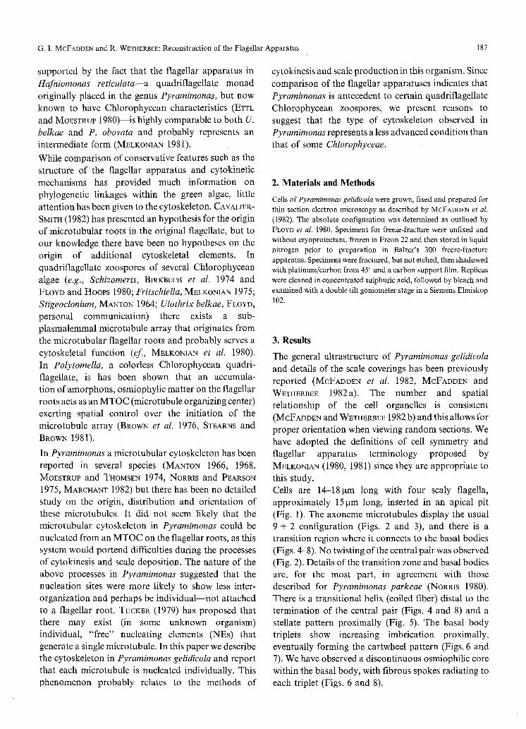
G, I. McFADDEN and R. WETHERBEE: Reconstruction of the Flagellar Apparatus 189
Fig. 11. Oblique LS through flagellum//showing synistosome (S), microbody (M), nucleus (N) and the right, ventral, four-stranded root (arrow). x 47,000
Fig. 12. LS of flagella III and/showing system II fibers (open arrow) attached to basal body III. The left, dorsal, four-stranded root is also visible (arrow). x 65,000
Fig. 13. LS of right, ventral, four-stranded root. A cross-section of this root is shown in the inset ( x 90,000). One cytoskeletal microtubule can be seen originating remote from the root (short arrow). • 50,000
Fig. i4. LS of flagella I and H showing nucleus (N) and microbody (m) situated beneath the basal bodies, x 35,000
The four basal bodies are set in a trapezoidal pat tern and two sets o f fibers (proximal and distal) interconnect the basal bodies directly or indirectly (Fig. 9). The connect ing fibers are very similar to those o f Pyramimonas obovata (MELKONIAN 1981) and need no t be described in detail here. Since they extend
approximately 200 nm into the cell (Figs. 8, 11, and 13) the connecting fibers apparent ly take the fo rm of sheets
with the major par t o f the distal connecting fibers being
a block, termed the synistosome (NORRIS and PEARSON 1975). The synistosome measures some 260 x 175rim
with a dark 30 nn band in the center (Fig. 9). There is a prominent proximal fiber connecting basal bodies I and IV with no visible striation pat tern (Fig. 10).
The flagellar roots comprise a cruciate 4-2-4-2
microtubular system and two system II fibers (rhizoplasts) (Figs. 9, 11-13, and 15). System I fibers were not observed. The microtubular roots ascend f rom

190 G.I. MCFADDEN and R. WETI-IERB~E: Reconstruction of the Flagellar Apparatus
between the basal bodies and underlie the cytoplasmic microtubules in the flagellar pit (Fig. 13). The four stranded roots display the usual 3 over 1 configuration proximally (Fig. 13, inset). All roots terminate distally in a small amorphous area of electron dense material within the flagellar pit region (Fig. 13).
The position of the roots relative to the other organelles is consistent. The left, ventral two stranded root shows an association with the opening of the scale reservoir. A fibrous band connects this root to a circular fibrous investment surrounding the reservoir opening. None of the microtubular roots was observed to contact the eyespot or chloroplast envelope.
The system II fibers (rhizoplasts) originate from the proximal ends of basal bodies II and III and ensheath the microbody (Figs. 11 and 12). The system II fibers are relatively fine and indistinct but display a striation periodicity of 100 nm at their proximal end (Fig. 12). The microbody is directly beneath basal bodies I and II with an anterior protrusion around basal body IV (Figs. 11 and 14)o The proximal portion of the microbody is adjacent the nucleus (Fig. 14) and the distal portion projects into the pyrenoid, but no connections between these organelles were observed (cf., Fig. 11, McFaDDEN et al. 1982). A diagramatic reconstruction of the flagettar apparatus is given in Fig.
15. The cytoplasmic microtubule network comprises between 70 and 80 microbubules that run roughly parallel (Figs. 16-24, 26, and 27). Each microtubule originates individually in a small amorphous mass of electron dense material (Figs. 16, 21-25). The microtubule endings do not display any inter- organization or connect to any cytoplasmic structure, although occasionally coated vesicles were observed close to microtubules. In the flagellar pit the microtubules are spaced at intervals of ca. 140 nm just beneath the plasmalemma (Fig. 16). The microtubules transcend the edge of the flagellar pit spreading to a distance of 200-250 nm at the anterior end (Fig. 17). The network curves around the rim of the pit and runs down the outside of the cell between the plasmalemma and chloroplast (Fig. 18) making a complete 180 ~ turn in the space of 1 gm. The number of microtubules on the outside exactly equals the number in the pit for each cell (excluding flagellar roots), and the ratio of outer and inner circumferences is approximately equal to the ratio of the distances between the microtubules in the two regions. In thin sections glancing the outside of the cell the microtubules are seen to be closely associated with
the chloroplast envelopes, lying in grooves in these membranes (Fig. 26). In freeze-fracture replicas of the outer chloroplast envelope, this groove can be seen clearly (Figs. 19, 20, 28, and 29). The grooves in the E fracture face are approximately 45 nm wide (Fig. 20) and the corresponding bumps in the P fracture face are approximately 35nm wide (Fig. 19), which would account for the microtubular diameter and the membrane leaflet around it. The bumps in the P fracture face of the outer chloroplast membrane often exhibit a fibrillar substructure with 5-6 longitudinal fibers averaging 7 nm in thickness (Fig. 28). Fracture faces at the posterior end, where the cell tapers to become conical, show microtubules in this region converging and some microtubules terminate on or very near to others, thereby maintained the spacing between microtubules (Fig. 29). A pattern of large square impressions (ca. 300nm square) from the external layer of box scales can be seen in most fracture faces (Figs. 19 and 20). Fracture faces of the plasmalemma, though rarely observed, showed square impressions of the box scales but no microtubular impressions.
4. Discussion
The absolute configuration of the flagellar apparatus in Pyrarnirnonas gelidicola appears identical to that of P. obovata (MELKONIAN 1981) and P. orientalis (MOESTRUP and TnOMSEN 1974) indicating that these species are closely related. Comparisons of organelle structure and fine details of the small scale varieties lead MELKONIAN and ROBEYEK (1981) to propose that P. obovata and P. orientalis belong together in a subgroup of the genus, which should now include P. gelidicola. This proposal is supported by the fact that P. obovata, P. gelidicola (see McFADDEN and WETnERBEE 1982a) and P. orientalis (MCFADDEN, unpublished) all possess footprint scales. The connecting fibers between the four basal bodies of Pyramimonas gelidicola appear completely asymmetrical. The asymmetry of the flagellar apparatus is further compounded by the association of one microtubular root with the duct opening of the scale reservoir. While this association was not reported for P. obovata (MELKONIAN 1981), it was observed by MANTON (1966, 1968), in P. arnylifera and P. tetrarhyncus, who suggested that the root may play some role in deploying the scale reservoir outlet. Although the significance of this association is still not entirely clear, it seems to be a further instance of a functional role for microtubular roots (cf., MEL~:ONIAN 1982 b).

G. I. MCFADDEN and R. WETHERBEE: Reconstruction of the Flagellar Apparatus 191
V E N T R A L S IDE
�9 . �9 �9 / / �9 �9 �9 . , ,
* " * . J
Fig. 15. Drawing showing arrangement of basal bodies (I--IV), 4-2-4-2 microtubular root system (arrows), synistosome (53, connecting fibers, microbody (M) and nucleus (N). The ventral side as defined by MELKONIAN (1981) is indicated (adapted from MELKONIAN 1981)
FLOYD and HooPs (1980) and FLOYD el al. (1980) point out that quadrifiagellate zoospores of Chlorophycean algae forming the Chaetophorales (sensu STEWART and MAT-COX 1975) (Ulothrfx belkae, U. fimbriata, Sch&orneris, Fritschiella, and Stigeoclonium) have an identical arrangement of fiagellar apparatus components. There are cruciate microtubular roots and of the four basal bodies, two are connected by a large banded distal fiber, while a ring of peripheral fibers connects all basal bodies. This arrangement is
essentially similar to that of P. gelidicola, particularly if one considers the synistosome to be homologous to the distal connecting fiber as proposed by MELKONIAN (1980, 1981) and RO~ERS et at. (1981) (see Fig. 30). We believe this identity to be indicative of a direct phylogenetic link between P. gelidicola and the Chaetophorales and support the proposal that Pyramimonas is antecedent to some quadriflagellate Chlorophyceae (MELKONIAN 1981). Cytoplasmic microtubules in Pyramimonas gelidicola

Fig. 16. One of a series of glancing sections of the flagellar pit showing the origin of several cytoskeletai microtubules at the base of the pit on the left hand side of the micrograph (arrows). x 47,000
Fig. 17. Cytoskeletal microtubules transcending the rim of the fiagellar pit at the anterior of the cell. x 105,000
Fig. 18, One of a series of glancing sections of the cell exterior showing microtubules converging toward posterior end (left hand side of the micrograph). One microtubule (arrow) ends, maintaining the spacing between microtubules, x 33,000

Fig. 19. Freeze-fracture of outer chloroplast envelope (PF) showing square impressions of scale periplast and longitudinal impressions of cytoskeletal microtubules (arrows). In this, and subsequent freeze-fracture micrographs, an encircled arrow-head indicates the direction of
shadowing, x 67,000
Fig. 20. Freeze-fracture of the outer chloroplast envelope (EF) showing corresponding impressions of scales and microtubules (arrows). x 64,000
194 G. L MCFADDEN and R. WETHERBEE: Reconstruction of the Flagellar Apparatus
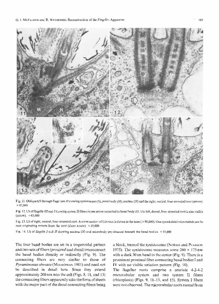
follow the cell contour, forming a precise parallel array directly beneath the plasmalemma. Preliminary experiments using the microtubule depolymerizing agents colchicine, oryzalin and hydrostatic pressure indicate that the microtubules act as a cytoskeleton. The characteristic cell shape of P. gelidicola is lost in cells observed to have lost their cytoplasmic microtubules (McFADDEN, unpublished). Microtubule grooves in the chloroplast envelopes were observed in both thin sections and freeze-fractures and may represent the effect of cell turgor pressing the chloroplast envelopes against the cytoskeleton in regions where the chloroplast envelopes are adjacent the plasmalemma (see Fig. 31). The fibrils observed in the microtubule impressions are of approximately the same dimensions as tubulin molecules composing protofilaments (AMos 1979). That an impression as small as a protofilament can be observed in a membrane leaflet seems quite remarkable and a specific microtubule/membrane association cannot be ruled out. It is possible that some molecular specialization occurs, such as a parallel organization of membrane proteins, or alternatively, that a modification of the lipid layer in the zone of microtubule/membrane contact allows visualization of the protofilaments in freeze-fracture. A microtubule/membrane association would serve in anchoring the cytoskeleton, which is otherwise unattached, and would maintain spatial relationships of internal components. Effective experimentation on the cytoskeleton was prevented by the variable response of cells to anti-microtubule agents. Using the microtubule depolymerizing techniques outlined by BROWN and BOUCK (1973) and STEARNS and BROWN (1981), a maximum of only 30% of cells in a population of P. gelidicola lost the cyto- skeleton (MCFADDEN, unpunished). This result is at variance with that of BURCH and MARCHANT (1983) but we can offer no explanation for this at present, The variability was apparently not related to cell cycle as
cells in various stages of cytokinesis may or may not round up when exposed to colchicine (McFADDEN, unpublished). This variable response has prevented us from determining if the cytoskeleton is re-established after removal of the depolymerizing agents. The square impressions in the membrane layers appear to result from cell turgor pressing against the box scale layer (see Fig. 31). The scales remain closely appressed to one another indicating that there is some cohesive force between individual box scales allowing them to act, to a limited extent, as an exoskeleton. The nature of the scale interconnections is unknown, but possibly similar to that observed in Tetraselmis where divalent cations interconnect scales on the flagella (MELKON1AN et al. 1981, MELKONIAN et al. 1983). The box scales are known to form an almost continuous covering on the plasmalemma in P. gelidicola and a protective function has been suggested (McFADDEN and WETHER~EE 1982 a) but this is the first evidence for an exoskeletal function. Cytoplasmic microtubules in Pyramirnonas gelidicola do not show any association with the flagellar roots, and initiation sites are individually located around the flagellar pit. This statement is supported by the observation that in those cells susceptible to microtubule depolymerizing agents, the flagellar pit is the last part of the characteristic shape to be lost (McFADDEN, unpublished). We propose that the small osmiophylic area observed at the end of each microtubule in the flagellar pit is an example of the hypothetical individual nucleating elements (NEs) postulated by TUCKER (1979). Studying serial thin sections, we have not been able to visualize any inter- organization or structuring of these NEs nor any affiliations with cell organelles. The NEs are evenly spaced in a ring encircling the base of the flagellar pit. Perhaps some organization of the nucleating elements might be visible by other forms of analysis such as high voltage EM (see PALEVITZ 1981) or possibly the NEs
Figs. 21-24. SeriaI sections glancing the base of the flagellar pit showing the origin of several cytoskeletaI microtubules. Three tubules are marked with arrow-heads in consecutive sections to assist in tracing the separate points of origin, x 34,000
Fig. 25, Section at the base of the flagellar pit showing one flagellum and the origin of a cytoskeletal microtubule in an amorphous osmiophilic region (arrow). x 111,000
Fig. 26. Vertical section of ceilexterior showing box scales, plasmalemma (PM), cytoskeletal microtubule profiles, and the chloroplast envelopes which are closely appressed to the microtubules (arrow-heads). x 67,000
Fig. 27. Vertical section of cell surface within the pit showing scales, plasmalemma (PM) and cytoskeletal microtubules (arrow-heads). x 62,000
Fig. 28. Freeze-fracture of the outer chloroplast envelope (PF) showing fibrillar sub-units of microtubule impressions (arrows). x 160,000
Fig. 29. Freeze-fracture of outer chloroplast envelope (Eft) at the posterior of the cell showing impressions of cytoskeletal microtubules converging and terminating (arrows). x 15,000
G. I. MCFADDEN and R. WETHERBEE" Reconstruction of the Flagellar Apparatus 195
Figs. 21-29

196 G.I. McFADDEN and R. WewH~aBee: Reconstruction of the Flagellar Apparatus
't i
b
Fig. 30. Diagramatic representations of the absolute configurations of the flagellar apparatuses and cytoskeletons in Pyramimonas (a) and the quadriflagellate zoospores of Chaetophoralean algae (b) (after FLOYD et al. 1980, FLOYD and HOOPS 1980, FLOYD, personal communication) allowing comparison of possible homologues [i.e., the synistosome (S) in Pyramimonas and the distal connecting fiber (DF) in the ChloropDyceae]. We support the proposal that the Chaetophoralean zoospores could have developed by the modification of the flagella to project laterally and the roots posteriorly (MELKONIAN I981) and suggest that at this time the nucleating elements for cytoskeletal microtubules (arrows) became organized onto the microtubular fiagellar roots. A star marks the region, void of cytoskeletal microtubules, where the scale reservoir opens into the flagellar pit in Pyramimonas. (Not all cytoskeletal microtubules shown)
scales
plasmalemma m icrotubules ch loroplast envelope(o l chloroplast envelope( i )
Fig. 31. Diagramatic representation of the cell surface layers in Pyramimonas, showing how impressions of the scale periplast and cytoskdeton might be formed by turgor pressure on both the inner (i) and outer (o) chloroplast envelopes where they are adjacent the plasmalemma
dissociate f rom a cytoplasmic structure after
establishment o f the cytoskeleton. In Pyramimonas
the cytoskeleton is retained th roughout cytokinesis (PEARSOY and NORRIS 1975, WOODS and TRIEMER 1981)
and a study o f the nucleating elements during cytokinesis should provide informat ion on the establishment o f this system.
We believe that a cytoskeleton of this type is present in Pyramimonas due to bo th the methods o f cytokinesis and scale deposition. Firstly, Pyramimonas cells only reproduce by longitudinal fission (NORRIS 1980) while
retaining their motility and cytoskeleton th roughout division (WooDs and TRmMER 1981, PEARSON and NORRIS 1975). It seems unlikely that a cytoskeleton nucleated f rom the microtubular roots could remain
intact during the process o f division as the four new basal bodies are formed de novo during prophase, each daughter cell acquiring two new basal bodies and two old (BELCHER 1969). Secondly, a cytoskeleton nucleated f rom the flagellar roots would effectively bar the release o f scales to the cell surface, since box and crown scales exceed the width o f the spaces between microtubules.
G. I. MCFADDEN and R. WETHERBEE: Reconstruction of the Flagellar Apparatus 197
The scales, manufactured internally, are known to be released via a duct opening into the base of the flagellar pi t (MANTON 1966, MANTON 1968, MOESTRUP and THOMSEN 1974). Our observations show that the cytoskeleton in Pyramimonas is nucleated above this region leaving this small ring of surface area available for scale release (see Fig. 30). It now becomes relevant to ask why the Chlorophyceae have a flagellar root MTOC. MATTOX and STEWART (1977) believe that the development of the cell wall in the Chlorophyceae induced the evolution of phycoplast cytokinesis. Since most of the Chlorophyceae dismantle their cytoskeleton to divide (possibly SQ that the tubulin may be used in the phycoplast) an MTOC seems highly suitable, particularly in re-establishing the cytoskeleton when swarmers are produced. Although members of the Chlorophyceae do not produce scales, some quadriflagellate zoospores of the Ulvophyceae, sensu STEWART and MATTOX (1978)--which produce scales but lack a phycoplast--also possess flagellar root MTOCs (Ulothrix zonata, SLUIMAN et al. 1980; Ulva, MELKONIAN 1979; Enteromorpha, EVANS and CHRISTIE 1970). However, nothing is known of the deposition method for these very small scales, making it difficult to rationalize this observation in terms of the above hypothesis. This leads us to the question of whether the cytoskeleton of the Chlorophyceae could have arisen from that of a Pyramimonas-like organism. TUCKER (1977) and BROWN et al. (1976) have proposed that an MTOC represents the organization of nucleating elements onto an existing cytoplasmic structure (e.g., centriole, rhizoplast, microtubular root) to give ordered initiating sites able to generate pre-patterned microtubular arrays. We suggest that during the development of the Chlorophyceae from a py- ramimonad ancestor, the requirements of the cytoskeleton changed due to the loss of the pit and scales and the development of a wall and phycoplast. The individual nucleating elements became organized onto the flagellar roots, probably in conjunction with the loss of the flagellar pit when the flagellar roots were modified to project posteriorly (see Fig. 30). This hypothesis could be tested by an examination of the cytoskeleton in Mesostigma which appears to be similar to Pyramimonas (cf., Fig. 3, ROGERS et al. 1981). Studies of the cytoskeleton in organisms such as Hafniomonas (a possible intermediary form between Pyramimonas and the ChIorophyceae--see Introduction) or Tetraselmis (a scale producing prasinophyte with a wall and phycoplast) will also be useful in validating this hypothesis.
Acknowledgements
We are grateful to Dr. T. MANDEL and Mr. A. ABBOT of the Walter and Eliza Hall Institute for the provision of their freeze-fracture facility. The first author was supported by a Commonwealth Postgraduate Award. Miss YARBO kindly processed the manuscript.
References
AMOS, L. A., 1979: Structure of microtubules. In: Microtubules (ROBERTS, K., HYAMS, J. S., eds.), pp. 1--64. London: Academic Press.
BELCHER, J. M., 1969: Further observations on the type species of Pyramimonas (P. tetrarhyncus Schmarda) (Prasinophyceae): an examination by light microscopy, together with notes on its taxonomy. Bot. J. Linn. Soc. 62, 241--253.
BIRKBECtt, T. E., STEWART, K. D., MATTOX, K. R., 1974: The cytology and classification of Schizomeris teibleinii (Chlorophyceae). II. The structure of quadriflagellate zoospores. Phycologia 13, 71--79.
BROWN, D. L., BOUCK, G. B., 1973: Microtubule biogenesis and cell shape in Ochromonas II. J. Cell. Biol. 56, 359--378.
- - MASSALSKI, A., PATENAUDE, B., 1976: Organization of the flagellar apparatus and associated cytoplasmic microtubules in the quadriflagellate alga Polytomella agilis. J. Cell. Biol. 69, 106-- 125.
BURCH, M. D., MARCHANT, H. J., 1983: Motility and microtubule stability of Antarctic algae at sub-zero temperatures. Protoplasms 115, 240--242.
CAVALIER-SMITH, W., 1982: The evolutionary origin and phylogeny of eukaryote flagella. In: Prokaryotic and eukaryotic flagella (AMOS, W. B., DUCNETT, J. G., eds.), pp. 465~494. London: Cambridge University Press.
ETTL, H., MOESTRUP, 0., 1980: Light and electron microscopical studies on Hafniomonas sen. nov. (Chlorophyceae, VolvocaIes), a genus resembling Pyramimonas (Prasinophyceae). P1. Syst. Evol. 135, 177--210.
EVANS, L. V., CHRISTIE, A. O., 1970: Studies on the ship fouling alga Enteromorpha. I. Aspects of the fine structure and biochemistry of swimming and newly settled zoospores. Ann. Bot. (Lond.) 34, 451~66 .
FLOYD, G. L., HOOPS, H. J., 1980: Schizomeris leibleinii revisited: ultrastructure of the flagellar apparatus. J. Phycol. 16 (suppl.), 11.
- - - - S W A N S O N , J . A . , 1980: Fine structure of the zoospore of Ulothrix belkae with emphasis on the flagellar apparatus. Protoplasms 104, 17--32.
MANTON, I., 1964: Observations on the fine structure of the zoospore and young germling of Stigeoclonium. J. exp. Bot. 15, 399--411.
- - 1965: Some phyletic implications of flagellar structure in plants. Advan. Botan. Res. 2, 1--34.
- -1966: Observations on scale production in Pyramimonas amylifera Conrad. J. Cell. Sci. 1, 429---438.
- - 1968: Observations on the microanatomy of the type species of Pyramimonas (P. tetrarhyncus Schmarda). Proc. Linn. Soc. (Lond.) 179, 147--152.
- - ETTL, H., 1965: Observations of the fine structure of Mesostigma viride Lauterborn. J. Linn. Soc. (hot.) 59, 175 184.
MARCHANT, H. J., 1982: The establishment and maintenance of plant cell shape by microtubules. In: The cytoskeleton in plant growth and development (LLOYD, C., ed.), pp. 295--319. London: Academic Press..
198 G.I . MCFADDEN and R. WETHERBEE: Reconstruction of the Flagellar Apparatus
MATTOX, K. R., STEWAgT, K. D., 1977: Cell division in the scaly green flagellate Heteromastix angulata and its bearing on the origin of the Chlorophyceae. Amer. J. Bot. 64, 931--945.
MCFADDEN, G. I., MOESTRUP, 0., WETHERBEE, R., 1982: Pyramimonas gelidicola sp. nov. (Prasinophyceae), a new species isolated from Antarctic sea ice. Phycologia 21, 103--i 11.
- - WETnERBE~, R., 1982a: An investigation of the periplast scale layers in the Antarctic flagellate, Pyrarnimonas gelidicola (Prasinophyceae). Micron 13, 329--330.
- - - 1982 b: Serial reconstruction ofthe mitochondrial reticulum in the Antarctic flagellate, Pyramimonas gelidicola (Prasinophyceae, Chlorophyta). Protoplasms 111, 79--82.
MELKONIAN, M., I975: The fine structure ofzoospores of Fritschiella tuberosa Iyeng (Chaetophorineae, Chlorophyceae) with special reference to the flagellar apparatus. Protoplasms 86, 3 9 1 4 0 4 .
- - 1979: Structure and significance ofcruciate flagellar root systems in green algae: Zoospores of Ulva lactuca (Ulvales, Chlorophyceae). Helgol~nder wiss. Meeresunters. 32, 425--435.
- - 1980: Ultrastructural aspects of basal body associated fibrous structures in green algae: A critical review. BiG Systems 12, 85-- 104.
- - I981: The flagellar apparatus of the scaly green flagellate Pyrarnirnonas obovata: Absolute configuration. Protoplasms 108, 341--355.
- - 1 9 8 2 a : Structural and evolutionary aspects of the flagellar apparatus in green algae and land plants. Taxon 31, 255--265.
- - 1982b: The functional analysis of the flagellar apparatus in green algae. In: Prokaryotic and eukaryotic flagella (AMos, W. B., DUCKETT, J. G., eds.), pp. 589--606. London: Cambridge University Press.
- - 1983: Mesostigma, a key organism in the evolution of two major classes of green algae and related to the ancestory of land plants. Brit. phycol. J. 18, 206.
- - KROGER, K.-H., MARQUARDT, K. G., 1980: Cell shape and microtubules in zoospores of the green alga Chlorosarcinopsis gleactinosa (Chlorosarcinales): Effects of low temperature. Protoplasms 104, 283--293.
- - ROBENEK, H., i981: Comparative ultrastructure of underlayer scales in four species of the green flagellate Pyrarnimonas: a freeze fracture and thin section study. Phycologia 20, 365--376.
- - - - RASSAT, J., MARX, M., 1981: Experimental studies on the cell surface-associated organic scales in some green algae. In: Cell walls '81 (ROBINSON, D. G., QUADER, H., eds.), pp. 261--272. Stuttgart: Wiss. Verlagsges.
- - Sb-REK, B., SALISBURY, J. L., SATIR, P., 1983: Isolation and characterization of flagellar scales from the scaly green flagellate Tetraselmis. Brit. phycol. J. 18, 207.
MOESTRUP, 0., 1982: Flagellar structure in algae. A review with new observations in the Chrvsophyceae, Phaeophyceae (Fucophyceae), Euglenophyceae, and in Reckertia. Phycologia 21, 427--528.
- - ETTL, H., 1979: A light and electron microscopical study of Nephroselrnis olivacea (Prasinophyceae). Opera Botanica 49, 1-- 40.
- - THOMSEN, H. A., 1974: An ultrastructural study of the flagellate Pyramimonas orientalis with particular emphasis on Golgi apparatus activity and the flagellar apparatus. Protoplasms 81, 247--269.
NORRIS, R. E., 1980: Prasinophytes. In: Phytoflagellates (Developments in marine biology, Vol. 2) (Cox, E., ed.), pp. 85-- 147. New York: Elsevier North Holland Inc.
- - PEARSON, B. R., 1975: Fine structure ofPyramimonasparkeae, sp. nov. (Chlorophyta, Prasinophyceae). Arch. Protistenk. 117, 192-- 213.
PALEVITZ, B. A., 1981: Microtubules and possible microtubule nucleation centers in the cortex of stomatal cells as visualized by high voltage electron microscopy. Protoplasms 107, l 15--125.
PEARSON, B. R., NORRIS, R. E., i975: Fine structure ofcelI division in Pyramimonas parkeae Norris and Pearson (Chlorophyta, Prasinophyceae). J. Phycol. 11, 113--124.
PICKETT-HEAPS, J. D., 1975: Green algae. Structure, reproduction and evolution in selected genera. 606 pp. Sunderland, Mass.: Sinauer Assoc.
ROGERS, C. E., DOMOZYCH, D. S., STEWART, K. D., MATTOX, K. R., 198 I: The flageller apparatus of Mesostigma viride: multilayered structures in a scaly green flagellate. P1. Syst. Evol. 138, 330---340.
SLUIMAN, H. J., ROBERTS, K. R., STEWART, K. D., MATTOX, K. R., 1980: Comparative cytology and taxonomy of the Ulvaphyceae. 1. The zoospore of Ulothrix zonata (Chlorophyta). J. PhycoI. 16, 537--545.
STEARNS, M. E., BROWN, D. L., 1981 : Microtubule organizing centres (MTOCs) of the alga Polytomella exert spatial control over microtubules initiation in vivo and in vitro. J. Ultrastruct. Res. 77, 366~378.
STEWART, K. D., MATTOX, K. R., 1975: Comparative cytology, evolution and classification of the green algae with some consideration of the origin of other organisms with chlorophylls a and b. Bot. Rev. 41, 104--135.
- - - 1978: Structural evolution in the flagellated cells ofgreen algae and land plants. BiG Systems 10, I45--152.
TUCKER, J. B., 1977: Shape and pattern specification during microtubule bundle assembly. Nature 266, 22--26.
- - 1979: Spatial organization of microtubules. In: Microtubules (ROBERTS, K., HYAMS, J. S., eds.), pp. 315--358. London: Academic Press.
WOODS, J. K., TRIEMER, R. E., 1981: Mitosis in the octoflagellate prasinophyte, Pyramimonas amyl(fera ( Chlorophyta). J. Phycol. 17, 81--90.