reconstructive - migraine-surgery-centre.com · reconstructive indications and outcomes for...
TRANSCRIPT
RECONSTRUCTIVE
Indications and Outcomes for SurgicalTreatment of Patients with Chronic MigraineHeadaches Caused by Occipital Neuralgia
Ivica Ducic, M.D., Ph.D.Emily C. Hartmann, M.D.
Ethan E. Larson, M.D.
Washington, D.C.
Background: Occipital neuralgia is a headache syndrome characterized by par-oxysmal headaches localizing to the posterior scalp. The critical diagnosticfeature is symptomatic response to local anesthetic blockade of the greater orlesser occipital nerve. Further characterization is debated in the literature re-garding the diagnosis and optimal management of this condition. The authorspresent the largest reported series of surgical neurolysis of the greater occipitalnerve in the management of occipital neuralgia.Methods: A retrospective chart review was conducted to identify 206 consecu-tive patients undergoing neurolysis of the greater or, less commonly, excisionof the greater and/or lesser occipital nerves. A detailed description of theprocedure is presented, as is the algorithm for patient selection and timing ofsurgery. Preoperative and postoperative visual analogue pain scores and mi-graine headache indices were measured. Success was defined as a reduction inpain of 50 percent or greater.Results: Of 206 patients, 190 underwent greater occipital nerve neurolysis (171bilateral). Twelve patients underwent greater and lesser occipital nerve excision,whereas four underwent lesser occipital nerve excision alone. The authors foundthat 80.5 percent of patients experienced at least 50 percent pain relief and 43.4percent of patients experienced complete relief of headache. Mean preoper-ative pain score was 7.9 � 1.4. Mean postoperative pain was 1.9 � 1.8. Minimumduration of follow-up was 12 months. There were two minor complications.Conclusion: Neurolysis of the greater occipital nerve appears to provide safe,durable pain relief in the majority of selected patients with chronic headachescaused by occipital neuralgia. (Plast. Reconstr. Surg. 123: 1453, 2009.)
Occipital neuralgia is a refractory and dis-abling disorder characterized by recurrentheadaches of moderate to severe intensity
localized to the occipital region, with occasionalradiation to the neck and face. Many patients suf-fer for years. This translates into diminished pro-ductivity, dependence on pain medications, andfrustration on the part of the patient and practi-tioner. Described in 1821,1 numerous causes havebeen theorized and a variety of interventions usedin the treatment of occipital neuralgia. Currently,
there is no clear consensus on diagnosis andmanagement.2
The nomenclature can be confusing. Thereare over 180 types of headache recognized by theInternational Headache Society.3 Occipital neu-ralgia is a subset, existing along a continuum ofposttraumatic pain, whiplash, cervical spine ab-normality, tension headache, chronic daily head-ache, and migraine. Monikers applied have in-cluded Arnold neuralgia, syndrome sympatiquecervicale posterieur,45 migraine cervicale, occipi-tal neuritis,4 cervicogenic headache,5 and spinallytransformed migraine.6 Although there is littleagreement on diagnostic criteria, the pertinentFrom the Department of Plastic Surgery, Georgetown Uni-
versity.Received for publication August 27, 2008; accepted Novem-ber 11, 2008.Presented at the IVth Congress of the World Society for Re-constructive Microsurgery, in Athens, Greece, June 24through 26, 2007.Copyright ©2009 by the American Society of Plastic Surgeons
DOI: 10.1097/PRS.0b013e3181a0720e
Disclosure: None of the authors has a financialinterest in any of the products mentioned in thisarticle.
www.PRSJournal.com 1453
findings tend to be tenderness over the occipitalnerves and headache elimination by anestheticblock of the occipital nerve on the affected side(s).
Various treatment modalities have been used.These include nerve stimulators,6–8 C2 gangliotomy,9C2 gangliectomy,10 C2 to C3 rhizotomy,11 C2 to C3root decompression,12 radiofrequency lesioning,13
subdermal denervation,14 neurectomy,15 and neu-rolysis with or without section of the inferior obliquemuscle.16–19 These have enjoyed varying degrees ofsuccess and often suffer from a limited sample size.We report a series of 206 patients treated for occip-ital neuralgia with surgical neurolysis of the greateroccipital nerve or excision of the lesser occipitalnerve.
PATIENTS AND METHODSAfter institutional review board approval, a ret-
rospective chart review was conducted of 206 con-secutive patients presenting to the senior author(I.D.) with occipital neuralgia between Februaryof 2005 and June of 2007 undergoing surgicaltreatment for occipital neuralgia.
There were 38 men and 168 women (18.4percent and 81.6 percent, respectively). Averageage of the patients was 45 years. Headaches weretypically bilateral (171 of 206). Average years withheadache was 17 (range, 0.6 to 60 years). Ninety-two percent of patients demonstrated tendernessover the greater occipital nerve on examination,with 90 percent responding to nerve block [2 mlof 0.5% bupivacaine with 0.5 ml of 40 mg Kenalog(Bristol-Myers Squibb, New York, N.Y.)]. Twentypatients had been treated with Botox (Allergan,Inc., Irvine, Calif.), with 17 (85 percent) demon-strating an interval of improvement (Table 1).
Pain was typically stabbing/lancinating incharacter, originating at the occiput and radiatingover the posterior scalp and occasionally templeor face. Often, diminished sensation or dysesthe-sia was present, and pressure over the greater oc-cipital nerve could recreate symptoms. Patientsoccasionally reported pain with hyperextension orrotation of the neck, preventing some from lyingon a pillow (“pillow sign”).
All patients had a workup performed by a neu-rologist before treatment to rule out other causes.No patients were treated surgically unless they hadhad symptoms for 6 months or longer. Nerveblocks were performed 2 to 3 cm inferior to theoccipital protuberance and 2 cm lateral to themidline. All patients had attempted other treat-ment modalities, including pharmacotherapy, an-esthetic and steroid injection, Botox, acupunc-ture, and electrostimulation, with limited success.
Pain was assessed using a visual analogue scalefrom 0 to 10, with 0 being no pain and 10 being theworst pain imaginable. Minimum follow-up was 12months. At follow-up, all patients were interviewedindependently by a medical student with a standardquestionnaire. Preoperative/postoperative pain lev-els were compared. The migraine headache indexwas measured as well [days/months � intensity (0–10) � duration (fraction of 24 hours)]. The resultswere compared statistically using Wilcoxon signedrank and Fisher’s exact tests (p � 0.05 indicatesstatistical significance). Therapeutic success was de-fined as a reduction of pain by at least 50 percent.
Surgical TechniqueThe midline of the neck and posterior scalp is
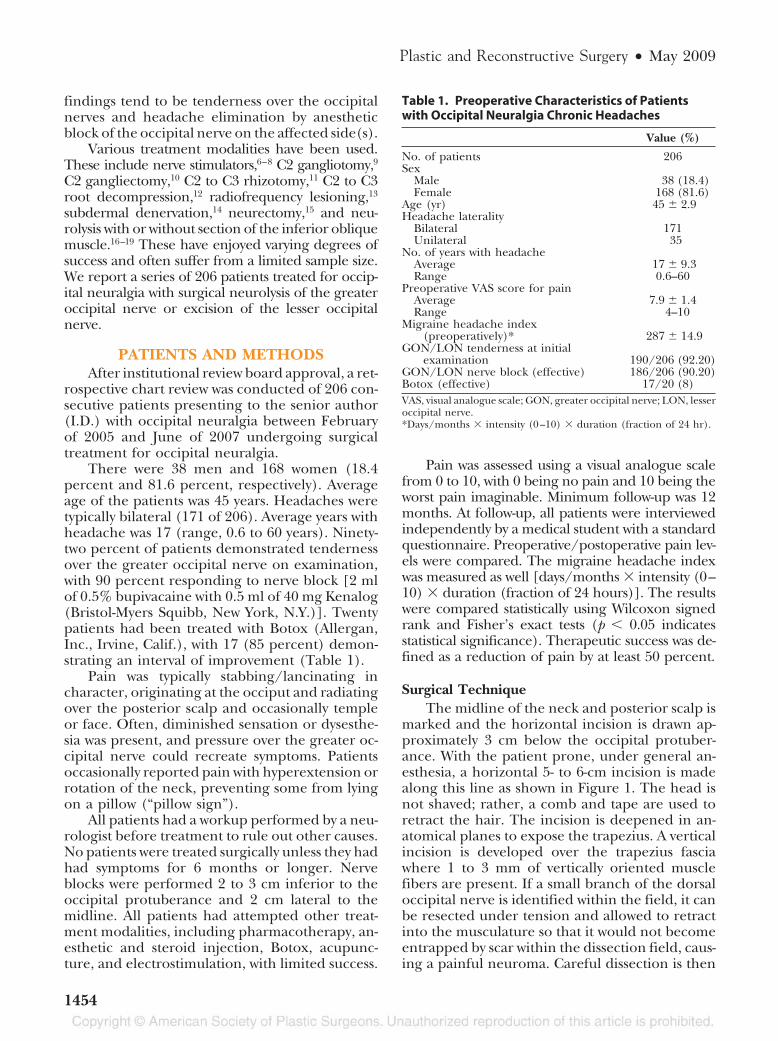
marked and the horizontal incision is drawn ap-proximately 3 cm below the occipital protuber-ance. With the patient prone, under general an-esthesia, a horizontal 5- to 6-cm incision is madealong this line as shown in Figure 1. The head isnot shaved; rather, a comb and tape are used toretract the hair. The incision is deepened in an-atomical planes to expose the trapezius. A verticalincision is developed over the trapezius fasciawhere 1 to 3 mm of vertically oriented musclefibers are present. If a small branch of the dorsaloccipital nerve is identified within the field, it canbe resected under tension and allowed to retractinto the musculature so that it would not becomeentrapped by scar within the dissection field, caus-ing a painful neuroma. Careful dissection is then
Table 1. Preoperative Characteristics of Patientswith Occipital Neuralgia Chronic Headaches
Value (%)
No. of patients 206Sex
Male 38 (18.4)Female 168 (81.6)
Age (yr) 45 � 2.9Headache laterality
Bilateral 171Unilateral 35
No. of years with headacheAverage 17 � 9.3Range 0.6–60
Preoperative VAS score for painAverage 7.9 � 1.4Range 4–10
Migraine headache index(preoperatively)* 287 � 14.9
GON/LON tenderness at initialexamination 190/206 (92.20)
GON/LON nerve block (effective) 186/206 (90.20)Botox (effective) 17/20 (8)VAS, visual analogue scale; GON, greater occipital nerve; LON, lesseroccipital nerve.*Days/months � intensity (0–10) � duration (fraction of 24 hr).
Plastic and Reconstructive Surgery • May 2009
1454
used within the vertical incision to identify thegreater occipital nerve, typically as it emerges fromthe semispinalis capitis muscle. Similar to the pub-lished report, a dissection is first carried proximally,removing the small medial piece of the semispinaliscapitis muscle abutting the greater occipital nerve,and then inferiorly to release the obliquus capitismuscle fibers overlying the greater occipital nerve.19
In approximately 6 percent of patients, the nerve isfound to be split within the substance of the semi-spinalis capitis muscle. In this case, the muscle fiberssplitting the nerve are also released. Dissection iscarried distally, releasing the nerve within the tra-pezial tunnel (the site where the greater occipitalnerve penetrates through the trapezial fascial attach-ments to the occiput). This 1- to 2-cm tunnel has anoblique superolateral direction and often containsangiolymphatics, another possible compression vari-able to be acknowledged.4 Usually, at its superolat-eral distal end, the occipital artery and vein cross thegreater occipital nerve. If the vessel is found to im-pinge the nerve, it is dissected free and ligated. En-larged and abutting lymph nodes are also removedfrom the tunnel, further decompressing the nerve.The same incision is used to treat the opposite sidewhen needed. The wound is closed in anatomicallayers without drains.
If unilateral lesser occipital nerve excision isperformed concurrently, a 3-cm incision is madeat a separate site lateral to the first incision, overthe path of the lesser occipital nerve, which is iden-tified along the posterior sternocleidomastoid at itsmiddle third. The lesser occipital nerve can be de-
compressed or excised and its proximal stumpimplanted into muscle.
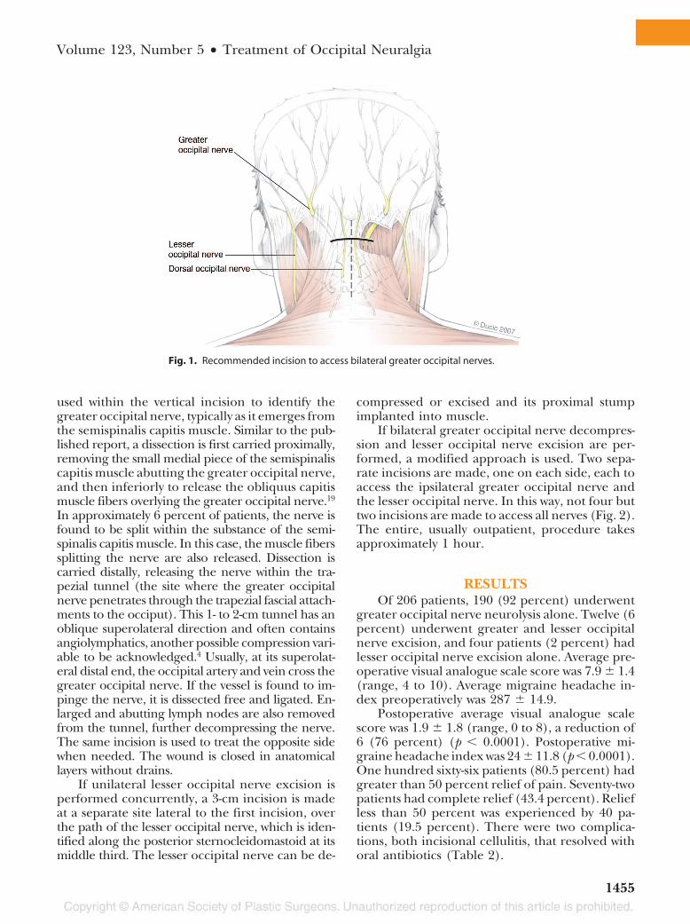
If bilateral greater occipital nerve decompres-sion and lesser occipital nerve excision are per-formed, a modified approach is used. Two sepa-rate incisions are made, one on each side, each toaccess the ipsilateral greater occipital nerve andthe lesser occipital nerve. In this way, not four buttwo incisions are made to access all nerves (Fig. 2).The entire, usually outpatient, procedure takesapproximately 1 hour.
RESULTSOf 206 patients, 190 (92 percent) underwent
greater occipital nerve neurolysis alone. Twelve (6percent) underwent greater and lesser occipitalnerve excision, and four patients (2 percent) hadlesser occipital nerve excision alone. Average pre-operative visual analogue scale score was 7.9 � 1.4(range, 4 to 10). Average migraine headache in-dex preoperatively was 287 � 14.9.
Postoperative average visual analogue scalescore was 1.9 � 1.8 (range, 0 to 8), a reduction of6 (76 percent) (p � 0.0001). Postoperative mi-graine headache index was 24 � 11.8 (p � 0.0001).One hundred sixty-six patients (80.5 percent) hadgreater than 50 percent relief of pain. Seventy-twopatients had complete relief (43.4 percent). Reliefless than 50 percent was experienced by 40 pa-tients (19.5 percent). There were two complica-tions, both incisional cellulitis, that resolved withoral antibiotics (Table 2).
Fig. 1. Recommended incision to access bilateral greater occipital nerves.
Volume 123, Number 5 • Treatment of Occipital Neuralgia
1455
Factors correlated with a positive outcomeincluded tenderness over the greater occipitalnerve, positive response to greater occipital nerveblock or Botox, a history of direct occipital trauma,and being under the care of a neurologist or painspecialist preoperatively. Negative outcomes wereassociated with a lack of greater occipital nervetenderness, poor response to nerve block, mentalillness, presence of other headache syndromes inaddition to occipital neuralgia, or absence of con-comitant treatment by a headache specialist.
DISCUSSIONThe greater occipital nerve has been described
as the largest purely sensory nerve in the body.20
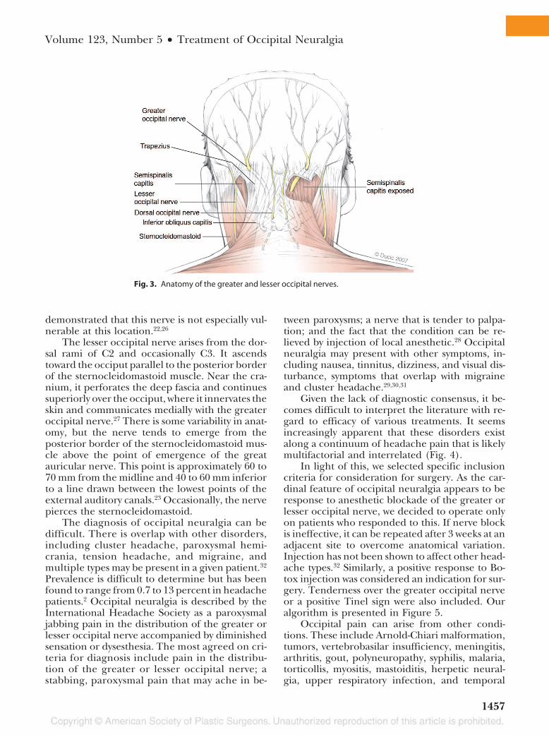
It arises from the dorsal ramus of C2 deep to theinferior oblique muscle where it branches. Themedial branch is the greater occipital nerve, whichruns transversely along the inferior oblique and iscovered by the splenius capitis, the longissimus,and the semispinalis muscles. Occasionally, thenerve travels within the substance of the inferioroblique muscle.21 The nerve then turns upward topierce the semispinalis capitis. Here, the nerveruns rostrolaterally before emerging into the scalpby piercing the aponeurotic fibrous attachment ofthe trapezius and sternocleidomastoid to the su-perior nuchal line. In this aperture, the occipitalartery and greater occipital nerve are in intimateassociation. Immediately below the superior nu-chal line, the nerve divides into several terminalbranches; medial branches innervate occipitalskin and the lateral branches pass into the regionbehind the pinna22 (Fig. 3).
It has been demonstrated that the greateroccipital nerve most commonly emerges fromthe semispinalis muscle at a point 3 cm below theoccipital protuberance and 1.5 cm lateral to themidline.21,23 There are occasionally variations inthis anatomy, particularly in the vertical axis.24,25 Ithas been postulated that the C2 ramus could becompressed between the posterior arch of the at-las and the lamina of the axis; however, it has been
Fig. 2. Recommended incisions to access the greater and lesser occipital nervesbilaterally.
Table 2. Postoperative Characteristics andData for Patients with Occipital NeuralgiaChronic Headache
Value (%)
GON neurolysis 190/206 (92)GON plus LON excision 12/206 (6)LON excision 4/206 (2)Postoperative VAS headache pain
Average 1.9 � 1.8Range 0–8
Migraine headache index(postoperatively)* 24 � 11.8
Outcome (same numbers standfor QOL improvement)
Positive (�50% relief) 166/206 (80.5)Complete ON headache relief 72/166 (43.4)Failure (no relief, or �50%) 40/206 (19.5)
Complications2/206 (incision
cellulitis) (0.009)Outpatient surgery 201/206 (97)GON, greater occipital nerve; LON, lesser occipital nerve; VAS, visualanalogue scale; QOL, quality of life; ON, occipital neuralgia.*Days/mo � intensity (0–10) � duration (fraction of 24 hr).
Plastic and Reconstructive Surgery • May 2009
1456
demonstrated that this nerve is not especially vul-nerable at this location.22,26
The lesser occipital nerve arises from the dor-sal rami of C2 and occasionally C3. It ascendstoward the occiput parallel to the posterior borderof the sternocleidomastoid muscle. Near the cra-nium, it perforates the deep fascia and continuessuperiorly over the occiput, where it innervates theskin and communicates medially with the greateroccipital nerve.27 There is some variability in anat-omy, but the nerve tends to emerge from theposterior border of the sternocleidomastoid mus-cle above the point of emergence of the greatauricular nerve. This point is approximately 60 to70 mm from the midline and 40 to 60 mm inferiorto a line drawn between the lowest points of theexternal auditory canals.23 Occasionally, the nervepierces the sternocleidomastoid.
The diagnosis of occipital neuralgia can bedifficult. There is overlap with other disorders,including cluster headache, paroxysmal hemi-crania, tension headache, and migraine, andmultiple types may be present in a given patient.32
Prevalence is difficult to determine but has beenfound to range from 0.7 to 13 percent in headachepatients.2 Occipital neuralgia is described by theInternational Headache Society as a paroxysmaljabbing pain in the distribution of the greater orlesser occipital nerve accompanied by diminishedsensation or dysesthesia. The most agreed on cri-teria for diagnosis include pain in the distribu-tion of the greater or lesser occipital nerve; astabbing, paroxysmal pain that may ache in be-
tween paroxysms; a nerve that is tender to palpa-tion; and the fact that the condition can be re-lieved by injection of local anesthetic.28 Occipitalneuralgia may present with other symptoms, in-cluding nausea, tinnitus, dizziness, and visual dis-turbance, symptoms that overlap with migraineand cluster headache.29,30,31
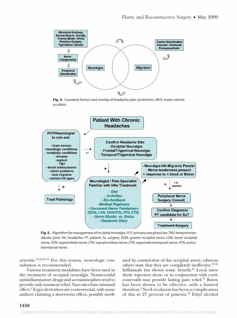
Given the lack of diagnostic consensus, it be-comes difficult to interpret the literature with re-gard to efficacy of various treatments. It seemsincreasingly apparent that these disorders existalong a continuum of headache pain that is likelymultifactorial and interrelated (Fig. 4).
In light of this, we selected specific inclusioncriteria for consideration for surgery. As the car-dinal feature of occipital neuralgia appears to beresponse to anesthetic blockade of the greater orlesser occipital nerve, we decided to operate onlyon patients who responded to this. If nerve blockis ineffective, it can be repeated after 3 weeks at anadjacent site to overcome anatomical variation.Injection has not been shown to affect other head-ache types.32 Similarly, a positive response to Bo-tox injection was considered an indication for sur-gery. Tenderness over the greater occipital nerveor a positive Tinel sign were also included. Ouralgorithm is presented in Figure 5.
Occipital pain can arise from other condi-tions. These include Arnold-Chiari malformation,tumors, vertebrobasilar insufficiency, meningitis,arthritis, gout, polyneuropathy, syphilis, malaria,torticollis, myositis, mastoiditis, herpetic neural-gia, upper respiratory infection, and temporal
Fig. 3. Anatomy of the greater and lesser occipital nerves.
Volume 123, Number 5 • Treatment of Occipital Neuralgia
1457
arteritis.12,26,33,34 For this reason, neurologic con-sultation is recommended.
Various treatment modalities have been used inthe treatment of occipital neuralgia. Nonsteroidalantiinflammatory drugs and acetaminophen tend toprovide only transient relief. Narcotics have minimaleffect.2 Ergot derivatives are controversial, with someauthors claiming a short-term effect, possibly medi-
ated by constriction of the occipital artery, whereasothers state that they are completely ineffective.29,35
Infliximab has shown some benefit.36 Local anes-thetic injection alone or in conjunction with corti-costeroids may provide lasting pain relief.37 Botoxhas been shown to be effective, with a limitedduration.2 Neck weakness has been a complicationof this in 27 percent of patients.19 Ethyl alcohol
Fig. 4. Causative factors and overlap of headache pain syndromes. MVA, motor vehicleaccident.
Fig. 5. Algorithm for management of occipital neuralgia. PCP, primary care physician; TMJ, temporoman-dibular joint; HA, headache; PT, patient; Sx, surgery; GON, greater occipital nerve; LON, lesser occipitalnerve; SON, supraorbital nerve; STN, supratrochlear nerve; ZTN, zygomaticotemporal nerve; ATN, auricu-lotemporal nerve.
Plastic and Reconstructive Surgery • May 2009
1458
injection may improve symptoms but suffers fromhigh recurrence rates.38 Epidural corticosteroidshave not been shown to be effective.39
More invasive modalities include radiofrequencylesioning of the greater occipital nerve. One casereport followed a patient who demonstrated painrelief of 60 to 70 percent over 5 months. The treat-ment had to be repeated for continued efficacy, andlong-term results are not known.13 Another study of15 patients demonstrated good relief of pain at 8.8months after radiofrequency neurotomy of C3 to C6dorsal rami; however, there was a tendency for thepain to recur during follow-up.40 It is our observationthat a number of patients who previously had radio-frequency ablation elsewhere failed decompressionand subsequently required nerve excision to dimin-ish their pain. It appears that patients who continueto have headache following the radiofrequency ab-lation often have increased pain, presenting similarto neuroma in continuity. We find it important todiscuss this with the patient, who might then opt fornerve excision instead of decompression.
Implanted nerve stimulators have been eval-uated. One study found that 16 of 30 patients (53percent) experienced greater than 50 percentpain improvement at a mean follow-up of 35months.6 There were several complications re-lated to the implanted device. Another study of sixpatients found pain relief to be greater than 50percent in all patients at a follow-up of 3 months.7In a series of 14 patients, seven experiencedgreater than 50 percent pain relief at a mean fol-low-up of 22 months.8 The expense of these sys-tems and the hazards of implanting a foreign bodyshould be considered against other options. Inaddition, the nerve stimulator treatment focus ison symptoms, whereas the peripheral nerve sur-gery addresses the anatomical cause of the head-ache—compression.
Surgical approaches have been varied. C2 andC3 nerve root decompression was reported in asingle case in which the nerve was compressed bya C3 facet spur, leading to complete pain relief at11 months’ follow-up.12 In this case, a proximalbony abnormality was identified by polytomogra-phy. Microsurgical C2 gangliotomy has been advo-cated following a series of four patients with com-plete pain relief after 24 months, with one patientsuffering pain recurrence thereafter.19 These pa-tients experienced transient nausea and dizzinesspostoperatively, and one patient had a cerebrospinalfluid leak. C1 to C4 rhizotomy has been reported in17 patients with occipital neuralgia, with 68.8 per-cent of patients reporting the procedure as worth-
while.11 Numbness in the affected dermatomes waspresent after the procedure.
Subdermal denervation of the affected skin seg-ments has been reported.14 In these patients, twolarge scalp flaps were elevated, containing the oc-cipitofrontalis muscle, to expose the greater andlesser occipital nerves. These were then excised andthe flaps replaced. Three patients were reportedto have received “satisfactory results” from thisprocedure. Avulsion neurectomy of the greateroccipital nerve has been performed in 22 patients,with 70 percent demonstrating pain relief at 18months.15 Thirty percent of these patients expe-rienced scalp hypersensitivity, dysesthesia, neu-roma, or recurrence.
Surgical neurolysis with sectioning of the infe-rior oblique muscle was performed on 10 patients,with a 70 percent rate of patient satisfaction at amean follow-up of 37 months.17 In another series of50 patients, neurolysis of the greater occipital nerveat the deep neck fascia and trapezial tunnel wasfound to provide short-term pain relief in 66 percentof patients. At 18-month follow-up, however, painwas found to have recurred in 46 of 50 patients.16
Interestingly, 80 percent of patients did not regretthe surgery and 40 percent wanted to undergo theidentical operation again. These authors postulatedthat they may not have released the nerve deepenough, as the semispinalis muscle was not ad-dressed. The semispinalis muscle was sectioned byGuyuron et al. in 34 patients, with 100 percent dem-onstrating improvement in headache pain.19 Simi-larly, in a series of 13 patients with whiplash traumaand occipital headache, release of the nerve at thetrapezial tunnel and the semispinalis resulted in 72.2percent reporting good or excellent pain relief.18 Ananecdotal review of 150 patients undergoing surgeryfor occipital neuralgia by means of release of thetrapezial tunnel found that approximately one-thirdof patients improved dramatically, one-third was def-initely improved but still had some pain, and one-third did not benefit.4
The humoral and cellular mechanisms ofheadache have been well-studied, yet no consen-sus exists as to the exact pathway of pain genera-tion. Interleukin-1� and tumor necrosis factor-�have been implicated as promoting hyperalgesia.2Nitric oxide may also play a role. Despite the com-plex molecular-biological mechanisms, it seemsclear when reviewing the body of literature on thistopic that there is often a peripheral trigger, suchas nerve irritation, that triggers the pain cascade.Five potential sources of potential entrapment ofthe greater occipital nerve are observed: C2 nerveroot (rarely), inferior oblique (rarely), within the
Volume 123, Number 5 • Treatment of Occipital Neuralgia
1459
semispinalis muscle, within the trapezial tunnel(trapezius muscle/aponeurosis), and angiolym-phatics (occipital artery/vein crosses the greateroccipital nerve; lymph node presence, within ordistal to trapezial tunnel, respectively). Nerve ir-ritation and hyperexcitability of peripheral noci-ceptors may lead to central sensitization and painevoked by nonnoxious stimuli.41 The response ofthese types of headaches to peripheral nerve blockstrengthens this concept. Similarly, connectionsbetween the trigeminal nucleus and the upperfour cervical roots may form the anatomical sub-strate for the spread of cervical pain from the neckto the head.42 The proximity of the occipital arteryto the greater occipital nerve has also been pos-tulated to cause nerve compression and paroxys-mal, throbbing pain.43
In our series of 206 patients, 80 percent ex-perienced meaningful pain relief at a minimum of12 months’ follow-up. In comparison with otherprocedures, the greater occipital nerve is not dam-aged and complications are rare. When the lesseroccipital nerve is excised (2 percent of cases), theresultant sensory defect is minor. These resultssuggest that occipital neuralgia is stimulated byperipheral nerve entrapment.
Approximately 20 percent of patients experi-enced less than 50 percent relief after 1 year. Wesuspect several reasons for treatment failure. Di-agnostic error may have played a role. Many of thesetreatment failures had other, coexistent headachesyndromes that may have come to predominate andovershadow any effect of the procedure. In somefailures, abnormal branching of the greater occipitalnerve was found, arousing suspicion that aberrantbranches might still have been active. Atypical anat-omy has been reported.44 The third occipital nervewas not addressed routinely in these patients at thebeginning of the study although, when identifiedin the operative field, it was cut and allowed toretract into muscle. Headache symptoms havebeen attributed to entrapment of this nerve.23 Wenow address this intraoperatively with either de-compression or excision. Although initially moreattention was given to the semispinalis capitis mus-cle and trapezius aponeurosis, it became apparentthat these two sites are among five possible sites ofcompression. More thorough release is now per-formed, including the inferior oblique, occipitalartery, and lymph nodes, if present, in the trape-zial tunnel. Ultimately, it is probably a combinationof the above factors, in addition to a lack of regen-erative potential in the long-compressed nerve, thatis responsible for the treatment failures.
Interestingly, many of these patients whom weclassified as treatment failures experienced somepostoperative pain relief. Often, they request asecond operation. In these cases, if nerve block isstill effective, we offer neurectomy as a secondalternative. The outcomes of this will be presentedin a future work.
This procedure has limitations. Although 80.5percent of patients benefited from surgery, only43 percent of patients experienced complete re-lief. Still, these odds may be attractive to manypatients, with partial abatement of symptoms al-lowing a more productive and enjoyable lifestyle.A detailed preoperative discussion must be un-dertaken to maintain reasonable expectations aspart of a multidisciplinary team. We look forwardto reporting these data and the results of a largercohort in the future. The importance of multidis-ciplinary coordination in patient care cannot beoveremphasized, including a headache-focusedneurologist or anesthesia pain specialist. The ad-ministration of nerve blocks or Botox requiresexperience for valid, reproducible results, oftenused as important inclusion criteria for surgery.Lastly, it is important for the surgeon to haveappropriate peripheral nerve surgery training andexperience to minimize complications and opti-mize outcomes.
CONCLUSIONSThe diagnosis and management of headache
is a controversial topic. Of 206 patients undergo-ing surgical neurolysis, 80 percent had meaningfulpain relief after at least 1 year of follow-up. Al-though many might argue that surgery is an in-vasive treatment for occipital neuralgia, the failureof medical management in many patients man-dates exploration of other options. Surgical neu-rolysis provides safe, durable pain relief to a sub-set of patients with occipital neuralgia–relatedchronic migraine headaches by addressing thecausative compression sites. Clearly, more workneeds to be performed before a consensus isreached. However, we hope that this study, bystandardizing indications and technique, con-firms the importance of surgical treatment forfuture work and discussion on this topic.
Ivica Ducic, M.D., Ph.D.Department of Plastic Surgery
Georgetown University HospitalWashington, D.C. 20007
REFERENCES1. Beruto LJ, Ramos MM. Decades de med y cirug pract. Madrid
1821;3:145–169.
Plastic and Reconstructive Surgery • May 2009
1460
2. Martelletti P, van Suijlekom H. Cervicogenic headache: Prac-tical approaches to therapy. CNS Drugs 2004;18:793–805.
3. Hecht JS. Occipital nerve blocks in postconcussive head-aches. J Head Trauma Rehabil. 2004;19:58–71.
4. Pantaloni M, Sullivan P. Relevance of the lesser occipitalnerve in facial rejuvenation surgery (Discussion). Plast Re-constr Surg. 2000;105:2600–2603.
5. Ballasteros-Del Rio B, Ares-Luque A, Tejada-Garcia J, Muela-Molinaro A. Occipital (Arnold) neuralgia secondary togreater occipital nerve schwannoma. Headache 2003;43:804–807.
6. Slavin KV, Colpan ME, Munawar N, Wess C, Nersesyan H.Trigeminal and occipital peripheral nerve stimulation forcraniofacial pain: A single institution experience and reviewof the literature. Neurosurg Focus 2006;21:1–5.
7. Kapural L, Mekhail N, Hayek SM, Stanton-Hicks M, Malak O.Occipital nerve electric stimulation via the midline approachand subcutaneous surgical leads for treatment of severe occip-ital neuralgia: A pilot study. Anesth Analg. 2005;101:171–174.
8. Slavin KV, Nersesyan H, Wess C. Peripheral neurostimulationfor treatment of intractable occipital neuralgia. Neurosurgery2006;58:112–119.
9. Stechison MT, Mullin BB. Surgical treatment of greater oc-cipital neuralgia: An appraisal of strategies. Acta Neurochir.1994;131:236–240.
10. Wand MY, Levi AD. Ganglionectomy of C-2 for the treatmentof medically refractory occipital neuralgia. Neurosurg Focus2002;12:1–3.
11. Kapoor V, Rothfus WE, Grahovac SC, Amin Kassam SZ,Horowitz MB. Refractory occipital neuralgia: Preoperativeassessment with CT-guided nerve block prior to dorsal cer-vical rhizotomy. AJNR Am J Neuroradiol. 2003;24:2105–2110.
12. Poletti CE. A proposed operation for occipital neuralgia: C-2and C-3 root decompression. Neurosurgery 1983;12:221–224.
13. Navani A, Mahajan G, Kreis P, Fishman SM. A case of pulsedradiofrequency lesioning for occipital neuralgia. Pain 2006;7:453–456.
14. Martin BC, Fagan PJ. The surgical therapy of certain occipitalheadaches. Plast Reconstr Surg. 1964;33:266–268.
15. Sharma RR, Devadas RV, Pawar SJ, Lad SD, Mahapatra AK.Current status of peripheral neurectomy for occipital neu-ralgia. Neurosurg Q. 2005;15:232–238.
16. Bovim G, Fredriksen TA, Stolt-Nielsen A, Sjaastad O. Neu-rolysis of the greater occipital nerve in cervicogenic head-ache: A follow up study. Headache 1992;32:175–179.
17. Gille O, Lavignolle B, Vital JM. Surgical treatment of greateroccipital neuralgia by neurolysis of the greater occipitalnerve and sectioning of the inferior oblique muscle. Spine2004;29:828–832.
18. Manusson T, Ragnarsson T, Bjornsson A. Occipital nerverelease in patients with whiplash trauma and occipital neu-ralgia. Headache 1996;36:32–36.
19. Guyuron B, Kriegler JS, Davis J, Amini SB. Comprehensivesurgical treatment of migraine headaches. Plast Reconstr Surg.2005;115:1–9.
20. Rifat SF, Lombardo JA. Occipital neuralgia in a footballplayer: A case report. Clin J Sports Med. 1995;5:251–253.
21. Mosser SW, Guyuron B, Janis JE, Rohrich RJ. The anatomy ofthe greater occipital nerve: Implications for the etiology ofmigraine headaches. Plast Reconstr Surg. 2004;113:693–697.
22. Bogduk N. The clinical anatomy of the cervical dorsal rami.Spine 1982;7:319–330.
23. Dash KS, Janis JE, Guyuron B. The lesser and third occipitalnerves and migraine headaches. Plast Reconstr Surg. 2005;115:1752–1758.
24. Natsis K, Baraliakos X, Appel HJ, Tsikaras P, Gigis I, Koebke J.The course of the greater occipital nerve in the suboccipitalregion: A proposal for setting landmarks for local anesthesia inpatients with occipital neuralgia. Clin Anat. 2006;19:332–336.
25. Becser N, Bovim G, Sjaastad O. Extracranial nerves in theposterior part of the head: Anatomic variations and theirpossible clinical significance. Spine 1998;23:1435–1441.
26. Weinberger LM. Cervico-occipital pain and its surgical treat-ment. Am J Surg. 1978;135:243–247.
27. Tubbs RS, Salter EG, Wellons JC, Blount JP, Oakes WJ. Land-marks for the identification of the cutaneous nerves of theocciput and nuchal regions. Clin Anat. 2007;20:235–238.
28. Ward JB. Greater occipital nerve block. Semin Neurol. 2003;23:59–61.
29. Trescot AM. Headache management in an interventionalpain practice. Pain Physician 2000;3:197–200.
30. Rozen TD. Non-hypothalamic cluster headache: The role ofthe greater occipital nerve in cluster headache pathogenesis.J Headache Pain 2005;6:149–151.
31. Scattoni L, Di Stani F, Villani V, et al. Great occipital nerveblockade for cluster headache in the emergency department:Case report. J Headache Pain 2006;7:98–100.
32. Yi X, Cook AJ, Hamill-Ruth RJ, Rowlingson JC. Cervicogenicheadache in patients with presumed migraine: Missed diag-nosis or misdiagnosis? J Pain 2005;6:700–703.
33. Tancredi A, Caputi F. Greater occipital neuralgia and ar-throsis of C1-2 lateral joint. Eur J Neurol. 2004;11:573–574.
34. Jundt JW, Mock D. Temporal arteritis with normal erythro-cyte sedimentation rates presenting as occipital neuralgia.Arthritis Rheum. 1991;34:217–219.
35. Martelletti P. Proinflammatory pathways in cervicogenicheadache. Clin Exp Rhematol. 2000;18:S33–S39.
36. Martelletti P. Inflammatory mechanisms in cervicogenicheadache: An integrative view. Curr Pain Headache Rep. 2002;6:315–319.
37. Naja ZM, El-Rajab M, Al-Tannir MA, Ziade FM, Tawfik OM.Occipital nerve blockade for cervicogenic headache: A dou-ble blind randomized clinical controlled trial. Pain Pract.2006;6:89–95.
38. Koch D, Wakhloo AK. CT-guided chemical rhizotomy of the C1root for occipital neuralgia. Neuroradiology 1992;34:451–452.
39. Marteletti P, Di Sabato F, Granata M, et al. Failure of long-term epidural steroid injection in cervicogenic headache.Eur Rev Med Pharmacol Sci. 1998;2:10–14.
40. Van Suijlekom J, Van Kleef M, Barendse G, Sluijter ME,Sjaastad O, Weber WE. Radiofrequency cervical zygapophy-seal joint neurotomy for cervicogenic headache: A prospec-tive study in 15 patients. Funct Neurol. 1998;13:297–303.
41. Ashkenazi A. Three common neuralgias. Postgrad Med. 2004;116:16–32.
42. Kerr FW. Central relationships of trigeminal and cervicalprimary afferents in the spinal cord and medulla. Brain Res.1972;43:561–572.
43. Shimizu S, Oka H, Osawa S, et al. Can proximity of theoccipital artery to the greater occipital nerve act as a causeof idiopathic greater occipital neuralgia? An anatomical andhistological evaluation of the artery-nerve relationship. PlastReconstr Surg. 2007;119:2029–2034.
44. Madhavi C, Holla SJ. Triplication of the lesser occipitalnerves. Clin Anat. 2004;17:667–671.
45. Bovim G, Bonamico L, Fredriksen TA, Lindboe CF, Stolt-Nielsen A, Sjaastad O. Topographic variations in the periph-eral course of the greater occipital nerve: Autopsy study withclinical correlations. Spine 1991;16:475–478.
Volume 123, Number 5 • Treatment of Occipital Neuralgia
1461