recovery simulator - sir oftalmica wetlab espianto cornea... · stabilize the scleral rim using...
TRANSCRIPT

www.bioniko.com | [email protected]
The CORDELIA recovery model was made possible by the valuable guidance and support of LIONS Vision Gift.
Recovery Simulator
The CORDELIA Recovery simulator is based on an in-situ excision scenario. Eye bank technicians must master this technique to successfully recover delicate corneal tissue from
donors in the field. CORDELIA is a task trainer that allows students to learn and practice the
tissue recovery technique without using valuable donor tissue, in a realistic
yet simple manner. Its repeatability and availability makes it ideal for developing standardized methods of instruction and assessment.
CORDELIA models are used with the Bioniko ORBIT Model, which
provides support, frame of reference and provides the challenges posed by
facial features surrounding the eye. ORBIT is available in Left and Right models
(sold separately).See at AAO Booth 2577
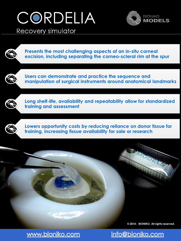
Presents the most challenging aspects of an in-situ corneal excision, including separating the corneo-scleral rim at the spur
Users can demonstrate and practice the sequence and manipulation of surgical instruments around anatomical landmarks
Long shelf-life, availability and repeatability allow for standardized training and assessment
Lowers opportunity costs by reducing reliance on donor tissue for training, increasing tissue availability for sale or research
© 2014- BIONIKO. All rights reserved.
Skills for eye-banking

www.bioniko.com
Watch an instructional video: www.youtube.com/user/BionikoDesign
The CORDELIA recovery simulator focuses on the most challenging aspects of an in-situ corneal excision.
Learning to dissect and separate the cornea from the donor eye, without stress to the endothelium, is a
fundamental skill for eye banking.
The model anatomy includes a scleral layer (1) and choroid layer (2). These layers are separated posterior to
the limbus (3), simulating the supra-choroidal space (4), and are attached at the spur (5), allowing a realistic
dissection to be performed. After trephination and dissection of the scleral layer, the user can practice
stabilizing the scleral rim while detaching the tissue at the spur with minimal stress to the endothelium. Cornea
(6) and iris (7) features provide additional landmarks that add to the model's realism. The structural ring (8) at
the base of the model maintains the model's shape and the notches (9) mate with tabs in the ORBIT(or EXOS)
model to prevent unwanted rotation of the model during trephination.
The CORDELIA recovery simulator can be used with two different platforms:
1. Standard ORBIT platform (Anterior segment only setup)
The ORBIT model (sold separately) serves as a base for the CORDELIA model
and provides reference and realism by challenging the user to manipulate
instruments according to the facial structures around the eye. There are
both right (R) and left (L) ORBIT models to practice both approaches.
2. FLEX-ORBIT platform (Whole Globe setup – Requires EXOS model)
The FLEX-ORBIT (sold separately)allows use of whole globe models for
greater fidelity. The EXOS Enucleation Simulator features a Posterior segment
model and a detachable and replaceable CORDELIA. CORDELIA models
can be replaced and used with the EXOS model on the FLEX-ORBIT.
1- SCLERAL LAYER 2- CHOROID LAYER 3- LIMBUS 4- SUPRA-CHOROIDAL
SPACE 5- SPUR 6- CORNEA 7- IRIS 8- STRUCTURAL RING 9- NOTCHES
INSTRUCTIONS FOR USE
Do not use dry. Lubricate with water. Do not use BSS. Soak CORDELIA for aprox 5 minutes in luke warm water (aprox 95F) immediately before use.

www.bioniko.com
1. Soak CORDELIA for aprox 5 minutes in luke warm water (aprox 95F) immediately before use.
(Note: cornea looks "milky" when hydrated)
2. Load CORDELIA model in the ORBIT. The ORBIT has a flexible “eyelid” that receives and secures the
CORDELIA model. Use a couple of water drops to moisten the eyelid and socket area. Insert the edge of
the model under the superior eyelid (1) and push the other side down into the socket (2) to load the
model. The small tabs on the ORBIT (3) will mate with the notches to limit model rotation.
3. Fix the ORBIT in place by pressing downward on a smooth surface to engage the suction-cup. Orient the
ORBIT according to the desired approach: Superior (S), Nasal (N), Inferior (I) or Temporal (T).
4. Trephinate the sclera to gain access to the supra-choroidal space (Ø18 mm max).(Tip: Stain trephine
edge for better visualization of score-line)
5. Complete the scleral incision parallel to the limbus with a scalpel or scissors. Take care to cut the scleral
layer without perforating the choroid layer underneath.(Note: the choroid layer on the model is not black.
Careful inspection is necessary to ensure there is no damage to the choroid layer during dissection
6. Check 360 degree separation of the scleral layer.
7. Detach cornea from spur. Stabilize the scleral rim using dressing forceps and gently grasp and push the
choroid/iris layer away with tissue forceps until separation is complete. Minimize stress and manipulation of
the cornea during separation.(Note: Spur attachment is firm as with young or dark eyed donors)
8. Remove used model from the ORBIT by inserting a closed instrument in either corner of the eyelid and
leveraging the model out.
9. Lift the suction release tab to remove FLEX-ORBIT from surface. DO NOT PULL ON THE ORBIT!
Refer to the EXOS model instructions for use.
In-situ excision simulation – Standard ORBIT Platform Setup
In-situ excision simulation – FLEX-ORBIT Platform Setup

www.bioniko.com
Follow these recommendations to maximize the shelf life of the models:
Store in a cool, dry and dark place (a drawer will be fine).
Extended exposure to some indoor lights or sunlight (UV) may
affect material properties. Prolonged exposure to humidity or
high temperatures may adversely affect material properties.
Do not place heavy objects on top of the model’s box.
Prolonged compression may deform the models.
FAQ
x Q: Why do the models smell sweet?
A: The gummy bear smell is not added. It is a property of the material ... to extend the life of your product
refrain from biting!
x Q:Is there a conjunctival layer?
A:The model currently does not have conjunctiva. These are on our list of future developments.
x Q:How can I tell if the CORDELIA has been soaked enough time?
A:The cornea looks "milky" when hydrated.
Instructions for care
FAQ