red brain, blue brain: evaluative processes differ in ...bluebrains.pdfred brain, blue brain:...
TRANSCRIPT

Red Brain, Blue Brain: Evaluative Processes Differ in Democrats and Republicans
Darren Schreiber1,2*, Alan N. Simmons3,4, Christopher T. Dawes1, Taru Flagan3,5, James H. Fowler1, Martin P. Paulus3,4,5 1 Department of Political Science, University of California San Diego, La Jolla, CA 92103 USA 2 School of Law, University of San Diego, San Diego, CA 92110 USA 3 Department of Psychiatry, University of California San Diego, La Jolla, CA 92093, USA 4 Veterans Affairs San Diego Healthcare System, San Diego, CA 92161, USA 5 Laboratory of Biological Dynamics and Theoretical Medicine, University of California San Diego, La Jolla, CA 92093, USA *To whom correspondence should be addressed. Email: [email protected] We matched public voter records to 54 subjects who performed a risk-taking task during functional imaging. We find that Democrats and Republicans had significantly different patterns of brain activation during processing of risky decisions. Amygdala activations, associated with externally directed reactions to risk, are stronger in Republicans, while insula activations, associated with internally directed reactions to affective perceptions, are stronger in Democrats. These results suggest an internal vs. external difference in evaluative process that illuminates and resolves a discrepancy in the existing literature. This process-based approach to political partisanship is distinct from the policy-based approach that has dominated research for at least the past half century. In fact, a two parameter model of partisanship based on amygdala and insula activations achieves better accuracy in predicting whether someone is a Democrat or a Republican than a well-established model in political science based on parental socialization of party identification.
Ideological differences between political partisans have been attributed to logical,
psychological, or social constraints 1. These differences are thought to be driven by institutional
political processes or individual policy preferences, rather than biological differences in
evaluative processes. To test a conjecture that ideological differences between partisans reflect
distinctive neural processes, we matched publicly available party registration records with the
names of participants (28 males, 26 females) who had previously taken part in an experiment
designed to examine risk-taking behavior during functional brain imaging.
Individuals completed a simple risk-taking decision-making task 2 during which
participants were presented with three numbers in ascending order (20, 40, and 80) for one

second each. While pressing a button during the presentation of the number 20 on the screen
always resulted in a gain of 20 cents, waiting to select 40 or 80 was associated with a pre-
determined possibility of losing 40 or 80 cents. Therefore, participants chose between a lower
“safe” payoff and a higher risky payoff. The probabilities of receiving a negative 40 or 80 were
selected such that there was no advantage of choosing 20, 40 or 80 during the task, i.e. the
overall pay-off would have been the same for each pure strategy.
Participant groups were composed of 31 Democrats and 23 Republicans who did not
differ with regard to age (F(52,1)=1.73, p=0.19) or gender (χ2=1.31, p=0.253) and gave informed
written consent approved by the University of California San Diego Human Research Protection
Program. Imaging data was processed using AFNI 3 and extracted for a priori regions of interest
within the amygdala, insula, and anterior cingulate. Significantly greater activation was
observed in the bilateral amygdala and ventral anterior cingulate for Republicans and in the right
anterior insula in Democrats when making winning risky versus winning safe decisions (see
SOM).
The amygdala plays a critical role in fear conditioning, however, this structure is also
important for other emotional information processing and behavior 4. Functional neuroimaging
studies have shown amygdala activation in fear conditioning 5, reward related processing 6,
encoding of emotionally salient information 7, risk-taking 8, processing positively valenced
stimuli9, and appetitive or aversive olfactory learning10. In comparison, neuroimaging studies of
insular cortex have found critical involvement of this neural structure in pain 11, interoceptive 12,
emotion-related 13, cognitive 14, and social processes 15. In particular, the insular cortex is
important for subjective feeling states and interoceptive awareness 12, 16. Thus, it appears in our
experiment that Republican participants, when making a risky choice, are predominantly

externally oriented, reacting to the fear-related processes with a tangible potential external
consequence. In comparison, risky decisions made by Democratic participants appear to be
associated with monitoring how the selection of a risky response might feel internally.
The association between party identification and ideology is well known, with
Republicans typically exhibiting more political conservatism than Democrats 17. Prior studies
have shown that self-reported liberals demonstrate stronger levels of physiological sensitivity to
cognitive conflict 17 and that supporters of socially conservative policies have higher skin
conductance response levels when exposed to startle stimuli 18. The present study resolves the
apparent conflict in these results. If Republicans are utilizing externally oriented processes in
reacting to risks while Democrats are internally directed, then we would expect the one group to
be more supportive of socially conservative policies and the other to be more sensitive to internal
conflict.
A critical unresolved problem common to studies of the formation of ideology on both
individual and institutional levels is the process through which a high dimensional space of
distinct values, preferences, or issues is reduced to a low dimensional ideological space 19. It is
even less clear why voters and their representatives in government should organize political
attitudes into apparently constrained bundles that are relatively consistent over time 20. While it
has been suggested that biological factors may lead liberals and conservatives to have different
sets of politically relevant values 21, the evidence presented here suggests that the processes of
evaluation themselves are distinct, perhaps leading to differentiable values, as well as differing
preferences for issues, candidates, and parties.
Perhaps the strongest finding to come out of the “Michigan school” when the behavioral
revolution spread to political science in the 1950s is that parents socialize their children to

identify with the same political parties that they do. In fact, the correlation between parent and
child is “so familiar and well established” that it is often taken as one of the few “axioms” of
political science 22. Indeed, a simple model of partisanship that includes mother’s and father’s
party accurately predicts about 69% of self-reported choices between the Democratic and
Republican party (see SOM). Yet, a simple two-parameter model of partisanship using
activations in the amygdala and the insular cortex during the risk task significantly out-performs
this longstanding model, correctly predicting about 79% of the observed choices (see SOM).
A central question unanswerable by this present study is the direction of causality.
Although political ideology 21 and strength of partisanship appear to be genetically heritable 23,
identification with a particular party does not. Thus, it is possible that neurobiological
differences between Republicans and Democrats drive their identification with different parties.
And, it is also possible that the mental processes that distinguish the world views of Republicans
and Democrats are reflected in the different neural mechanisms they utilize. Further untangling
the roles of party, ideology, genes, and neurocognition will be essential for advancing our
understanding of political attitudes and behavior. Being able to accurately predict party
identification using only neural activity during a risk-taking task suggests that investigating basic
neuropsychological differences between partisans may provide us with more powerful insights
than have been available using the traditional tools of political science.

Figure 1. Republicans and Democrats differ in the neural mechanisms activated while
performing a risk-taking task. Republicans more strongly activate their ventral anterior cingulate
(Region of Interest 1 in the brain image and bar graphs above) and bilateral amgydala (3 above),
associated with a more externally oriented reaction to risk. Democrats have higher activity in
their right insula (2 above), associated with internally directed reactions to affective perceptions.

Reference List
1. Converse, P. The Nature of Belief Systems in Mass Publics. in Ideology and Discontent (ed. D. Apter) 206-261 (Free Press, New York, 1964). 2. Paulus, M.P., Rogalsky, C., Simmons, A., Feinstein, J.S. & Stein, M.B. Increased activation in the right insula during risk-taking decision making is related to harm avoidance and neuroticism. Neuroimage 19, 1439-1448 (2003). 3. Cox, R.W. AFNI: software for analysis and visualization of functional magnetic resonance neuroimages. Comput Biomed Res 29, 162-173 (1996). 4. LeDoux, J.E. Brain mechanisms of emotion and emotional learning. Curr Opin Neurobiol 2, 191-197 (1992). 5. Buchel, C., Morris, J., Dolan, R.J. & Friston, K.J. Brain systems mediating aversive conditioning: an event-related fMRI study. Neuron 20, 947-957 (1998). 6. Breiter, H.C. & Rosen, B.R. Functional magnetic resonance imaging of brain reward circuitry in the human. Ann N Y Acad Sci 877, 523-547 (1999). 7. Canli, T., Zhao, Z., Brewer, J., Gabrieli, J.D. & Cahill, L. Event-related activation in the human amygdala associates with later memory for individual emotional experience. J Neurosci 20, RC99 (2000). 8. Ernst, M., et al. Decision-making in a risk-taking task: a PET study. Neuropsychopharmacology 26, 682-691 (2002). 9. Garavan, H., Pendergrass, J.C., Ross, T.J., Stein, E.A. & Risinger, R.C. Amygdala response to both positively and negatively valenced stimuli. Neuroreport 12, 2779-2783 (2001). 10. Gottfried, J.A., O'Doherty, J. & Dolan, R.J. Appetitive and aversive olfactory learning in humans studied using event-related functional magnetic resonance imaging. J Neurosci 22, 10829-10837 (2002). 11. Tracey, I., et al. Noxious hot and cold stimulation produce common patterns of brain activation in humans: a functional magnetic resonance imaging study. Neurosci Lett 288, 159-162 (2000). 12. Critchley, H.D., Wiens, S., Rotshtein, P., Ohman, A. & Dolan, R.J. Neural systems supporting interoceptive awareness. Nat Neurosci 7, 189-195 (2004). 13. Phan, K.L., Wager, T., Taylor, S.F. & Liberzon, I. Functional neuroanatomy of emotion: a meta-analysis of emotion activation studies in PET and fMRI. Neuroimage 16, 331-348 (2002). 14. Huettel, S.A., Misiurek, J., Jurkowski, A.J. & McCarthy, G. Dynamic and strategic aspects of executive processing. Brain Res 1000, 78-84 (2004). 15. Eisenberger, N.I., Lieberman, M.D. & Williams, K.D. Does rejection hurt? An FMRI study of social exclusion. Science 302, 290-292 (2003). 16. Craig, A.D. How do you feel? Interoception: the sense of the physiological condition of the body. Nat Rev Neurosci 3, 655-666 (2002). 17. Amodio, D.M., Jost, J.T., Master, S.L. & Yee, C.M. Neurocognitive correlates of liberalism and conservatism. Nat Neurosci 10, 1246-1247 (2007). 18. Oxley, D.R., et al. Political attitudes vary with physiological traits. Science 321, 1667-1670 (2008).

19. Jost, J.T. The end of the end of ideology. Am Psychol 61, 651-670 (2006). 20. Poole, K.T. Spatial models of parliamentary voting (Cambridge University Press, Cambridge ; New York, 2005). 21. Alford, J.R., Funk, C., L. & Hibbing, J.R. Are Political Orientations Genetically Transmitted? American Political Science Review 99, 153-167 (2005). 22. Achen, C. Parental socialization and rational party identification. Political Behavior 24, 151-170 (2002). 23. Settle, J.E., Dawes, C.T. & Fowler, J.H. The Heritability of Partisan Attachment. Political Research Quarterly (2009 (in press)).
1
Supporting Online Material Red Brain, Blue Brain: Evaluative Processes Differ in Democrats and Republicans
Darren Schreiber1,2*, Alan N. Simmons3,4, Christopher T. Dawes1, Taru Flagan3,5, James H. Fowler1, Martin P. Paulus3,4,5 1 Department of Political Science, University of California San Diego, La Jolla, CA 92103 USA 2 School of Law, University of San Diego, San Diego, CA 92110 USA 3 Department of Psychiatry, University of California San Diego, La Jolla, CA 92093, USA 4 Veterans Affairs San Diego Healthcare System, San Diego, CA 92161, USA 5 Laboratory of Biological Dynamics and Theoretical Medicine, University of California San Diego, La Jolla, CA 92093, USA
*To whom correspondence should be addressed. Email: [email protected]
Participants
The UCSD Institutional Review Board approved study procedures. All participants
provided written informed consent and were paid for their participation. Fifty-four individuals
were studied, including 31 Democrats and 24 Republicans. We acquired voter registration
records from San Diego County in March 2008 that included party of registration and electoral
turnout history, and names, addresses, and phone numbers to ensure exact matches to subjects
who participated in the functional brain imaging study. The groups of Democrats and
Republicans did not differ in age (Dem= 27.5, SD=10.1; Rep=31.4, SD=11.7; F(52,1)=1.73,
p=0.19) and gender (Dem: 17 females and 14 males; Rep: 9 females and 14 males; χ2=1.31,
p=0.25). Functional imaging data was collected across 1.5T (n=12) and 3T (n=42) scanners.
There was no difference between Democrats and Republicans on which scanner the data was
acquired on (χ2=0.47, p=0.50).
Task

2
For the Risky-Gains task 1, participants were presented with three numbers in ascending
order (20, 40, and 80). Each number was presented on the screen for one second and if the
participant pressed a button when the number was shown on the screen, he/she received the
number of points shown on the screen. The participants were informed that for both 40 and 80
points there was a chance that a 40 or 80 in red color might appear on the computer screen which
signaled that the participant lost 40 or 80 points, respectively. Thus, although the participant
may have gained more points per trial by waiting until a 40 or 80 appears on the screen, there
was also a risk of losing 40 or 80 points. The probabilities of presenting a negative 40 or 80 are
such that a participant's final score would be identical were they to consistently select 20, 40, or
80. Thus, there was no inherent advantage to select the risky response (40 or 80) over the safe
response (20). Each trial lasted 3.5 s irrespective of the participant's choice and the participant
received rewarding feedback (stimulus on the screen and auditory sound) immediately after
selecting a response.
Image acquisition
For 42 participants, during the task a BOLD-fMRI run was collected for each participant
using a Signa EXCITE (GE Healthcare, Milwaukee) 3.0T scanner (T2 * weighted echo planar
imaging, TR=2000ms, TE=32ms, FOV=250x250 mm3, 64×64 matrix, 30 2.6mm axial slices
with a 1.4mm gap, 290 scans). Functional MRI acquisitions were time-locked to the onset of
functional run. During the same experimental session, a high resolution T1-weighted image
(SPGR, TI=450ms, TR=8ms, TE=4ms, flip angle=12°, FOV=250x250, ~1 mm3 voxels) was
obtained for anatomical reference. For 12 participants, during the task a BOLD-fMRI run was
collected for each participant using a 1.5-T Siemens (Erlangen, Germany) scanner (T2*-

3
weighted echo planar imaging, TR=2,000 ms, TE=40 ms, 64×64 matrix, 20 4-mm axial slices,
256 repetitions). During the same experimental session, a T1-weighted image (MPRAGE,
TR=11.4 ms, TE=4.4 ms, flip angle=10°, FOV=256×256, 1 mm3 voxels) was obtained for
anatomical reference.
fMRI analysis pathway
The data were preprocessed and analyzed with the software AFNI 2. The echo-planar
images were realigned to the temporal center of the longest stable head position and time-
corrected for slice acquisition order. To exclude the voxels showing an artifact related to signal
drop, a combined threshold/cluster-growing algorithm was applied to the mean of the functional
images to compute a region of interest brain mask. This screened out non-brain voxels and
voxels falling within the artifact region. A randomized, fast-event related design was used with
six resting trials interspersed between the 96 risky-gains trials. The preprocessed time series data
for each individual were analyzed using a multiple regression model where five regressors of
interest were constructed from the behavioral data obtained from each participant during the task.
Specifically, response regressors were defined from the onset of the trial until the individual
selected an option and, for punished trials, until the appearance of negative 40 or 80. These five
regressors are referred to as (1) selecting 20 (safe response), (2) selecting 40 (risky response), (3)
selecting 80 (risky response), (4) punished with -40, and (5) punished with -80. The subsequent
time period, which included outcome and intertrial interval, as well as the null trials, served as
the baseline condition for this analysis. The regressors of interest were convolved with a
modified gamma variate function modeling a prototypical hemodynamic response 3 before
inclusion in the regression model. In addition, three regressors were used to account for residual

4
motion (in the roll, pitch, and yaw direction). Regressors for baseline and linear trends were
used to eliminate slow signal drifts. The AFNI program 3dDeconvolve was used to calculate the
estimated voxel-wise response amplitude. Finally, a participant-specific voxel-based linear
contrast was used to identify brain activation associated with selecting a winning risky response
(40 or 80) vs a safe response (20). A Gaussian filter with FWHM 6 mm was applied to the
voxel-wise percent signal change data to account for individual variations of the anatomical
landmarks. Data of each participant were normalized to Talairach coordinates.
Statistical analyses
A priori regions of interest (defined by the Talairach Daemon atlas 4) in the bilateral
amygdala, bilateral insula, and anterior cingulate (Brodmann Areas 24 and 32) were used as
masks for a between groups win risky versus safe decisions (contrasting regressors 2 and 3 with
regressor 1 in the list of regressors given above). On the basis of these areas of interest, a voxel-
wise a priori probability of 0.05 was determined via simulations, which would result in a
corrected cluster-wise activation probability of 0.05 if a minimum volume of 128 μl and two
connected voxels (in the amygdala) or 512 μl and eight connected voxels (in all other regions of
interest) was considered. Using the thresholding and clustering techniques described above, the
corrected voxel-wise probabilities are as follows: amygdala p<0.012, insular cortex p<0.000069,
and anterior cingulate cortex p<0.00014. The areas of interest were superimposed on each
individual’s voxel-wise percent signal change brain image. Only activations within the areas of
interest, which also satisfied the volume and voxel connection criteria, were extracted and used
for further analysis. Significance values reported in the cluster table were corrected for age and
gender. Behavioral analyses were carried out with SPSS 12.0 (Chicago, Il).

5
Several analyses were carried out to determine the degree to which brain activation
predicted partisanship. First, receiver-operator characteristic (ROC) curves were determined for
each functional region of interest as well as for the combination of the two most predictive areas.
Second, a step-wise linear discriminant function analysis (Fenter: p < 0.05) was computed with
partisanship as the dependent measure and the activation patterns in the areas that differed across
democrats and republicans as independent measures. A cross-validation procedure using a
leave-one-out classification method (predictions were generated by resampling with one subject
removed) was used to determine sensitivity and specificity of the activation patterns to predict
partisanship.
Results
Significant greater activation was seen in the bilateral amygdala and ventral anterior
cingulate for Republicans and in the right anterior insula in Democrats when making winning
risky versus winning safe decisions.
Table S1 Volume x y z Side Area BA F p Dem>Rep
512 30 17 -2 Right Insula 6.886 0.011 Rep>Dem
960 6 32 -7 Right Anterior Cingulate 32/24 8.320 0.006 768 23 -3 -19 Right Amygdala 6.081 0.017 256 -18 -5 -14 Left Amygdala 6.347 0.015
6
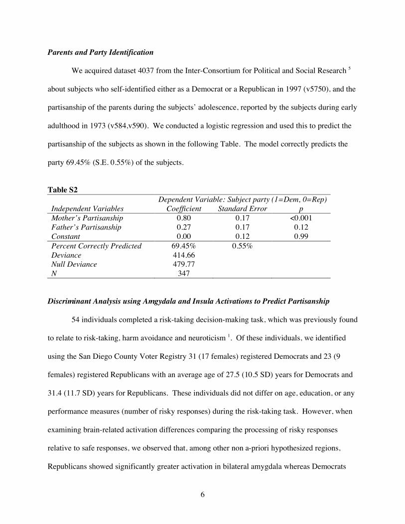
Parents and Party Identification
We acquired dataset 4037 from the Inter-Consortium for Political and Social Research 5
about subjects who self-identified either as a Democrat or a Republican in 1997 (v5750), and the
partisanship of the parents during the subjects’ adolescence, reported by the subjects during early
adulthood in 1973 (v584,v590). We conducted a logistic regression and used this to predict the
partisanship of the subjects as shown in the following Table. The model correctly predicts the
party 69.45% (S.E. 0.55%) of the subjects.
Table S2 Dependent Variable: Subject party (1=Dem, 0=Rep) Independent Variables Coefficient Standard Error p Mother’s Partisanship 0.80 0.17 <0.001 Father’s Partisanship 0.27 0.17 0.12 Constant 0.00 0.12 0.99 Percent Correctly Predicted 69.45% 0.55% Deviance 414.66 Null Deviance 479.77 N 347
Discriminant Analysis using Amgydala and Insula Activations to Predict Partisanship
54 individuals completed a risk-taking decision-making task, which was previously found
to relate to risk-taking, harm avoidance and neuroticism 1. Of these individuals, we identified
using the San Diego County Voter Registry 31 (17 females) registered Democrats and 23 (9
females) registered Republicans with an average age of 27.5 (10.5 SD) years for Democrats and
31.4 (11.7 SD) years for Republicans. These individuals did not differ on age, education, or any
performance measures (number of risky responses) during the risk-taking task. However, when
examining brain-related activation differences comparing the processing of risky responses
relative to safe responses, we observed that, among other non a-priori hypothesized regions,
Republicans showed significantly greater activation in bilateral amygdala whereas Democrats

7
showed relatively greater activation in right anterior insular cortex. In a subsequent step-wise
discriminant function analysis to determine whether brain activation patterns related to risk-
taking would be useful to predict party affiliation, we found that using a cross-validation method,
brain activation in right amygdala and insula correctly predicted the party affiliation of 79% of
the study participants (for further test details see Table S3). A Receiver Operator Curve
revealed that we achieved significantly greater predictive accuracy (AUC = 0.804 +/- 0.06) than
chance.
Table S3
Crossvalidation Results Group Democrat Republican
Democrat 70.97% 29.03% Positive Predictive Value 0.71 Republican 13.04% 86.96% Negative Predictive Value 0.87
Sensitivity Specificity Correct Prediction 0.845 0.750 78.96% (S.E. 0.74%)
Figure S3
ROC Curve (R amygdala , R Insula)
0
0.1
0.2
0.3
0.4
0.5
0.6
0.7
0.8
0.9
1
0 0.1 0.2 0.3 0.4 0.5 0.6 0.7 0.8 0.9 1
1 - Specificity
Sens
itivi
ty
8
Supporting Online Material References
1. Paulus, M.P., Rogalsky, C., Simmons, A., Feinstein, J.S. & Stein, M.B. Increased activation in the right insula during risk-taking decision making is related to harm avoidance and neuroticism. Neuroimage 19, 1439-1448 (2003). 2. Cox, R.W. AFNI: software for analysis and visualization of functional magnetic resonance neuroimages. Comput Biomed Res 29, 162-173 (1996). 3. Boynton, G.M., Engel, S.A., Glover, G.H. & Heeger, D.J. Linear systems analysis of functional magnetic resonance imaging in human V1. J Neurosci 16, 4207-4221 (1996). 4. Lancaster, J.L., et al. Automated Talairach atlas labels for functional brain mapping. Hum Brain Mapp 10, 120-131 (2000). 5. Jennings, M.K., Markus, G.B., Niemi, R.G. & Stoker, L. Youth-Parent Socialization Panel Study, 1965-1997: Four Waves Combined. in ICPSR04037-v1 (University of Michigan, Center for Political Studies/Survey Research Center, 2005).