redefining identity of disease, tissues and cells a
TRANSCRIPT

Redefining Identity of Disease, Tissues and Cells – A Biomaterials Paradigm
Abhay Pandit
Director, CÚRAM- SFI Research Centre for Medical Devices; National University of Ireland; Galway, Ireland
Abstract
Biomaterials are no longer considered innate structures and using functionalisation and
biofabrication strategies to modulate a desired response whether it is a host or implant is
currently an important focus in current research paradigms. Fundamentally, a thorough
understanding of the host response will enable us to design appropriate strategies. The input
from the host response needs to be weighed in depending on the host disease condition. Our
current inputs have been through a thorough understanding of glyco-proteomics based tools
which we are developing in our laboratory. In addition, biomaterials themselves provide
immense therapeutic benefits which needs to be accounted in the design paradigm. Using
functionalisation strategies such as enzymatic and hyperbranched linking systems, we have been
able to link biomolecules to different structural moieties. The programmed assembly of
biomolecules into higher-order self-organized systems is central to innumerable biological
processes and development of the next generation of biofabricated scaffolds. Recent design
efforts have utilized a glycobiology and developmental biology approach toward both
understanding and engineering supramolecular protein and sugar assemblies.
Biography
Professor Abhay Pandit is the Established Professor in Biomaterials and the Director of a Science Foundation Ireland funded Centre for Research in Medical Devices (CÚRAM) at the National University of Ireland, Galway. Professor Pandit’s research integrates material science and biological paradigms in developing solutions for chronic diseases including neural, musculoskeletal and cardiovascular clinical targets with numerous other targets currently under development. His research is funded by Science Foundation Ireland (SFI), EU Framework program, Enterprise Ireland, Health Research Board, the AO Foundation and industry sources, and in excess of €84 million. He has also established a critical mass of biomaterial expertise in Ireland by securing funding for an SFI funded Strategic Research Cluster. He is the author of 22 patents and has licensed three technologies to medical device companies and authored more than 260 manuscripts. Prof Pandit has successfully supervised 26 Ph.D. students, 17 Master’s students and mentored 25 postdoctoral fellows. He is currently leading the team in the supervision of 15 Ph.D. students, 15 Post doctorates and three research associates.

Acellular Biomaterials for Dental Tissue Repair
Adam D. Celiz
UKRI Future Leaders Fellow and Lecturer, Department of Bioengineering; Imperial College
London, UK
Abstract
Dental disease annually affects billions of patients, and while regenerative dentistry aims to heal
dental tissue after injury, existing polymeric restorative materials, or fillings, do not directly
participate in the healing process in a bioinstructive manner. There is a need for restorative
materials that can support native functions of dental pulp stem cells (DPSCs), which are capable
of regenerating dentin. A polymer microarray formed from commercially available monomers to
rapidly identify materials that support DPSC adhesion is used. Based on these findings, thiol-ene
chemistry is employed to achieve rapid light-curing and minimize residual monomer of the lead
materials. Several triacrylate bulk polymers support DPSC adhesion, proliferation, and
differentiation in vitro, and exhibit stiffness and tensile strength similar to existing dental materials.
Conversely, materials composed of trimethacrylates or bisphenol A glycidyl methacrylate
(BisGMA), which is a monomer standard in dental materials, do not support stem cell adhesion
and negatively impact matrix and signaling pathways. Furthermore, thiol-ene polymerized
triacrylates are used as permanent filling materials at the dentin-pulp interface in direct contact
with irreversibly injured pulp tissue. These acellular materials have potential to enable novel
regenerative dental therapies in the clinic by both restoring teeth and providing a supportive niche
for DPSCs.
Biography
Dr. Celiz’s research focusses on the development of
acellular biomaterials to repair or regenerate tissues. Dr.
Celiz gained his PhD in Chemistry in the Melville
Laboratory for Polymer Synthesis at the University of
Cambridge. Dr. Celiz gained postdoctoral training at the
University of Nottingham and the Wyss Institute for
Biologically Inspired Engineering at Harvard University via
a Marie Curie International Outgoing Fellowship (IOF). In
2017, Dr. Celiz was appointed as Lecturer (Assistant
Professor) in Department of Bioengineering at Imperial
College London. Dr. Celiz’s research has been published in
journals including Science, Nature Materials and Advanced
Materials. Dr Celiz has been awarded several early-career awards including the Larry Hench
Young Investigators Prize from the UK Society for Biomaterials and is currently a holder of a
UKRI Future Leader Fellowship.

Bioactive glasses with tuned ion releasing capability to stimulate stem cells for
tissue engineering
Aldo R. Boccaccini
Institute of Biomaterials, University of Erlangen-Nuremberg, 91058 Erlangen, Germany
Abstract
Biochemical reactions occurring at the interface between bioactive glasses (BGs) and the
biological environment, involving the release of BG ionic dissolution products, are relevant for
both hard and soft tissue regeneration. The development and characterization of ion doped BGs
will be discussed with focus on the effect of different biologically active ions released from BGs
on stem cells, mainly umbilical cord and adipose derived stem cells as well as bone marrow-
derived mesenchymal stem cells (BMSCs). BGs incorporating biologically active ions, such as B,
Sr, Cu, Nb, Co, Li, Mn, will be considered. Indirect cell culture methods using endothelial cells
with or without BMSCs in cell culture inserts exposed to ion dissolution products from BG
scaffolds (e.g. Cu doped) will be presented to show that BMSCs secrete an increased concentration
of vascular endothelial growth factor, thus confirming the angiogenic potential of such BGs. The
results are evaluated regarding the stimulating effect of metallic ions on stem cells, also based on
literature results. The variation of ion concentration in medium as function of time and the time
dependent effects on stem cells will be discussed, which is required for the comprehensive
assessment of BG biological performance with implication for clinical applications.
Biography
Aldo R. Boccaccini is Professor of Biomaterials and Head
of the Institute of Biomaterials at University of Erlangen-
Nuremberg, Germany. He is also Visiting Professor at
Imperial College London. His research activities are in the
broad area of glasses, ceramics and composites for
biomedical applications. He has co-authored more than 850
scientific papers. His work has been cited more than 36,000
times (Scopus®). Boccaccini is Fellow of the Institute of
Materials, Minerals and Mining, American Ceramic
Society, Society of Glass Technology and European
Ceramic Society. He is the Editor-in-Chief of the journal
“Materials Letters” and founding Editor of “Biomedical
Glasses”. He has received numerous international awards, including the Materials Science Prize
of German Materials Society and Turner Award of International Commission on Glass. He is also
a member of the World Academy of Ceramics, National Academy of Engineering and Applied
Sciences of Germany and advisor to the Science and Technology Ministry of Argentina.
Boccaccini serves in the Executive Committee of the Federation of European Materials Societies
and in the Council of the European Society for Biomaterials.
Harnessing proteins and supramolecular events to build advanced
biomaterials
Alvaro Mata
Queen Mary University of London, UK
Abstract
Nature has evolved to grow and heal materials and tissues through self-assembling processes capable of
organizing a wide variety of molecular building-blocks at multiple size scales. While advances in fields
such as nanotechnology and biofabrication are enhancing our capacity to emulate some of these biological
structures, it is increasingly evident that recreation of the complexity and functionality of living systems
will require new ways to build with proteins. This talk will present our laboratory’s efforts to harness
supramolecular events found in nature such as multicomponent self-assembly, protein order-disorder
synergies, and diffusion-reaction processes to engineer advanced protein-based materials. The resulting
materials exhibit properties such as hierarchical organization1-3, the capacity to grow and heal2,3, tuneable
mechanical properties1,2, and spatially controlled bioactivity4,5.
References
1. Elsharkawy et al (2018). Nature Communications, 10.1038/s41467-018-04319-0.
2. Wu et al (2020). Nature Communications, 1182, 10.1038/s41467-020-14716-z.
3. Inostroza-Brito et al (2015). Nature Chemistry, 10.1038/nchem.2349.
4. Hedegaard et al (2018). Advanced Functional Materials, 10.1002/adfm.201703716.
5. Hedegaard et al (2020). Science Advances, 10.1126/sciadv.abb3298.
Biography
Alvaro Mata is Professor in Biomedical Engineering and Biomaterials in the School of Pharmacy
and the Department of Chemical and Environmental Engineering at the University of Nottingham.
He holds a Bachelor's Degree from the University of Kansas, a Master's Degree from the
University of Strathclyde, and a Doctor of Engineering Degree from Cleveland State University
working with Prof. Shuvo Roy at the Cleveland Clinic. He conducted his postdoctoral training
with Prof. Samuel Stupp at Northwestern University. His group focuses on developing innovative
ways to build with biomolecules to engineer active, hierarchical, and living materials that can
recreate complex biological environments. Before joining the University of Nottingham, he helped
established and served as Director of the Institute of Bioengineering at Queen Mary University of
London between 2015-2018 and is now the Chair of the Manufacturing Commercial and
Regulatory Committee of the UK Regenerative Medicine Platform (UKRMP2) – Acellular / Smart
Materials – 3D Architecture Hub. His work has led to seven patents or patent applications;
publications in journals including Nature Chemistry, Nature Communications, Science Advances,
and Advanced Functional Materials; and awards such as a Ramon y Cajal Fellowship and an ERC
Staring Grant.

Harnessing the Host Response for In Situ Cardiovascular Tissue Engineering
– the Challenge of Elastogenesis
Anthal I.P.M. Smits
Assistant Professor and Group Leader ImmunoRegeneration Group, Department of Biomedical
Engineering and the Institute for Complex Molecular Systems (ICMS), Eindhoven University of
Technology, Eindhoven, The Netherlands.
Abstract
The use of acellular resorbable synthetic scaffolds for replacing diseased cardiovascular tissues is
an attractive strategy that has shown great promise in recent preclinical studies and ongoing
clinical trials. These scaffolds are designed to instantaneously take over the functionality of the
replaced tissue upon implantation, and maintain functionality while they are gradually resorbed
and replaced by autologous new tissue by infiltrating cells, directly in situ. This process of in situ
tissue engineering is poorly understood to date, leading to unpredictable variability in outcome.
Moreover, one of the biggest unmet challenges is the in situ regeneration of a functional, native-
like elastin network, which is critical for sustaining long-term functionality of cardiovascular
tissues. In this talk, I will present our efforts in the understanding and controlling of the in situ
formation of functional new cardiovascular tissues (i.e. blood vessels and heart valves) by
modulating the host immune response using resorbable supramolecular elastomers. Specifically, I
will elaborate on our recent results on the influence of biomechanical loads on the inflammatory
and regenerative processes to such scaffolds, and how these may dictate tissue formation and
elastin deposition in particular, both in vitro and in vivo.
Biography
Dr.ir. Anthal Smits was appointed Assistant Professor at
Eindhoven University of Technology in 2016, where he
since initiated and leads the ImmunoRegeneration Group as
one of the main research pillars of the Department of
Biomedical Engineering and the Institute for Complex
Molecular Systems (ICMS). His research is aimed at
modulating the immune response using biomaterials in
order to induce functional, homeostatic tissue regeneration.
His group performs interdisciplinary work, dedicated to
gaining a mechanistic understanding of the interactions
between immune cell behavior, biomaterial design, and
biomechanical loads, in conditions of health and disease.
The main target applications are cardiovascular replacements (e.g. heart valves and blood vessels),
yet the research is curiosity-driven, and applicable to a wide variety of clinical applications. Dr.
Smits is a member of the Heart Valve Society and the Tissue Engineering Young Investigator
Council, among others. He is a leading researcher in various international research programs and
public-private partnerships, such as the Materials-Driven Regeneration program (NWO
Gravitation; 25 M€) and the Cardiac Moonshot within the RegMed-XB program (800,000 €).

Tropoelastin promotes elastic tissue regeneration and restoration
Anthony S. Weiss
McCaughey Chair in Biochemistry, Professor of Biochemistry & Molecular Biotechnology,
Charles Perkins Centre School of Life and Environmental Sciences,
University of Sydney, Australia
Abstract
Elastic tissue does not typically regenerate in adults, so there is demand for ways to restore these
tissues following damage. The stages through which tropoelastin self-assembles into elastin and,
in turn, elastic fibers, are hierarchical and the topic of extensive, ongoing research. It is this
capacity for self-assembly that is of interest for a class of materials that promote the formation of
new elastic tissue.
Processes and a hybrid biomaterial, developed in association with Dr. Suzanne Mithieux in my
lab, are intended to deliver tunable levels of histologically detectable patient elastin into full-
thickness wound sites. This approach addresses a persistent unmet need because repairing wounds
lack this elastic substratum. Previously, dogma asserted that elastin synthesis is attenuated in early
childhood, but we found that we can overcome this restriction by adding exogenous tropoelastin,
regardless of the age of the dermal fibroblast donor. We found how to further enhance synthesis
with older cells by using conditioned media. This approach delivers elastin as a layer on the leading
dermal repair template for contact with the deep dermis in order to deliver prefabricated elastic
fibers to the physiologically appropriate site during surgery to repair scar tissue at sites of healing
full thickness wounds.
Biography
Professor Anthony Weiss PhD AM FRSC FTSE FRSN
FRACI FAIMBE FAICD FBSE FTERM is the McCaughey
Chair in Biochemistry at the University of Sydney. His
research focuses on the assembly of human elastic tissue,
damage and its repair. His awards include the Order of
Australia, Clunies Ross National Science and Technology
Award, Eureka Prize for Innovation in Medical Research,
Premier’s Prize for Science & Engineering Leadership in
Innovation, Roslyn Flora Goulston Prize, NIH Fogarty
International Fellow, David Syme Research Medal,
Amersham Pharmacia Biotechnology Medal, NSW
Commercialization Expo Prize, Australian Innovation
Challenge Award, Fondation des Treilles Scholar, Pauling Prize Medal, Barry Preston Award,
ASBTE Research Excellence Award, FAOBMB Entrepreneurship Award and RACI Applied
Research Medal. Professor Weiss founded the biotechnology clinical stage company Elastagen Pty
Ltd which was sold to Allergan in one of the largest transactions ever completed in the Australian
life science sector. He is an inventor with 105 awarded international patents in 17 patent families.
He is on eleven editorial boards comprising leading journals in the field and is global President-
Elect of TERMIS.

‘Mix and match’: local delivery of protein-based biologics
using responsive microgels
April M. Kloxin
Centennial Development Professor of Chemical and Biomolecular Engineering, Chemical and
Biomolecular Engineering and Materials Science and Engineering, University of Delaware,
Newark, DE, 19716, USA
Abstract
Protein-based biologics, particularly antibodies, are of growing interest owing to their specificity
and therapeutic efficacy, especially for many conditions that traditionally have been difficult to
treat (e.g., metastatic cancer, misregulated wound healing). The use of multiple therapeutics,
combination therapies, that target different aspects of disease mechanisms can be particularly
effective; however, such therapies have a significant risk of systemic toxicity owing to the high
total doses that must be used. Responsive hydrogels offer a facile platform for the local, controlled
release of these large, hydrophilic proteins for the design of personalized combination therapies
while minimizing adverse side effects. Specifically, in this talk, I report the development of mixed
populations of hydrogel microparticles, or microgels, for achieving tunable and tailorable release
profiles of antibodies in vitro and in vivo. Microgels of uniform size and relevance for local
injection were created using microfluidic devices. To achieve tunable and on-demand release
profiles, microgels that respond to either internal (i.e., reducing microenvironments) or external
(i.e., light) cues were designed. Modular building blocks, multifunctional polymers with a variety
of chemical handles, were used to create mixed populations of microgels that localize to desired
tissues and release multiple therapeutics across a range of time scales.
Biography
Professor April M. Kloxin, Ph.D., is Centennial
Development Professor of Chemical and Biomolecular
Engineering at the University of Delaware (UD) and a
member of the Breast Cancer Research Program at the
Helen F. Graham Cancer Center and Research Institute. She
obtained her B.S. (Summa Cum Laude) and M.S. in
Chemical Engineering from North Carolina State University
and Ph.D. in Chemical Engineering from the University of
Colorado, Boulder, as a NASA Graduate Student Research
Program Fellow. She trained as a Howard Hughes Medical
Institute postdoctoral research associate at the University of
Colorado before joining the faculty at UD in 2011. Her
group aims to create unique materials with multiscale property control for addressing outstanding
problems in human health. Her research currently focuses on the design of responsive biomaterials
with multiscale properties and development of controlled, dynamic models of disease and
regeneration. Her honors include a NIH Director’s New Innovator Award, ACS PMSE Arthur K.
Doolittle Award, Susan G. Komen Foundation Career Catalyst Research award, NSF CAREER
award, and a Pew Scholars in Biomedical Sciences award.

Success and Challenges in Biofabrication
Brian Derby
Director, Centre for Digital Fabrication; Professor of Materials Science; Department of
Materials; University of Manchester, Oxford Road, Manchester, M13 9PL, UK.
Abstract
Biofabrication as a scientific/engineering term is now 16 years old. In this period it has rapidly
progressed from being seen as a topic for Science Fiction to now a method for the production of
proto- or in vitro model tissues for tissue-on-a-chip applications. Indeed there are several
commercial organizations that have developed and market commercial Bioprinters. However,
despite these not inconsiderable advances, the original target of printed cell-laden implants or
tissue patches have still not been realized.
This talk will present a review of the key advances that have occurred over the recent past and
identify the key challenge of the dimensions of practical printed tissue models. It is well known
that the diffusion limit in 3D culture of cells limits spheroid size to a few hundred m and that
the capillaries in healthy tissue have a diameter of 5 – 10 m and are typically spaced around 50
m. However, current additive manufacturing routes that are in widespread use in biofabriction
have a resolution > 100 m. New approaches to producing high resolution vascular structures
and models for angiogenesis will be reviewed.
Biography
Professor Derby’s research focusses on the use of a range of
printing methods compatible with the delivery of cells for
applications in regenerative medicine and organ-on-a-chip
development. This has been developed by interdisciplinary
university work, international multi-institute collaborations
and collaboration with industry. In 2004 he hosted, in
Manchester, the First International Workshop in
Biofabrication and has been active in the field since then.
Prof. Derby has authored > 300 peer-reviewed journal
publications. His work has been funded through a range of
agencies including EPSRC, BBSRC and MRC from Research Councils UK, the Wellcome Trust,
British Heart Foundation, Innovate UK, the European Commission and the Office of Naval
research (USA)

Nanofilled electrospun fibers inspired by elastin and natural polymers
Nicola Ciarfaglia, Antonietta Pepe, Antonio Laezza, Brigida Bochicchio*
Associate Professor of Organic Chemistry; Department of Science; University of Basilicata, Via
Ateneo Lucano 10, Potenza, Italy
Abstract
Electrospinning is an emerging technique with applications in tissue engineering for the high
surface area to volume ratio and the interconnected pores of nanofibers. Blending of synthetic
polymers as polylactic acid (PLA) and poly--caprolactone (PCL), together with natural proteins
(Gelatin, Elastin) were conceived until now in order to obtain scaffolds with combination of
strength and biocompatibility usually used in soft-tissue regeneration. Furthermore, the use of
nanoreinforcements as bioglass and/or nanocellulose is aimed to overcome the limited mechanical
performances of the scaffolds. In this work, we have successfully electrospun highly hydrophobic
(PDLLA) and hydrophilic polymers (gelatin and a human tropoelastin-inspired sequence) together
with bioglass microparticles and crystalline nanocellulose as nanoreinforcements in the
perspective of future applications in hard-tissue regeneration (bone). Additionally, PDLLA was
herein adoperated for its faster degradation times in comparison to the most used PLA. The
matrices were cross-linked in order to confer stability in water. Afterward, the matrices were
tested concerning wettability, swelling and Young's elastic modulus. They show complete
wettability and a significative decrease of Young's Modulus after swelling (comprised between 27
and 23 MPa). The value is analogous to that found for electrospun scaffolds composed of collagen,
elastin and PCL. Funding: PON R&I 2014-2020 (572 PON_ARS01_01081).
Biography
Professor Bochicchio’s research focusses on biopolymers
and peptides inspired by elastomeric proteins for tissue
engineering. Professor Bochicchio has authored 60
international peer-reviewed journal publications and Invited
Speaker at 24 International Meetings. Professor
Bochicchio’s research has been supported by Italian
Ministry of University and European Community and also
attracts interest from multinational industrial partners. To
date, she has secured ≈€1.8M (Co-I) research funding from
National Operational Program "Research and Innovation"
2014-2020 in health sector. Professor Bochicchio has
successfully supervised to completion 2 PhD student and 20
MSc students all of whom have remained in engineering/science and or academia. She is currently
involved in the supervision of 1 PhD student. Additionally, she is currently mentoring one PhD
research fellow.

Encanpsulation of ICOS-Fc in biocompatible and biodegradable
nanoparticles elicits novel therapeutic activities
C. Luca Gigliotti
Novaicos s.r.l.s., Spin-off of Università del Piemonte Orientale (UPO), 28100 Novara, Italy
Abstract
ICOS is a T cell co-stimulatory molecule binding ICOSL expressed on several cell types, including
osteoclasts, vascular endothelial cells and several tumor cell types. ICOSL triggering by a soluble
recombinant form of ICOS (ICOS-Fc) inhibits osteoclast activity in vitro and development of
osteoporosis in mice in vivo. Moreover, it inhibits migration of tumor cells in vitro and tumor
metastatization in vivo, but it has no effect on the growth of primary tumors. In this work, we show
that loading of ICOS-Fc into either cross-linked cyclodextrin nanosponges (CDNS) or poly(lactic-
co-glycolic acid) (PLGA) nanoparticles (NP), in order to increase stability, sustain release, and
increase tumor-delivery of the drug, elicits a strong antitumor activity ascribable to inhibition of
tumor neoangiogenesis and resetting of the anti-tumor immune response. The substantial in vivo
activity of ICOS-Fc in NP makes these nanoformulations attractive candidates for modulating
ICOS-Fc activity and eliciting novel therapeutic activities.
Biography
The research of Dr. Gigliotti is outlined in different fields
having as a key point the immune system and the role of the
interaction of the costimulatory molecule ICOS and its
ligand ICOSL in vitro and in vivo. His studies also include
the bone system and, in particular, osteoclasts which are
cells of immune origin. The research gave rise to two
patented drugs inhibiting bone resorption and tumor growth,
respectively. He is currently CEO of Novaicos srls, a
biotech company aimed to develop innovative approaches
to correct tissue dysfunctions.

Nanoscale regulation of adhesion and growth factor signaling by material
mechanical and chemical properties
E. Ada Cavalcanti-Adam
Group Leader, Max Planck Institute for Medical Research; Faculty, Institute of Physical
Chemistry, Heidelberg University; Heidelberg, Germany
Abstract
Mechanical and chemical cues present in the extracellular environment regulate cell adhesion-
mediated responses, such as migration, proliferation and differentiation. Designing materials
which combine adhesive ligands and growth factors at the nanoscale is of great interest to address
how local changes in the extracellular environment regulate cell responses through specific
receptor-ligand interactions. I will present the development of surface functionalization strategies
to control integrin clustering and the generation of cellular forces at the interface during adhesion.
The nanoscale presentation of integrin ligands is also combined with growth factors, namely BMP-
2 and BMP-6, to modulate the osteogenic differentiation of cells. In this talk, I will also discuss
the synergistic effect of BMPs and mechanical cues on osteogenic signaling and
mechanotransduction. By varying the stiffness of polyethylene glycol (PEG)- and hyaluronan
(HA)- based hydrogels which are functionalized with integrin ligands and BMPs, it is possible to
elucidate the interdependency of Smad 1/5 and YAP/TAZ signaling.
Biography
Ada Cavalcanti’s research is centered on the
mechanobiology of cell-matrix adhesion. She combines
surface patterning and functionalization approaches to study
the nanoscale regulation of cell adhesion structures and
signaling. She has authored >60 publications in
international peer-reviewed journals. Her research is
supported by the Max Planck Society and by the German
Science Foundation (DFG) and she has been awarded in
2008 with the prize “for women in science” from UNESCO-
L’Orèal.

Active ageing and osteoporosis: the challenge of the GIOTTO project
Chiara Vitale Brovarone
Department of Applied Science and Technology
Politecnico di Torino
Italy
Osteoporosis is a systemic, degenerative disorder, predominantly affecting postmenopausal
women (1 out of 3) but also men at an advanced age (1 out of 5) that increases the fracture risk. A
number of anti-osteoporotic drugs are available and decrease the fracture risk between 50 and 70%
but they also have important side effects and they do not promote fracture healing.
GIOTTO aims to develop a platform of technologies and materials to treat different types of
osteoporotic fractures and to support the prevention of new fractures.
In particular, we will design and validate three different solutions:
1) A 3D graded scaffold allowing fixations with screws to treat periprosthetic fractures
2) A collagen based fibrous scaffold produced via electrospinning to deal with small, not
confined pelvis fractures
3) A radiopaque, bioresorbable, injectable cement to stabilise vertebral fractures
The three devices will share smart nanobiomaterials able to release chemical and biological cues
to stimulate bone regeneration while reducing bone loss.
For further information please visit: http://www.giottoproject.eu/
Full Professor in Materials Science and Technology, Applied Science and Technology
Department, Politecnico di Torino
Prof. Chiara Vitale-Brovarone, Full Professor in Materials
Science and Technology, Politecnico di Torino where she leads
the IRIS group (Improving Regeneration by Intelligent
Scaffolds). Scopus: 160 papers including research articles and
book chapters, H-index 37, 4000 citations.
She has coordinated the EU projects (FP6 – BIORESS, FP7 -
MATCH and H2020 - MOZART) and she has been Team
leader for the FP7 project RESTORATION. At present, she is
coordinating the H2020 project GIOTTO that aims to develop
innovative devices to treat osteoporotic fractures and she is the
PI of the ERC consolidator grant BOOST.
Her research interests are mainly related to the development of innovative biomaterials ranging
from the macro to the nanoscale (3D-scaffolds, micro and nanoparticles, injectable cement and
smart surfaces with osteoproductive, antibacterial and biomolecule release properties).
She is developing novel approaches to target bone and wound healing and osteoporotic fractures
as well as the fabrication of smart bone scaffolds through rapid prototyping approaches including
bioextrusion, ink-jet printing and electrospinning.

Logical Breakdown: Programming Boolean-based Responsiveness into
Hydrogel Biomaterials
Cole A. DeForest
Dan Evans Assistant Professor, Departments of Chemical Engineering and Bioengineering;
Core Member, Institute for Stem Cell & Regenerative Medicine;
University of Washington, Seattle, Washington, USA
Abstract
The successful transport of drug- and cell-based therapeutics to diseased sites represents a major
barrier in the development of clinical therapies. Targeted delivery can be mediated through
degradable biomaterial vehicles that utilize disease biomarkers to trigger payload release. Here,
we report a modular chemical framework for imparting hydrogels with precise degradative
responsiveness by using multiple environmental cues to trigger reactions that operate user-
programmable Boolean logic. By specifying the molecular architecture and connectivity of
orthogonal stimuli-labile moieties within material crosslinkers, we show selective control over gel
dissolution and therapeutic delivery. To illustrate the versatility of this methodology, we
synthesized seventeen distinct stimuli-responsive materials that collectively yielded all possible
YES/OR/AND logical outputs from input combinations involving enzyme, reductant, and light.
Using these hydrogels, we demonstrate the first sequential and environmentally stimulated release
of small molecules, site-specifically modified proteins, and multiple cell lines in well-defined
combinations from a material. We expect these platforms will find utility in several diverse fields
including drug delivery, diagnostics, and regenerative medicine.
Biography
Dr. Cole A. DeForest is the Dan Evans Career Development
Assistant Professor in the Departments of Chemical
Engineering and Bioengineering, and a core faculty member
of the Institute for Stem Cell & Regenerative Medicine at
the University of Washington (UW) where he began in
2014. He received his B.S.E. degree from Princeton
University in 2006, majoring in Chemical Engineering and
minoring in Material Science Engineering and
Bioengineering. He earned his Ph.D. degree under the
guidance of Dr. Kristi Anseth from the University of
Colorado in Chemical and Biological Engineering with an
additional certificate in Molecular Biophysics. His
postdoctoral research was performed with Dr. David Tirrell in the Divisions of Chemistry and
Chemical Engineering at Caltech. He has authored ~45 peer-reviewed articles, including as the
corresponding author for those appearing in Nature Materials, Nature Chemistry, Advanced
Materials, JACS, and Nature Reviews Materials. Dr. DeForest has received numerous research
awards including the Safeway Early Career Award (2018), NSF CAREER Award (2017), AIChE
35-Under-35 Award (2017), and the Presidential Distinguished Teaching Award (2016, UW’s
highest teaching award).
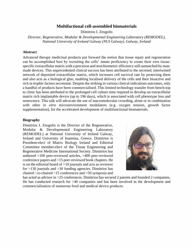
Multifactional cell-assembled biomaterials
Dimitrios I. Zeugolis
Director, Regenerative, Modular & Developmental Engineering Laboratory (REMODEL),
National University of Ireland Galway (NUI Galway), Galway, Ireland
Abstract
Advanced therapy medicinal products put forward the notion that tissue repair and regeneration
can be accomplished best by recruiting the cells’ innate proficiency to create their own tissue-
specific extracellular matrix with a precision and stoichiometric efficiency still unmatched by man-
made devices. This unprecedented clinical success has been attributed to the secreted, intertwined
network of deposited extracellular matrix, which increases cell survival rate by protecting them
and also acts as a biological glue, enabling localised delivery of the cells and their bioactive and
rich in trophic factors secretome. Despite the striking in various clinical indications outcomes, only
a handful of products have been commercialised. This limited technology transfer from bench-top
to clinic has been attributed to the prolonged cell culture time required to develop an extracellular
matrix rich implantable device (up to 196 days), which is associated with cell phenotype loss and
senescence. This talk will advocate the use of macromolecular crowding, alone or in combination
with other in vitro microenvironment modulators (e.g. oxygen tension, growth factor
supplementation), for the accelerated development of multifunctional biomaterials.
Biography
Dimitrios I. Zeugolis is the Director of the Regenerative,
Modular & Developmental Engineering Laboratory
(REMODEL) at National University of Ireland Galway,
Ireland and University of Ioannina, Greece. Dimitrios is
President-elect of Matrix Biology Ireland and Editorial
Committee member-elect of the Tissue Engineering and
Regenerative Medicine International Society. Dimitrios has
authored >100 peer-reviewed articles, >400 peer-reviewed
conference papers and >15 peer-reviewed book chapters. He
is on the editorial board of >10 journals and acts as reviewer
for >130 journals and >30 funding agencies. Dimitrios has
chaired / co-chaired >15 conferences and >50 symposia and
has acted as advisor in >25 conferences. Dimitrios has secured 2 patents and founded 2 companies.
He has conducted research for >40 companies and has been involved in the development and
commercialisation of numerous food and medical device products.
Triggerable Self-immolative Polymer Delivery Systems: From Polyglyoxylates
to Polyglyoxylamides
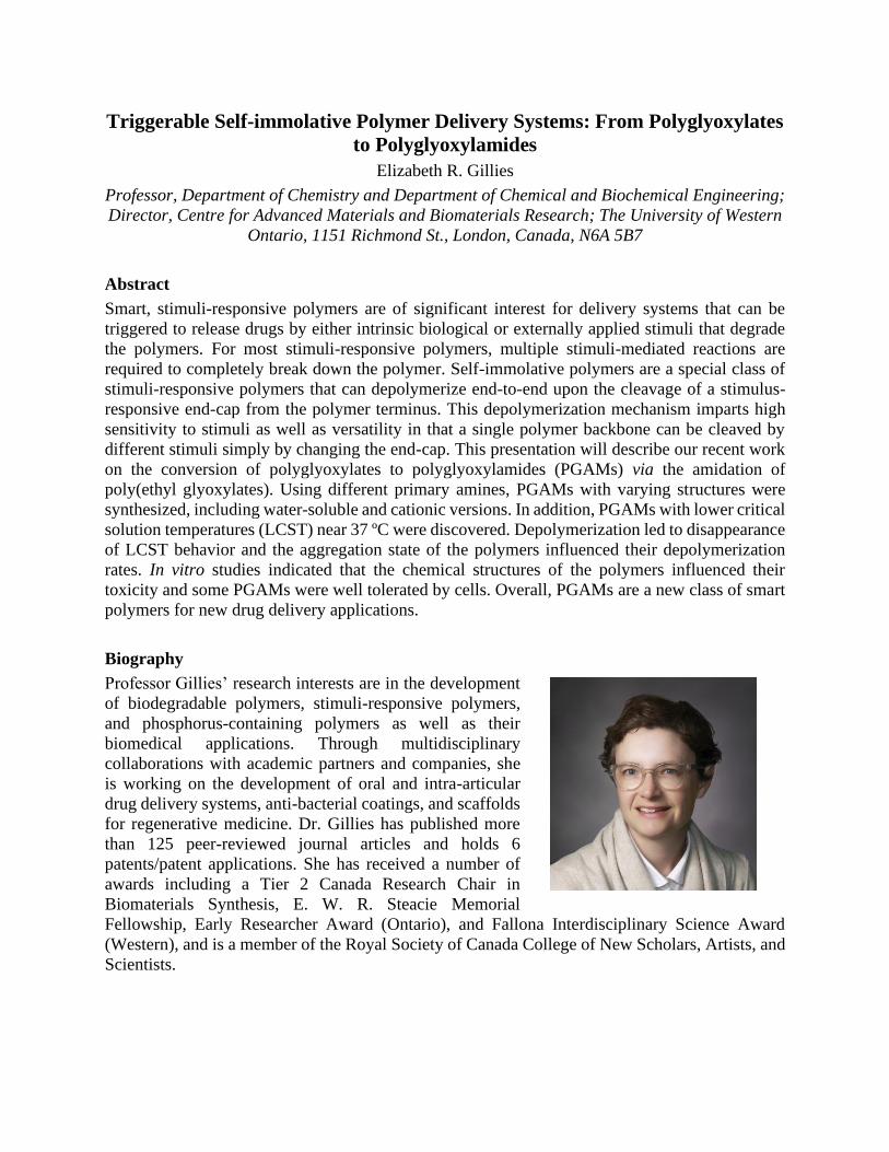
Elizabeth R. Gillies
Professor, Department of Chemistry and Department of Chemical and Biochemical Engineering;
Director, Centre for Advanced Materials and Biomaterials Research; The University of Western
Ontario, 1151 Richmond St., London, Canada, N6A 5B7
Abstract
Smart, stimuli-responsive polymers are of significant interest for delivery systems that can be
triggered to release drugs by either intrinsic biological or externally applied stimuli that degrade
the polymers. For most stimuli-responsive polymers, multiple stimuli-mediated reactions are
required to completely break down the polymer. Self-immolative polymers are a special class of
stimuli-responsive polymers that can depolymerize end-to-end upon the cleavage of a stimulus-
responsive end-cap from the polymer terminus. This depolymerization mechanism imparts high
sensitivity to stimuli as well as versatility in that a single polymer backbone can be cleaved by
different stimuli simply by changing the end-cap. This presentation will describe our recent work
on the conversion of polyglyoxylates to polyglyoxylamides (PGAMs) via the amidation of
poly(ethyl glyoxylates). Using different primary amines, PGAMs with varying structures were
synthesized, including water-soluble and cationic versions. In addition, PGAMs with lower critical
solution temperatures (LCST) near 37 ºC were discovered. Depolymerization led to disappearance
of LCST behavior and the aggregation state of the polymers influenced their depolymerization
rates. In vitro studies indicated that the chemical structures of the polymers influenced their
toxicity and some PGAMs were well tolerated by cells. Overall, PGAMs are a new class of smart
polymers for new drug delivery applications.
Biography
Professor Gillies’ research interests are in the development
of biodegradable polymers, stimuli-responsive polymers,
and phosphorus-containing polymers as well as their
biomedical applications. Through multidisciplinary
collaborations with academic partners and companies, she
is working on the development of oral and intra-articular
drug delivery systems, anti-bacterial coatings, and scaffolds
for regenerative medicine. Dr. Gillies has published more
than 125 peer-reviewed journal articles and holds 6
patents/patent applications. She has received a number of
awards including a Tier 2 Canada Research Chair in
Biomaterials Synthesis, E. W. R. Steacie Memorial
Fellowship, Early Researcher Award (Ontario), and Fallona Interdisciplinary Science Award
(Western), and is a member of the Royal Society of Canada College of New Scholars, Artists, and
Scientists.

Biomaterials to Eliminate Bacterial Infections
Andrés J. García
Executive Director, Parker H. Petit Institute for Bioengineering and Bioscience
Regents’ Professor, George W. Woodruff School of Mechanical Engineering
Georgia Institute of Technology, Atlanta, GA, U.S.A.
Abstract
Device-associated infections result in substantial morbidity and mortality and contribute
significantly to the high cost of caring for patients. Current therapies to eliminate biofilm formation
in medical devices have shown low levels of success due to the inherent resistance of the biofilm
towards antimicrobial agents, as well as the diverse methods in which bacteria can be introduced
to the site of infection. Lysostaphin is an enzyme that cleaves the pentaglycine cross-links of the
staphylococcal cell wall, leading to cell lysis, making it a potentially useful agent to eradicate
infection in both sites of bacterial growth, as well as established biofilms. We have engineered a
synthetic hydrogel for the encapsulation and delivery of lysostaphin to the site of infection.
Lysostaphin-delivering hydrogels eradicated S. aureus infection and out-performed prophylactic
antibiotic and soluble lysostaphin therapy after 7 days in a murine model of femur fracture.
Infected fractures treated with lysostaphin-delivering hydrogels completely healed by 5 weeks
with equivalent bone formation and mechanical properties to uninfected fractures, whereas
fractures treated without the hydrogel carrier were equivalent to untreated infections. Finally,
lysostaphin-delivering hydrogels were effective against methicillin-resistant S. aureus infections.
Biography
Prof. García’s research program integrates innovative
engineering, materials science, and cell biology concepts
and technologies to create cell-instructive biomaterials for
regenerative medicine and generate new knowledge in
mechanobiology. This cross-disciplinary effort has resulted
in new biomaterial platforms that elicit targeted cellular
responses and tissue repair in various biomedical
applications, innovative technologies to study and exploit
cell adhesive interactions, and new mechanistic insights into
the interplay of mechanics and cell biology. In addition, his
research has generated intellectual property and licensing
agreements with start-up and multi-national companies. He
has received several distinctions, including the Young Investigator Award from the Society for
Biomaterials, the Clemson Award for Basic Science from the Society for Biomaterials, and the
International Award from the European Society for Biomaterials. He is an elected Fellow of
Biomaterials Science and Engineering, Fellow of the American Association for the Advancement
of Science, Fellow of the American Society of Mechanical Engineers, and Fellow of the American
Institute for Medical and Biological Engineering. He served as President for the U.S. Society for
Biomaterials in 2018-2019.

Directing tissue regeneration (and stem cell differentiation) with ions released
from materials.
Gavin Jell
Associate Professor of Nanotechnology & Regenerative Medicine; Division of Surgery and
Interventional Science, University College London
Abstract
Ions released (in a controlled manner) from materials can promote desirable cellular behavior and
tissue regeneration. A range of materials have been designed to release different therapeutic ions
(including Ag, B, Ca, Co, Cu, K, Li, Mg, P and Si), which have (either individually or in
combination with other ions) been reported to promote tissue regeneration including; antimicrobial
activity, angiogenesis, cell survival, desirable ECM production, stem cell recruitment and stem
cell differentiation. The performance and application of these ion releasing materials could,
however, be improved by gaining a greater understanding of the intracellular role of the ions in
directing cell behavior (including stem cell differentiation), by understanding their effect on
different stages on tissue regeneration, and by tailoring the ion release profile for patient-specific
characteristics. Indeed, despite passing the 50th year anniversary since the creation of arguably the
first ion releasing material in medicine, Bioglass® by Prof. L. Hench, there remains a lack of
understanding of how these ions interact with cells, how they regulate gene expression and how
they can be used to direct cellular differentiation.
Biography
Gavin’s research focusses on understanding material-
biological interactions, to create improved biomaterials;
materials that have reduced failure rates and increased
functionality (e.g. promote tissue regeneration or improved
nanoparticle targeting). He is an interdisciplinary scientist
with over 60 publications in bone tissue engineering, soft
and hard tissue implant failure, and nanomedicine. Among
his successes is the invention of Co releasing of bioactive
glasses in 2009 at Imperial College, which is (possibly) the
first bioceramic to target a particular intracellular pathway,
namely the HIF-1α pathway. He’s a Trustee for the British
Society of Nanomedicine and a passionate educator. He has
created a number of successful post-graduate and undergraduate courses and is currently the course
director for MSc in Nanotechnology & Regenerative Medicine (with over 200 graduates), the iBSc
in Surgical Science and a new cross faculty BSc in Medical Innovation and Enterprise. He
currently supervises 9 PhD students and 2 postdoctoral researchers.

Bio-mimetic Tissue Models for Disease Modelling and Testing New Materials
Ghaemmaghami, Amir
Immunology & Immune-bioengineering, Faculty of Medicine & Health Sciences, University of
Nottingham, United Kingdom
Despite emergence of many promising new drug leads and biomaterials there is still a substantial gap
between biological effects observed in in vitro and pre-clinical animal models and the safety and efficacy
of new drugs and materials when tested and used in patients. This has substantial cost and time implications
making introducing efficient and safe new drugs and medical devices and expensive and lengthy endeavour.
These issues are partly due limited physiological relevance of animal models to human and the lack of bio-
mimetic human based tissue models that can be sued for disease modelling and/or testing new drugs and
materials for both safety and efficacy.
In this presentation we discuss our approach for developing biomimetic models of human barrier tissues
with emphasis on the importance of stromal-epithelial cross-talk and the role of immune cells in
investigating inflammatory responses. By simulating key aspects of structural and functional features of
these tissues it would be possible to use in vitro models for disease modelling and testing the efficacy and
safety of new drug leads or chemicals. We will discuss some examples of such applications. Furthermore,
monitoring cellular responses and microenvironmental changes in 3D tissue models is not straightforward
and often involves physical probing or terminating experiments to collect cells or supernatants. Here, we
discuss different strategies for real-time and non-invasive monitoring of cellular responses and changes in
microenvironment in 3D tissue models. This capability will enable the use of these models in repeat-dose
and longer-term experiments providing higher physiological relevance and more in depth understanding of
cellular responses.
Biography
Amir Ghaemmaghami is Professor of Immunology & Immuno-
bioengineering at the Faculty of Medicine and Health Sciences,
University of Nottingham, United Kingdom. He obtained his
MD (1996) before studying for a PhD in Immunology (2002) at
the University of Nottingham. After a period of postdoctoral
training in universities of Leicester and Nottingham, in 2006 he
was appointed as a Lecturer in Immunology in the Institute of
Infection, Immunity and Inflammation at the Faculty of Medicine
and Health Sciences, University of Nottingham followed by his
promotion to Chair in Immunology & Immuno-bioengineering in
2014. Professor Ghaemmaghami’s research is focused on
understanding the interaction between the immune system and
environmental stimuli with an emphasis on the role of antigen
presenting cells in immune regulation. His group’s work in the area of innate immune recognition has led
to many novel findings in the field. More recently his work has focused on developing ‘immune-competent’
tissue models and ‘immune-instructive’ biomaterials for applications in regenerative medicine,
vaccinations and implantable medical devices. Professor Ghaemmaghami is a Follow of the Royal Society
of Biologists and the UK Higher Education Academy and has served on various Editorial/Advisory Boards
and international research funding panels.

Improve Patients’ Lives Using Biomaterials and Tissue Engineered Solutions
for Complex Limb Reconstruction – a Surgeons View of the History and
Current Opportunities
Andrew Hart
Consultant Hand, Plastic & Reconstructive Surgeon, Canniesburn Plastic Surgery Unit,
Glasgow; The Scottish National Brachial Plexus Injury Service.
Professor of Plastic Surgery Research, College of Medical Veterinary & Life Sciences, The
University of Glasgow
Editor-in-Chief, the Journal of Plastic Reconstructive & Aesthetic Surgery
Abstract
Hand, Plastic & Reconstructive Surgery has a long history of using bioengineering knowledge and
engineered solutions to enhance clinical practice, from fracture management and microsurgical
equipment, to the behaviour of cartilage and tendon mechanics. As an interface specialty with a
high prevalence of doctoral research experience, we are optimally placed to aid translational
development and implementation of new technologies.
Glasgow has seen internationally significant developments in transplant immunology and
microsurgery led by its plastic surgeons, amongst others. The need for clinicians, engineers, and
scientists to work within shared research pathways to build understanding and common purpose is
central to the future evolution of limb reconstruction. In this talk, salient aspects of significant
historical interaction between local clinicians and engineers / scientists will be detailed, to shed
light upon the ways forward in the fields of bone and peripheral nerve regeneration using
bioengineered and tissue engineered approaches.
The place for these technologies in the reconstruction of complex oncological, traumatic, and
congenital limb abnormalities will be considered. From enhancing autologous reconstructive
techniques through use of engineered products, to broadening the future horizons of reconstructive
transplantation and intelligent motile prosthetics, the potential for a new, shared, approach to
reconstruction will be elucidated.
Biography
Professor Hart is an internationally trained academic Consultant Hand,
Plastic & Reconstructive Surgeon. He has subspecialty clinical expertise
in major nerve injury, and the microsurgical reconstruction and
reanimation of the hand and upper limb. As Editor of Europe’s leading
Reconstructive Surgery Journal (JPRAS) he has an overview of cutting
edge developments in the specialty, and high level engagement in national
and international societies focused upon developing the clinical
technologies and translational research base for reconstruction (e.g.
BAPRAS, EPSRC, EURAPS, Sunderland Society). In collaboration with
academic groups in Glasgow, London, Manchester and Umeå, he has
focused upon the neurobiology of peripheral nerve injury and regeneration,
and tissue engineering approaches, and guided research direction and
supervision of 14 doctoral theses, with four ongoing. He has recruited

major grant funding including the NC3R’s CrackIt DRGNet contract. A regular invited speaker
to national/international societies and teaching programmes, he also works to build interaction and
understanding between surgeons, bioengineers, and tissue scientists within the EPSRC lifetime
Centre for Doctoral Training in the University of Glasgow’s Centre for the Cellular
Microenvironment.

Nanomedicine within Biomaterials for Healthcare Applications
Helen O McCarthy
Associate Dean of the Graduate School; Professor of Nanomedicine;
School of Pharmacy, Queen’s University Belfast, Belfast BT9 7BL
Abstract
This talk will focus on the application of a non-viral peptide to deliver nucleic acid therapies from
two different biomaterials for vaccine and wound healing applications. Nucleic acid vaccination
holds appeal for those patients with a particular disease, particularly as both a prophylactic and
therapeutic effect can occur. However, the bottle-neck in nucleic acid vaccination lies in an
effective delivery technology. Our ‘solution’ to this problem is a two-pronged approach of; i) a
peptide delivery system, termed RALA, that is able to wrap the nucleic acids into nanoparticles,
protect the nucleic acid from degradation, enter cells, disrupt endosomes and deliver the cargo to
the cytoplasm (mRNA) or nucleus (DNA) ii) a microneedle patch (MN) that will house the
nanoparticles within the polymer matrix, painlessly breach the skin’s stratum corneum barrier and
dissolve upon contact with skin interstitial fluid thus releasing the nanoparticles into the skin to
the antigen presenting cells. Using our novel technology platform we have created both DNA and
RNA vaccine for cervical cancer in a dissolvable microneedle patch and demonstrated both
prophylactic and therapeutic responses in vivo. We have also developed an electrospun
nanofibrous wound healing patch loaded with RALA nanoparticles with siRNA designed give
temporal downregulation of the FK506-binding protein-like (FKBPL) gene in order to promote
angiogenesis. The nanofibrous patch was designed to accommodate the nanomedicine specifically
for wound healing and in vivo studies demonstrated a functional prototype.
Biography
Professor McCarthy’s research team focuses on the development of
non-viral delivery systems for nanomedicine applications. These
biomimetic systems are designed to overcome the extra and
intracellular barriers, so that the macromolecular payload can be
delivered at the destination site in order to exert the optimal
therapeutic effect. We have designed and patented a peptide delivery
system, termed RALA that is able condense large and small anionic
entities into nanoparticles, protect the cargo from degradation, cross
cell membranes, escape endosomes and deliver the cargo to the
cytoplasm and nucleus. Current research projects involve gene
therapy for metastatic deposits; miRNA therapeutics for oncology and
wound healing applications; mRNA and DNA vaccination strategies;
repurposing of bisphosphonates and regeneration of bone by increasing the bioavailability of
ceramics. The wide-spread utility of RALA delivery system has led to a spin-out company pHion
Therapeutics www.phiontx.co.uk. Phion have commenced two lead therapeutic pre-clinical
development programs supported by Innovate UK funding (i) a RALA/mRNA therapeutic vaccine
for HPV; and (ii) a tumour targeted chemotherapeutic for pancreatic cancer.
https://scholar.google.co.uk/citations?user=-4C6BasAAAAJ&hl=en

Clinical Translation in Biofabrication
James J. Yoo
Professor, Wake Forest Institute for Regenerative Medicine; Wake Forest School of Medicine;
Winston-Salem, North Carolina, USA
Abstract
The US Department of Health and Human Services has recognized regenerative medicine as an
innovative scientific field that would become the standard of care for replacing tissue/organ
systems in the human body. As such, recent advances in this field have offered new therapeutic
opportunities that facilitate the restoration and maintenance of normal tissue function. Various
engineering strategies have been developed and applied to build functional tissues and organs for
clinical applications. While techniques developed for tissue engineering and regenerative medicine
applications have had initial successes in building a number of tissues clinically, challenges still
exist in developing complex tissue systems. In recent years, 3D bioprinting has emerged as an
innovative tool that enables the rapid construction of complex 3D tissue structures with precision
and reproducibility. In this session novel and versatile approaches to building tissue structures
using 3D printing technology will be discussed. Clinical perspectives unique to 3D printed
structures will also be discussed.
Biography
Dr. Yoo is Professor and Associate Director of the Wake
Forest Institute for Regenerative Medicine (WFIRM), with
a cross-appointment to the Departments of Urology,
Physiology and Pharmacology, and the Virginia Tech-Wake
Forest School of Biomedical Engineering and Sciences. Dr.
Yoo's research efforts have been directed toward the clinical
translation of tissue engineering technologies and cell-based
therapies. Dr. Yoo's background in cell biology and
medicine has facilitated the transfer of several cell-based
technologies from the bench-top to the bedside. A few
notable examples of successful clinical translation include
the bladder, urethra, vagina, and muscle cell therapy for incontinence. Dr. Yoo has been a lead
scientist in the bioprinting program at WFIRM and has been instrumental in developing skin
bioprinting and integrated tissue and organ printing (ITOP) systems for preclinical and clinical
applications.

What Value Can Bio-based Materials Bring to the Life Science and Clinical
Markets?
Johana Kuncová-Kallio
Director, UPM Biomedicals, UPM, Alvar Aallon katu 1, PO Box 380 | 00101 Helsinki,
FINLAND
Abstract
In the search for alternatives to synthetic or animal-based products for research and clinical
applications, attention has turned to the potential offered by bio-based materials. Nanofibrillar
cellulose in particular, has specific physical characteristics which make it an ideal candidate for
applications such as 3D cell culture, 3D printing and wound care and other extractives such as
lignin have shown promise in electrospinning. An overview of bio-based materials, specifically
those derived from wood, will be presented and their potential investigated.
Biography
Johana works in the fields of cell technology and diagnostics
since 2000. The topics ranged from single cell handling and
analysis, biomimetic environments for cell cultivation and
stem cell differentiation, to point-of-care diagnostics,
biosensors, nanoparticle characterization and surface
modifications.
She acted as an evaluator for Dutch Technology Foundation
and for the European Commission. She has been involved
in the Finnish national initiatives of Research Tissue Bank
Finland, Infrastructure for Personalized Treatments, as well
as in the building of BioMediTech institute combining the
medical doctors with engineers. She has established several
courses and still gives lectures in the fields where biology meets technology. Besides that, she acts
as a mentor and advances the skills of others in the fields of presentation skills, career development
and nanotechnology start-ups.
During her career, she was involved as a business advisor, investor and/or board member in several
spin-offs from universities as well as from companies. Before joining UPM, she was a CEO of a
scientific instrumentation manufacturer. At UPM, she is responsible for the development of
business in Life Science and Clinical sectors.

Abstract 1
New approaches for immunological medical product testing – Analysis of macrophage
phenotypes in direct contact with the material surface
Susanne Kurz, Juliane Spohn
Fraunhofer Institute for Ceramic Technologies and Systems IKTS, Dresden
Macrophages play a central role in tissue healing and regeneration at the implant site. The transition
from an inflammatory (M1) to an anti-inflammatory (M2) macrophage phenotype is required to ensure
a normal wound healing process, thus promoting the functional integration and the long-term stability
of the implant. The implant itself, with its physical and chemical characteristics, may influence this
transition in a positive or negative way. Safety and functionality has to be demonstrated for each
implantable medical product. Biological safety testing, as for example testing for cytotoxicity according
to ISO 10993 thereby concentrates on two scenarios: 1) Studies on extractable and leachable substances,
and 2) studies focusing on the direct cell-material-contact. With regard to macrophage activation both
test scenarios are of importance as macrophage polarization might be influenced by compounds released
by the implant itself as well as by physical- and chemical features of the implant surface. We concentrate
on developing test scenarios and new possible analysis strategies. Especially in the field of direct
implant-cell-interaction we work on new analytical methods and tools which allow for fast and
automatable determination of macrophage phenotype. We here introduce our approaches that may help
to develop standardizable test scenarios for bio- and immunological testing of medical products.
Abstract 2
Biomaterial Testing 2.0 – New in vitro test system for the quantification of cell response in direct
contact to the material
Constantin Ißleib, Susanne Kurz, Juliane Spohn
Fraunhofer Institute for Ceramic Technologies and Systems IKTS, Dresden
For biocompatibility tests scientists rely on standardized, biocompatible plastic cell culture plates with
known geometry and size of culture area. However, testing of solid biomaterial and implant material in
this standardized plates is often challenging. First the biomaterial has to fit in the standardized well size.
One major disadvantage of placing the biomaterial into a well is that plastic culture area surrounding
the biomaterial is left and needs to be taken into account when evaluating the collected data. Additionally
accurate prediction of cellular behavior around the edges and the bottom side of the biomaterial is
complicated. This may also be a reason why quantitative analysis of biological response of cells in the
direct contact to medical products are underrepresented in the biological testing guidelines like the ISO
10993. In this work, we introduce our lab solution for creating standardized wells on a wide range of
solid biomaterials, which are fully comparable to cell culture well geometry. The system is adaptable to
different material sizes and roughness as well as standard plastic and glass slides. Applications like
quantification of direct cell contact assays, cell secretion assays or protein adsorption assays are easy to
handle in our device thereby allowing replicable and multiple assaying on the same biomaterial. Here
we describe the use of our device for the quantitative testing of macrophage polarization in direct contact
limited to the biomaterial surface of interest.

Abstract 3
Specific cell model for immunological medical product testing – Cell line based macrophage
model (M1/M2 switch)
Samuel Scholl, Susanne Kurz, Constantin Ißleib, Juliane Spohn
Fraunhofer Institute for Ceramic Technologies and Systems IKTS, Dresden
One of the main reason for aseptic loosening of implants, as the most important cause for implant failure,
is the unsuccessful resolution of acute inflammation after implantation. Macrophages at the implant site
drive this inflammation due to their activated inflammatory phenotype. Macrophages are known to
remove pathogens causing an inflammatory environment and thereby recruiting other immune cells to
the area of infection. The surgery as the initial event of implantation is said to cause a sterile
inflammation mainly caused by damaged cells and tissue and to a less extent by pathogens. The cell
damage provides a macrophage activating environment enriched on damage associated molecular
pattern (DAMPs) leading to the activation and polarization of macrophages to the inflammatory (M1)
phenotype. The transition of the inflammatory M1 phenotype to an anti-inflammatory phenotype (M2)
represents the successful resolution of the inflammation and the initiation of the normal wound healing
process. Implant materials should therefore ideally promote this transition of M1 to M2 macrophages or
at least avoid influencing it (functionality and/or safety of the implant material). Why not to test this
macrophage influencing properties of implantable medical products before they get in contact with the
test animal or human body at the in vitro cellular level? For such a test the cell model should resemble
the in vivo like conditions at the implant site as good as possible. Thereby the test should be fast,
transferable, standardized and easy to analyze. Here we describe and show first results of our test
scenario, which is based on a compilation of scientific approaches. It is appropriate for the testing of
macrophage modulating properties of implantable medical products in the test scenario with direct cell-
material-contact as well as in the indirect material test scenario (studies on extractable and leachable
substances).
Biosketch: Juliane Spohn
Dr. rer. nat. Juliane Spohn earned her doctorate in 2013 at the University of Rostock in the working
group Clinical Immunology. During her postdoc phase she worked in numerous projects with
industrial partners and research institutions at OUK (Orthopediatric Clinic and Polyclinic) in Rostock
till 2015 and gained knowledge in the field of immunobiological testing and development of
biomaterials and appropriate test methods. In addition to the subject-specific suitability in the field of
implantology and immunology, she is experienced in the planning, implementation and evaluation /
documentation of in vitro and in vivo examinations. Juliane Spohn has been working at the Fraunhofer
IKTS since 2016 and established her own Research group “Biological Materials Analysis” at the
external location (Fraunhofer Institute for Cell Therapy and Immunology, IZI) in Leipzig.

Biofabrication – Chances, Challenges and Current Limitations
Jürgen Groll
Full Professor, Chair and head of the Department of Functional Materials in Medicine and
Dentistry, University of Würzburg, and Head of the KeyLab Polymers for Medicine, Bavarian
Polymer Institute, Würzburg, Germany
Abstract
Biofabrication is a young field of research that aims at the automated generation of hierarchical
tissue-like structures from cells and materials through automated procedures in Bioprinting or
Bioassembly [1]. This approach has the potential to overcome a number of classical challenges in
relating to organization, personalized shape and mechanical integrity of generated constructs.
Despite some remarkable early successes, the lack of variety in materials that can be formulated
together with cells for Bioprinting, so called Bioinks [2], has for long been one major drawback
for the field [3]. However, recent years have seen tremendous progress, and current changes rather
lie in the transformation of the now existing fabrication power to structures with biological
function. This contribution will give an introduction to the field, critically review the current status,
including examples of our recent work, and also concern some of the current challenges.
References
[1] J. Groll, et al.: Biofabrication: Reappraising the definition of an evolving field. Biofabrication,
8, 013001 (2016)
[2] J. Groll, et al.: A Definition of Bioinks and their Distinction from Biomaterial Inks.
Biofabrication, 11, 013001 (2019)
[3] T. Jüngst, et al.: Strategies and Molecular Design Criteria for 3D Printable Hydrogels.
Chemical Reviews, 116 (3), 1496 (2016)
Biography
Jürgen Groll’s research interest comprises applied polymer
chemistry, nanobiotechnology, immuno-modulatory and
regenerative materials and biofabrication. He coordinated the
large-scale integrated European project HydroZONES (contract
no 309962; 2012 – 2017) and was awarded an ERC consolidator
grant (Design2Heal, contract no° 617989). Currently he acts as
founding spokesman of the Collaborative research Center TRR
225 “From the fundamentals of biofabrication towards func-
tional tissue models” (http://trr225biofab.de/), an integrated
funding scheme of the German Research Foundation that is
running between the Universities of Würzburg, Bayreuth and
Erlangen-Nürnberg, comprising 18 research projects and 36
PhD students that are supervised by 36 PIs. He has published over 170 papers in peer-reviewed
scientific journals. For his work, he received a number of awards, such as the Henkel Innovation
Award (2007), the Bayer Early Excellence in Science Award (2009), the Reimund-Stadler Award
of the German Chemical Society (2010) and the Unilever Prize of the Polymer Networks Group
(2014). He currently serves as editorial board member of the journal biofabrication and as board
member of the international society for biofabrication.

Immune response deciphering interactions on the biomaterial-body interface by the
advanced immunoprofiling: impacts on implants and medical devices
Kaia Palm, Protobios, Estonia
Mariliis Jaago1, 2, Arno Pihlak1, Helle Sadam1,2, Valentina Bozok1, Miljana Bacevic3, France
Lambert,3 Nihal Engin Vrana4,5 and Kaia Palm1,2*
1 Protobios Llc, Mäealuse 4, 12618 Tallinn, Estonia; 2 Department of Chemistry and Biotechnology, Tallinn University of Technology, Akadeemia tee 15,
12618 Tallinn, Estonia; 3 Department of Periodontology and Oral Surgery at Liège University, Domaine Universitaire du Sart
Tilman, Bat B 35, B-4000 Liège, France
4SPARTHA Medical, 14B Rue de la Canardiere, 67100, Strasbourg, France; 5INSERM UMR 1121, 11 Rue Humann, 67000, Strasbourg, France.
Abstract
The demand for biomaterials has increased rapidly due to societal ageing and accompanying surge in the
demand of anti-ageing agents. However, introducing biomaterials to the body induces early immunogenic
effects and further down chronic immune response due to the degradation products released by devices
(tissue engineered scaffolds, orthopedic implants, biomedical devices) that combined determine the
outcome of the integration and the biological performance of the implant. Currently, clinical uptake of
biomaterials is poor.
In recent years, quantitative immunomics has developed rapidly, offering systems level immune response
analysis and personalized medicine at high-throughput. Mimotope Variation Analysis (MVA) is a powerful
tool to characterize the immune response profiles in a blood sample against millions of synthetic peptide
antigens simultaneously. Here, this approach has been used to delineate immunoprofiles of biomaterials
and to predict the response to biomaterial (BM) accommodation by the body.
Using MVA immunoprofiling technology, several specific amino acid patterns were predicted to be
associated with certain antibody response profiles to BMs. The immune response to any foreign material
was found to be highly individual. The individual epitopes displayed by different peptide sequences varied
in abundance. These were quite frequently detected in some individuals while not detected at all in others.
The observed high variance of specific epitope targeting patterns across studied cohort (one BM might elicit
a very different response in one individual vs the other) and across BMs (for one person, a certain BM
might be more compatible with the body than the other) highlighted the necessity for a multiplexed, robust
and fast assay system to be used in the BM selection process for biomedical development and clinical care.
In sum, MVA data allowed to define antibody response signatures, whose presence might be enhanced due
to their pre-existing nature by implanted BM and thus misdirect immune responses against self, against
microbiota or environmental derived antigens.
Acknowledgement (optional): The research was funded by the institutional research grant of Protobios (nr
5.1-4/1373) and by personal research grant of KP (PRG578) from the Estonian Research Council. Protobios
team was supported by EU H2020 RIA research funding (PanBioRa, EU760921).
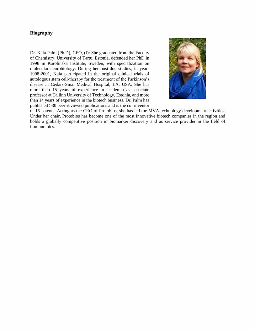
Biography
Dr. Kaia Palm (Ph.D), CEO, (f): She graduated from the Faculty
of Chemistry, University of Tartu, Estonia, defended her PhD in
1998 in Karolinska Institute, Sweden, with specialization on
molecular neurobiology. During her post-doc studies, in years
1998-2001, Kaia participated in the original clinical trials of
autologous stem cell-therapy for the treatment of the Parkinson’s
disease at Cedars-Sinai Medical Hospital, LA, USA. She has
more than 15 years of experience in academia as associate
professor at Tallinn University of Technology, Estonia, and more
than 14 years of experience in the biotech business. Dr. Palm has
published >30 peer-reviewed publications and is the co- inventor
of 15 patents. Acting as the CEO of Protobios, she has led the MVA technology development activities.
Under her chair, Protobios has become one of the most innovative biotech companies in the region and
holds a globally competitive position in biomarker discovery and as service provider in the field of
immunomics.

High content imaging of cells and 3D in vitro tissues
Katja Schenke-Layland1,2,3
Director of 1The Natural and Medical Sciences Institute (NMI) at the University of Tübingen,
Reutlingen, Germany; Professor in the 2Department of Women’s Health, Research Institute for
Women’s Health, Eberhard Karls University Tübingen, Tübingen, Germany and the 3Department
of Medicine/Cardiology, Cardiovascular Research Laboratories (CVRL), University of
California (UCLA), Los Angeles, CA, USA
As the field of regenerative and personalized medicine matures, the need for novel enabling
technologies to characterize cells and engineered constructs (i.e. cells/tissue combined with
biomaterials and three-dimensional (3D) scaffolds) as well as their individual components in a
more insightful, quantitative and preferably non-invasive manner becomes imperative. Raman
microspectroscopy and imaging as well as fluorescence lifetime imaging (FLIM) are emerging
techniques that allow the assessment of molecular interactions and the biochemical structure of a
sample in a non-invasive, marker-independent manner. Specifically for tissue engineering
applications, it has been proven to allow determining biochemical information on cells, tissues
and/or material-cell tissue constructs without the need for labels. The presentation aims to show
the applicability of Raman technologies and FLIM for regenerative and personalized medicine
applications, and to discuss the added value of the generated data for tissue engineering construct
design optimization and preclinical as well as clinical applications.
Biography
Professor Schenke-Layland’s main research interests
revolve around the role of the extracellular matrix in tissue
engineering and regenerative medicine, the translation of
human developmental biological data into therapeutic
strategies, and the non-invasive monitoring of biological
processes. Prof. Katja Schenke-Layland currently holds a
dual appointment as a full professor (W3) at the University
Women’s Hospital and as Director of the Natural and
Medical Sciences Institute (NMI) Reutlingen in Germany.
She is also affiliated with the Department of
Medicine/Cardiology at the University of California in Los Angeles, USA. She is an executive
editor for Advanced Drug Delivery Reviews, which is one of the top journals in the field of
advanced gene and drug delivery, and co-editor-in-chief of Tissue Engineering, Part B. To date,
she has secured more than in 11.8 Mio € individual research funding as PI, has published 129
peer-reviewed manuscripts with more than 4600 citationsScopus and has an h-Index of 39Scopus.
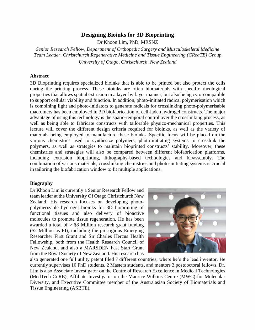
Designing Bioinks for 3D Bioprinting
Dr Khoon Lim, PhD, MRSNZ
Senior Research Fellow, Department of Orthopedic Surgery and Musculoskeletal Medicine
Team Leader, Christchurch Regenerative Medicine and Tissue Engineering (CReaTE) Group
University of Otago, Christchurch, New Zealand
Abstract
3D Bioprinting requires specialized bioinks that is able to be printed but also protect the cells
during the printing process. These bioinks are often biomaterials with specific rheological
properties that allows spatial extrusion in a layer-by-layer manner, but also being cyto-compatible
to support cellular viability and function. In addition, photo-initiated radical polymerisation which
is combining light and photo-initiators to generate radicals for crosslinking photo-polymerisable
macromers has been employed in 3D biofabrication of cell-laden hydrogel constructs. The major
advantage of using this technology is the spatio-temporal control over the crosslinking process, as
well as being able to fabricate constructs with tailorable physico-mechanical properties. This
lecture will cover the different design criteria required for bioinks, as well as the variety of
materials being employed to manufacture these bioinks. Specific focus will be placed on the
various chemistries used to synthesize polymers, photo-initiating systems to crosslink the
polymers, as well as strategies to maintain bioprinted constructs’ stability. Moreover, these
chemistries and strategies will also be compared between different biofabrication platforms,
including extrusion bioprinting, lithography-based technologies and bioassembly. The
combination of various materials, crosslinking chemistries and photo-initiating systems is crucial
in tailoring the biofabrication window to fit multiple applications.
Biography
Dr Khoon Lim is currently a Senior Research Fellow and
team leader at the University Of Otago Christchurch New
Zealand. His research focuses on developing photo-
polymerizable hydrogel bioinks for 3D bioprinting of
functional tissues and also delivery of bioactive
molecules to promote tissue regeneration. He has been
awarded a total of > $3 Million research grant funding
($2 Million as PI), including the prestigious Emerging
Researcher First Grant and Sir Charles Hercus Health
Fellowship, both from the Health Research Council of
New Zealand, and also a MARSDEN Fast Start Grant
from the Royal Society of New Zealand. His research has
also generated one full utility patent filed 7 different countries, where he’s the lead inventor. He
currently supervises 10 PhD students, 2 Masters students, and mentors 3 postdoctoral fellows. Dr.
Lim is also Associate Investigator on the Centre of Research Excellence in Medical Technologies
(MedTech CoRE), Affiliate Investigator on the Maurice Wilkins Centre (MWC) for Molecular
Diversity, and Executive Committee member of the Australasian Society of Biomaterials and
Tissue Engineering (ASBTE).

Creating regenerative environments with materials
Liam M Grover
Director, Healthcare Technologies Institute; Full Professor of Biomaterials Science
University of Birmingham. Edgbaston, UK, @DWLiam
Abstract
The long-term outcome following wounding depends strongly on the environment that is created
within the damaged tissue. Acute inflammation results in the upregulation of cytokines such as
TGF-1, which cause the differentiation of cells within the wound into myofibroblasts, cells which
contract the wound bed and deposit collagen more rapidly than the fibroblasts that are normally
present in a tissue. Although this rapid production of collagen and contraction of the wound results
in closure, it also results in scarring, which can cause psychosocial impacts, as well as limiting
movement and if on the surface of the eye, blindness. We have developed a range of technologies
that aim to modulate the wound environment and thereby limit or even eliminate scarring on the
surface of the skin and in the eye. This talk will describe the development of these technologies
that have been designed to limit the scarring process, by sequestering pro-fibrotic growth factors
or reducing their activity in the site of a wound. It will describe how we have taken these
technologies through the translational pathway, from preclinical testing through to manufacture
using GMP processes. It will also highlight some of the hurdles that we have had to overcome in
taking our novel technologies through to the clinic.
Biography
Liam M Grover is a Professor of Biomaterials Science at the
University of Birmingham. His research focusses on
investigating the interactions between materials and
biological systems, a greater understanding of which has
enabled him to develop a host of novel materials that have
been used for skeletal reconstruction and the treatment of
scarring. He has published more than 180 peer reviewed
publications and filed more than 10 patents. He has raised
in excess of £30m of research funding, and is the founder-
director of the Healthcare Technologies Institute, which is
based in the Institute of Translational Medicine at the
University of Birmingham. The institute seeks to take novel
interventions to the point of clinical trial, supporting the full translational process. He has taken
three new medical interventions to the point of clinical trial. He was the youngest full Professor
ever appointed to the University of Birmingham at the age of 32.

Advanced Biofabrication Methods for Cellular Grafts
Marcy Zenobi-Wong
Director, Institute for Biomechanics; Associate Professor of Tissue Engineering +
Biofabrication;ETH Zürich, Zürich, Switzerland
Abstract
Of the many evolving biofabrication approaches, microextrusion bioprinting has been the most
commonly applied in tissue engineering. In order to recreate complex geometries and large scale,
the typical bioink used in extrusion bioprinting has required the use of viscosity enhancers and/or
support materials to be effective. We have recently explored the class of bioinks called ‘jammed’,
granular or microgel materials for extrusion bioprinting. In our case, we work with microgels with
high aspect ratio and ability to entangle with each other. These entangled bioprinting materials are
less subject to flow instabilities, have natural macroporosity and orientation and can be used in a
modular, multimaterial and multicellular approach. In this lecture, entangled microgel bioinks are
explored in the context of cartilage engineering.
Biography
Dr Marcy Zenobi-Wong is an Associate Professor of Tissue
Engineering and Biofabrication at ETH Zürich,
Switzerland. She obtained her PhD from Stanford
University and then did a post-doctoral fellowship in the
Orthopaedic Research Laboratories, University of
Michigan. In 2012, she moved to the Department of Health
Sciences & Technology at ETH Zürich. The Zenobi-Wong
research group is focused on the development of advanced
biomaterials for tissue regeneration using biofabrication
technologies including bioprinting, two-photon
polymerization, casting and electrospinning. Dr Zenobi-
Wong is the author of over 95 peer-reviewed publications and co-inventor on four licensed patents.
She is currently President of the Swiss Society for Biomaterials and Regenerative Medicine and
General Secretary for the International Society of Biofabrication (ISBF). She serves on the
editorial board for Biofabrication and Advanced Healthcare Materials.
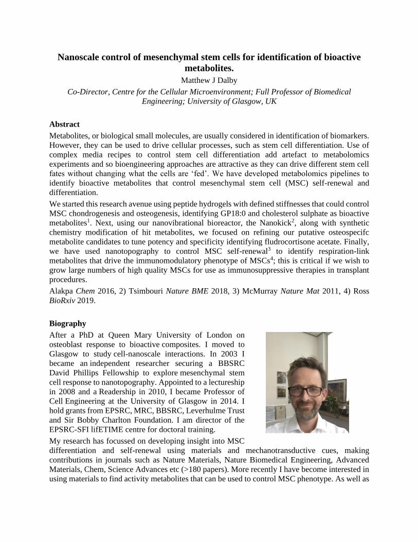
Nanoscale control of mesenchymal stem cells for identification of bioactive
metabolites.
Matthew J Dalby
Co-Director, Centre for the Cellular Microenvironment; Full Professor of Biomedical
Engineering; University of Glasgow, UK
Abstract
Metabolites, or biological small molecules, are usually considered in identification of biomarkers.
However, they can be used to drive cellular processes, such as stem cell differentiation. Use of
complex media recipes to control stem cell differentiation add artefact to metabolomics
experiments and so bioengineering approaches are attractive as they can drive different stem cell
fates without changing what the cells are ‘fed’. We have developed metabolomics pipelines to
identify bioactive metabolites that control mesenchymal stem cell (MSC) self-renewal and
differentiation.
We started this research avenue using peptide hydrogels with defined stiffnesses that could control
MSC chondrogenesis and osteogenesis, identifying GP18:0 and cholesterol sulphate as bioactive
metabolites1. Next, using our nanovibrational bioreactor, the Nanokick2, along with synthetic
chemistry modification of hit metabolites, we focused on refining our putative osteospecifc
metabolite candidates to tune potency and specificity identifying fludrocortisone acetate. Finally,
we have used nanotopography to control MSC self-renewal3 to identify respiration-link
metabolites that drive the immunomodulatory phenotype of MSCs4; this is critical if we wish to
grow large numbers of high quality MSCs for use as immunosuppressive therapies in transplant
procedures.
Alakpa Chem 2016, 2) Tsimbouri Nature BME 2018, 3) McMurray Nature Mat 2011, 4) Ross
BioRxiv 2019.
Biography
After a PhD at Queen Mary University of London on
osteoblast response to bioactive composites. I moved to
Glasgow to study cell-nanoscale interactions. In 2003 I
became an independent researcher securing a BBSRC
David Phillips Fellowship to explore mesenchymal stem
cell response to nanotopography. Appointed to a lectureship
in 2008 and a Readership in 2010, I became Professor of
Cell Engineering at the University of Glasgow in 2014. I
hold grants from EPSRC, MRC, BBSRC, Leverhulme Trust
and Sir Bobby Charlton Foundation. I am director of the
EPSRC-SFI lifETIME centre for doctoral training.
My research has focussed on developing insight into MSC
differentiation and self-renewal using materials and mechanotransductive cues, making
contributions in journals such as Nature Materials, Nature Biomedical Engineering, Advanced
Materials, Chem, Science Advances etc (>180 papers). More recently I have become interested in
using materials to find activity metabolites that can be used to control MSC phenotype. As well as

basic science, I am interested in translational science and have been involved in veterinary bone
regeneration trials and am working now towards a human bone regeneration trail.
In 2016 I was elected a Fellow of the Royal Society of Edinburgh and have won a number of
awards – most recently the Biochemical Society Industrial-Academic Collaboration Award.

BIOMATERIAL RISK ASSESSMENT AND PERSONALISED
BIOMATERIAL TESTING: FROM NANOSCALE TO MACROSCALE
Nihal Engin Vrana
CEO, SPARTHA Medical, 67100, Strasbourg France; Affiliated Researcher, INSERM UMR
1121 University of Strasbourg, 67000, Strasbourg, France
Abstract
The increasing number of implantable biomedical devices and novel biomaterials creates a double pressure
on the current framework on the biomaterial related risk management. The significant variation of the shape,
size and mechanical properties of the organs between the patients necessitates the development of
personalized solutions which can diminish the level of reaction by the host. However, beyond the
anatomical match, the patient’s immunological profile and the specific reactions to a given material must
also be considered for better clinical outcomes. This is particularly relevant for new biomaterials where
unforeseeable side effects due to the unintentional similarities with antigens or unknown biological effects
are possible (such as synthetic polypeptides). The determination of patient specific components of the
adverse immune reactions enables the development of safer personalized solutions. H2020 PANBioRA
project aims to provide the necessary tools for such assessments for decreasing the complications related to
the available implantable devices and facilitating the uptake of new biomaterials for clinical use. The
multiscale approach in the project assesses biomaterials at antibody level, cell level, miniaturized organ and
finally at macro level. This introductory talk will focus on the proposed risk management tools by
PANBioRA system and the introduction of the current version and its components.
ACKNOWLEDGEMENTS: This project has received funding from Horizon 2020 research and
innovation programme under grant agreement No 760921 (PANBioRA).
Biography
Dr. Nihal Engin VRANA is CEO of SPARTHA Medical and an
affiliated researcher in INSERM UMR 1121. He obtained his
PhD in 2009 at Dublin City University as a Marie Curie ESR
fellow. His major research interests are implants, antimicrobial
coatings, tissue engineering, cell encapsulation,
immunomodulation, real-time monitoring of implants,
biomaterial assessment and cell biomaterials interactions. He has
been the scientific coordinator of 2 European Projects
(EuroTransBio Bimot and FP7 IMMODGEL) and he currently
coordinates H2020 PANBioRA project (11 countries, 17 partners,
www.panbiora.eu). He has published 73 articles in peer-reviewed
academic journals (over 2200 citations, h index: 26), 8 book chapters and holds 5 European patents. He
edited two books for Taylor and Francis on Cell Material Interactions (2015) and Immune response to
Biomaterials (2018). His awards include ESB Translational Research award (2011), BPI i-Lab (2019) and
Fond’Action Alsace Future Talents Award (2019) for SPARTHA Medical.

Effect of bioactive glass cotton wool like fibre conditioned medium on bone
marrow derived stem cells
Olga Tsigkou
Lecturer in Biomaterials; Biomaterials Subject Lead; Department of Materials, University of
Manchester, Oxford Road, Manchester, UK
Abstract
Biomaterials science focuses on engineering smart materials that can direct cellular behaviour. In
vivo bone tissue resident adult stem cells are encompassed by a 3D microenvironment that presents
a repertoire of signals, such as matrix rigidity, soluble factors, cell-cell interactions and
extracellular matrix ligands. Ultimately, bone stem cells or bone progenitors respond to these
components by initiating signaling pathways, which control and direct cell function and
differentiation into osteogenic lineage. Bioactive glasses have shown the ability to form
hydroxyapatite through exchange of silicon and calcium ions with interstitial fluids. Silica and
calcium have been reported to be highly beneficial for bone and cartilage health and their dietary
intake has been implicated with increased bone mineral density. In this talk I will report the
development of a bioactive glass electrospun cotton wool like fibres and the effect of their
conditioned medium on bone marrow derived mesenchymal stem cells. I will demonstrate that the
bioactive glass conditioned medium enhance osteogenic differentiation and ECM deposition and
mineralization in vitro 3 times faster than conventional osteogenic supplements. Additionally,
activation of specific intracellular pathways activated and the effect of the 70S30C bioactive glass
conditioned media on bone mass and bone remodeling will be discussed.
Biography
Dr Olga Tsigkou is a Lecturer in Biomaterials (Assistant
Professor) and Programme Director for the MSc in
Biomaterials at the Department of Materials, School of
Natural Sciences, Faculty of Science and Engineering at the
University of Manchester, UK. Her research focuses
predominantly in the application of stem cells, 3D porous
scaffolds and hydrogels on tissue regeneration applications.
She has developed robust protocols for 3D cell culture
models in static and shear stress (bioreactor) culture
conditions. Her research interests include cell-substrate
interactions, vascularization strategies, in vitro tissue
modeling and substrate directed differentiation of stem
cells. She has published highly cited papers on these topics
in leading international journals (e.g. PNAS, Biomaterials,
Advanced Functional Materials and Acta Biomaterialia). Her published work includes more than
30 original research papers in international peer-review journals with over 1200 citations focusing
in the fields of bone tissue engineering, bioactive glasses, vascularization, as well as imaging and
material characterization. Dr Tsigkou has successfully supervised 18 Master students and is
currently involved in the supervision of 6 PhD students and an EPSRC fellow.
‘Mix and match’: local delivery of protein-based biologics
using responsive microgels
Paige LeValley
Centennial Development Professor of Chemical and Biomolecular Engineering, Chemical and
Biomolecular Engineering and Materials Science and Engineering, University of Delaware,
Newark, DE, 19716, USA
Abstract
Protein-based biologics, particularly antibodies, are of growing interest owing to their specificity
and therapeutic efficacy, especially for many conditions that traditionally have been difficult to
treat (e.g., metastatic cancer, misregulated wound healing). The use of multiple therapeutics,
combination therapies, that target different aspects of disease mechanisms can be particularly
effective; however, such therapies have a significant risk of systemic toxicity owing to the high
total doses that must be used. Responsive hydrogels offer a facile platform for the local, controlled
release of these large, hydrophilic proteins for the design of personalized combination therapies
while minimizing adverse side effects. Specifically, in this talk, I report the development of mixed
populations of hydrogel microparticles, or microgels, for achieving tunable and tailorable release
profiles of antibodies in vitro and in vivo. Microgels of uniform size and relevance for local
injection were created using microfluidic devices. To achieve tunable and on-demand release
profiles, microgels that respond to either internal (i.e., reducing microenvironments) or external
(i.e., light) cues were designed. Modular building blocks, multifunctional polymers with a variety
of chemical handles, were used to create mixed populations of microgels that localize to desired
tissues and release multiple therapeutics across a range of time scales.

Nitric Oxide vs Bacteria: NO means NO!
Raechelle A D’Sa
Head of Antimicrobial Biomaterials Group, School of Engineering, University of Liverpool
Brownlow Hill, Liverpool, L69 3GH
Abstract
Nitric oxide (NO) plays a significant role in many biological processes, including the body’s
own immune response to fighting off infections. Given the rise in multi drug resistant bacteria,
conventional antibiotics are failing, which demands the development of new antimicrobial agents
that are not antibiotics. NO holds substantial promise in this regard as it has shown potent
antimicrobial activity against both planktonic and biofilm bacteria and has a low tendency to
develop microbial resistance. NO is a short-lived, lipophilic gas that can easily diffuse across a
cell membrane and combines with reactive oxygen species to cause nitrosative damage on
invading pathogens. The activity of NO is dose dependent with low concentrations dispersing
biofilms and high concentrations having more of a biocidal effect. Despite the potency of NO as
antimicrobial agent, delivery of the gas in a controlled and sustained manner for the desired
clinical application is where the challenge lies. This talk will describe a variety of delivery
platforms that have been developed to provide controlled and sustained delivery for an elongated
lifetime of NO release. The platforms include the hydrogels, nanoparticles, electrospun fibers
and thin film coatings for treatment of dermal, ocular surface or orthopaedic infections.
Biography
Dr. D’Sa’s leads the Antimicrobial Biomaterials Group at
the University of Liverpool. Her research focuses on the
development of nitric oxide delivery platforms for treatment
of multidrug resistant infections. Her research focuses on
the development of these antimicrobial platforms to be used
for skin, ocular and bone tissue engineering applications.
She has over £2 million in research funding with over £1.5
million as a PI. She currently sits on the Editorial Board and
Topic Board for the Journal Polymers and Antibiotics. She
has been the recipient of the Arthur D Chambers and Buhle
Endowment Fellowships. Dr D’Sa is passionate about
supporting the next generation of scientists and has supervised students from various backgrounds
to deliver new solutions for infection control and tissue engineering applications. She is currently
involved with the supervision of 7 PhD students, and 2 postdoctoral fellows. She engages widely
with clinicians, health professionals, patients, charities, has a significant interest in communication
of science to the general public through artistic media.

Engineered microenvironments for stem cell engineering
Manuel Salmeron-Sanchez
Co-Director, Centre for the Cellular Microenvironment; Full Professor of Biomedical
Engineering; University of Glasgow, UK
Abstract
Physical properties of the extracellular matrix (ECM) and the use of growth factors are powerful
tools to control cell behaviour, including stem cell differentiation. Integrins are
mechanotransductors that feel and respond to the viscoelastic properties of the ECM. We have
shown that cells respond to pure viscous interfaces and that results are explained by the classical
clutch model for cell adhesion. At the same time, growth factors are important molecules that
trigger signalling cascades that control e.g osteogenesis and vascularisation. We have developed
material systems that allow simultaneous stimulation of integrins and growth factors receptors. We
have engineered polymers that unfold and assemble fibronectin to allow exposure of the integrin
(FNIII9-10) and growth factor (FNIII12-14) binding regions, leading to highly efficiency
presentation of growth factors, to maximise effects with minimal doses. In parallel, we have also
engineered dynamic systems that allow control over temporal release of adhesion molecules and
growth factors. Unconventionally, we have used non-pathogenic bacteria that have been
engineered to control release of a fribronectin fragment and BMP-2 in a dynamic way. We present
bacteria-based materials in which symbiotic bacteria/mammalian cell interactions occur and their
use for stem cell engineering.
Biography
Prof Manuel Salmeron-Sanchez is co-Director of the Centre
for the Cellular Microenvironment at the University of
Glasgow. He did a PhD in Valencia and postdoctoral work
at the Institute for Macromolecular Chemistry in Prague and
the Katholieke Universiteit Leuven. He was Assistant
Professor at Universitat Politècnica de València, promoted
to Associate Professor in 2008 and Full Professor in 2010.
He did a sabbatical year at the Georgia Institute of
Technology and moved to the University of Glasgow in
2013 as the Chair of Biomedical Engineering. Manuel is a
European Research Council (ERC) investigator. His work
has attracted significant funding from the ERC (PoC, 2
awards) and Research Councils in the UK (MRC) and has set up the basis for a programme of
research to help civilians affected by landmines, funded by the Sir Bobby Charlton Foundation.
Overall, his work spans fundamental mechanisms at the cell/material interface as well as
translational research that has saved from amputation the leg of a first veterinary patient, a dog
called Eva (https://goo.gl/1Z3r8t). He has authored more than 160 papers in major journals
including PNAS, Science Advances, Nature Biomedical Engineering and Advanced Materials. He
has had his research featured in newspapers and TV channels around the world.

Flexible and injectable fibrous scaffolds for the regeneration of pelvic
fractures
Sandra Camarero-Espinosa
MERLN Institute for for Technology-Inspired Regenerative Medicine, Maastricht University,
Maastricht, The Netherlands.
Abstract
Pelvic fractures in the elderly population account for 7% of the total osteoporosis-associated
fractures. Current treatments rely on extensive and invasive surgery and are therefore, not
recommended to elderly patients. Common advice is total rest until natural bone healing occurs,
with the associated development of “dysmobility syndromes” upon long periods of bed
confinement. Bone tissue regeneration with the aid of injectable osteoinductive materials would
avoid invasive surgeries, providing a solution to this problem.
Collagen, the main component of the non-mineralized fraction of bone, and nano-hydroxyapatite
(nHA) have been shown to have beneficial effects on the regeneration of bone, promoting an
enhance mineralized matrix deposition. Similarly, bioactive mesoporous glasses (BMGs) account
for a very high specific surface are (200-500 m2/g) that enhances the deposition of mineralized
matrix. To promote a pro-osteogenic and anti-osteoclastogenic behavior, these materials can be
doped with strontium and selenium. Here, we report on the fabrication of electrospun UV-
crosslinked collagen scaffolds with fiber diameters between 140-180 nm, reinforced with Sr- and
Se-doped nHA and BMGs that are exploited for the regeneration of pelvic bone fractures.
Biography
Sandra Camarero-Espinosa`s research revolves around the
regeneration of complex tissues. The design, synthesis,
fabrication and investigation of novel hierarchical
polymeric bio(nano)materials whose physicochemical
properties can be tuned mimicking nature from the
molecular to the macro scale and, the effect of these ones on
cell phenotype and matrix deposition.
Sandra Camarero-Espinosa was educated at the University
of the Basque Country (Spain) where she obtained her BSc.
degree as Chemical Engineer and M.Sc. in Engineering of
Advanced Materials. Sha developed her doctoral studies at
the Adolphe Merkle Institute (Fribourg, Switzerland) and
was recognized with an award to an outstanding PhD thesis by the Swiss Chemical Society. After
gaining a fellowship from the Swiss National Science Foundation, she moved to Brisbane
(Australia) to work at the Australian Institute for Bioengineering and Nanotechnology. Sandra is
now a post-doctoral researcher at the MERLN institute where she works on the development of
instructive hierarchical biomaterial scaffolds for the regeneration of complex tissues.

The platform of materials and functionalization routes for the
biofabrication of GIOTTO devices
Sonia Fiorilli
Politecnico di Torino, Department of Applied Science and Technologies, Corso Duca degli
Abruzzi 24, Turin, Italy
Abstract
The incidence of osteoporotic fractures is expected to double rapidly due to progressive population
ageing. In this context, the GIOTTO project aims to develop three different devices to treat specific
osteoporosis fractures through the synergistic combination of smart nanomaterials and 3D
fabrication technologies. The three devices will share the use of novel bioactive inorganic phases,
nano-hydroxyapatites and mesoporous bioactive glasses, substituted with biologically active ions
able to stimulate bone production (e.g. Sr2+). The developed inorganic phases will be dispersed in
a resorbable matrix to produce composites with the desired resorption kinetics and matching the
fracture specificities at different body sites. In particular, the bioactive materials will be combined
with the following different matrices:
- an optimised blend of biodegradable polyesters (e.g. PLLA, PCL) to fabricate 3D scaffolds
by extrusion-based printing
- collagen matrix to produce a flexible, fibrous injectable scaffold through electrospinning
- calcium sulphate hemihydrate to produce an injectable resorbable radiopaque cement
GIOTTO materials will be also ad-hoc functionalised through different strategies, with the dual
aim to impart specific properties (e.g. mechanical resistance, degradation kinetics) to the final
devices and to couple them with a novel recombinant biomolecule able to inactivate the osteoclast
activity (ICOS-Fc).
Biography
Sonia Lucia Fiorilli graduated in Industrial Chemistry and took her
PhD in Materials Science and Technology at Politecnico di Torino in
2005. Currently she is Associate Professor at Politecnico di Torino,
where she is lecturer of “Chemistry”, co-lecture of “Physical chemistry
of materials for nanotechnologies”. Her research activity mainly
focuses on the synthesis, characterization and functionalization of
bioceramics, as coatings and 3D scaffolds, for bone and soft tissue
regeneration. More recently, her research interests also include the
design of 3D printed biomimetic scaffolds based on the combination
of collagen and inorganic bioactive phases, properly optimised through
different cross-linking methods.
Prof. Fiorilli is involved in several funded projects as principal investigator or WP/task leader,
including EU-H2020 projects (e.g. EU-H2020- NMBP-22-2018- GIOTTO, EU-H2020-NMP6-
2015 MOZART, MSCA-ITN Action POLYSTORAGE) and national projects (e.g. PI of
ZODIAC “Zwitterionic mesOstructureD glAsses: powerful deviCes for bone regeneration”).
She is currently involved in the supervision/co-supervision of 6 PhD students.

Nanofilled electrospun fibers inspired by elastin and natural polymers
Nicola Ciarfaglia, Antonietta Pepe, Antonio Laezza, Brigida Bochicchio*
Associate Professor of Organic Chemistry; Department of Science; University of Basilicata, Via
Ateneo Lucano 10, Potenza, Italy
Abstract
Electrospinning is an emerging technique with applications in tissue engineering for the high
surface area to volume ratio and the interconnected pores of nanofibers. Blending of synthetic
polymers as polylactic acid (PLA) and poly--caprolactone (PCL), together with natural proteins
(Gelatin, Elastin) were conceived until now in order to obtain scaffolds with combination of
strength and biocompatibility usually used in soft-tissue regeneration. Furthermore, the use of
nanoreinforcements as bioglass and/or nanocellulose is aimed to overcome the limited mechanical
performances of the scaffolds. In this work, we have successfully electrospun highly hydrophobic
(PDLLA) and hydrophilic polymers (gelatin and a human tropoelastin-inspired sequence) together
with bioglass microparticles and crystalline nanocellulose as nanoreinforcements in the
perspective of future applications in hard-tissue regeneration (bone). Additionally, PDLLA was
herein adoperated for its faster degradation times in comparison to the most used PLA. The
matrices were cross-linked in order to confer stability in water. Afterward, the matrices were
tested concerning wettability, swelling and Young's elastic modulus. They show complete
wettability and a significative decrease of Young's Modulus after swelling (comprised between 27
and 23 MPa). The value is analogous to that found for electrospun scaffolds composed of collagen,
elastin and PCL. Funding: PON R&I 2014-2020 (572 PON_ARS01_01081).
Biography
Professor Bochicchio’s research focusses on biopolymers
and peptides inspired by elastomeric proteins for tissue
engineering. Professor Bochicchio has authored 60
international peer-reviewed journal publications and Invited
Speaker at 24 International Meetings. Professor
Bochicchio’s research has been supported by Italian
Ministry of University and European Community and also
attracts interest from multinational industrial partners. To
date, she has secured ≈€1.8M (Co-I) research funding from
National Operational Program "Research and Innovation"
2014-2020 in health sector. Professor Bochicchio has
successfully supervised to completion 2 PhD student and 20
MSc students all of whom have remained in engineering/science and or academia. She is currently
involved in the supervision of 1 PhD student. Additionally, she is currently mentoring one PhD
research fellow.

The Effect of Elastin Degradation Products and Elastin Fibers on COPD and Control Lung Mesenchymal Stromal cells
Willeke F. Daamen* Dept. of Biochemistry, Radboud Institute for Molecular Life Sciences,
Radboud university medical center, Nijmegen, the Netherlands Abstract Chronic obstructive pulmonary disease (COPD) is characterized by chronic inflammation and an irreversible loss of alveolar architecture and extracellular matrix. Novel strategies aimed at the regeneration of the lost alveolar tissue are needed and may include the use of lung mesenchymal stromal cells. These cells produce anti-inflammatory factors, growth factors and extracellular matrix components, including elastin, thereby providing a potential niche for alveolar repair. The reparative capacity of mesenchymal stromal cells from the lungs of COPD patients, however, may be hampered due to oxidative stress and extracellular matrix loss, e.g. by the presence of degradation products of elastic fibers. We investigated whether degradation products of the extracellular matrix, such as hydrolyzed elastin, affect the regenerative capacity of lung mesenchymal stromal cells and whether a supporting micro-environment consisting of intact collagen fibrils and elastin fibers improves the function of lung mesenchymal stromal cells. Biography Willeke Daamen PhD is a scientific researcher at Radboud university medical center. She aims to promote the intrinsic regenerative capacity of patients, mostly by using cell-free biodegradable biomaterials that stimulate the endogenous healing response of tissues. Her group has designed biomaterials that indeed influence infiltrating cells. One example is that the incorporation of solubilized elastin enhances angiogenesis and elastic fiber formation in vivo. Her straightforward biomaterial designs in combination with close collaborations with clinicians, researchers and entrepreneurs will facilitate the translation to the clinic, so that patients will indeed benefit from her research achievements. Willeke Daamen is secretary of the Netherlands Society for Biomaterials and Tissue Engineering (NBTE) and organized and chaired the 10th European Elastin Meeting. She supervised 15 past and present PhD students and has published >95 peer-reviewed papers.
* Full author list: Danique J Hof1, Dennis MLW Kruk2,3, Rob TC Meuwese1, Elly MM Versteeg1, Nick HT ten Hacken3,4, Irene H Heijink2,3,4, Toin H van Kuppevelt1 & Willeke F Daamen1. 1Radboud university medical center, Radboud Institute for Molecular Life Sciences, Dept. of Biochemistry, Nijmegen, The Netherlands 2University of Groningen, University Medical Center Groningen, Dept. of Pathology and Medical Biology, Groningen, The Netherlands 3University of Groningen, University Medical Center Groningen, Groningen Research Institute for Asthma and COPD, Groningen, The Netherlands 4University of Groningen, University Medical Center Groningen, Dept. of Pulmonary Diseases, Groningen, The Netherlands

Polyglycolic acid---an old polymer for new scaffolds and implants
Ying Yang
Professor in Biomaterials and Tissue Engineering; Course director of MSc Biomedical Engineering; School of
Pharmacy and Bioengineering; Keele University, Stoke-on-Trent ST4 7QB; UK
Abstract
Polyglycolide (PGA) is one of the earliest biodegradable polymers explored for biomedical application, date back
1970s for the first synthetic absorbable suture. PGA is highly crystalline polymer, endowing its high tensile modulus.
In addition, PGA’s rapid degradation via bulk hydrolysis makes it as appropriate materials for scaffolds in regenerative
medicine. However, the application of PGA as scaffolds in tissue engineering is hampered by its high crystallinity
(45-55%) property. PGA is insoluble in most organic solvents except the highly toxic solvent, hexafluoroisopropanol.
Currently the practically feasible fabrication technique for porous PGA scaffolds is making PGA nonwoven fibers via
melt-spinning, which has poor compressive properties and difficulty to control porosity, pore size and distribution.
Working with the research partners, we explored a novel fabrication technique, supercritical carbon dioxide (scCO2)
assisted melt-foaming, to generate porous PGA scaffolds. In this talk, the uniqueness of the scCO2 fabrication
processing and the cellular response to the scaffolds will be demonstrated. The comparison study of in vitro and in
vivo degradation and immunoresponse triggered by PGA degraded products will be presented. It is confirmed that
PGA could be explored as unique scaffolds and implants through the new fabrication technique.
Biography
Dr Ying Yang is a Professor in biomaterials and tissue engineering. Her
main research interests/activities are the design and fabrication of
biomaterials to provoke desired cellular response at materials and cell
interface including bioactive scaffolds for tissue engineering,
anticoagulant surface for implanted biosensor, and the bioorganic metal
surface for anti-fouling application. She has established systematic
study methods including smart nanofiber applications, detection of
variation of cell adhesion capacities and structures of collagen based
matrices, in order to develop new strategies in regenerative medicine
and clinical diagnostics. She has undertaken diverse clinical projects from colony growth of stem cells as a diagnosis
assay for osteogenic potency assessment, pathological calcification of heart valves, the relation of pelvic organ
prolapse and ageing, cartilage/blood vessel regeneration, eye models (glaucoma and retina) to pseudoislets generation.
She also heavily engages in development of non-destructive and on-line monitoring systems for tissue engineering
products and diagnostics. As the PI and co-PI, her research has been financially supported by BBSRC, EPSRC,
European frame work FP5-7 and various charity. She has published over 130 full peer-reviewed papers, 11 chapters
in books and filed 5 patents.