redescription and phylogenetic position of the enigmatic ... · pdf file458 redescription and...
TRANSCRIPT

Neotropical Ichthyology, 9(3): 457-469, 2011Copyright © 2011 Sociedade Brasileira de Ictiologia
457
Redescription and phylogenetic position of the enigmatic Neotropical
electric fish Iracema caiana Triques (Gymnotiformes: Rhamphichthyidae)
using x-ray computed tomography
Tiago P. Carvalho and James S. Albert
We redescribe Iracema caiana, a monotypic genus of the gymnotiform electric fish family Rhamphichthyidae. Iracema isknown only from the type series (holotype and three paratypes) collected from the rio Jauaperi, affluent to the rio Negro in theAmazon basin, and was never collected again. Previous morphological studies were limited to features of external morphology.To study the osteology of Iracema we examined two specimens of different sizes using high-resolution x-ray computedtomography, a non-invasive and non-destructive technique to visualize internal anatomical structures. We describe andillustrate the osteology of Iracema caiana, and present data on morphometrics and external morphology. Contrary to previoushypotheses we propose that Iracema is the sister group to Rhamphichthys based on four synapomorphies: intermuscularbones present in the adductor mandibulae, reticulated texture of opercles, fully ossified Baudelot’s ligaments, and elongatescales above the lateral line in the posterior portion of the body.
Iracema caiana, um genêro monotípico de peixes elétricos Gymnotiformes da família Rhamphichthyidae, é aqui redescrito.Iracema é conhecido apenas da série-tipo (holótipo e três parátipos) coletado no rio Jauaperi, afluente do rio Negro na baciaamazônica, nunca sendo coletado novamente. Estudos morfológicos prévios de Iracema foram limitados somente à morfologiaexterna. Neste trabalho, a osteologia do gênero foi examinada com base em dois espécimes de diferentes tamanhos usando-se tomografia computadorizada de alta resolução, uma técnica não invasiva para a visualização de estruturas anatômicasinternas. Caracteres externos e morfométricos são apresentados e a osteologia é ilustrada e descrita. Contrariamente a hipótesesprévias, Iracema é proposto como grupo irmão de Rhamphichthys com base em quatro sinapomorfias: presença de ossosintermusculares no adductor mandibulae, textura reticulada do opérculo, ligamento de Baudelot completamente ossificado eescamas alongadas na região posterior do corpo acima da linha lateral.
Key words: Amazon, Biodiversity, Guiana Shield, Osteology, Rare fish, Rhamphichthys.
University of Louisiana at Lafayette, Department of Biology, P.O. Box 42451, Lafayette, LA 70504, USA. [email protected](TPC), [email protected] (JSA).
Introduction
Iracema caiana Triques is a member of theRhamphichthyidae, a clade of Gymnotiformes with 15 validspecies distributed in three genera (Ferraris, 2003; Lundberg,2005; Carvalho et al., 2011). Rhamphichthyidae andHypopomidae constitute the Rhamphichthyoidea, a wellsupported clade within Gymnotiformes (Mago-Leccia, 1978;Alves-Gomes et al., 1995; Albert, 2001). Iracema Triques wasoriginally based on a unique combination of characters ofexternal morphology. According to Triques (1996a) Iracemacan be differentiated from other rhamphichthyids by thepresence of one series of irregular to roundish blotches onthe sides of the body; a broad and uniformly dark pigmented
stripe along the dorsal margin of the body; and an intermediatenumber of anal-fin rays between Rhamphichthys Muller andTroschel, and Gymnorhamphichthys Ellis. Triques (1996a)briefly discussed putative relationships of Iracema withinother rhamphichthyids, hypothesizing it as the sister groupto Gymnorhamphichthys based on the shared loss of scaleson the anterior portion of the body.
Iracema was first included in a formal phylogeneticanalysis by Albert & Campos-da-Paz (1998), who were able tocode only 46 characters-states in a data matrix of 170 charactersdue to absence of osteological information. Most osteologicaldata on Iracema were not available due to a lack of specimensavailable for clearing and staining; the genus remains knownonly from four specimens in the type series. To avoid
Redescription and phylogenetic position of Iracema caiana458
dissection and staining procedures with a limited type serieswe used an alternative method of visualizing skeletalmorphology, high-resolution x-ray computed tomography(HRXCT). HRXCT is a non-destructive technique that allowsreconstruction of a virtual skeleton in digital form. Similarapproaches to recover osteological data from rare or extinctspecies have been previously adopted by ichthyologists(Schaefer, 2003; Schaefer & Fernández, 2009; Fink &Humphries, 2010).
Material and Methods
Two specimens were scanned at the High-Resolution X-ray Computed Tomography Facility, The University of Texas,Austin. The head and cleithral region of a preserved paratypeof Iracema caiana (MZUSP 49205, 345 mm SL) was scannedusing the following parameters. FeinFocus microfocal X-raysource operating at 200 kV and 0.16 mA with no X-ray prefilterwas employed. An empty container wedge was used. Slicethickness corresponded to two lines in a CCD image intensifierimaging system and equaled 0.0641 mm, with a source-to objectdistance of 92 mm. For each slice, 1,400 views were taken withthree samples per view. The field of image reconstruction was28 mm, and an image reconstruction offset of 4600 was usedwith a reconstruction scale of 7000. The dataset consists of761 1024x1024-pixel HRXCT slices. A second specimen(MZUSP 49205, 235 mm SL) was scanned using the followingparameters. An Xradia microXCT scanner operating at 40 kVand 7 W with a 0.15 mm glass X-ray prefilter was employed.An air wedge was used. Source-object distance was 125.3mm and detector-object distance was 37.1 mm. A total of 521views were gathered over a rotation of ±104 degrees. Theacquisition time per view was 4 seconds. The dataset consistsof 909 1024x1024-pixel HRXCT slices, with cubic voxelsmeasuring 43.84 microns. The scan was taken along the longaxis of the specimen from the tip of the snout to about the 8-9 vertebrae. Visualizations were produced in the commercialsoftware package VGStudio Max®, at the Ichthyologydepartment of the Academy of Natural Sciences ofPhiladelphia. Although the renderings appear similar tophotographs, they represent the density differences of thebiological materials as reflected in their X-ray opacity. Figureswere based on still frames captured from digital animations ofHRXCT and were prepared using Adobe Photoshop®CS.Homologous bone depicted in figures 3A and 3B wererepresented by the same spectral colors (hues) in VG StudioMax. These colors differ only in terms of tint (shading density).The shadowing option in VG Studio, used to help visualizeobjects in 3D produced different effects in these images dueto shape differences. Some bones of Iracema caiana are verythin and overlap each other, and therefore it was impossiblein the reconstructions to separate them into different colorschemes (e.g. preopercle and interopercle). Differing fromother fish with published Computed Tomography (CT) scanreconstructions, Iracema has thin and weakly ossified bones(e.g. those from the branchial basket), which are not clearly
identified in the CT scans. In addition to the imagesreconstructed using the software we use the raw X-ray slicesdata to describe the morphology (e.g. contact betweenretroarticular and quadrate), however we did not includeillustrations, because they have little value compared to the3d images. The branchial basket was illustrated in themidsagittal plane instead of the usual coronal plane becausethere is a better resolution and less overlap of bones in theseviews.
Measurements were made to the nearest 0.1 mm with digitalcalipers. Standard length (SL), head length (HL) and snout length(SN) follow Nijssen et al. (1976: fig. 1). Standard length as usedhere is the same measurement as length to end of anal fin (LEA;Mago-Leccia, 1994). Caudal-appendage length (CL) and pectoral-fin length (PL) follow Schwassmann (1989: fig. 1). Anal-fin length(AL) is the distance from the anterior origin of the anal fin to theposterior end. Body depth (BD) is the greatest depth of thebody, which is usually at about half the length of the standardlength posterior to end of precaudal vertebrae. Caudal-filamentdepth (CD) is the depth of caudal appendage at the vertical ofthe last anal fin ray. Interorbital distance (IO) is the distancebetween the bony orbits, in the middle part of the orbit.Postorbital length (PO) is measured from the posterior limit ofthe eyeball to the posterior edge of bony opercle. Distance toposterior nares (PN) is the distance from the tip of snout to theanterior margin of the posterior nares (= measurement Prenasal2 of Triques, 1996b; fig. 1). Opercular opening (BO) is thedistance between the dorsal and the ventral corners of thebranchial aperture (measurement 11 of Triques, 1996b; fig. 2).Morphometric data are expressed as percentages of standardlength, except subunits of the head, which are expressed aspercentages of head length. Osteological terminology is thatof Fink & Fink (1981) and Fink & Fink (1996). Cleared and stainedspecimens (cs) were prepared according to the method of Taylor& Van Dyke (1985). Myological nomenclature followsWinterbottom (1974) and Datovo & Bockmann (2010).Electrocyte rows at the end of anal fin were observed under thetransmitted light source in the stereomicroscope. One scale, atabout the second third of body (above the lateral line) wasremoved from MZUSP 49205 (345 mm SL) and stained in alizarinred. Photographs of fish scales were taken using a digital cameraattached to an Olympus SZX 12 microscope. Drawings weremade using a camera lucida attached to an Olympus SZX 12microscope. The number of vertebrae to the end of anal finincludes the four of the Weberian apparatus and end with thelast vertebra with its hemal spine associated with the last anal-fin pterygiophore (Lundberg, 2005). Institutional abbreviationsare as listed at http://research.calacademy.org/research/ichthyology/catalog/collections.asp.
Results
Iracema Triques, 1996
Type species. Iracema caiana Triques, 1996a: 91 by originaldesignation and monotypy.
T. P. Carvalho & J. S. Albert 459
Diagnosis. Iracema is distinguished from otherrhamphichthyids by the following characteristics: 240-257 anal-fin rays (vs. 139-211 in Gymnorhamphichthys and 304-470 inRhamphichthys); 15 or 16 pectoral-fin rays (vs. 10-13 inGymnorhamphichthys and 17-22 in Rhamphichthys); a seriesof dark, rounded pigment blotches on the side of the bodyalong the lateral line (vs. no pigmentation inGymnorhamphichthys and dark oblique transverse bands orno pigmentation in Rhamphichthys); caudal filament ovoid incross section, its depth slightly greater than its width (vs. caudalfilament laterally compressed in cross section, its depth aboutthree times its width in Gymnorhamphichthys andRhamphichthys).
Iracema caiana Triques, 1996Figs. 1-2, Table 1
Iracema caiana Triques, 1996a: 91 [original description]. -Albert & Campos-da-Paz, 1998: 423, 425 [phylogeny anddiagnosis]. -Albert, 2001: 63, 68 [phylogeny and diagnosis].-Ferraris, 2003: 495 [listed]. -Albert & Crampton, 2005: 363[listed]. -Crampton & Albert, 2006: 650 [listed and habitatdescription]. -Crampton 2011: 175 [listed]. -Winemiller &Willis, 2011: 231 [listed].
Diagnosis. The same as for genus.
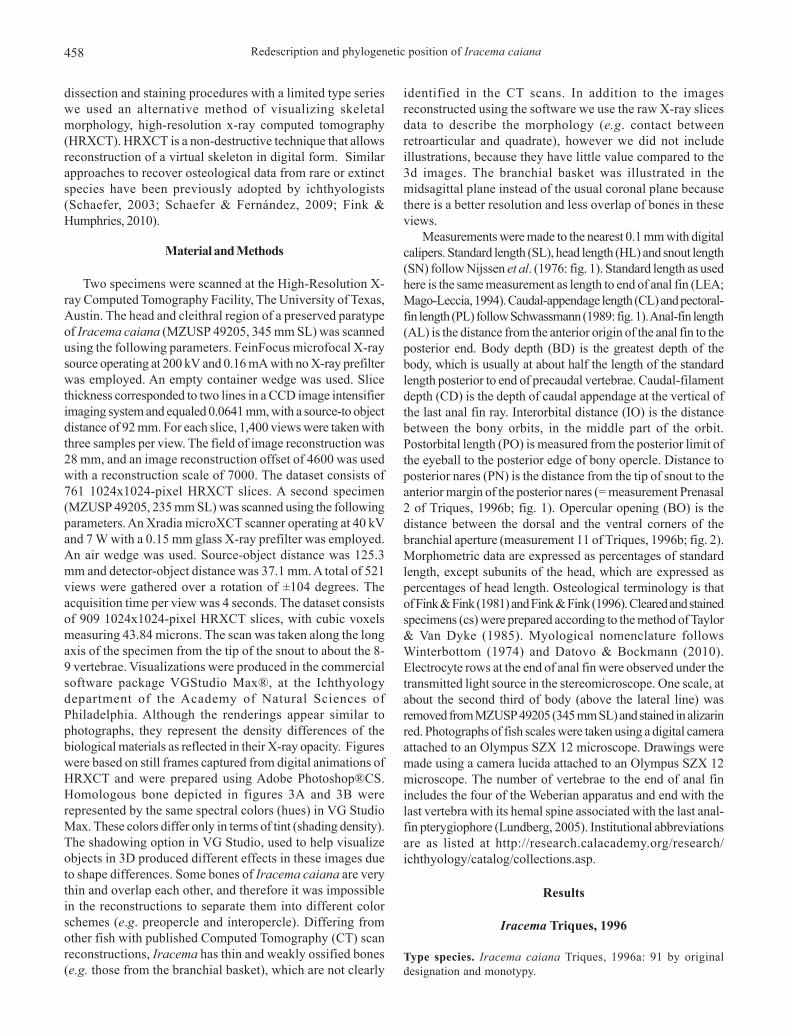
Description. Morphometrics and meristics in Table 1. Adultbody size moderate for Rhamphichthyidae (maximum size 360mm SL). Dorsal profile of snout strongly concave anterior toeye, head profile slightly convex post-orbitally. Interorbitalregion slightly convex. Dorsal margin of body almost straight,slightly ascending at end of body cavity. Ventral body margin
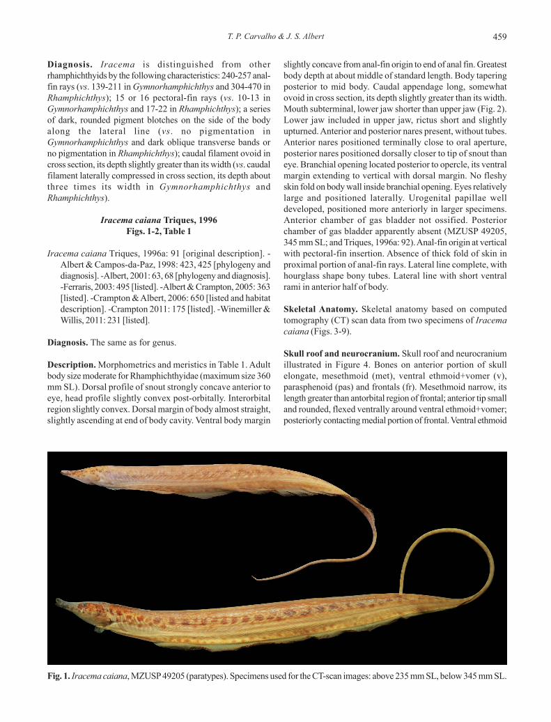
slightly concave from anal-fin origin to end of anal fin. Greatestbody depth at about middle of standard length. Body taperingposterior to mid body. Caudal appendage long, somewhatovoid in cross section, its depth slightly greater than its width.Mouth subterminal, lower jaw shorter than upper jaw (Fig. 2).Lower jaw included in upper jaw, rictus short and slightlyupturned. Anterior and posterior nares present, without tubes.Anterior nares positioned terminally close to oral aperture,posterior nares positioned dorsally closer to tip of snout thaneye. Branchial opening located posterior to opercle, its ventralmargin extending to vertical with dorsal margin. No fleshyskin fold on body wall inside branchial opening. Eyes relativelylarge and positioned laterally. Urogenital papillae welldeveloped, positioned more anteriorly in larger specimens.Anterior chamber of gas bladder not ossified. Posteriorchamber of gas bladder apparently absent (MZUSP 49205,345 mm SL; and Triques, 1996a: 92). Anal-fin origin at verticalwith pectoral-fin insertion. Absence of thick fold of skin inproximal portion of anal-fin rays. Lateral line complete, withhourglass shape bony tubes. Lateral line with short ventralrami in anterior half of body.
Skeletal Anatomy. Skeletal anatomy based on computedtomography (CT) scan data from two specimens of Iracemacaiana (Figs. 3-9).
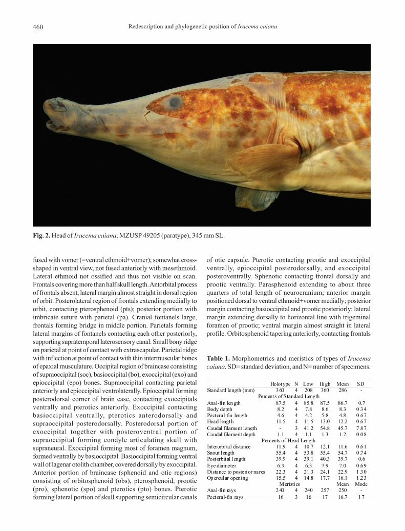
Skull roof and neurocranium. Skull roof and neurocraniumillustrated in Figure 4. Bones on anterior portion of skullelongate, mesethmoid (met), ventral ethmoid+vomer (v),parasphenoid (pas) and frontals (fr). Mesethmoid narrow, itslength greater than antorbital region of frontal; anterior tip smalland rounded, flexed ventrally around ventral ethmoid+vomer;posteriorly contacting medial portion of frontal. Ventral ethmoid
Fig. 1. Iracema caiana, MZUSP 49205 (paratypes). Specimens used for the CT-scan images: above 235 mm SL, below 345 mm SL.
Redescription and phylogenetic position of Iracema caiana460
fused with vomer (=ventral ethmoid+vomer); somewhat cross-shaped in ventral view, not fused anteriorly with mesethmoid.Lateral ethmoid not ossified and thus not visible on scan.Frontals covering more than half skull length. Antorbital processof frontals absent, lateral margin almost straight in dorsal regionof orbit. Posterolateral region of frontals extending medially toorbit, contacting pterosphenoid (pts); posterior portion withimbricate suture with parietal (pa). Cranial fontanels large,frontals forming bridge in middle portion. Parietals forminglateral margins of fontanels contacting each other posteriorly,supporting supratemporal laterosensory canal. Small bony ridgeon parietal at point of contact with extrascapular. Parietal ridgewith inflection at point of contact with thin intermuscular bonesof epaxial musculature. Occipital region of braincase consistingof supraoccipital (soc), basioccipital (bo), exoccipital (exo) andepioccipital (epo) bones. Supraoccipital contacting parietalanteriorly and epioccipital ventrolaterally. Epioccipital formingposterodorsal corner of brain case, contacting exoccipitalsventrally and pterotics anteriorly. Exoccipital contactingbasioccipital ventrally, pterotics anterodorsally andsupraoccipital posterodorsally. Posterodorsal portion ofexoccipital together with posteroventral portion ofsupraoccipital forming condyle articulating skull withsupraneural. Exoccipital forming most of foramen magnum,formed ventrally by basioccipital. Basioccipital forming ventralwall of lagenar otolith chamber, covered dorsally by exoccipital.Anterior portion of braincase (sphenoid and otic regions)consisting of orbitosphenoid (obs), pterosphenoid, prootic(pro), sphenotic (spo) and pterotics (pto) bones. Pteroticforming lateral portion of skull supporting semicircular canals
of otic capsule. Pterotic contacting prootic and exoccipitalventrally, epioccipital posterodorsally, and exoccipitalposteroventrally. Sphenotic contacting frontal dorsally andprootic ventrally. Parasphenoid extending to about threequarters of total length of neurocranium; anterior marginpositioned dorsal to ventral ethmoid+vomer medially; posteriormargin contacting basioccipital and prootic posteriorly; lateralmargin extending dorsally to horizontal line with trigeminalforamen of prootic; ventral margin almost straight in lateralprofile. Orbitosphenoid tapering anteriorly, contacting frontals
Fig. 2. Head of Iracema caiana, MZUSP 49205 (paratype), 345 mm SL.
Table 1. Morphometrics and meristics of types of Iracemacaiana. SD= standard deviation, and N= number of specimens.
Holotype N Low High Mean SD Standard length (mm) 3 60 4 208 360 286 -
Percents of Standard Length Anal-fin len gth 87.5 4 85.8 87.5 86.7 0.7 Body depth 8.2 4 7.8 8.6 8.3 0 .34 Pectoral-fin length 4.6 4 4.2 5.8 4.8 0 .67 Head length 11.5 4 11.5 13.0 12.2 0 .67
Caudal filament length - 3 41.2 54.8 45.7 7 .87 Caudal filament depth 1.1 4 1.1 1.3 1.2 0 .08
Percents of Head Length Interorbi tal distance 11.9 4 10.7 12.1 11.6 0 .61 Snout length 55.4 4 53.8 55.4 54.7 0 .74 Postorbital length 39.9 4 39.1 40.3 39.7 0.6 Eye diameter 6.3 4 6.3 7.9 7.0 0 .69 Distance to posterior nares 22.3 4 21.3 24.1 22.9 1 .30 Opercular opening 15.5 4 14.8 17.7 16.1 1 .23
Meristics Mean Mode Anal-fin rays 2 40 4 240 257 250 -
Pectoral-fin rays 16 3 16 17 16.7 17

T. P. Carvalho & J. S. Albert 461
dorsally and parasphenoid ventrally. Orbitosphenoid notcontacting pterosphenoid, probably connected by cartilage(as in Rhamphichthys). Orbitosphenoid and pterosphenoid withporous, reticulated surfaces. Pterosphenoid contactingsphenotic posteriorly and parasphenoid ventrally. Sphenoticsomewhat rectangular with anterodorsal process contactingfrontal. Prootic, sphenotic and anteroventral portion of pteroticarticulating with hyomandibula. Prootic contacting basioccipital,exoccipital, pterotics, sphenotics, and parasphenoid.Dorsolateral margin of sphenotic straight, anterior marginventral to frontal. Prootic ossifying around numerous foraminafor nerves and blood vessels. Prootic encapsulatinganteriormost pair of otoliths.
Cephalic sensory canals. Small and tubular ossificationssurrounding sensory canals of cephalic sensory canalsystem. Weakly ossified canals of preopercular-mandibularsensory canals; posteriorly contacting preopercle.Infraorbital series represented by five or six long bony tubes;series curved dorsoposteriorly at about third infraorbital.Two or three ossifications of supraorbital sensory canalanteriorly, canal entering frontal posteriorly. One or two
ossifications in supratemporal sensory canal overlyingparietals, with variable and asymmetrical appearance.Antorbital (ant) laminar; without canal (Fig. 5).
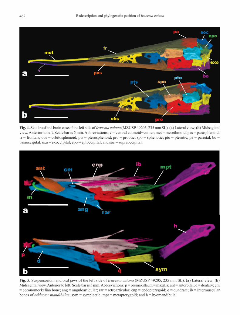
Suspensorium and oral jaws. Suspensorium consisting ofhyomandibula (h), symplectic (sym), quadrate (q),metapterygoid (mpt) and endopterygoid (enp) bones (Fig. 5).Long axis of hyomandibula highly oblique, almost parallel tomain axis of neurocranium (Fig. 3). Dorsal (proximal) head ofhyomandibula narrow and robustly ossified, with twoarticulating surfaces. Ventral (distal) limb of hyomandibulanarrow and thinly ossified, with slender shelf on its anteriormargin at middle of bone. Ventral limb of hyomandibulaarticulating with metapterygoid anterodorsally, and symplecticanteroventrally (almost certainly by cartilage as in otherrhamphichthyoids). Hyomandibula with single large foramenon dorsomedial surface, two foramina on lateral surface, andone on posterodorsal margin, all through which pass cranialnerves. Posterior lateral line nerve with separate lateral foramenlocated on posterodorsal margin of hyomandibula. Largeforamen on lateral surface of dorsal portion of hyomandibula(laterosensory, Trigeminal, and Facial nerve components).
Fig. 3. Lateral view of the skull and cleithral region of Iracema caiana, (a) MZUSP 49205, 235 mm SL, scale bar is 5 mm; (b)MZUSP 49205, 345 mm SL, snout bent upwards due to preservation artifact. Scale bar is 10 mm.
Redescription and phylogenetic position of Iracema caiana462
Fig. 4. Skull roof and brain case of the left side of Iracema caiana (MZUSP 49205, 235 mm SL). (a) Lateral view; (b) Midsagittalview. Anterior to left. Scale bar is 5 mm. Abbreviations: v = ventral ethmoid+vomer; met = mesethmoid; pas = parasphenoid;fr = frontals; obs = orbitosphenoid; pts = pterosphenoid; pro = prootic; spo = sphenotic; pto = pterotic; pa = parietal, bo =basioccipital; exo = exoccipital; epo = epioccipital; and soc = supraoccipital.
Fig. 5. Suspensorium and oral jaws of the left side of Iracema caiana (MZUSP 49205, 235 mm SL). (a) Lateral view; (b)Midsagittal view. Anterior to left. Scale bar is 5 mm. Abbreviations: p = premaxilla; m = maxilla; ant = antorbital; d = dentary; cm= coronomeckelian bone; ang = anguloarticular; rar = retroarticular; enp = endopterygoid; q = quadrate; ib = intermuscularbones of adductor mandibulae; sym = symplectic; mpt = metapterygoid; and h = hyomandibula.
T. P. Carvalho & J. S. Albert 463
Preopercular-mandibular (also laterosensory, Trigeminal, andFacial nerve components) foramen located on lateral surfaceof ventral hyomandibula about 2/3 distance to its tip.Metapterygoid narrow and triangular, with anterior dorsalprocess contacting endopterygoid. Posterior margin ofmetapterygoid directly abutting hyomandibula, anteroventralmargin contacting symplectic. Symplectic triangular andnarrow, about same size as metapterygoid. Anterior portionof symplectic contacting medial surface of quadrate betweenascending blade and posteroventral process of quadrate.Quadrate contacting anguloarticular (ang) anteriorly andendopterygoid dorsally, with interdigitated contact margin.Quadrate anterodorsal process somewhat squared, notrounded. Endopterygoid elongate anteriorly, edentulous.Ascending process of endopterygoid long and thin, directedobliquely backward towards frontal and orbitosphenoid, notcontacting these bones. Ascending endopterygoid processlocated about midlength of bone. Autopalatine not ossifiedand thus not visible in scan. Several isolated filamentousbones in adductor mandibulae (ib), located lateral toendopterygoid, extending from posterodorsal portion ofdentary (d) obliquely to articulation of hyomandibula withneurocranium. Intermuscular ossifications absent in levatoroperculi, dilator operculi and protractor hyoidei.
Upper jaw consisting of maxilla (m) and premaxilla (p).Maxilla elongate, sickle shape, its dorsal margin concave andventral margin convex; anterior margin rounded; anteriormaxillary process absent; descending process of maxilla narrow,tapering distally. Premaxilla gracile, with small ascendingprocess. Bones of lower jaw consist in dentary, anguloarticular,retroarticular (rar), and coronomeckelian (cm) bones. Dorsalportion of dentary straight; posterior margin deeply forked;anteroventral portion with small hook-shaped process directedposteroventrally. Anguloarticular in close contact with dentaryanteriorly and extensively contacting quadrate posteriorly.Anguloarticular condyle socket positioned along posterodorsalmargin of lower jaw. Anguloarticular contributing more thanretroarticular to joint with quadrate. Retroarticular triangular inshape, anterior margin pointed, dorsoposterior portioncontacting quadrate (Fig. 5A). Coronomeckelian bone elongateand teardrop shaped, located at dorsoposterior portion ofdentary posterior concavity. Teeth absent in both jaws.
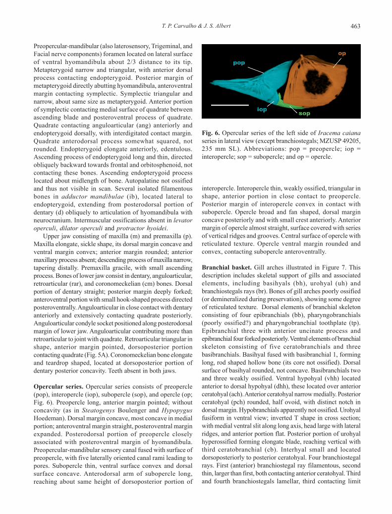
Opercular series. Opercular series consists of preopercle(pop), interopercle (iop), subopercle (sop), and opercle (op;Fig. 6). Preopercle long, anterior margin pointed; withoutconcavity (as in Steatogenys Boulenger and HypopygusHoedeman). Dorsal margin concave, most concave in medialportion; anteroventral margin straight, posteroventral marginexpanded. Posterodorsal portion of preopercle closelyassociated with posteroventral margin of hyomandibula.Preopercular-mandibular sensory canal fused with surface ofpreopercle, with five laterally oriented canal rami leading topores. Subopercle thin, ventral surface convex and dorsalsurface concave. Anterodorsal arm of subopercle long,reaching about same height of dorsoposterior portion of
interopercle. Interopercle thin, weakly ossified, triangular inshape, anterior portion in close contact to preopercle.Posterior margin of interopercle convex in contact withsubopercle. Opercle broad and fan shaped, dorsal marginconcave posteriorly and with small crest anteriorly. Anteriormargin of opercle almost straight, surface covered with seriesof vertical ridges and grooves. Central surface of opercle withreticulated texture. Opercle ventral margin rounded andconvex, contacting subopercle anteroventrally.
Branchial basket. Gill arches illustrated in Figure 7. Thisdescription includes skeletal support of gills and associatedelements, including basihyals (bh), urohyal (uh) andbranchiostegals rays (br). Bones of gill arches poorly ossified(or demineralized during preservation), showing some degreeof reticulated texture. Dorsal elements of branchial skeletonconsisting of four epibranchials (bb), pharyngobranchials(poorly ossified?) and pharyngobranchial toothplate (tp).Epibranchial three with anterior uncinate process andepibranchial four forked posteriorly. Ventral elements of branchialskeleton consisting of five ceratobranchials and threebasibranchials. Basihyal fused with basibranchial 1, forminglong, rod shaped hollow bone (its core not ossified). Dorsalsurface of basihyal rounded, not concave. Basibranchials twoand three weakly ossified. Ventral hypohyal (vhh) locatedanterior to dorsal hypohyal (dhh), these located over anteriorceratohyal (ach). Anterior ceratohyal narrow medially. Posteriorceratohyal (pch) rounded, half ovoid, with distinct notch indorsal margin. Hypobranchials apparently not ossified. Urohyalfusiform in ventral view; inverted T shape in cross section;with medial ventral slit along long axis, head large with lateralridges, and anterior portion flat. Posterior portion of urohyalhyperossified forming elongate blade, reaching vertical withthird ceratobranchial (cb). Interhyal small and locateddorsoposteriorly to posterior ceratohyal. Four branchiostegalrays. First (anterior) branchiostegal ray filamentous, secondthin, larger than first, both contacting anterior ceratohyal. Thirdand fourth branchiostegals lamellar, third contacting limit
Fig. 6. Opercular series of the left side of Iracema caianaseries in lateral view (except branchiostegals; MZUSP 49205,235 mm SL). Abbreviations: pop = preopercle; iop =interopercle; sop = subopercle; and op = opercle.

Redescription and phylogenetic position of Iracema caiana464
between anterior and posterior ceratohyal, fourth contactingposterior ceratohyal.
Weberian Apparatus. Anterior vertebrae and WeberianApparatus illustrated in Figure 8. Neural arches (na) of thirdand fourth vertebrae and supraneural (sn) meet togetherforming roof of neural canal. Supraneural adjacent tosupraoccipital and neural arch of third vertebra adjacent toexoccipital. Bones of Weberian complex forming socket fordorsal contact between occipital bones of skull and axialskeleton (in Gymnorhamphichthys only supraoccipitalarticulates with neural complex; Mago-Leccia, 1994: 40).Scaphium (s) barrel-shaped and located anterior to, andslightly ventral to third neural arch. Intercalarium (i) small andround, positioned posterior to scaphium and anterior to tripus(t). Tripus positioned obliquely and descending posteriorlybetween scaphium and ventral portion of fourth centrum.Tripus narrow anteriorly, broad in median region and taperingposteriorly. First centrum (c1) anteroposteriorly compressedand disk-like shape. Second centrum (c2) larger and withprominent parapophysis behind ossified Baudelot’s ligament(bl), extending posterolaterally and fused with fourthparapophysis (pp4) and pleural rib complex. Third centrumlocated posterior to other components of Weberian complex.Fourth vertebra with fourth parapophysis and pleural ribcomplex fused to second parapophysis anteriorly. Ossuspensorium (os) fused with ventral portion of fourth
vertebrae, which extends anteriorly, tapering to reach ventralportion of second centrum. Fifth centrum bearing fifth rib(r5), slightly expanded along antero-posterior axis inmidregion. Fifth vertebra bears first neural spine with spinousmorphology, located in anterior portion of vertebra, andoriented obliquely posterodorsally. Subsequent ribs widestclose to articulation with vertebrae; tapering distally. Anteriorchamber of gas bladder not enclosed in bony capsule as inRhamphichthys (Mago-Leccia, 1994: 40).
Vertebral column. Eighteen precaudal vertebrae (n=2; x-rayedspecimens) including vertebrae of Weberian apparatus; 84vertebrae to end of anal fin (n=1; x-rayed specimen). Neuraland hemal spines positioned anteriorly on centrum in anteriorvertebrae, transitioning to posterior position on centrum atabout vertebra 62. Intermuscular bones (observed in CTscans) composed by dorsal series of myorhabdoi, epicentralbones and series of filamentous bones of anterior epaxialmuscles. Epicentral bones in contact with dorsoposteriorportion of ribs, rod shaped and somewhat sinusoidal, directedtowards posterodorsal portion of body. First two epicentralbones not contacting ribs. Anterior portion of first epicentralbones located slightly anteriorly to vertical with scaphium;second slightly anterior to contact between neural archesthree and four, third contacting fourth rib. Dorsal series ofmyorhabdoi obliquely positioned, anterior and posterior endsbranched multiply filamentous bones in anterior dorsal
Fig. 7. Branchial basket and branchiostegals rays of leftside of Iracema caiana (MZUSP 49205, 235 mm SL). (a)Lateral view; (b) Midsagittal view. Anterior to left. Scale baris 5 mm. Abbreviations: bh+bb1 = basihyal +basibranchial1; bb2 = basibranchial 2; bb3 = basibranchial 3; vhh = ventralhypohyal; dhh = dorsal hypohyal; ach = anterior ceratohyal;pch = posterior ceratohyal; uh = urohyal; br =branchiostegals; cb = ceratobranchial; eb = epibranchial;and tp = pharyngobranchial tooth plate.
Fig. 8. Weberian apparatus of right side (image reversed) ofIracema caiana (MZUSP 49205, 235 mm SL). Left side ribsand parapophysis removed. Anterior to left. Scale bar is 5mm. Abbreviations: c1-c2 = centrum; s= scaphium; i =intercalarium; os = os suspensorium; t = tripus; bl = Baudelot’sligament; na3-na4 = neural arches; sn = supraneural; ns =neural spine; pp4 = parapophysis 4 and r5-r6 = ribs.

T. P. Carvalho & J. S. Albert 465
muscles positioned posterior to epioccipital simple orsometimes branched.
We were not able to count precisely number of displacedhemal spines, but radiographs show at least more than six inMZUSP 49205, 345 mm SL. Displaced hemal spines all of similarsize; absence of enlarged anterior displaced hemal spine(Albert, 2001: fig. 35c-d; posteroventral abdominal bone ofHilton et al., 2007: fig. 16b).
Anal fin and supports. Anal-fin rays range from 240-257 rays(n=4). All rays posterior to rays 23-42 branched, no raysbranched more than once. Branched portions of rays alwaysadnate from branching origin to distal end of fin. Three anal-fin pterygiophores between adjacent hemal spines.
Pectoral fin and girdle. Pectoral fin with 16-17 rays (n=4),anomalous specimen with 12 fin rays on left side (MZUSP49205, 235 mm SL; as noted by Triques 1996: 92). Pectoral finlanceolate, dorsal rays longer than ventral rays, distal finmargin slightly rounded. One or two dorsalmost raysunbranched.
Dermal elements of pectoral girdle consisting ofposttemporal (pt), supracleithrum (scl) and cleithrum (cl; Fig.9); postcleithrae apparently absent. Posttemporal bone fusedwith supracleithrum (pt+scl), bearing large foramen forpassage of posterior lateral line nerve. Dorsal portion ofposttemporal + supracleithrum bones positioned over
exoccipital and epioccipital. Cleithrum large, dorsal and ventralportions of similar size.
Chondral components of pectoral girdle consist of scapula(sc), coracoid (co) and mesocoracoid (mco; Fig. 9). Scapulanot well ossified; axe-blade shape. Coracoid with long, thinand curved ventral process directed anteriorly. Dorsal processof coracoid hatchet-shaped with hook-shaped process ondorsal surface. Mesocoracoid present, rod-like hourglassshaped structure, obliquely oriented anterodorsally toposteroventrally. Support for pectoral fin rays composed offour radials (ra; MZUSP 49205, 345 mm SL). Baudelot’s ligamentossified, branching anteriorly and posteriorly; extending fromposteroventral portion of basioccipital and posterolateralportion of exoccipital to cleithrum; with “><“ shape in lateralview.
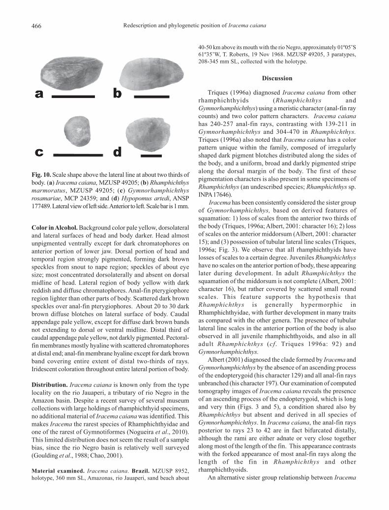
Scales. Absence of scales on anterior two-thirds of body.Scales covering entire caudal appendage. Absence of scalesin anterior portion of lateral line. Lateral line ossicles embeddedin thick layer of skin. Scales above lateral line in posteriorportion of body ovoid in shape, elongate on horizontal axis,about twice as long as deep (Fig. 10).
Electric organs. No information available regarding electricorgan discharges (EOD) in Iracema caiana. Hypaxial electricorgan extending anteriorly to mental region. Four rows ofelectrocytes present above posterior end of anal fin.
Fig. 9. Pectoral girdle of left side of Iracema caiana (MZUSP 49205, 235 mm SL; pectoral fin rays digitally removed). (a) Lateralview; (b) Midsagittal view. Anterior to left. Scale bar is 5 mm. Abbreviations: pt+scl = posttemporal + supracleithrum; cl =cleithrum; co = coracoid; sc = scapula; mco = mesocoracoid; and ra = radials.
Redescription and phylogenetic position of Iracema caiana466
Color in Alcohol. Background color pale yellow, dorsolateraland lateral surfaces of head and body darker. Head almostunpigmented ventrally except for dark chromatophores onanterior portion of lower jaw. Dorsal portion of head andtemporal region strongly pigmented, forming dark brownspeckles from snout to nape region; speckles of about eyesize; most concentrated dorsolaterally and absent on dorsalmidline of head. Lateral region of body yellow with darkreddish and diffuse chromatophores. Anal-fin pterygiophoreregion lighter than other parts of body. Scattered dark brownspeckles over anal-fin pterygiophores. About 20 to 30 darkbrown diffuse blotches on lateral surface of body. Caudalappendage pale yellow, except for diffuse dark brown bandsnot extending to dorsal or ventral midline. Distal third ofcaudal appendage pale yellow, not darkly pigmented. Pectoral-fin membranes mostly hyaline with scattered chromatophoresat distal end; anal-fin membrane hyaline except for dark brownband covering entire extent of distal two-thirds of rays.Iridescent coloration throughout entire lateral portion of body.
Distribution. Iracema caiana is known only from the typelocality on the rio Jauaperi, a tributary of rio Negro in theAmazon basin. Despite a recent survey of several museumcollections with large holdings of rhamphichthyid specimens,no additional material of Iracema caiana was identified. Thismakes Iracema the rarest species of Rhamphichthyidae andone of the rarest of Gymnotiformes (Nogueira et al., 2010).This limited distribution does not seem the result of a samplebias, since the rio Negro basin is relatively well surveyed(Goulding et al., 1988; Chao, 2001).
Material examined. Iracema caiana. Brazil. MZUSP 8952,holotype, 360 mm SL, Amazonas, rio Jauaperi, sand beach about
40-50 km above its mouth with the rio Negro, approximately 01º05’S61º35’W, T. Roberts, 19 Nov 1968. MZUSP 49205, 3 paratypes,208-345 mm SL, collected with the holotype.
Discussion
Triques (1996a) diagnosed Iracema caiana from otherrhamphichthyids (Rhamphichthys andGymnorhamphichthys) using a meristic character (anal-fin raycounts) and two color pattern characters. Iracema caianahas 240-257 anal-fin rays, contrasting with 139-211 inGymnorhamphichthys and 304-470 in Rhamphichthys.Triques (1996a) also noted that Iracema caiana has a colorpattern unique within the family, composed of irregularlyshaped dark pigment blotches distributed along the sides ofthe body, and a uniform, broad and darkly pigmented stripealong the dorsal margin of the body. The first of thesepigmentation characters is also present in some specimens ofRhamphichthys (an undescribed species; Rhamphichthys sp.INPA 17646).
Iracema has been consistently considered the sister groupof Gymnorhamphichthys, based on derived features ofsquamation: 1) loss of scales from the anterior two thirds ofthe body (Triques, 1996a; Albert, 2001: character 16); 2) lossof scales on the anterior middorsum (Albert, 2001: character15); and (3) possession of tubular lateral line scales (Triques,1996a; Fig. 3). We observe that all rhamphichthyids havelosses of scales to a certain degree. Juveniles Rhamphichthyshave no scales on the anterior portion of body, these appearinglater during development. In adult Rhamphichthys thesquamation of the middorsum is not complete (Albert, 2001:character 16), but rather covered by scattered small roundscales. This feature supports the hypothesis thatRhamphichthys is generally hypermorphic inRhamphichthyidae, with further development in many traitsas compared with the other genera. The presence of tubularlateral line scales in the anterior portion of the body is alsoobserved in all juvenile rhamphichthyoids, and also in alladult Rhamphichthys (cf. Triques 1996a: 92) andGymnorhamphichthys.
Albert (2001) diagnosed the clade formed by Iracema andGymnorhamphichthys by the absence of an ascending processof the endopterygoid (his character 129) and all anal-fin raysunbranched (his character 197). Our examination of computedtomography images of Iracema caiana reveals the presenceof an ascending process of the endopterygoid, which is longand very thin (Figs. 3 and 5), a condition shared also byRhamphichthys but absent and derived in all species ofGymnorhamphichthys. In Iracema caiana, the anal-fin raysposterior to rays 23 to 42 are in fact bifurcated distally,although the rami are either adnate or very close togetheralong most of the length of the fin. This appearance contrastswith the forked appearance of most anal-fin rays along thelength of the fin in Rhamphichthys and otherrhamphichthyoids.
An alternative sister group relationship between Iracema
Fig. 10. Scale shape above the lateral line at about two thirds ofbody. (a) Iracema caiana, MZUSP 49205; (b) Rhamphichthysmarmoratus, MZUSP 49205; (c) Gymnorhamphichthysrosamariae, MCP 24359; and (d) Hypopomus artedi, ANSP177489. Lateral view of left side. Anterior to left. Scale bar is 1 mm.
T. P. Carvalho & J. S. Albert 467
and Rhamphichthys is supported by the followingsynapomorphies: 1) filamentous intermuscular bones in themusculus adductor mandibulae; 2) central surface of operclereticulated; 3) Baudelot’s ligament ossified from its origin inthe exoccipital and basioccipital to attachment with cleithrum;and 4) scales above lateral line in the posterior portion of bodyelongate in the horizontal axis, about twice as long as deep.
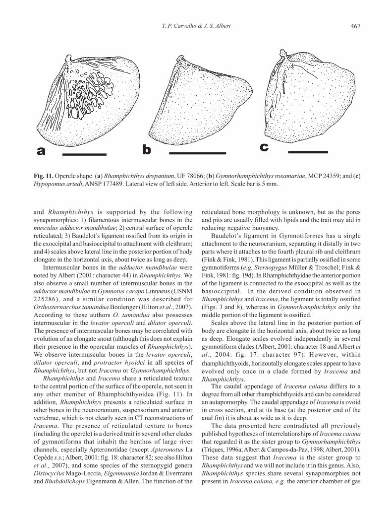
Intermuscular bones in the adductor mandibulae werenoted by Albert (2001: character 44) in Rhamphichthys. Wealso observe a small number of intermuscular bones in theadductor mandibulae in Gymnotus carapo Linnaeus (USNM225286), and a similar condition was described forOrthosternarchus tamandua Boulenger (Hilton et al., 2007).According to these authors O. tamandua also possessesintermuscular in the levator operculi and dilator operculi.The presence of intermuscular bones may be correlated withevolution of an elongate snout (although this does not explaintheir presence in the opercular muscles of Rhamphichthys).We observe intermuscular bones in the levator operculi,dilator operculi, and protractor hyoidei in all species ofRhamphichthys, but not Iracema or Gymnorhamphichthys.
Rhamphichthys and Iracema share a reticulated textureto the central portion of the surface of the opercle, not seen inany other member of Rhamphichthyoidea (Fig. 11). Inaddition, Rhamphichthys presents a reticulated surface inother bones in the neurocranium, suspensorium and anteriorvertebrae, which is not clearly seen in CT reconstructions ofIracema. The presence of reticulated texture to bones(including the opercle) is a derived trait in several other cladesof gymnotiforms that inhabit the benthos of large riverchannels, especially Apteronotidae (except Apteronotus LaCepède s.s.; Albert, 2001: fig. 18: character 82; see also Hiltonet al., 2007), and some species of the sternopygid generaDistocyclus Mago-Leccia, Eigenmannia Jordan & Evermannand Rhabdolichops Eigenmann & Allen. The function of the
reticulated bone morphology is unknown, but as the poresand pits are usually filled with lipids and the trait may aid inreducing negative buoyancy.
Baudelot’s ligament in Gymnotiformes has a singleattachment to the neurocranium, separating it distally in twoparts where it attaches to the fourth pleural rib and cleithrum(Fink & Fink, 1981). This ligament is partially ossified in somegymnotiforms (e.g. Sternopygus Müller & Troschel; Fink &Fink, 1981: fig. 19d). In Rhamphichthyidae the anterior portionof the ligament is connected to the exoccipital as well as thebasioccipital. In the derived condition observed inRhamphichthys and Iracema, the ligament is totally ossified(Figs. 3 and 8), whereas in Gymnorhamphichthys only themiddle portion of the ligament is ossified.
Scales above the lateral line in the posterior portion ofbody are elongate in the horizontal axis, about twice as longas deep. Elongate scales evolved independently in severalgymnotiform clades (Albert, 2001: character 18 and Albert etal., 2004: fig. 17: character 97). However, within
rhamphichthyoids, horizontally elongate scales appear to haveevolved only once in a clade formed by Iracema andRhamphichthys.
The caudal appendage of Iracema caiana differs to adegree from all other rhamphichthyoids and can be consideredan autapomorphy. The caudal appendage of Iracema is ovoidin cross section, and at its base (at the posterior end of theanal fin) it is about as wide as it is deep.
The data presented here contradicted all previouslypublished hypotheses of interrelationships of Iracema caianathat regarded it as the sister group to Gymnorhamphichthys(Triques, 1996a; Albert & Campos-da-Paz, 1998; Albert, 2001).These data suggest that Iracema is the sister group toRhamphichthys and we will not include it in this genus. Also,Rhamphichthys species share several synapomorphies notpresent in Iracema caiana, e.g. the anterior chamber of gas
Fig. 11. Opercle shape. (a) Rhamphichthys drepanium, UF 78066; (b) Gymnorhamphichthys rosamariae, MCP 24359; and (c)Hypopomus artedi, ANSP 177489. Lateral view of left side. Anterior to left. Scale bar is 5 mm.
Redescription and phylogenetic position of Iracema caiana468
bladder enclosed in a bony capsule (Mago-Leccia, 1994), andthe presence of a fleshy skin fold on the body wall, inside theopercular opening (Triques, 2005), which reinforce itsmonophyly.
Comparative material. Apteronotus albifrons: Venezuela.Monagas. UF 29921, 11 (1 cs), Laguna Grande. Brachyhypopomusoccidentalis: Costa Rica. Limón. ANSP 163176, 9 (1 cs), creekclose to Shiroles. Eigenmannia virescens: Venezuela. Guarico. UF78117, 27 (2 cs), río Tiznados at Guardatinajas.Gymnorhamphichthys bogardusi: Venezuela. Delta Amacuro.ANSP 187349, 7 (2 cs), río Orinoco, downstream from cañoRemolina. Gymnorhamphichthys hypostomus: Brazil. Amazonas.MCP 24292, 2 (1 cs), rio Negro, 18.5 miles above Manaus. Pará.MCP 26580, 3 (1 cs), rio Trombetas 0.8 miles upstream Vila Aracua.Gymnorhamphichthys petiti: Brazil. Mato Grosso. MCP 30373, 4(1 cs), ribeirão Macuco on highway BR-163, about 74 km N ofSinop. MCP 40189, 5 (1 cs), creek tributary to rio Suiazinho, onhighway BR-158 N of Ribeirão Cascalheira. Gymnorhamphichthysrondoni: Brazil. Pará. MCP 15161, 3 (1 cs), rio Tapajós, beach atisland 5 km upstream Itaituba. MZUSP 97147, 9, (1cs), rio Curuátributary of rio Iriri at Vila Castelo dos Sonhos.Gymnorhamphichthys rosamariae: Brazil. Amazonas. MCP24359, 2 (1 cs), rio Negro 18.5 miles upstream Manaus. MCP24872, 9 (2 cs), rio Negro upstream rio Branco mouth, betweenCarvoeiro and Vila Guajara. Gymnotus carapo: Suriname. Nickerie.USNM 225286, 15 (1 cs), Koekwie Kreek. Hypopomus artedi:Suriname. ANSP 177489, 2 (1 cs), Burro-Burro River. CAS 72233,2 of 4, Parwapa Kreek. Hypopygus lepturus: Brazil. Amazonas.MCP 41121, 10 (2 cs), creek on highway BR-319. Rhamphichthysapurensis: Venezuela. Bolivar. ANSP 162300, 60 (1 cs), río Orinoconear mouth of río Caura. Rhamphichthys drepanium: Venezuela.Guarico. UF 78066, 2 (1 cs), Morichal 2.3 km N of San Fernando deApure. Rhamphichthys hahni: Brazil. Mato Grosso do Sul. MZUSP59297, 2 (1 cs), rio Novo tributary to rio Negro at brejo de SantaSofia. Rhamphichthys lineatus: Brazil. Amazonas. MCP 33457, 2(1 cs), paraná Maiana, lago Mamirauá. Rhamphichthys marmoratus:Brazil. Pará. MZUSP 44493, 2 (1 cs), rio Apeu, at Boa Vista deApeu. Rhamphichthys rostratus: Brazil. Amazonas. MCP 27756,23 (1 cs), rio Negro 15 miles downstream Moura. Rhamphichthyssp.: Brazil. Amazonas. INPA 17646, 7 (1 cs), Costa do Catalão.Steatogenys elegans: Brazil. Amazonas. MCP 24290, 8 (2 cs), rioAmazonas downstream rio Negro. Sternopygus macrurus:Venezuela. Apure. MCNG 3733, 4 cs, río Apure.
Acknowledgements
We thank J. Maisano and M. Colbert (UT Austin) forpreparation of computed tomography x-rays, K. Luckenbill(ANSP) for help in learning image editing with VGStudio Max,and O. Oyakawa (MZUSP) for the loan of types specimens.We also thank C. Lucena (MCP), O. Oyakawa and J. Figueiredo(MZUSP), M. Sabaj-Pérez, K. Luckenbill and J. Lundberg(ANSP), L. Py-Daniel and M. Rocha (INPA), and R. Robbins(UF) for the loan of specimens or support in the fish collections.We acknowledge J. Lundberg and E. Maxime for discussionsof gymnotiform morphology. This project was supported inpart by the Student Exchange Program of DeepFin. TPC ispartially supported by a research assistantship of NSF-0741450 to JSA.
Literature Cited
Albert, J. S. 2001. Species diversity and phylogenetic systematicsof American knifefishes (Gymnotiformes, Teleostei).Miscellaneous Publications, Museum of Zoology, Universityof Michigan, 190: 1-127.
Albert, J. S. & R. Campos-da-Paz. 1998. Phylogenetic systematicsof Gymnotiformes with diagnoses of 58 clades: a review ofavailable data. Pp. 419-446. In: Malabarba, L. R., R. E. Reis, R. P.Vari, Z. M. S. Lucena & C. A. S. Lucena (Eds.). Phylogeny andclassification of Neotropical fishes. Porto Alegre, Edipucrs, 603p.
Albert J. S. & W. G. R. Crampton. 2005. Diversity and phylogenyof Neotropical electric fishes (Gymnotiformes). Pp. 360-409.In:Bullock, T. H., C. D. Hopkins, A. N. Popper & R. R. Fay(Eds.). Electroreception. New York, Springer Handbook ofAuditory Research, 485p.
Albert, J. S., W. G. R. Crampton, D. H. Thorsen & N. R. Lovejoy.2004. Phylogenetic systematics and( historical biogeographyof the Neotropical electric fish Gymnotus (Teleostei:Gymnotiformes). Systematic and Biodiversity, 2: 375-417.
Alves-Gomes, J. A., G. Orti, M. Haygood, A. Meyer & W.Heiligenberg. 1995. Phylogenetic analysis of the South Americanelectric fishes (Order Gymnotiformes) and the evolution of theirelectrogenic system: a synthesis based on morphology,electrophysiology, and mitochondrial sequence data. MolecularBiology and Evolution, 12: 298-318.
Carvalho T. P., C. S. Ramos & J. S. Albert. 2011. A new species ofGymnorhamphichthys (Gymnotiformes: Rhamphichthyidae)from Paraná-Paraguay system. Copeia, 2011(3): 400-406.
Chao, N. L. 2001. The fishery, diversity and conservation of or-namental fishes in the Rio Negro Basin, Brazil – a review ofthe Project Piaba (1989-99). Pp. 161-204. In: Chao N. L., P.Petry, G. Prang. L. Sonneschien & M. Tlusty (Eds.).Conservation and management of ornamental fish resourcesof the Rio Negro Basin, Amazonia, Brazil – Project Piaba.Manaus, EDUA, 310p.
Crampton, W. G. R. 2011. An ecological perspective on diversityand distributions. Pp. 165-189. In: Albert, J. S. & R. E. Reis(Eds.). Historical Biogeography of Neotropical FreshwaterFishes. Berkeley, UCPress, 388p.
Crampton, W. G. R. & J. S. Albert. 2006. Evolution of electricsignal diversity in gymnotiform fishes. Pp. 647-731. In: Ladich,F., S. P. Collin, P. Moller & B. G. Kapoor (Eds.). Communicationin fishes. Enfield, Science Publishers, 870p.
Datovo, A. & F. A. Bockmann. 2010. Dorsolateral head muscles ofthe catfish families Nematogenyidae and Trichomycteridae(Siluriformes: Loricarioidei): comparative anatomy andphylogenetic analysis. Neotropical Ichthyology, 8: 193-246.
Ferraris Jr., C. J. 2003. Rhamphichthyidae (sand knifefishes). Pp.495-497. In: Reis, R. E., S. O. Kullander & C. J. Ferraris, Jr.(Eds.). Checklist of the Freshwater Fishes of the South andCentral America. Porto Alegre, Edipucrs, 729p.
Fink S. V. & W. L. Fink. 1981. Interrelationships of theOstariophysan fishes (Teleostei). Zoological Journal of theLinnean Society, 72: 297-353.
Fink S. V. & W. L. Fink. 1996. Interrelationships of Ostariophysanfishes (Teleostei). Pp. 209-249. In: Stiassny, M. L. J., L. R.Parenti & D. Johnson (Eds.). San Diego, Academic Press, 496p.
Fink, W. L. & J. H. Humphries. 2010. Morphological descriptionof the extinct North American sucker Moxostoma lacerum(Ostariophysi, Catostomidae), based on high-resolution x-raycomputed tomography. Copeia, 2010: 5-13.
T. P. Carvalho & J. S. Albert 469
Goulding, M., M. L. Carvalho & E. G. Ferreira, 1988. Rio Negro,rich life in poor water: Amazonian diversity and foodchainecology as seen through fish communities. The Hague, SPBAcademic Publishing, 200p.
Hilton E. J., C. Cox Fernandes, J. P. Sullivan, J. G. Lundberg & R.Campos-da-Paz. 2007. Redescription of Orthosternachustamandua (Boulenger, 1998) (Gymnotiformes, Apteronotidae),with reviews of its ecology, electric organ discharges, externalmorphology, osteology, and phylogenetic affinities. Proceedingsof the Academy of Natural Sciences of Philadelphia, 156: 1-25.
Lundberg, J. G. 2005. Gymnorhamphichthys bogardusi, a new speciesof sand knifefish (Gymnotiformes: Rhamphichthyidae) from theRio Orinoco, South America. Notula Naturae, 479: 1-4.
Mago-Leccia, F. 1978. Los Peces de la familia Sternopygidae deVenezuela, incluyendo una descripción de la osteología deEigenmannia virescens y una nueva definición y classificación delOrden Gymnotiformes. Acta Cientifica Venezuelana, 29: 1-51.
Mago-Leccia, F. 1994. Electric fishes of the continental waters ofAmerica. Caracas, Fundación para el Desarollo de las CienciasFísicas, Matemáticas y Naturales, Electric Fishes: 1-206.
Nijssen, H., I. J. H. Isbrucker & J. Géry. 1976. On the species ofGymnorhamphichthys Ellis, 1912, translucent sand-dwellinggymnotid fishes from South America (Pisces, Cypriniformes,Gymnotoidei). Studies on Neotropical Fauna and Environment,11: 37-63.
Nogueira C., P. A. Buckup, N. A. Menezes, O. T. Oyakawa, T. P.Kasecker, M. B. Ramos Neto & J. M. C. da Silva. 2010.Restricted-range fishes and the conservation of Brazilianfreshwaters. PLOS One, 5: 1-10.
Schaefer, S. A. 2003 Relationships of Lithogenes villosus Eigenmann,1909 (Siluriformes, Loricariidae): evidence from high-resolutioncomputed microtomography. American Museum Novitates,3401: 1-55.
Schaefer S. A. & L. Fernández. 2009. Redescription of the PezGraso, Rhizomichthys totae (Trichomycteridae) of Lago Tota,Colombia, and aspects of cranial osteology revealed bymicrotomography. Copeia, 2009: 510-522.
Schwassmann, H. O. 1989. Gymnorhamphichthys rosamariae, anew Species of knifefish (Rhamphichthyidae, Gymnotiformes)from the upper Rio Negro, Brazil. Studies on Neotropical Faunaand Environment, 24: 157-167.
Taylor, W. R. & G. C. Van Dyke. 1985. Revised procedures forstaining and clearing small fishes and other vertebrates for boneand cartilage study. Cybium, 9: 107-119.
Triques, M. L. 1996a. Iracema caiana, a genus and species ofelectrogenic Neotropical freshwater fish (Rhamphichthyidae:Gymnotiformes: Ostariophysi: Actionpterygii). RevueFrançaise d’Aquariologie, 23: 91-92.
Triques, M. L. 1996b. Eigenmannia vicentespelea, a new species ofcave dwelling electrogenic neotropical fish (Ostariophisi:Gymnotiformes: Sternopygidae). Revue Françaised’Aquariologie, 23: 1-4.
Triques, M. L. 2005. Novas sinapomorfias para RhamphichthysMuller & Troschel, 1848 (Teleostei: Rhamphichthyidae).Lundiana, 6: 35-39.
Winemiller, K. O. & S. C. Willis. 2011. The Vaupes Arch andCasiquiare Canal: barriers and passages. Pp. 225-242. In: Albert,J. S. & R. E. Reis (Eds.). Historical Biogeography of NeotropicalFreshwater Fishes. Berkeley, UCPress, 388p.
Winterbottom, R. 1974. A descriptive synonymy of the striatedmuscles of the Teleostei. Proceedings of the Academy of Natu-ral Sciences of Philadelphia, 125(12): 225-317.
Submitted January 4, 2011Accepted August 19, 2011
Published September 16, 2011