reduction in hemoglobin–oxygen affinity results in the … · · 2016-11-12objectives this...
TRANSCRIPT

T(i
FSSoJToOfaa
a
Journal of the American College of Cardiology Vol. 52, No. 9, 2008© 2008 by the American College of Cardiology Foundation ISSN 0735-1097/08/$34.00P
PRECLINICAL RESEARCH
Reduction in Hemoglobin–OxygenAffinity Results in the Improvementof Exercise Capacity in Mice With Chronic Heart Failure
Tetsuya Watanabe, MD, PHD,* Toshihiro Takeda, MD, PHD,* Shigemiki Omiya, MD,*Shungo Hikoso, MD, PHD,* Osamu Yamaguchi, MD, PHD,* Yuko Nakano, DDS,†Yoshiharu Higuchi, MD, PHD,* Atsuko Nakai, PHD,* Yusuke Abe, MS,‡ Yayoi Aki-Jin, PHD,§Masayuki Taniike, MD, PHD,* Isamu Mizote, MD,* Yasushi Matsumura, MD, PHD,�Takahiko Shimizu, PHD,‡ Kazuhiko Nishida, MD, PHD,* Kiyohiro Imai, PHD,§Masatsugu Hori, MD, PHD, FACC,* Takuji Shirasawa, MD, PHD,‡ Kinya Otsu, MD, PHD*
Osaka and Tokyo, Japan
Objectives This study examined whether a reduction in hemoglobin–oxygen affinity improves exercise capacity in mice withheart failure.
Background Exercise intolerance is a major determinant of quality of life in patients with chronic heart failure. One of themajor goals of the treatment for chronic heart failure is to improve quality of life.
Methods Four weeks after left coronary ligation, we transplanted bone marrow cells isolated from the transgenic miceexpressing a hemoglobin variant with low oxygen affinity, Presbyterian, into the lethally irradiated mice withheart failure or administered a synthetic allosteric modifier of hemoglobin. The mice were then exercised on atreadmill.
Results Four weeks after the left coronary artery ligation, mice showed cardiac dysfunction and chamber dilation, whichwere characteristics of heart failure. The transplantation led to a reduction in hemoglobin–oxygen affinity and anincrease in oxygen supply for skeletal muscle without changes in muscle properties. The transplanted miceshowed improved running performance on a treadmill despite impaired cardiac contractility. Furthermore, ad-ministration of the synthetic allosteric modifier of hemoglobin showed a similar effect.
Conclusions Allosteric modification of hemoglobin represents a therapeutic option for improving exercise capacity in patientswith chronic heart failure. One mechanism of improvement in exercise capacity is enhanced oxygen delivery inthe skeletal muscle. (J Am Coll Cardiol 2008;52:779–86) © 2008 by the American College of CardiologyFoundation
ublished by Elsevier Inc. doi:10.1016/j.jacc.2008.06.003
oqib
rcpaP�pt
he 3 major goals of treatment for chronic heart failureCHF) are to reduce symptoms, prolong survival, andmprove quality of life (1). A nearly universal manifestation
rom the *Department of Cardiovascular Medicine, Osaka University Graduatechool of Medicine, Osaka, Japan; †First Department of Oral and Maxillo-Facialurgery, Osaka University Graduate School of Dentistry, Osaka, Japan; ‡Departmentf Molecular Gerontology, Tokyo Metropolitan Institute of Gerontology, Tokyo,apan; §Department of Frontier Bioscience, Faculty of Engineering and Micro-Nanoechnology Research Center, Hosei University, Tokyo, Japan; and the �Departmentf Medical Information Science, Osaka University Graduate School of Medicine,saka, Japan. This work was supported by a Grant-in-Aid for Scientific Research
rom the Ministry of Education, Culture, Sports, Science and Technology, Japan, andresearch grant from Nakatomi Foundation to Dr. Otsu. Drs. Watanabe, Takeda,
nd Omiya contributed equally to this work.
aManuscript received October 29, 2007; revised manuscript received April 29, 2008,
ccepted June 3, 2008.
f CHF is a reduction in exercise capacity, which diminishesuality of life. The pathophysiology of exercise intolerancen this syndrome is incompletely understood, but it iselieved to be multifactorial.The oxygen (O2) affinity of hemoglobin (Hb), a tet-
americ protein consisting of �- and �-globin subunits, isritical for gas exchange in the lung and O2 delivery in theeripheral tissues. A variety of mutations in the genes of �-nd �-globin have been reported, one of which, hemoglobinresbyterian (HbPres), carries lysine at asparagine-108 of-globin and shows a low Hb–O2 affinity in vitro (2). Wereviously reported that mutant mice, carrying the Presby-erian mutation generated with a targeted knock-in strategy,
lso showed a low Hb–O2 affinity (2). The mutant mice
tt
imfWip
M
TAGaMBflhg1aDbspompSftac(wTo
td1uarpAfitMgc
gpoafPwdTa(hpvicslmmadmtm5SbPotors
R
Apfw
780 Watanabe et al. JACC Vol. 52, No. 9, 2008Hemoglobin and Physical Activity in Heart Failure August 26, 2008:779–86
showed various phenotypes, in-cluding mild anemia, respiratoryacidosis, enhanced tissue oxygen-ation, increased O2 consump-tion, a higher ratio of type IIA/IIB muscle fibers, and increasedspontaneous physical activity.
The 2-[4-[(3,5-dimethyl-anilino carbonyl)-methyl]phen-oxy]-2-methylpropionic acid,known as RSR13, is a syntheticallosteric modifier of Hb that actsto increase the release of O2 fromHb to the surrounding tissues(3,4). This agent markedly shifts theHb–O2 curve rightward in both invitro and in vivo rodent models
hrough the stabilization of deoxyhemoglobin in a manner similaro the natural allosteric effecter of Hb, 2, 3-diphosphoglycerate.
In the present study, we investigated whether a reductionn Hb–O2 affinity could improve exercise capacity of CHF
ice by transplanting bone marrow cells (BMCs) obtainedrom the HbPres mice or by the administration of RSR13.
e showed that a reduction in Hb–O2 affinity leads tomproved running performance on a treadmill despite im-aired cardiac function.
ethods
his study was carried out under the supervision of thenimal Research Committee in accordance with theuidelines on Animal Experiments of Osaka University
nd the Japanese Government Animal Protection andanagement Law (no. 105).
MC transplantation. The BMCs were obtained byushing the bone marrow cavity of femurs of 6-week-oldeterozygous Presbyterian mice with a C57Bl6/J back-round (2) and C57Bl6/J (wild-type [WT]) mice. The 5 �06 cells were intravenously injected into WT mice irradi-ted with a single whole-body dose of 10 Gy (5).
etection and quantification of �Pres-globin and redlood cell oxygen dissociation curve. Erythrocytes lysateample containing 50 �g Hb was subjected to reversed-hase high-performance liquid chromatography, as previ-usly described (6). Oxygen dissociation curve measure-ents were performed at pH 6.9 and 7.4 at 37°C as we
reviously reported (7).urgical procedure and in vivo assessment of cardiac
unctions. Eight-week-old male WT mice were anesthe-ized with sodium pentobarbital (50 mg/kg, intraperitone-lly), and silk thread (7-0 type) was passed around the leftoronary artery (LCA) about 1 mm distal to the LCA origin8). Echocardiography and hemodynamic measurementsere performed on mice as we previously described (8,9).raining and exercise protocol. Exercise was performed
Abbreviationsand Acronyms
BMC � bone marrow cell
BMT � bone marrow celltransplantation
CHF � chronic heart failure
Hb � hemoglobin
HbPres � hemoglobinPresbyterian
LCA � left coronary artery
MI � myocardialinfarct/infarction
SDH � succinatedehydrogenase
WT � wild type
n a treadmill (10). Four weeks after bone marrow cell p
ransplantation (BMT), mice were initially trained 3 timesaily for 10 min for 3 days. The velocity of the treadmill was0 m/min. Then, the exercise test was performed. The micenderwent 1 exercise session, which consisted of running ontreadmill at 10 m/min at a gradient of 0° for 30 min. The
unning of mice was video recorded for measuring theeriods for running and resting.nalysis of tissue O2 and characterization of muscleber. Four weeks after BMT, an O2 electrode with ahermocouple (Needle-type electrode, Laboratory and
edical Supplies, Tokyo, Japan) was inserted into the leftastrocnemius muscle to measure tissue O2 in a double-hamber plethysmograph (2).
Muscle fiber type classification and succinate dehydro-enase (SDH) activity were examined as previously re-orted (2). Total SDH activity was measured as previ-usly described (11). Capillary density from tibialisnterior muscle was measured using von Willebrandactor antibody.hysical activity after RSR13 intravenous injection. Twelve-eek-old male WT mice were initially trained 3 times peray for 10 min. The velocity of the treadmill was 15 m/min.he mice were then exercised 5 times per week for 30 min
t 15 m/min for 3 weeks. The RSR13 was synthesized3), dissolved in dimethyl sulfoxide and 2-amino-2-ydroxymethyl-1,3-propanediol/4-(2-hydroxyethyl)-1-iperazineethanesulfonic acid (pH 7.4), and injected intra-enously (150 mg/kg) (12). Thirty minutes after thenjection, the exercise test was performed. The exercise testonsisted of running at 15 m/min for 30 min on a levelurface, during the next 30 min on a 5° incline, and for theast 30 min on a 10° incline. The run was calculated every 30
in. To examine the effect of RSR13 on exercise capacity inyocardial infarct (MI) mice, male mice were used 4 weeks
fter LCA ligation. The mice were initially trained 3 timesaily for 10 min for 3 days at a treadmill velocity of 10/min, followed by the exercise test. The graded exercise
est was performed for 90 min at a constant speed of 12.5/min for first 30 min, followed by 30 min at an incline of
°, and another 30 min at an angle of 10°.tatistical analysis. All experiments were performed in alinded fashion. Results are shown as mean � SEM.aired data were evaluated by the Student t test. Ane-way analysis of variance with the Bonferroni post hocest was used for multiple comparisons. For the analysisf exercise capacity, data were evaluated by nonparamet-ic methods. A value of p � 0.05 was consideredtatistically significant.
esults
nalysis of peripheral blood cells in the mice trans-lanted with Presbyterian BMCs. The BMC obtainedrom femurs of heterozygous Presbyterian or WT miceere transplanted into WT mice. Reversed-phase high-
erformance liquid chromatography profiles of purified Hb
pptbthba1ef
6HEmlaeifcitmswdsa(ttwccfievfbagtb(wcMpiPaegsa
t(rmfiwSM
781JACC Vol. 52, No. 9, 2008 Watanabe et al.August 26, 2008:779–86 Hemoglobin and Physical Activity in Heart Failure
repared from Presbyterian BMT mice showed doubleeaks for the �-chain (Fig. 1A), which enabled the estima-ion that 32.2 � 5.8% (n � 5) of the Hb in the peripherallood consisted of HbPres. Genotyping of BMC revealedhat 43.2% of BMC obtained from Presbyterian BMT micead the Presbyterian allele. In O2 dissociation plots, redlood cells prepared from Presbyterian BMT mice showedrightward shift in comparison with control subjects (Fig.
B). At pH 6.9, Presbyterian BMT mice showed a morextensive rightward shift than at pH 7.4 (P50 � 99 mm Hg
Figure 1 Characterization of Transplanted BMCs
(A) Detection of Presbyterian � (�Pres)-globin. Reversed-phase high-performanceliquid chromatography profiles of hemolysate prepared from WT (top) or Presby-terian bone marrow cell (BMC)-transplanted (bottom) mice. The peaks of �-glo-bin, WT (�WT-globin), and �Pres-globin are indicated. (B) The O2 dissociationcurves of the red blood cells from WT and Presbyterian BMC-transplanted micemeasured at pH 7.4 (open circles and triangles) or at pH 6.9 (closed circlesand triangles). Circles and triangles indicate WT and Presbyterian BMC trans-planted mice, respectively. BMT � bone marrow cell transplantation;Pres � Presbyterian; WT � wild type.
or Presbyterian BMT and 86 mm Hg for WT BMT at pH B
.9; P50 � 54 mm Hg for Presbyterian BMT and 48 mmg for WT BMT at pH 7.4).ffect of Presbyterian BMT on exercise capacity of CHFice. The 8-week-old WT mice were subjected to LCA
igation (Fig. 2A). Four weeks later, echocardiographicnalysis indicated that the left ventricular end-diastolic andnd-systolic diameters showed comparatively significantncreases (Table 1). Cardiac contractility as assessed byractional shortening was significantly reduced. Infarctsausing this degree of CHF did not result in early mortalityn our mouse model (8). The BMC isolated from theransgenic mice expressing Presbyterian Hb or from WTice were then transplanted into the lethally irradiated
ham-operated or MI WT mice. No mice died during 4eeks after BMT. Echocardiography showed no significantifferences in chamber size or cardiac function betweenham-operated WT BMC-transplanted (Sham-WT BMT)nd sham-operated Presbyterian BMC-transplantedSham-Pres BMT) groups, nor between MI WT BMC-ransplanted (MI-WT BMT) and MI Presbyterian BMC-ransplanted (MI-Pres BMT) groups either before or 4eeks after BMT (Table 1, Fig. 2B). In addition, the
hamber size and cardiac function of any group showed nohanges between before and 4 weeks after BMT. Thesendings suggest that either WT or mutant BMT had noffect on cardiac remodeling. Catheterization of the leftentricle revealed a similar extent of impaired cardiacunction in both MI groups (Fig. 2C). The heart weight toody weight ratio and cross-sectional area of cardiomyocytest remote area in the MI-WT BMT or MI-Pres BMTroups were increased compared with sham-operated con-rol subjects, but there were no significant differencesetween the MI-WT BMT and MI-Pres BMT groupsFigs. 2D and 2E). The lung weight to body weight ratioas not different among groups. We observed enhanced
ardiac fibrosis at the border area of MI-WT BMT andI-Pres BMT hearts to a similar degree (Fig. 2F). The
eripheral blood cell analysis performed 4 weeks after BMTndicated that Sham-WT BMT, MI-WT BMT, Sham-res BMT, and MI-Pres BMT mice did not show anemia,nd the number of red blood cells, Hb level, and serumrythropoietin level did not significantly differ among allroups (data not shown). Furthermore, there were noignificant differences in the pH, PaCO2, and PaO2 level ofrterial blood among all groups (data not shown).
Four weeks after BMT, the mice were exercised on areadmill at 10 m/min and at a 0° gradient for 30 minFig. 3A). The MI-WT BMT mice used a longer totalesting time during the exercise test than sham-WT BMTice, whereas Presbyterian BMT enabled MI mice to run
or a longer time (Fig. 3A). The Presbyterian BMT resultedn decreased total resting time during the exercise test,hich was not significantly different from that forham-WT BMT or Sham-Pres BMT mice. As a result,I-Pres BMT mice could run 292.6 � 4.2 m, but MI-WT
MT mice could only run 189.3 � 37.7 m (Fig. 3B).

782 Watanabe et al. JACC Vol. 52, No. 9, 2008Hemoglobin and Physical Activity in Heart Failure August 26, 2008:779–86
Figure 2 Characterization of MI and BMT Mouse Hearts
(A) Experimental protocol. The LCA ligation operation was performed on 8-week-old WT mice. Four weeks after LCA ligation, echocardiographic analysis was performed andBMCs were transplanted into the lethally irradiated WT mice. Then, 4 weeks after BMT, the mice were echocardiographically analyzed again and subjected to the treadmill test.(B) Echocardiographic analysis findings before BMT (4 weeks post-operatively) and before the treadmill test (8 weeks post-operatively). (C) Hemodynamic data of the mice 4weeks after BMT. Light blue, blue, yellow, and red bars represent Sham-WT BMT, MI-WT BMT, Sham-Pres BMT, and MI-Pres BMT, respectively. (D) Morphometric data from themice 4 weeks after BMT. (E) Hematoxylin-eosin stain of heart section and cross-sectional area of cardiomyocytes at remote area. (F) Cardiac fibrosis in border and remote areawith Azan-Mallory staining. BMC � bone marrow cell; HR � heart rate; HW/BW � heart weight to body weight ratio; LCA � left coronary artery; lungW/BW � lung weight-to-bodyweight ratio; LVSP � left ventricular systolic pressure; MI � myocardial infarction; other abbreviations as in Figure 1.
CmmtarMcet
CmPntspaTt
Pa
F†
l ; MI � mw
783JACC Vol. 52, No. 9, 2008 Watanabe et al.August 26, 2008:779–86 Hemoglobin and Physical Activity in Heart Failure
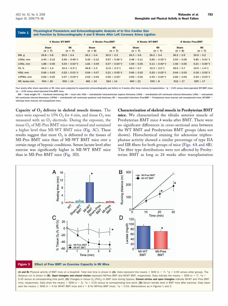
apacity of O2 delivery in skeletal muscle tissues. Theice were exposed to 15% O2 for 4 min, and tissue O2 waseasured with an O2 electrode. During the exposure, the
issue O2 of MI-Pres BMT mice was retained and sustainedhigher level than MI-WT BMT mice (Fig. 3C). These
esults suggest that more O2 is delivered to the tissues ofI-Pres BMT mice than of MI-WT BMT mice over a
ertain range of hypoxic conditions. Serum lactate level afterxercise was significantly higher in MI-WT BMT micehan in MI-Pres BMT mice (Fig. 3D).
Figure 3 Effect of Pres BMT on Exercise Capacity in MI Mice
(A and B) Physical activity of BMT mice on a treadmill. Total rest time is shown indistance run is shown in (B). Open triangles and closed circles represent MI-Pres0.05 versus at corresponding time point. (C) Changes in tissue O2 (PtiO2) in BMTmice, respectively. Data show the means � SEM (n � 3). *p � 0.05 versus at cosent the means � SEM (n � 6 for MI-WT BMT mice and n � 8 for MI-Pres BMT m
hysiological Parameters and Echocardiographic Analysis of In Vivond Function by Echocardiography 4 and 8 Weeks After Left Coron
Table 1 Physiological Parameters and Echocardiographic Analyand Function by Echocardiography 4 and 8 Weeks Afte
4 Weeks WT-BMT 4 Weeks Pres-B
Sham(n � 7)
MI(n � 7)
Sham(n � 6) (n
BW, g 25.8 � 0.6 25.9 � 0.4 26.1 � 0.4 25.9
LVIDd, mm 3.40 � 0.12 5.56 � 0.46*† 3.42 � 0.12 5.67
LVIDs, mm 1.88 � 0.06 5.04 � 0.53*† 1.81 � 0.09 5.07
FS, % 44.3 � 1.4 10.4 � 2.3*† 46.8 � 1.3 11.6
IVSd, mm 0.66 � 0.03 0.22 � 0.01*† 0.64 � 0.07 0.21
LVPWd, mm 0.66 � 0.02 0.47 � 0.03*† 0.63 � 0.04 0.50
HR, beats/min 504 � 20 500 � 14 481 � 34 524
our weeks after sham operation or MI, mice were subjected to sequential echocardiography justp � 0.05 versus sham-operated Pres-BMT mice.BW � body weight; FS � fractional shortening; HR � heart rate; IVSd � end-diastolic intravent
eft ventricular internal dimension; LVPWd � end-diastolic left ventricular posterior wall thicknessild-type bone marrow cell transplanted mice.
haracterization of skeletal muscle in Presbyterian BMTice. We characterized the tibialis anterior muscle ofresbyterian BMT mice 4 weeks after BMT. There wereo significant differences in cross-sectional area betweenhe WT BMT and Presbyterian BMT groups (data nothown). Histochemical staining for adenosine triphos-hatase activity showed a similar percentage of type IIAnd IIB fibers for both groups of mice (Figs. 4A and 4B).he fiber type distributions were not affected by Presby-
erian BMT as long as 24 weeks after transplantation
ata represent the means � SEM (n � 7). *p � 0.05 versus other groups. Theand MI-WT BMT, respectively. Data indicate the means � SEM (n � 7). *p �
during hypoxia. Closed circles and open triangles indicate MI-WT and -Pres BMTnding time point. (D) Serum lactate level in BMT mice after exercise. Data repre-p � 0.05. Abbreviations as in Figures 1 and 2.
diac Sizertery Ligation
f In Vivo Cardiac Sizet Coronary Artery Ligation
8 Weeks WT-BMT 8 Weeks Pres-BMT
Sham(n � 7)
MI(n � 7)
Sham(n � 6)
MI(n � 7)
26.3 � 0.6 26.2 � 0.4 26.2 � 0.5 25.9 � 0.2
*† 3.48 � 0.11 5.66 � 0.33*† 3.53 � 0.09 5.80 � 0.41*†
*† 1.94 � 0.05 5.11 � 0.40*† 1.92 � 0.05 5.21 � 0.45*†
† 44.0 � 0.7 10.3 � 2.2*† 45.5 � 0.7 10.5 � 1.4*†
*† 0.66 � 0.02 0.20 � 0.02*† 0.64 � 0.03 0.18 � 0.02*†
* 0.65 � 0.02 0.43 � 0.02*† 0.60 � 0.04 0.44 � 0.03*†
490 � 21 530 � 8 512 � 17 529 � 17
or 4 weeks after bone marrow transplantation. *p � 0.05 versus sham-operated WT-BMT mice;
eptum thickness; LVIDd � end-diastolic left ventricular internal dimension; LVIDs � end-systolicyocardial infarction; Pres-BMT � Presbyterian bone marrow cell–transplanted mice; WT-BMT �
(A). DBMTmicerrespoice). *
Carary A
sis or Lef
MT
MI� 7)
� 0.3
� 0.46
� 0.53
� 2.3*
� 0.05
� 0.03
� 14
before
ricular s

(mfi(b(d
cPEeBp
784 Watanabe et al. JACC Vol. 52, No. 9, 2008Hemoglobin and Physical Activity in Heart Failure August 26, 2008:779–86
data not shown). There were no significant differences initochondrial SDH activity in either type IIA or IIB
bers between the WT and Presbyterian BMT groupsFigs. 4A and 4C). Total SDH activity measured byiochemical methods was not different in each groupFig. 4D). These results indicate that Presbyterian BMT
Figure 4 Histological and Biochemical Analyses of Skeletal Mu
(A) Transverse sections of the deep region in the tibialis anterior muscle were sta(SDH) activity (bottom). Scale bars indicate 50 �m. Quantitative analysis resultsTotal SDH activity from tibialis anterior muscle measured by enzymological methodidentified by staining with von Willebrand factor antibody. Capillary density was calpower). Scale bars indicate 50 �m. Abbreviations as in Figures 1 and 2.
id not affect oxidative energy metabolism. Furthermore, u
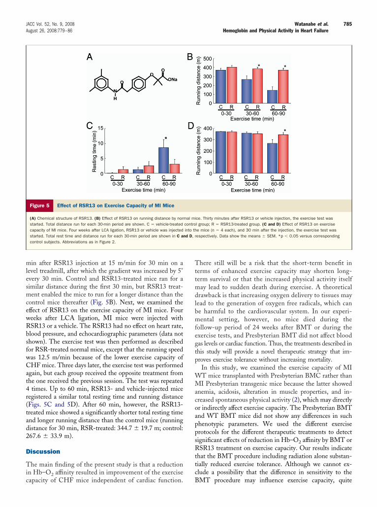
apillary density of the muscle was not altered in theresbyterian BMT groups (Figs. 4E and 4F).ffect of RSR13 on exercise capacity. We examined the
ffect of RSR13 (Fig. 5A) on exercise capacity of WT mice.ecause both RSR13- and vehicle-treated mice could com-lete a run for 90 min on a treadmill under the conditions
in BMT Mice
r adenosine triphosphatase activity (top) and for succinate dehydrogenasescle type distribution and SDH activity are shown in B and C, respectively. (D)
3 for WT-BMT and n � 5 for Pres-BMT mice). (E and F) Endothelial cells wered by counting the numbers of capillaries in 20 random high-power fields (�400
scles
ined fofor mus (n �
culate
sed for the BMT treatment, the exercise test was started 30
mlesmcewRbsfwCat4r(tad2
D
Tic
Tttmdlbmfegtp
WMacoappsRttc
785JACC Vol. 52, No. 9, 2008 Watanabe et al.August 26, 2008:779–86 Hemoglobin and Physical Activity in Heart Failure
in after RSR13 injection at 15 m/min for 30 min on aevel treadmill, after which the gradient was increased by 5°very 30 min. Control and RSR13-treated mice ran for aimilar distance during the first 30 min, but RSR13 treat-ent enabled the mice to run for a longer distance than the
ontrol mice thereafter (Fig. 5B). Next, we examined theffect of RSR13 on the exercise capacity of MI mice. Foureeks after LCA ligation, MI mice were injected withSR13 or a vehicle. The RSR13 had no effect on heart rate,lood pressure, and echocardiographic parameters (data nothown). The exercise test was then performed as describedor RSR-treated normal mice, except that the running speedas 12.5 m/min because of the lower exercise capacity ofHF mice. Three days later, the exercise test was performed
gain, but each group received the opposite treatment fromhe one received the previous session. The test was repeated
times. Up to 60 min, RSR13- and vehicle-injected miceegistered a similar total resting time and running distanceFigs. 5C and 5D). After 60 min, however, the RSR13-reated mice showed a significantly shorter total resting timend longer running distance than the control mice (runningistance for 30 min, RSR-treated: 344.7 � 19.7 m; control:67.6 � 33.9 m).
iscussion
he main finding of the present study is that a reductionn Hb–O2 affinity resulted in improvement of the exercise
Figure 5 Effect of RSR13 on Exercise Capacity of MI Mice
(A) Chemical structure of RSR13. (B) Effect of RSR13 on running distance by normstarted. Total distance run for each 30-min period are shown. C � vehicle-treatedcapacity of MI mice. Four weeks after LCA ligation, RSR13 or vehicle was injectedstarted. Total rest time and distance run for each 30-min period are shown in C acontrol subjects. Abbreviations as in Figure 2.
apacity of CHF mice independent of cardiac function. B
here still will be a risk that the short-term benefit inerms of enhanced exercise capacity may shorten long-erm survival or that the increased physical activity itselfay lead to sudden death during exercise. A theoretical
rawback is that increasing oxygen delivery to tissues mayead to the generation of oxygen free radicals, which cane harmful to the cardiovascular system. In our experi-ental setting, however, no mice died during the
ollow-up period of 24 weeks after BMT or during thexercise tests, and Presbyterian BMT did not affect bloodas levels or cardiac function. Thus, the treatments described inhis study will provide a novel therapeutic strategy that im-roves exercise tolerance without increasing mortality.In this study, we examined the exercise capacity of MIT mice transplanted with Presbyterian BMC rather thanI Presbyterian transgenic mice because the latter showed
nemia, acidosis, alteration in muscle properties, and in-reased spontaneous physical activity (2), which may directlyr indirectly affect exercise capacity. The Presbyterian BMTnd WT BMT mice did not show any differences in suchhenotypic parameters. We used the different exerciserotocols for the different therapeutic treatments to detectignificant effects of reduction in Hb–O2 affinity by BMT orSR13 treatment on exercise capacity. Our results indicate
hat the BMT procedure including radiation alone substan-ially reduced exercise tolerance. Although we cannot ex-lude a possibility that the difference in sensitivity to the
ce. Thirty minutes after RSR13 or vehicle injection, the exercise test wasl group; R � RSR13-treated group. (C and D) Effect of RSR13 on exercisee mice (n � 4 each), and 30 min after the injection, the exercise test wasespectively. Data show the means � SEM. *p � 0.05 versus corresponding
al micontrointo th
nd D, r
MT procedure may influence exercise capacity, quite

drie
apsoifdvtsneiiIoCocsgtWaPFstbessHcemeafd
bdtw
Hb
ATt
Ram
R
1
1
1
1
1
1
786 Watanabe et al. JACC Vol. 52, No. 9, 2008Hemoglobin and Physical Activity in Heart Failure August 26, 2008:779–86
ifferent procedures such as RSR13 treatment gave similaresults in terms of exercise capacity of mice with CHF, andt is likely that Hb–O2 affinity is a major determinant inxercise capacity.
The most common causes of exercise intolerance associ-ted with CHF are the development of dyspnea caused byulmonary congestion and the failure of the heart to provideufficient blood flow to exercising muscles (1). A number ofther peripheral factors may explain exercise intolerance,ncluding abnormalities of vasodilatory capacity, endothelialunction, ergoreflex activation, muscular metabolism, andistribution of cardiac output (13). The impairment ofasodilatory capacity has been attributed to excessive sympa-hetic stimulation, activation of the plasma renin–angiotensinystem, higher levels of endothelin, and impaired release ofitric oxide. Anaerobic metabolism occurs early duringxercise in CHF and is likely an important cause of exercisentolerance (14). Patients with CHF have shown a decreasen oxidative type I fibers and an increase in glycolytic typeIB in the skeletal muscles (14). Levels of oxidative enzymesf mitochondrial enzymes, including SDH, are decreased inHF (15). In this study, the improved exercise performancef Presbyterian BMT mice was not accompanied withhanges in cardiac function, properties of skeletal muscle, orympathetic nerve activity (data not shown). In addition,rowth factors or endothelial stem/progenitor cells con-ained in BMC solution may function differently between
T and Presbyterian BMC. The fact that the acutedministration of RSR13 showed a similar effect withresbyterian BMT, however, excludes such possibilities.urthermore, it is unlikely that slow processes such astructural alterations or metabolic changes containing pro-ein synthesis are involved in the enhanced exercise capacityy Presbyterian BMT. Thus, the mechanism producingnhanced exercise capacity in response to the therapieseems to be derived from enhanced O2 delivery in thekeletal muscle. However, it is still possible that reducingb–O2 affinity may directly or indirectly affect a number of
entral or peripheral factors contributing to exercise intol-rance in CHF. Although the exact underlying molecularechanism for the improved exercise capacity remains to be
lucidated, this study clearly indicates that a syntheticllosteric modifier of Hb, such as an RSR13, will be usefulor the treatment of patients with CHF to improve theiraily physical activity.Because RSR13 is a relatively short-acting drug and can
e administered only intravenously, modification of therug is necessary for its clinical application. Further inves-igations using larger animals over a longer period of time
ill be necessary to determine whether the reduction in Kb–O2 affinity and increased physical activity actuallyenefit the lives of patients with CHF.
cknowledgmentshe authors thank Naoko Watanabe and Sunao Tanaka for
heir technical assistance.
eprint requests and correspondence: Dr. Kinya Otsu, 2-2 Yamad-oka, Suita, Osaka 565-0871, Japan. E-mail: [email protected].
EFERENCES
1. Braunwald E, editor. Heart Disease: A Textbook of CardiovascularMedicine. Philadelphia, PA: Saunders, 1997.
2. Shirasawa T, Izumizaki M, Suzuki Y, et al. Oxygen affinity ofhemoglobin regulates O2 consumption, metabolism, and physicalactivity. J Biol Chem 2003;278:5035–43.
3. Randad R, Mahran M, Mehanna A, et al. Allosteric modifiers ofhemoglobin. 1. Design, synthesis, testing, and structure-allostericactivity relationship of novel hemoglobin oxygen affinity decreasingagents. J Med Chem 1991;34:752–7.
4. Abraham D, Wireko F, Randad R, et al. Allosteric modifiers of hemo-globin: 2-[4-[[(3,5-disubstituted anilino)carbonyl]methyl]phenoxy]-2-methylpropionic acid derivatives that lower the oxygen affinity of hemo-globin in red cell suspensions, in whole blood, and in vivo in rats.Biochemistry 1992;31:9141–9.
5. Soares MB, Lima RS, Rocha LL, et al. Transplanted bone marrowcells repair heart tissue and reduce myocarditis in chronic chagasicmice. Am J Pathol 2004;164:441–7.
6. Suzuki Y, Shimizu T, Sakai H, et al. Model mice for Presbyterianhemoglobinopathy (Asn(beta108)¡Lys) confer hemolytic anemiawith altered oxygen affinity and instability of Hb. Biochem BiophysRes Commun 2002;295:869–76.
7. Mawjood A, Imai K. Automatic measurement of the red cell oxygendissociation curve identical with the whole blood curve. Jpn J Physiol1999;49:379–87.
8. Yamaguchi O, Higuchi Y, Hirotani S, et al. Targeted deletion ofapoptosis signal-regulating kinase 1 attenuates left ventricular remod-eling. Proc Natl Acad Sci U S A 2003;100:15883–8.
9. Nakayama H, Otsu K, Yamaguchi O, et al. Cardiac-specific overex-pression of a high Ca2� affinity mutant of SERCA2a attenuates in vivopressure overload cardiac hypertrophy. FASEB J 2003;17:61–3.
0. Yamashita N, Hoshida S, Otsu K, et al. Exercise provides directbiphasic cardioprotection via manganese superoxide dismutase activa-tion. J Exp Med 1999;189:1699–706.
1. Trounce IA, Kim YL, Jun AS, et al. Assessment of mitochondrialoxidative phosphorylation in patient muscle biopsies, lymphoblasts,and transmitochondrial cell lines. Methods Enzymol 1996;264:484–509.
2. Grocott HP BR, Sheng H, Miura Y, Steffen R, Pearlstein RD,Warner DS. Effects of a synthetic allosteric modifier of hemoglobinoxygen affinity on outcome from global cerebral ischemia in the rat.Stroke 1998;29:1650–5.
3. Pina I, Apstein C, Balady G, et al. Exercise and heart failure: a statementfrom the American Heart Association Committee on exercise, rehabili-tation, and prevention. Circulation 2003;107:1210–25.
4. Drexler H, Riede U, Munzel T, et al. Alterations of skeletal muscle inchronic heart failure. Circulation 1992;85:1751–9.
5. Sullivan M, Green H, Cobb F. Skeletal muscle biochemistry andhistology in ambulatory patients with long-term heart failure. Circu-lation 1990;81:518–27.
ey Words: hemoglobin y heart failure y oxygen y muscles.