ref lecture ejournal
TRANSCRIPT
7282019 Ref Lecture eJournal
httpslidepdfcomreaderfullref-lecture-ejournal 110
33013 eJournal
wwwjaypeejournalscomeJournalsShowTextaspxID=965ampType=FREEampTYP=TOPampIN=_eJournalsimagesJPLOGOgifampIID=84ampisPDF=NO
JAYPEE JOURNALS Subscribers Login
International Scientific Journals from Jaypee Home Instructions Editorial Board Current Issue Pubmed Archives Subscription Advertisement
Show Contents
DSJUOGREVIEW ARTICLE
Ultrasonographic Evaluation of the
Fetal Neural AxisSakshi Tomar
Department of Radiodiagnosis Sanjay Pathology Center Lakhimpur Kheri Uttar Pradesh India
Correspondence Sakshi Tomar Department of Radiodiagnosis Sanjay Pathology Center Lakhimpur Kheri Uttar Pradesh
India e -mail sakshi10tomaryahoocoin
ABSTRACT
The central nervous system is probably the first organ system to be investigated in utero by diagnostic ultrasound Anencephaly was the first
congenital anomaly to be recognized by this technique before viability Since then the investigation of the fetal neural axis has steadily
remained a central issue of antenatal sonography Such an interest is explained by a number of reasons CNS anomalies are frequent and often
have a severe prognosis In many cases they have a genetic background consequently a large number of couples are at risk and warrant
antenatal diagnosis Modern high resolution ultrasound equipment yields a unique potential in evaluating normal and abnormal anatomy of the
fetal neural axis beginning at very early stages of development
Keywords Central nervous system Antenatal sonography Congenital anomalies
NORMAL SONOGRAPHIC ANATOMY
The fetal brain undergoes major developmental changes throughout
pregnancy Fetal neurosonography demands a thorough knowledge of
ontogeny of the brain If one is unaware of the normal developmental
changes of the brain both missed as well as false positive diagnosis
will be the result1
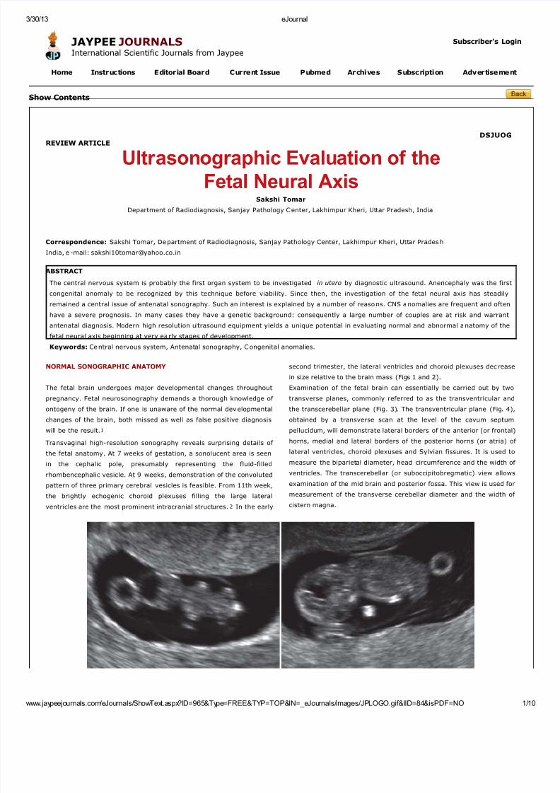
Transvaginal high-resolution sonography reveals surprising details of
the fetal anatomy At 7 weeks of gestation a sonolucent area is seen
in the cephalic pole presumably representing the fluid-filled
rhombencephalic vesicle At 9 weeks demonstration of the convoluted
pattern of three primary cerebral vesicles is feasible From 11th week
the brightly echogenic choroid plexuses filling the large lateral
ventricles are the most prominent intracranial structures2 In the early
second trimester the lateral ventricles and choroid plexuses decrease
in size relative to the brain mass (Figs 1 and 2)
Examination of the fetal brain can essentially be carried out by two
transverse planes commonly referred to as the transventricular and
the transcerebellar plane (Fig 3) The transventricular plane (Fig 4)
obtained by a transverse scan at the level of the cavum septum
pellucidum will demonstrate lateral borders of the anterior (or frontal)
horns medial and lateral borders of the posterior horns (or atria) of
lateral ventricles choroid plexuses and Sylvian fissures It is used to
measure the biparietal diameter head circumference and the width of
ventricles The transcerebellar (or suboccipitobregmatic) view allows
examination of the mid brain and posterior fossa This view is used for
measurement of the transverse cerebellar diameter and the width of
cistern magna
7282019 Ref Lecture eJournal
httpslidepdfcomreaderfullref-lecture-ejournal 210
33013 eJournal
wwwjaypeejournalscomeJournalsShowTextaspxID=965ampType=FREEampTYP=TOPampIN=_eJournalsimagesJPLOGOgifampIID=84ampisPDF=NO 2
Fig 1 Longitudinal views of normal embryos at 7 and 9 weeks At 7 weeks the rhombencephalic vesicle the primordium of the 4th ventricle is usually
the only discernible cerebral structure At 9 weeks the prosencepha lic vesicle primordium of the 3rd ventricle and the m ese ncephalic vesicle
primordium of the aqueduct of Sylvius can be seen as well
373 JAYPEE
Ultrasonographic Evaluation of the Fetal Neural Axis
Fig 2 Transverse views of the cephalic pole normal embryos at 7 to 12 weeks At 7 weeks the rhombencephalic vesicle is the dominant structure At 9
weeks the prosencephalic vesicle is seen infront of the rhombencephalon At 12 weeks the cerebral hemispheres have greatly developed and it is
possible to appreciate the midline as well as the large echogenic choroid plexus filling the cavity of the ventricles
Fig 3 The two axial planes recommended for the standard
sonographic examination of the fetal brain
Fig 5 Schematic representation of the scanning planes commonly
used for the evaluation of fetal cerebral anatomy
7282019 Ref Lecture eJournal
httpslidepdfcomreaderfullref-lecture-ejournal 310
33013 eJournal
wwwjaypeejournalscomeJournalsShowTextaspxID=965ampType=FREEampTYP=TOPampIN=_eJournalsimagesJPLOGOgifampIID=84ampisPDF=NO 3
Fig 4 Transventricular view showing the cavum septum pellucidum and
the thalami
Fig 6 Transabdo minal a xial view demo nstrating the circle of willis
Donald School Journal of Ultrasound in Obstetrics and Gynecology October-December 20104(4)373-382 374
Sakshi Tomar
Fig 7 Sagittal axial and coronal view of the normal fetal spine in the 2nd trimester
Additional scanning planes along with different orientations may be
required from time to time to better define subtle details of intracranial
anatomy better in selected cases Reverberation artifacts usually
obscure the cerebral hemisphere close to the transducer Visualizationof both cerebral hemispheres would require sagittal and coronal planes
that are often difficult to obtain and may require vaginal sonography
(Fig 5) Color Doppler sonography helps in the diagnosis of lesions
(Fig 6)
Luckily unilateral cerebral lesions are rare and often associated with a
shift in the midline echo Therefore we adhere to the approach that in
standard examination only one hemisphere is seen and symmetry is
assumed unless otherwise proven
A sagittal and coronal view of the entire fetal spine should be obtained
in each case In the sagittal plane the normal spine has a double
railway appearance and it is possible to appreciate the intact soft
tissues above it In the coronal plane three ossification centers of the
vertebra form three regular lines that tether down into the sacrumThese views are used to assess the integrity of the vertebrae (to rule
out spina bifida) and the presence and regularity of the whole spine (to
rule out sacral agenesis and scoliosis) (Fig 7)
NEURAL TUBE DEFECTS
These include anencephaly spina bifida and cephalocele In
anencephaly there is absence of the cranial vault (acrania) and
telencephalon with secondary degeneration of the brain Cephaloceles
(encephalocele) are cranial defects usually occipital with herniated
fluid-filled or bra in-filled cysts In spina bifida the neural arch usually
in the lumbosacral region is incomplete with secondary damage to the
ex osed nerves
-s are implicated in about 10 of the cases However the precise
etiology for the majority of these defects is unknown When a parent
or previous sibling has had a neural tube defect the risk of recurrence
is 5 to 10 Periconceptual supplementation of the maternal diet withfolate reduces by about half the risk of developing these defects
Diagnosis
The diagnosis of anencephaly during the second trimester of
pregnancy is based on the demonstration of absent cranial vault and
cerebral hemispheresHowever the facial bones brain stem and
portions of the occipital bones and midbrain are usually present
Associated spinal lesions are found in up to 50 of cases In the first
trimester the diagnosis can be made after 11 weeks when ossification
of the skull normally occurs Anencephaly is considered to be the final
stage of acrania as a consequence of disruption of abnormal brain
tissue unprotected by the calvarium (Figs 8 and 9) In the first
trimester the pathognomonic feature is acrania the brain being eitherentirely normal or at varying degrees of distortion and disruption Most
anencephalics have normal eyes The orbits are often shallow causing
protrusion of the eyes The forebrain is then replaced by an
angiomatous mass with multiple cavities containing CSF3
Diagnosis of spina bifida requires the systematic examination of each
neural arch from the cervical to the sacral region both transversely
and longitudinally In the transverse scan the normal neural arch
appears as a closed circle with an intact skin covering whereas in
spina bifida the arch is U shaped and there is an associated bulging
meningocele (thinwalled cyst) or myelomeningocele (Fig 10) The
extent of the defect and any associated kyphoscoliosis are best
assessed in the longitudinal scan (Fig 11)
7282019 Ref Lecture eJournal
httpslidepdfcomreaderfullref-lecture-ejournal 410
33013 eJournal
wwwjaypeejournalscomeJournalsShowTextaspxID=965ampType=FREEampTYP=TOPampIN=_eJournalsimagesJPLOGOgifampIID=84ampisPDF=NO 4
Prevalence
This is subject to large geographical and ethnic variations In the
United Kingdom the prevalence is about 5 per 1000 births
Anencephaly and spina bifida with an approximately equal
prevalence account for 95 of the cases and cephalocele for the
remaining 5
Etiology
Chromosomal abnormalities single mutant genes and maternaldiabetes mellitus or ingestion of teratogens such as antiepileptic drug-
recognition of associated abnormalities in the skull and brain These
abnormalities are secondary to the Arnold-Chiari malformation and
include frontal bone scalloping (lemon sign) and obliteration of the
cisterna magna with either an absent cerebellum or abnormal
anterior curvature of the cerebellar hemispheres (banana sign)4 A
variable degree of ventricular enlargement is present in virtually all
cases of open spina bifida at birth but in only about 70 of cases in
the midtrimester
Encephaloceles are recognized as cranial defects with herniated fluid-
filled or brain-filled cysts They are most commonly found in an
occipital location (75 of the cases) but alternative sites include the
375 JAYPEE
Ultrasonographic Evaluation of the Fetal Neural Axis
Fig 8 Anencephaly with acrania Note the absence of the calvarium
above the orbits The a ngiomatous stroma is seen
Fig 9 Anencephaly
Fig 12 Loculated paracranial cyst in the third trimester fetus Small
defect in the cranial vault is seen Cranial cephalocele
frontoethmoidal and parietal regions (Fig 12)
Prognosis
Anencephaly is fatal at or within the hours of birth In cephalocele the
prognosis is inversely related to the amount of herniated cerebral
tissue Overall the neonatal mortality is about 40 and more that 80
of survivors are intellectually and neurologically handicapped In spina
bifida the surviving infants are often severely handicapped with
paralysis in the lower limbs and double incontinence Despite the
associated hydrocephalus requiring surgery intelligence may be
normal5
Fetal Therapy
There is some experimental evidence that in utero closure of spina
bifida may reduce the risk of handicap because the amniotic fluid in
the third trimester is thought to be neurotoxic
VENTRICULOMEGALY AND HYDROCEPHALUS
In hydrocephalus there is pathological increase in the size o f cerebral
ventricles
Prevalence
Hydrocephalus is found in about 2 per 1000 births Ventriculomegaly
(lateral ventricle diameter of 10 mm or more at the level of the
glomus of choroid plexus) is found in 1 of pregnancies at the 20 to
23 week scan Therefore the majority of fetuses with
ventriculomegaly do not develop hydrocephalus
7282019 Ref Lecture eJournal
httpslidepdfcomreaderfullref-lecture-ejournal 510
7282019 Ref Lecture eJournal
httpslidepdfcomreaderfullref-lecture-ejournal 610
33013 eJournal
wwwjaypeejournalscomeJournalsShowTextaspxID=965ampType=FREEampTYP=TOPampIN=_eJournalsimagesJPLOGOgifampIID=84ampisPDF=NO 6
posterior horn 10-15 mm) and overt ventriculomegaly or
hydrocephalus (diameter greater than 15 mm)7 (Figs 13A and B)
Prognosis
Fetal or perinatal death and neurodevelopment in survivors are
strongly related to the presence of other malformations and
chromosomal defects Although mild also referred to as borderline
ventriculomegaly is generally associated with a good prognosis
affected fetuses form the group with the highest incidence of
chromosomal abnormalities (often trisomy 21) In addition in a few
cases with apparently isolated mild ventriculomegaly there may be an
underlying cerebral maldevelopment (such as lissencephaly) or
destructive lesion (such as periventricular leukomalacia) Recent
evidence suggests that in about 10 of cases there is mild to
moderate neurodevelopmental delay
Fetal Therapy
There is some experimental evidence that in utero cerebro spinal fluid
diversion may be beneficial However attempts in the 1980s to treat
hydrocephalic fetuses by ventriculoamniotic shunting have now been
abandoned because of poor results mainly because of inappropriate
selection of patients It is possible that intrauterine drainage may be
beneficial if all intraand extracerebral malformations and
chromosomal defects are excluded and if serial ultrasound scans de-
of the thalami In the semilobar type there is partial segmentation of
the ventricles and cerebral hemispheres posteriorly with incomplete
fusion of the thalami In lobar holoprosencephaly there is normal
separation of the ventricles and thalami but absence of the septum
pellucidum The first two types are often accompanied by
microcephaly and facial abnormalities8
Prevalence
Holoprosencephaly is found in about 1 per 10000 births
Etiology
Although in many cases the cause is a chromosomal abnormality
(usually trisomy 13) or a genetic disorder with an autosomal dominant
or recessive mode of transmission The etiology is unknown in many
cases The risk of recurrence for sporadic nonchromosomal
holoprosencephaly the empirical recurrence risk is 6
Diagnosis
In the standard transverse view of the fetal head for measurement of
the biparietal diameter there is a single dilated midline ventricle
replacing the two lateral ventricles or partial segmentation of the
ventricles The alobar and semilobar types are often associated with
facial defects such as hypotelorism or cyclopia facial cleft and nasal
hypoplasia or proboscis9 (Fig 14)
377 JAYPEE
Ultrasonographic Evaluation of the Fetal Neural Axis
Fig 14 In this fe tus at 11 weeks the absence of midline and presence
of fused choroid plexus are diagnostic of alobar holoprosencephaly
Prognosis
Alobar and semilobar holoprosencephaly are lethal Lobar
holoprosencephaly is associated with mental retardation
AGENESIS OF CORPUS CALLOSUM
The corpus callosum is a bundle of fibers that connects the two
cerebral hemispheres It develops at 12 to 18 weeks of gestation
Agenesis of the corpus callosum may be either complete or partial
(usually a ffecting the posterior part)
Prevalence
Agenesis of the corpus callosum is found in about 5 per 1000 births
Etiology
Agenesis of the corpus callosum may be due to maldevelopment or
secondary to a destructive lesion It is commonly associated with
chromosomal abnormalities usuall trisomies 18 13 and 8 and more
-tion of the lateral ventricles (enlargement of the posterior horns)10
Agenesis of the corpus callosum is demonstrated in the mid-coronal
and mid-sagittal views which may require vaginal sonography (Fig
15)
Prognosis
This depends on the underlying cause In about 90 of those with
apparently isolated agenesis of the corpus callosum development isnormal
DANDY-WALKER COMPLEX
The Dandy-Walker complex refers to a spectrum of abnormalities of
the cerebellar vermis cystic dilation of the fourth ventricle and
enlargement of the cisterna magna The condition is classified into (a)
Dandy-Walker malformation (complete or partial agenesis of the
cerebellar vermis and enlarged posterior fossa) (b) Dandy-Walker
variant (partial agenesis of the cerebellar vermis without enlargement
of the posterior fossa) and (c) mega-cisterna magna (normal vermis
and fourth ventricle)11
Prevalence
Dandy-Walker ma lformation is found in about 1 per 30000 births
Etiology
The Dandy-Walker complex is a non-specific endpoint of chromosomal
abnormalities (usually trisomy 18 or 13 and triploidy) more than 50
genetic syndromes congenital infection or teratogens such as
warfarin but it can also be an isolated finding
Diagnosis
Ultrasonographically the contents of the posterior fossa are visualized
through a transverse suboccipitobregmatic section of the fetal head In
the Dand -Walker malformation there is c stic dilatation of the fourth
7282019 Ref Lecture eJournal
httpslidepdfcomreaderfullref-lecture-ejournal 710
33013 eJournal
wwwjaypeejournalscomeJournalsShowTextaspxID=965ampType=FREEampTYP=TOPampIN=_eJournalsimagesJPLOGOgifampIID=84ampisPDF=NO 7
than 100 genetic syndromes
Diagnosis
The corpus callosum is not visible in the standard transverse views of
the brain but agenesis of the corpus callosum may be suspected by
the absence of cavum septum pellucidum and the teardrop configura-
Fig 15 Agenesis of corpus callosum
ventricle with partial or complete agenesis of the vermis (Figs 16A and
B) In more than 50 of the cases there is associated hydrocephalus
and other extracranial defects Enlarged cisterna magna is diagnosed
if the vertical distance from the vermis to the inner border of the skull
is more than 10 mm12
Figs 16A and B (A) Classic Dandy-Walker Malformation A large defect
of the cerebellar vermis connects the 4th ventricle with a cystic cisterna
magna (B) Megacisterna Magna
Donald School Journal of Ultrasound in Obstetrics and Gynecology October-December 20104(4)373-382 378
Sakshi Tomar Prenatal diagnosis of isolated partial agenesis of the vermis is difficult
and a false diagnosis can be made prior to 18 weeks gestation when
the formation of the vermis is incomplete and anytime in gestation if
the angle of insonation is too steep
Prognosis
Dandy-Walker malformation is associated with a high postnatal
mortality (about 20) and a high incidence (more than 50) of
impaired intellectual and neurological development Experience with
apparently isolated partial agenesis of the vermis or enlarged cisterna
magna is limited and the prognosis for these conditions is uncertain
DESTRUCTIVE CEREBRAL LESIONS
These lesions include hydranencephaly porencephaly and
schizencephaly In hydranencephaly there is absence of the cerebral
hemispheres with preservation of the midbrain and cerebellum In
porencephaly there are cystic cavities within the brain that usually
communicate with the ventricular system the subarachnoid space or
both Schizencephaly is associated with clefts in the fetal brain
connecting the lateral ventricles with the subarachnoid space
Prevalence
Destructive cerebral lesions are found in about 1 per 10000 births
Etiology
Hydranencephaly is a sporadic abnormality that may result from
widespread vascular occlusion in the distribution of internal carotid
arteries prolonged severe hydrocephalus or an overwhelming
infection such as toxoplasmosis and cytomegalovirus Porencephaly
may be caused by infarction of the cerebral arteries or hemorrhage
into the brain parenchyma Schizencephaly may be a primary disorder
of brain development or it may be due to bilateral occlusion of the
middle cerebral arteries
Diagnosis
Fig 17 Bilateral clefts typical of s chizencephaly are se en
Prognosis
Hydranencephaly is usually incompatible with survival beyond early
infancy The prognosis in porencephaly is related to the size and
location of the lesion and although there is increased r isk of impaired
neurodevelopment in some cases development is normal
Schizencephaly is associated with severe neurodevelopmental delay
and seizures
DISORDERS OF NERVE CELL PROLIFERATION
Microcephaly Microcephaly means small head and brain
Prevalence
Microcephaly is found in about 1 per 1000 births
Etiology
This may result from chromosomal and genetic abnormalities fetal
hypoxia congenital infection and exposure to radiation or other
teratogens such maternal anticoagulation with warfarin It is
7282019 Ref Lecture eJournal
httpslidepdfcomreaderfullref-lecture-ejournal 810
33013 eJournal
wwwjaypeejournalscomeJournalsShowTextaspxID=965ampType=FREEampTYP=TOPampIN=_eJournalsimagesJPLOGOgifampIID=84ampisPDF=NO 8
omp e e a sence o ec oes rom e an er or an m e ossa
distinguishes hydranencephaly from severe hydrocephalus in which a
thin rim of remaining cortex and the midline echo can always be
identified In porencephaly there is one or more cystic area in the
cerebral cortex which usually communicates with the ventricle The
differential diagnosis is from intracranial cysts (arachnoid
glioependymal) that are usually found either within the scissures or in
the midline and compress the brain In schizencephaly there are
bilateral clefts extending from the lateral ventricles to the
subarachnoid space and is usually associated with absence of the
cavum septum pellucidum13 (Fig 17)
commony oun n e presence o o er ra n a norma es suc as
cephalocele and holoprosencephaly
Diagnosis
The diagnosis is made by the demonstration of brain abnormalities
such as holoprosencephaly In cases with apparently isolated
microcephaly it is necessary to demonstrate progressive decrease in
the head to abdomen circumference ra tio to below the 1st centile with
advancing gestation14 Such diagnosis may not be apparent before
the third trimester In microcephaly there is a typical disproportion
between the size of the skull and face The brain is small with the cer- 379 JAYPEE
Ultrasonographic Evaluation of the Fetal Neural Axis
Fig 18 Fetal microcephaly Note the sloping of the forehead
-ebral hemispheres affected to a greater extent than the midbrain and
posterior fossa Demonstration of a sloping forehead also increases
the index of suspicion (Fig 18)
Prognosis
This depends on the underlying cause but in more than 50 of cases
there is severe mental retardation
B Megalencephaly Megalencephaly means large head and brain
PrevalenceMegalencephaly is a very rare abnormality
Etiology
This is usually familial with no adverse consequence However it may
also be the consequence of genetic syndromes such as Beckwith-
Wiedemann syndrome achondroplasia neurofibromatosis and
tuberous sclerosis Unilateral megalencephaly is a sporadic condition
Diagnosis
The diagnosis is made by the demonstration of a head to abdomen
circumference ratio above the 99th centile without the evidence of hydrocephalus or intracranial masses Unilateral megalencephaly is
characterized by macrocrania a shift in the midline echo borderline
enlargement of the lateral ventricle and a typical gyri of the affected
hemisphere15
Prognosis
Isolated megalencephaly is usually an asymptomatic condition
Unilateral megalencephaly is associated with severe mental
retardation and untreatable seizures
INTRACRANIAL HEMORRHAGE IN UTERO
Hemorrha e ma be seen in a t ical sube end mal or
junction
Perinatal asphyxia known to induce fluctuation in blood
pressure16
ARTERIOVENOUS MALFORMATION
VEIN OF GALEN ANEURYSM
This is a midline aneurysmal dilation of the vein of Galen due to an
arteriovenous malformation with major hemodynamic disturbances
Prevalence
Vein of Galen aneurysm is a rare abnormality
Etiology
Vein of Galen aneurysm is a sporadic abnormality
Diagnosis
The diagnosis is made by the demonstration of a supratentorial
midline translucent elongated cyst Color Doppler demonstrates active
arteriovenous flow within the cyst There may be associated evidence
of high-output heart failure17 (Fig 19)
Prognosis
In the neonatal period about 50 of the infants present with heart
failure and the rest are asymptomatic In later life hydrocephalus and
intracranial hemorrhage may develop Good results can be achieved
by catheterization and embolization of the malformation
CHOROID PLEXUS CYSTS
These cysts usually bilateral are in the choroid plexuses of the lateral
cerebral ventricles
PrevalenceChoro id plexus cysts are found in about 2 of fetuses at 20 weeks o f
gestation but in more than 90 of cases they resolve by 26 weeks
Etiology
The choroid plexus is easily visualized from 10 weeks of gestation
when it occupies almost the entire hemisphere Thereafter and until 26
weeks there is a rapid decrease in both the size of the choroid plexus
and of the lateral cerebral ventricle in relation to the hemisphere
Choro id plexus cysts contain cerebrospinal fluid and cellular debris
Diagnosis
Single or multiple cystic areas (greater than 2 mm in diameter) in one
or both choroid plexuses18 (Fig 20)
7282019 Ref Lecture eJournal
httpslidepdfcomreaderfullref-lecture-ejournal 910
33013 eJournal
wwwjaypeejournalscomeJournalsShowTextaspxID=965ampType=FREEampTYP=TOPampIN=_eJournalsimagesJPLOGOgifampIID=84ampisPDF=NO 9
intraparenchymal location
Etiology
Sudden changes in the cerebral blood pressure leading to
ruptured capillaries in the germinal matrix or capillary venous
Prognosis
They are usually of no pathological significance but they are
associated with an increased risk for trisomy 18 and possibly trisomy
21 In the absence of other markers of trisomy 18 the maternal age-
related risk is increased by a factor of 15
Donald School Journal of Ultrasound in Obstetrics and Gynecology October-December 20104(4)373-382 380
Sakshi Tomar
Fig 19 Vein of Galen aneurysm Color Doppler flow image of the fetal
head demonstrating a vein of galen aneurysm (arrow) supplied by the
feed ing vesse ls from the circle of willis
Fig 20 Bilateral choroid plexus cysts seen in a transverse axial
sonogram
ARACHNOID CYSTS
Arachnoid cysts are fluid-filled cyst contained within the arachnoid
space
Prevalence
Arachnoid cysts are extremely rare
Etiology
Unknown infectious process has been hypothesized but it is unlikely
that this may explain the congenital cysts
Diagnosis
Arachnoid cys ts appear on antenatal ultrasound as sonolucent lesions
Fig 21 Arachnoid Cyst Corresponding MRI T2W image is seen
difficult Interhemispheric cysts associated with agenesis of the corpus
callosum most likely are not arachnoid cysts but rather glioependymal
cysts
Prognosis
Large cysts may cause intracranial hypertension and require
neurosurgical treatment However a normal intellectual development
in the range of 80 to 90 is reported by most series Spontaneous
remission has been described both in the postnatal as well as in the
antenatal per iod Glioependymal cyst that should be suspected in the
cases with associated agenesis of corpus callosum probably re flects a
greater degree of derangement in the development of the brain and
this may be reflected in worse outcome
CONCLUSION
Modern ultrasound equipments yield a unique potential for evaluation
of normal and abnormal fetal CNS very early in pregnancy It should
be stressed however that some cerebral anomalies are the
consequence of disruptions or are characterized by intrauterine
development Counseling the parents and deciding on a sensible
obstetric management are frequently difficult
REFERENCES
Monteagudo A Fetal neurosonography Should it be routine
Should it be detailed [Editorial] Ultrasound Obstet Gynecol
1998121
Callen Ultrasonography in Obstetrics and Gynecology (4th ed)
277-304
Bronshtein M Ornoy A Acrania Anencephaly resulting from
secondary degeneration of a closed neural tube Two cases in
the same family J Clin Ultrasound 199119230
Levi S Hijazi Y Schapps J et al Sensitivity and specificity of
routine antenatal screening for congenital anomalies by
ultrasound The Belgian Multicentric Study Ultrasound Obstet
Gynecol 19911102
Goldstein RB LaPidus AS Filly RA Fetal cephaloceles Diagnosis
7282019 Ref Lecture eJournal
httpslidepdfcomreaderfullref-lecture-ejournal 1010
33013 eJournal
j j l J l Sh T t ID 965ampT FREEampTYP TOPampIN J l i JPLOGO ifampIID 84ampi PDF NO 10
with a thin regular outline and containing no blood flow do not
communicate with the lateral ventricles and anyhow are not associated
with the loss of brain tissue (Fig 21) They occur most frequently in
the area of cerebral fissure and midline Large cyst may cause
significant mass effect and the distinction from porencephaly may be
with US Radiology 1991180803
Filly RA Goldstein RB Callen PW Fetal ventricle Importance in
381 JAYPEE
Ultrasonographic Evaluation of the Fetal Neural Axis routine obstetric sonography Radiology 19911811
Cardoza JD Goldstein RB Filly RA Exclusion of fetal
ventriculomegaly with a single measurement The width of lateral
ventricular atrium Radiology 1988169711
DeMyer W Holoprosencephaly vol 30 Amsterdam Elsevier
1977
Bronshtein M Weiner Z Early transvaginal sonographic diagnosis
of a lobar holoprosencephaly Prenat Diagn 199111459
Bennett GL Bromley B Benacerraf BR Agenesis of corpus
callosum Prenatal detection usually is not possible before 22
week s o f ge station Ra diology 1996199447
Benda C The Dandy-Walker syndrome or the so called atresia of
the foramen Magendie J Neuropathol Exp Neurol 19541314
Pilu G Goldstein I Reece E et al Sonography of fetal Dandy
Walker Malformations A reappraisal Ultrasound Obstet Gynecol
19922151
Pilu G falco P Perolo A Differential diagnosis and outcome of
fetal intracranial hypoechoic lesions Report of 21 cases
Ultrasound Obstet Gynecol 19979229
Chervenak FA Jeanty P Cantraine F et al The diagnosis of
fetal microcephaly Am J Obstet Gynecol 1984149512
Bignami A Palladini G Zappella M Unilateral Megalencephaly
with nerve cell hypertrophy An anatomical and quantitative
histochemical study Brain Res 19689103
Vergani P Strobelt N Locatelli A et al Clinical significance of
fetal intracranial haemorrhage Am J Obstet Gynecol
1996175536
Comstock CH Kirk JS Arteriovenous malformations Location
and evolution in the fetal brain J Ultrasound Med 199110361
Snijders R Shawa L Nicolaides K Fetal choroid cysts a nd trisomy
18 Assessment of risk based on ultrasound findings and
maternal age Prenat Diagnosis 199414119
382
JAYPEE
copy Jaypee Brothers Medical Publishers (P) Ltd
7282019 Ref Lecture eJournal
httpslidepdfcomreaderfullref-lecture-ejournal 310
33013 eJournal
wwwjaypeejournalscomeJournalsShowTextaspxID=965ampType=FREEampTYP=TOPampIN=_eJournalsimagesJPLOGOgifampIID=84ampisPDF=NO 3
Fig 4 Transventricular view showing the cavum septum pellucidum and
the thalami
Fig 6 Transabdo minal a xial view demo nstrating the circle of willis
Donald School Journal of Ultrasound in Obstetrics and Gynecology October-December 20104(4)373-382 374
Sakshi Tomar
Fig 7 Sagittal axial and coronal view of the normal fetal spine in the 2nd trimester
Additional scanning planes along with different orientations may be
required from time to time to better define subtle details of intracranial
anatomy better in selected cases Reverberation artifacts usually
obscure the cerebral hemisphere close to the transducer Visualizationof both cerebral hemispheres would require sagittal and coronal planes
that are often difficult to obtain and may require vaginal sonography
(Fig 5) Color Doppler sonography helps in the diagnosis of lesions
(Fig 6)
Luckily unilateral cerebral lesions are rare and often associated with a
shift in the midline echo Therefore we adhere to the approach that in
standard examination only one hemisphere is seen and symmetry is
assumed unless otherwise proven
A sagittal and coronal view of the entire fetal spine should be obtained
in each case In the sagittal plane the normal spine has a double
railway appearance and it is possible to appreciate the intact soft
tissues above it In the coronal plane three ossification centers of the
vertebra form three regular lines that tether down into the sacrumThese views are used to assess the integrity of the vertebrae (to rule
out spina bifida) and the presence and regularity of the whole spine (to
rule out sacral agenesis and scoliosis) (Fig 7)
NEURAL TUBE DEFECTS
These include anencephaly spina bifida and cephalocele In
anencephaly there is absence of the cranial vault (acrania) and
telencephalon with secondary degeneration of the brain Cephaloceles
(encephalocele) are cranial defects usually occipital with herniated
fluid-filled or bra in-filled cysts In spina bifida the neural arch usually
in the lumbosacral region is incomplete with secondary damage to the
ex osed nerves
-s are implicated in about 10 of the cases However the precise
etiology for the majority of these defects is unknown When a parent
or previous sibling has had a neural tube defect the risk of recurrence
is 5 to 10 Periconceptual supplementation of the maternal diet withfolate reduces by about half the risk of developing these defects
Diagnosis
The diagnosis of anencephaly during the second trimester of
pregnancy is based on the demonstration of absent cranial vault and
cerebral hemispheresHowever the facial bones brain stem and
portions of the occipital bones and midbrain are usually present
Associated spinal lesions are found in up to 50 of cases In the first
trimester the diagnosis can be made after 11 weeks when ossification
of the skull normally occurs Anencephaly is considered to be the final
stage of acrania as a consequence of disruption of abnormal brain
tissue unprotected by the calvarium (Figs 8 and 9) In the first
trimester the pathognomonic feature is acrania the brain being eitherentirely normal or at varying degrees of distortion and disruption Most
anencephalics have normal eyes The orbits are often shallow causing
protrusion of the eyes The forebrain is then replaced by an
angiomatous mass with multiple cavities containing CSF3
Diagnosis of spina bifida requires the systematic examination of each
neural arch from the cervical to the sacral region both transversely
and longitudinally In the transverse scan the normal neural arch
appears as a closed circle with an intact skin covering whereas in
spina bifida the arch is U shaped and there is an associated bulging
meningocele (thinwalled cyst) or myelomeningocele (Fig 10) The
extent of the defect and any associated kyphoscoliosis are best
assessed in the longitudinal scan (Fig 11)
7282019 Ref Lecture eJournal
httpslidepdfcomreaderfullref-lecture-ejournal 410
33013 eJournal
wwwjaypeejournalscomeJournalsShowTextaspxID=965ampType=FREEampTYP=TOPampIN=_eJournalsimagesJPLOGOgifampIID=84ampisPDF=NO 4
Prevalence
This is subject to large geographical and ethnic variations In the
United Kingdom the prevalence is about 5 per 1000 births
Anencephaly and spina bifida with an approximately equal
prevalence account for 95 of the cases and cephalocele for the
remaining 5
Etiology
Chromosomal abnormalities single mutant genes and maternaldiabetes mellitus or ingestion of teratogens such as antiepileptic drug-
recognition of associated abnormalities in the skull and brain These
abnormalities are secondary to the Arnold-Chiari malformation and
include frontal bone scalloping (lemon sign) and obliteration of the
cisterna magna with either an absent cerebellum or abnormal
anterior curvature of the cerebellar hemispheres (banana sign)4 A
variable degree of ventricular enlargement is present in virtually all
cases of open spina bifida at birth but in only about 70 of cases in
the midtrimester
Encephaloceles are recognized as cranial defects with herniated fluid-
filled or brain-filled cysts They are most commonly found in an
occipital location (75 of the cases) but alternative sites include the
375 JAYPEE
Ultrasonographic Evaluation of the Fetal Neural Axis
Fig 8 Anencephaly with acrania Note the absence of the calvarium
above the orbits The a ngiomatous stroma is seen
Fig 9 Anencephaly
Fig 12 Loculated paracranial cyst in the third trimester fetus Small
defect in the cranial vault is seen Cranial cephalocele
frontoethmoidal and parietal regions (Fig 12)
Prognosis
Anencephaly is fatal at or within the hours of birth In cephalocele the
prognosis is inversely related to the amount of herniated cerebral
tissue Overall the neonatal mortality is about 40 and more that 80
of survivors are intellectually and neurologically handicapped In spina
bifida the surviving infants are often severely handicapped with
paralysis in the lower limbs and double incontinence Despite the
associated hydrocephalus requiring surgery intelligence may be
normal5
Fetal Therapy
There is some experimental evidence that in utero closure of spina
bifida may reduce the risk of handicap because the amniotic fluid in
the third trimester is thought to be neurotoxic
VENTRICULOMEGALY AND HYDROCEPHALUS
In hydrocephalus there is pathological increase in the size o f cerebral
ventricles
Prevalence
Hydrocephalus is found in about 2 per 1000 births Ventriculomegaly
(lateral ventricle diameter of 10 mm or more at the level of the
glomus of choroid plexus) is found in 1 of pregnancies at the 20 to
23 week scan Therefore the majority of fetuses with
ventriculomegaly do not develop hydrocephalus
7282019 Ref Lecture eJournal
httpslidepdfcomreaderfullref-lecture-ejournal 510
7282019 Ref Lecture eJournal
httpslidepdfcomreaderfullref-lecture-ejournal 610
33013 eJournal
wwwjaypeejournalscomeJournalsShowTextaspxID=965ampType=FREEampTYP=TOPampIN=_eJournalsimagesJPLOGOgifampIID=84ampisPDF=NO 6
posterior horn 10-15 mm) and overt ventriculomegaly or
hydrocephalus (diameter greater than 15 mm)7 (Figs 13A and B)
Prognosis
Fetal or perinatal death and neurodevelopment in survivors are
strongly related to the presence of other malformations and
chromosomal defects Although mild also referred to as borderline
ventriculomegaly is generally associated with a good prognosis
affected fetuses form the group with the highest incidence of
chromosomal abnormalities (often trisomy 21) In addition in a few
cases with apparently isolated mild ventriculomegaly there may be an
underlying cerebral maldevelopment (such as lissencephaly) or
destructive lesion (such as periventricular leukomalacia) Recent
evidence suggests that in about 10 of cases there is mild to
moderate neurodevelopmental delay
Fetal Therapy
There is some experimental evidence that in utero cerebro spinal fluid
diversion may be beneficial However attempts in the 1980s to treat
hydrocephalic fetuses by ventriculoamniotic shunting have now been
abandoned because of poor results mainly because of inappropriate
selection of patients It is possible that intrauterine drainage may be
beneficial if all intraand extracerebral malformations and
chromosomal defects are excluded and if serial ultrasound scans de-
of the thalami In the semilobar type there is partial segmentation of
the ventricles and cerebral hemispheres posteriorly with incomplete
fusion of the thalami In lobar holoprosencephaly there is normal
separation of the ventricles and thalami but absence of the septum
pellucidum The first two types are often accompanied by
microcephaly and facial abnormalities8
Prevalence
Holoprosencephaly is found in about 1 per 10000 births
Etiology
Although in many cases the cause is a chromosomal abnormality
(usually trisomy 13) or a genetic disorder with an autosomal dominant
or recessive mode of transmission The etiology is unknown in many
cases The risk of recurrence for sporadic nonchromosomal
holoprosencephaly the empirical recurrence risk is 6
Diagnosis
In the standard transverse view of the fetal head for measurement of
the biparietal diameter there is a single dilated midline ventricle
replacing the two lateral ventricles or partial segmentation of the
ventricles The alobar and semilobar types are often associated with
facial defects such as hypotelorism or cyclopia facial cleft and nasal
hypoplasia or proboscis9 (Fig 14)
377 JAYPEE
Ultrasonographic Evaluation of the Fetal Neural Axis
Fig 14 In this fe tus at 11 weeks the absence of midline and presence
of fused choroid plexus are diagnostic of alobar holoprosencephaly
Prognosis
Alobar and semilobar holoprosencephaly are lethal Lobar
holoprosencephaly is associated with mental retardation
AGENESIS OF CORPUS CALLOSUM
The corpus callosum is a bundle of fibers that connects the two
cerebral hemispheres It develops at 12 to 18 weeks of gestation
Agenesis of the corpus callosum may be either complete or partial
(usually a ffecting the posterior part)
Prevalence
Agenesis of the corpus callosum is found in about 5 per 1000 births
Etiology
Agenesis of the corpus callosum may be due to maldevelopment or
secondary to a destructive lesion It is commonly associated with
chromosomal abnormalities usuall trisomies 18 13 and 8 and more
-tion of the lateral ventricles (enlargement of the posterior horns)10
Agenesis of the corpus callosum is demonstrated in the mid-coronal
and mid-sagittal views which may require vaginal sonography (Fig
15)
Prognosis
This depends on the underlying cause In about 90 of those with
apparently isolated agenesis of the corpus callosum development isnormal
DANDY-WALKER COMPLEX
The Dandy-Walker complex refers to a spectrum of abnormalities of
the cerebellar vermis cystic dilation of the fourth ventricle and
enlargement of the cisterna magna The condition is classified into (a)
Dandy-Walker malformation (complete or partial agenesis of the
cerebellar vermis and enlarged posterior fossa) (b) Dandy-Walker
variant (partial agenesis of the cerebellar vermis without enlargement
of the posterior fossa) and (c) mega-cisterna magna (normal vermis
and fourth ventricle)11
Prevalence
Dandy-Walker ma lformation is found in about 1 per 30000 births
Etiology
The Dandy-Walker complex is a non-specific endpoint of chromosomal
abnormalities (usually trisomy 18 or 13 and triploidy) more than 50
genetic syndromes congenital infection or teratogens such as
warfarin but it can also be an isolated finding
Diagnosis
Ultrasonographically the contents of the posterior fossa are visualized
through a transverse suboccipitobregmatic section of the fetal head In
the Dand -Walker malformation there is c stic dilatation of the fourth
7282019 Ref Lecture eJournal
httpslidepdfcomreaderfullref-lecture-ejournal 710
33013 eJournal
wwwjaypeejournalscomeJournalsShowTextaspxID=965ampType=FREEampTYP=TOPampIN=_eJournalsimagesJPLOGOgifampIID=84ampisPDF=NO 7
than 100 genetic syndromes
Diagnosis
The corpus callosum is not visible in the standard transverse views of
the brain but agenesis of the corpus callosum may be suspected by
the absence of cavum septum pellucidum and the teardrop configura-
Fig 15 Agenesis of corpus callosum
ventricle with partial or complete agenesis of the vermis (Figs 16A and
B) In more than 50 of the cases there is associated hydrocephalus
and other extracranial defects Enlarged cisterna magna is diagnosed
if the vertical distance from the vermis to the inner border of the skull
is more than 10 mm12
Figs 16A and B (A) Classic Dandy-Walker Malformation A large defect
of the cerebellar vermis connects the 4th ventricle with a cystic cisterna
magna (B) Megacisterna Magna
Donald School Journal of Ultrasound in Obstetrics and Gynecology October-December 20104(4)373-382 378
Sakshi Tomar Prenatal diagnosis of isolated partial agenesis of the vermis is difficult
and a false diagnosis can be made prior to 18 weeks gestation when
the formation of the vermis is incomplete and anytime in gestation if
the angle of insonation is too steep
Prognosis
Dandy-Walker malformation is associated with a high postnatal
mortality (about 20) and a high incidence (more than 50) of
impaired intellectual and neurological development Experience with
apparently isolated partial agenesis of the vermis or enlarged cisterna
magna is limited and the prognosis for these conditions is uncertain
DESTRUCTIVE CEREBRAL LESIONS
These lesions include hydranencephaly porencephaly and
schizencephaly In hydranencephaly there is absence of the cerebral
hemispheres with preservation of the midbrain and cerebellum In
porencephaly there are cystic cavities within the brain that usually
communicate with the ventricular system the subarachnoid space or
both Schizencephaly is associated with clefts in the fetal brain
connecting the lateral ventricles with the subarachnoid space
Prevalence
Destructive cerebral lesions are found in about 1 per 10000 births
Etiology
Hydranencephaly is a sporadic abnormality that may result from
widespread vascular occlusion in the distribution of internal carotid
arteries prolonged severe hydrocephalus or an overwhelming
infection such as toxoplasmosis and cytomegalovirus Porencephaly
may be caused by infarction of the cerebral arteries or hemorrhage
into the brain parenchyma Schizencephaly may be a primary disorder
of brain development or it may be due to bilateral occlusion of the
middle cerebral arteries
Diagnosis
Fig 17 Bilateral clefts typical of s chizencephaly are se en
Prognosis
Hydranencephaly is usually incompatible with survival beyond early
infancy The prognosis in porencephaly is related to the size and
location of the lesion and although there is increased r isk of impaired
neurodevelopment in some cases development is normal
Schizencephaly is associated with severe neurodevelopmental delay
and seizures
DISORDERS OF NERVE CELL PROLIFERATION
Microcephaly Microcephaly means small head and brain
Prevalence
Microcephaly is found in about 1 per 1000 births
Etiology
This may result from chromosomal and genetic abnormalities fetal
hypoxia congenital infection and exposure to radiation or other
teratogens such maternal anticoagulation with warfarin It is
7282019 Ref Lecture eJournal
httpslidepdfcomreaderfullref-lecture-ejournal 810
33013 eJournal
wwwjaypeejournalscomeJournalsShowTextaspxID=965ampType=FREEampTYP=TOPampIN=_eJournalsimagesJPLOGOgifampIID=84ampisPDF=NO 8
omp e e a sence o ec oes rom e an er or an m e ossa
distinguishes hydranencephaly from severe hydrocephalus in which a
thin rim of remaining cortex and the midline echo can always be
identified In porencephaly there is one or more cystic area in the
cerebral cortex which usually communicates with the ventricle The
differential diagnosis is from intracranial cysts (arachnoid
glioependymal) that are usually found either within the scissures or in
the midline and compress the brain In schizencephaly there are
bilateral clefts extending from the lateral ventricles to the
subarachnoid space and is usually associated with absence of the
cavum septum pellucidum13 (Fig 17)
commony oun n e presence o o er ra n a norma es suc as
cephalocele and holoprosencephaly
Diagnosis
The diagnosis is made by the demonstration of brain abnormalities
such as holoprosencephaly In cases with apparently isolated
microcephaly it is necessary to demonstrate progressive decrease in
the head to abdomen circumference ra tio to below the 1st centile with
advancing gestation14 Such diagnosis may not be apparent before
the third trimester In microcephaly there is a typical disproportion
between the size of the skull and face The brain is small with the cer- 379 JAYPEE
Ultrasonographic Evaluation of the Fetal Neural Axis
Fig 18 Fetal microcephaly Note the sloping of the forehead
-ebral hemispheres affected to a greater extent than the midbrain and
posterior fossa Demonstration of a sloping forehead also increases
the index of suspicion (Fig 18)
Prognosis
This depends on the underlying cause but in more than 50 of cases
there is severe mental retardation
B Megalencephaly Megalencephaly means large head and brain
PrevalenceMegalencephaly is a very rare abnormality
Etiology
This is usually familial with no adverse consequence However it may
also be the consequence of genetic syndromes such as Beckwith-
Wiedemann syndrome achondroplasia neurofibromatosis and
tuberous sclerosis Unilateral megalencephaly is a sporadic condition
Diagnosis
The diagnosis is made by the demonstration of a head to abdomen
circumference ratio above the 99th centile without the evidence of hydrocephalus or intracranial masses Unilateral megalencephaly is
characterized by macrocrania a shift in the midline echo borderline
enlargement of the lateral ventricle and a typical gyri of the affected
hemisphere15
Prognosis
Isolated megalencephaly is usually an asymptomatic condition
Unilateral megalencephaly is associated with severe mental
retardation and untreatable seizures
INTRACRANIAL HEMORRHAGE IN UTERO
Hemorrha e ma be seen in a t ical sube end mal or
junction
Perinatal asphyxia known to induce fluctuation in blood
pressure16
ARTERIOVENOUS MALFORMATION
VEIN OF GALEN ANEURYSM
This is a midline aneurysmal dilation of the vein of Galen due to an
arteriovenous malformation with major hemodynamic disturbances
Prevalence
Vein of Galen aneurysm is a rare abnormality
Etiology
Vein of Galen aneurysm is a sporadic abnormality
Diagnosis
The diagnosis is made by the demonstration of a supratentorial
midline translucent elongated cyst Color Doppler demonstrates active
arteriovenous flow within the cyst There may be associated evidence
of high-output heart failure17 (Fig 19)
Prognosis
In the neonatal period about 50 of the infants present with heart
failure and the rest are asymptomatic In later life hydrocephalus and
intracranial hemorrhage may develop Good results can be achieved
by catheterization and embolization of the malformation
CHOROID PLEXUS CYSTS
These cysts usually bilateral are in the choroid plexuses of the lateral
cerebral ventricles
PrevalenceChoro id plexus cysts are found in about 2 of fetuses at 20 weeks o f
gestation but in more than 90 of cases they resolve by 26 weeks
Etiology
The choroid plexus is easily visualized from 10 weeks of gestation
when it occupies almost the entire hemisphere Thereafter and until 26
weeks there is a rapid decrease in both the size of the choroid plexus
and of the lateral cerebral ventricle in relation to the hemisphere
Choro id plexus cysts contain cerebrospinal fluid and cellular debris
Diagnosis
Single or multiple cystic areas (greater than 2 mm in diameter) in one
or both choroid plexuses18 (Fig 20)
7282019 Ref Lecture eJournal
httpslidepdfcomreaderfullref-lecture-ejournal 910
33013 eJournal
wwwjaypeejournalscomeJournalsShowTextaspxID=965ampType=FREEampTYP=TOPampIN=_eJournalsimagesJPLOGOgifampIID=84ampisPDF=NO 9
intraparenchymal location
Etiology
Sudden changes in the cerebral blood pressure leading to
ruptured capillaries in the germinal matrix or capillary venous
Prognosis
They are usually of no pathological significance but they are
associated with an increased risk for trisomy 18 and possibly trisomy
21 In the absence of other markers of trisomy 18 the maternal age-
related risk is increased by a factor of 15
Donald School Journal of Ultrasound in Obstetrics and Gynecology October-December 20104(4)373-382 380
Sakshi Tomar
Fig 19 Vein of Galen aneurysm Color Doppler flow image of the fetal
head demonstrating a vein of galen aneurysm (arrow) supplied by the
feed ing vesse ls from the circle of willis
Fig 20 Bilateral choroid plexus cysts seen in a transverse axial
sonogram
ARACHNOID CYSTS
Arachnoid cysts are fluid-filled cyst contained within the arachnoid
space
Prevalence
Arachnoid cysts are extremely rare
Etiology
Unknown infectious process has been hypothesized but it is unlikely
that this may explain the congenital cysts
Diagnosis
Arachnoid cys ts appear on antenatal ultrasound as sonolucent lesions
Fig 21 Arachnoid Cyst Corresponding MRI T2W image is seen
difficult Interhemispheric cysts associated with agenesis of the corpus
callosum most likely are not arachnoid cysts but rather glioependymal
cysts
Prognosis
Large cysts may cause intracranial hypertension and require
neurosurgical treatment However a normal intellectual development
in the range of 80 to 90 is reported by most series Spontaneous
remission has been described both in the postnatal as well as in the
antenatal per iod Glioependymal cyst that should be suspected in the
cases with associated agenesis of corpus callosum probably re flects a
greater degree of derangement in the development of the brain and
this may be reflected in worse outcome
CONCLUSION
Modern ultrasound equipments yield a unique potential for evaluation
of normal and abnormal fetal CNS very early in pregnancy It should
be stressed however that some cerebral anomalies are the
consequence of disruptions or are characterized by intrauterine
development Counseling the parents and deciding on a sensible
obstetric management are frequently difficult
REFERENCES
Monteagudo A Fetal neurosonography Should it be routine
Should it be detailed [Editorial] Ultrasound Obstet Gynecol
1998121
Callen Ultrasonography in Obstetrics and Gynecology (4th ed)
277-304
Bronshtein M Ornoy A Acrania Anencephaly resulting from
secondary degeneration of a closed neural tube Two cases in
the same family J Clin Ultrasound 199119230
Levi S Hijazi Y Schapps J et al Sensitivity and specificity of
routine antenatal screening for congenital anomalies by
ultrasound The Belgian Multicentric Study Ultrasound Obstet
Gynecol 19911102
Goldstein RB LaPidus AS Filly RA Fetal cephaloceles Diagnosis
7282019 Ref Lecture eJournal
httpslidepdfcomreaderfullref-lecture-ejournal 1010
33013 eJournal
j j l J l Sh T t ID 965ampT FREEampTYP TOPampIN J l i JPLOGO ifampIID 84ampi PDF NO 10
with a thin regular outline and containing no blood flow do not
communicate with the lateral ventricles and anyhow are not associated
with the loss of brain tissue (Fig 21) They occur most frequently in
the area of cerebral fissure and midline Large cyst may cause
significant mass effect and the distinction from porencephaly may be
with US Radiology 1991180803
Filly RA Goldstein RB Callen PW Fetal ventricle Importance in
381 JAYPEE
Ultrasonographic Evaluation of the Fetal Neural Axis routine obstetric sonography Radiology 19911811
Cardoza JD Goldstein RB Filly RA Exclusion of fetal
ventriculomegaly with a single measurement The width of lateral
ventricular atrium Radiology 1988169711
DeMyer W Holoprosencephaly vol 30 Amsterdam Elsevier
1977
Bronshtein M Weiner Z Early transvaginal sonographic diagnosis
of a lobar holoprosencephaly Prenat Diagn 199111459
Bennett GL Bromley B Benacerraf BR Agenesis of corpus
callosum Prenatal detection usually is not possible before 22
week s o f ge station Ra diology 1996199447
Benda C The Dandy-Walker syndrome or the so called atresia of
the foramen Magendie J Neuropathol Exp Neurol 19541314
Pilu G Goldstein I Reece E et al Sonography of fetal Dandy
Walker Malformations A reappraisal Ultrasound Obstet Gynecol
19922151
Pilu G falco P Perolo A Differential diagnosis and outcome of
fetal intracranial hypoechoic lesions Report of 21 cases
Ultrasound Obstet Gynecol 19979229
Chervenak FA Jeanty P Cantraine F et al The diagnosis of
fetal microcephaly Am J Obstet Gynecol 1984149512
Bignami A Palladini G Zappella M Unilateral Megalencephaly
with nerve cell hypertrophy An anatomical and quantitative
histochemical study Brain Res 19689103
Vergani P Strobelt N Locatelli A et al Clinical significance of
fetal intracranial haemorrhage Am J Obstet Gynecol
1996175536
Comstock CH Kirk JS Arteriovenous malformations Location
and evolution in the fetal brain J Ultrasound Med 199110361
Snijders R Shawa L Nicolaides K Fetal choroid cysts a nd trisomy
18 Assessment of risk based on ultrasound findings and
maternal age Prenat Diagnosis 199414119
382
JAYPEE
copy Jaypee Brothers Medical Publishers (P) Ltd
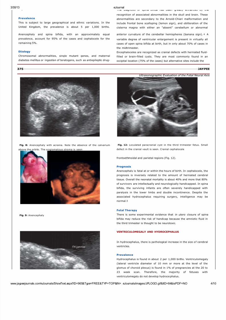
7282019 Ref Lecture eJournal
httpslidepdfcomreaderfullref-lecture-ejournal 410
33013 eJournal
wwwjaypeejournalscomeJournalsShowTextaspxID=965ampType=FREEampTYP=TOPampIN=_eJournalsimagesJPLOGOgifampIID=84ampisPDF=NO 4
Prevalence
This is subject to large geographical and ethnic variations In the
United Kingdom the prevalence is about 5 per 1000 births
Anencephaly and spina bifida with an approximately equal
prevalence account for 95 of the cases and cephalocele for the
remaining 5
Etiology
Chromosomal abnormalities single mutant genes and maternaldiabetes mellitus or ingestion of teratogens such as antiepileptic drug-
recognition of associated abnormalities in the skull and brain These
abnormalities are secondary to the Arnold-Chiari malformation and
include frontal bone scalloping (lemon sign) and obliteration of the
cisterna magna with either an absent cerebellum or abnormal
anterior curvature of the cerebellar hemispheres (banana sign)4 A
variable degree of ventricular enlargement is present in virtually all
cases of open spina bifida at birth but in only about 70 of cases in
the midtrimester
Encephaloceles are recognized as cranial defects with herniated fluid-
filled or brain-filled cysts They are most commonly found in an
occipital location (75 of the cases) but alternative sites include the
375 JAYPEE
Ultrasonographic Evaluation of the Fetal Neural Axis
Fig 8 Anencephaly with acrania Note the absence of the calvarium
above the orbits The a ngiomatous stroma is seen
Fig 9 Anencephaly
Fig 12 Loculated paracranial cyst in the third trimester fetus Small
defect in the cranial vault is seen Cranial cephalocele
frontoethmoidal and parietal regions (Fig 12)
Prognosis
Anencephaly is fatal at or within the hours of birth In cephalocele the
prognosis is inversely related to the amount of herniated cerebral
tissue Overall the neonatal mortality is about 40 and more that 80
of survivors are intellectually and neurologically handicapped In spina
bifida the surviving infants are often severely handicapped with
paralysis in the lower limbs and double incontinence Despite the
associated hydrocephalus requiring surgery intelligence may be
normal5
Fetal Therapy
There is some experimental evidence that in utero closure of spina
bifida may reduce the risk of handicap because the amniotic fluid in
the third trimester is thought to be neurotoxic
VENTRICULOMEGALY AND HYDROCEPHALUS
In hydrocephalus there is pathological increase in the size o f cerebral
ventricles
Prevalence
Hydrocephalus is found in about 2 per 1000 births Ventriculomegaly
(lateral ventricle diameter of 10 mm or more at the level of the
glomus of choroid plexus) is found in 1 of pregnancies at the 20 to
23 week scan Therefore the majority of fetuses with
ventriculomegaly do not develop hydrocephalus
7282019 Ref Lecture eJournal
httpslidepdfcomreaderfullref-lecture-ejournal 510
7282019 Ref Lecture eJournal
httpslidepdfcomreaderfullref-lecture-ejournal 610
33013 eJournal
wwwjaypeejournalscomeJournalsShowTextaspxID=965ampType=FREEampTYP=TOPampIN=_eJournalsimagesJPLOGOgifampIID=84ampisPDF=NO 6
posterior horn 10-15 mm) and overt ventriculomegaly or
hydrocephalus (diameter greater than 15 mm)7 (Figs 13A and B)
Prognosis
Fetal or perinatal death and neurodevelopment in survivors are
strongly related to the presence of other malformations and
chromosomal defects Although mild also referred to as borderline
ventriculomegaly is generally associated with a good prognosis
affected fetuses form the group with the highest incidence of
chromosomal abnormalities (often trisomy 21) In addition in a few
cases with apparently isolated mild ventriculomegaly there may be an
underlying cerebral maldevelopment (such as lissencephaly) or
destructive lesion (such as periventricular leukomalacia) Recent
evidence suggests that in about 10 of cases there is mild to
moderate neurodevelopmental delay
Fetal Therapy
There is some experimental evidence that in utero cerebro spinal fluid
diversion may be beneficial However attempts in the 1980s to treat
hydrocephalic fetuses by ventriculoamniotic shunting have now been
abandoned because of poor results mainly because of inappropriate
selection of patients It is possible that intrauterine drainage may be
beneficial if all intraand extracerebral malformations and
chromosomal defects are excluded and if serial ultrasound scans de-
of the thalami In the semilobar type there is partial segmentation of
the ventricles and cerebral hemispheres posteriorly with incomplete
fusion of the thalami In lobar holoprosencephaly there is normal
separation of the ventricles and thalami but absence of the septum
pellucidum The first two types are often accompanied by
microcephaly and facial abnormalities8
Prevalence
Holoprosencephaly is found in about 1 per 10000 births
Etiology
Although in many cases the cause is a chromosomal abnormality
(usually trisomy 13) or a genetic disorder with an autosomal dominant
or recessive mode of transmission The etiology is unknown in many
cases The risk of recurrence for sporadic nonchromosomal
holoprosencephaly the empirical recurrence risk is 6
Diagnosis
In the standard transverse view of the fetal head for measurement of
the biparietal diameter there is a single dilated midline ventricle
replacing the two lateral ventricles or partial segmentation of the
ventricles The alobar and semilobar types are often associated with
facial defects such as hypotelorism or cyclopia facial cleft and nasal
hypoplasia or proboscis9 (Fig 14)
377 JAYPEE
Ultrasonographic Evaluation of the Fetal Neural Axis
Fig 14 In this fe tus at 11 weeks the absence of midline and presence
of fused choroid plexus are diagnostic of alobar holoprosencephaly
Prognosis
Alobar and semilobar holoprosencephaly are lethal Lobar
holoprosencephaly is associated with mental retardation
AGENESIS OF CORPUS CALLOSUM
The corpus callosum is a bundle of fibers that connects the two
cerebral hemispheres It develops at 12 to 18 weeks of gestation
Agenesis of the corpus callosum may be either complete or partial
(usually a ffecting the posterior part)
Prevalence
Agenesis of the corpus callosum is found in about 5 per 1000 births
Etiology
Agenesis of the corpus callosum may be due to maldevelopment or
secondary to a destructive lesion It is commonly associated with
chromosomal abnormalities usuall trisomies 18 13 and 8 and more
-tion of the lateral ventricles (enlargement of the posterior horns)10
Agenesis of the corpus callosum is demonstrated in the mid-coronal
and mid-sagittal views which may require vaginal sonography (Fig
15)
Prognosis
This depends on the underlying cause In about 90 of those with
apparently isolated agenesis of the corpus callosum development isnormal
DANDY-WALKER COMPLEX
The Dandy-Walker complex refers to a spectrum of abnormalities of
the cerebellar vermis cystic dilation of the fourth ventricle and
enlargement of the cisterna magna The condition is classified into (a)
Dandy-Walker malformation (complete or partial agenesis of the
cerebellar vermis and enlarged posterior fossa) (b) Dandy-Walker
variant (partial agenesis of the cerebellar vermis without enlargement
of the posterior fossa) and (c) mega-cisterna magna (normal vermis
and fourth ventricle)11
Prevalence
Dandy-Walker ma lformation is found in about 1 per 30000 births
Etiology
The Dandy-Walker complex is a non-specific endpoint of chromosomal
abnormalities (usually trisomy 18 or 13 and triploidy) more than 50
genetic syndromes congenital infection or teratogens such as
warfarin but it can also be an isolated finding
Diagnosis
Ultrasonographically the contents of the posterior fossa are visualized
through a transverse suboccipitobregmatic section of the fetal head In
the Dand -Walker malformation there is c stic dilatation of the fourth
7282019 Ref Lecture eJournal
httpslidepdfcomreaderfullref-lecture-ejournal 710
33013 eJournal
wwwjaypeejournalscomeJournalsShowTextaspxID=965ampType=FREEampTYP=TOPampIN=_eJournalsimagesJPLOGOgifampIID=84ampisPDF=NO 7
than 100 genetic syndromes
Diagnosis
The corpus callosum is not visible in the standard transverse views of
the brain but agenesis of the corpus callosum may be suspected by
the absence of cavum septum pellucidum and the teardrop configura-
Fig 15 Agenesis of corpus callosum
ventricle with partial or complete agenesis of the vermis (Figs 16A and
B) In more than 50 of the cases there is associated hydrocephalus
and other extracranial defects Enlarged cisterna magna is diagnosed
if the vertical distance from the vermis to the inner border of the skull
is more than 10 mm12
Figs 16A and B (A) Classic Dandy-Walker Malformation A large defect
of the cerebellar vermis connects the 4th ventricle with a cystic cisterna
magna (B) Megacisterna Magna
Donald School Journal of Ultrasound in Obstetrics and Gynecology October-December 20104(4)373-382 378
Sakshi Tomar Prenatal diagnosis of isolated partial agenesis of the vermis is difficult
and a false diagnosis can be made prior to 18 weeks gestation when
the formation of the vermis is incomplete and anytime in gestation if
the angle of insonation is too steep
Prognosis
Dandy-Walker malformation is associated with a high postnatal
mortality (about 20) and a high incidence (more than 50) of
impaired intellectual and neurological development Experience with
apparently isolated partial agenesis of the vermis or enlarged cisterna
magna is limited and the prognosis for these conditions is uncertain
DESTRUCTIVE CEREBRAL LESIONS
These lesions include hydranencephaly porencephaly and
schizencephaly In hydranencephaly there is absence of the cerebral
hemispheres with preservation of the midbrain and cerebellum In
porencephaly there are cystic cavities within the brain that usually
communicate with the ventricular system the subarachnoid space or
both Schizencephaly is associated with clefts in the fetal brain
connecting the lateral ventricles with the subarachnoid space
Prevalence
Destructive cerebral lesions are found in about 1 per 10000 births
Etiology
Hydranencephaly is a sporadic abnormality that may result from
widespread vascular occlusion in the distribution of internal carotid
arteries prolonged severe hydrocephalus or an overwhelming
infection such as toxoplasmosis and cytomegalovirus Porencephaly
may be caused by infarction of the cerebral arteries or hemorrhage
into the brain parenchyma Schizencephaly may be a primary disorder
of brain development or it may be due to bilateral occlusion of the
middle cerebral arteries
Diagnosis
Fig 17 Bilateral clefts typical of s chizencephaly are se en
Prognosis
Hydranencephaly is usually incompatible with survival beyond early
infancy The prognosis in porencephaly is related to the size and
location of the lesion and although there is increased r isk of impaired
neurodevelopment in some cases development is normal
Schizencephaly is associated with severe neurodevelopmental delay
and seizures
DISORDERS OF NERVE CELL PROLIFERATION
Microcephaly Microcephaly means small head and brain
Prevalence
Microcephaly is found in about 1 per 1000 births
Etiology
This may result from chromosomal and genetic abnormalities fetal
hypoxia congenital infection and exposure to radiation or other
teratogens such maternal anticoagulation with warfarin It is
7282019 Ref Lecture eJournal
httpslidepdfcomreaderfullref-lecture-ejournal 810
33013 eJournal
wwwjaypeejournalscomeJournalsShowTextaspxID=965ampType=FREEampTYP=TOPampIN=_eJournalsimagesJPLOGOgifampIID=84ampisPDF=NO 8
omp e e a sence o ec oes rom e an er or an m e ossa
distinguishes hydranencephaly from severe hydrocephalus in which a
thin rim of remaining cortex and the midline echo can always be
identified In porencephaly there is one or more cystic area in the
cerebral cortex which usually communicates with the ventricle The
differential diagnosis is from intracranial cysts (arachnoid
glioependymal) that are usually found either within the scissures or in
the midline and compress the brain In schizencephaly there are
bilateral clefts extending from the lateral ventricles to the
subarachnoid space and is usually associated with absence of the
cavum septum pellucidum13 (Fig 17)
commony oun n e presence o o er ra n a norma es suc as
cephalocele and holoprosencephaly
Diagnosis
The diagnosis is made by the demonstration of brain abnormalities
such as holoprosencephaly In cases with apparently isolated
microcephaly it is necessary to demonstrate progressive decrease in
the head to abdomen circumference ra tio to below the 1st centile with
advancing gestation14 Such diagnosis may not be apparent before
the third trimester In microcephaly there is a typical disproportion
between the size of the skull and face The brain is small with the cer- 379 JAYPEE
Ultrasonographic Evaluation of the Fetal Neural Axis
Fig 18 Fetal microcephaly Note the sloping of the forehead
-ebral hemispheres affected to a greater extent than the midbrain and
posterior fossa Demonstration of a sloping forehead also increases
the index of suspicion (Fig 18)
Prognosis
This depends on the underlying cause but in more than 50 of cases
there is severe mental retardation
B Megalencephaly Megalencephaly means large head and brain
PrevalenceMegalencephaly is a very rare abnormality
Etiology
This is usually familial with no adverse consequence However it may
also be the consequence of genetic syndromes such as Beckwith-
Wiedemann syndrome achondroplasia neurofibromatosis and
tuberous sclerosis Unilateral megalencephaly is a sporadic condition
Diagnosis
The diagnosis is made by the demonstration of a head to abdomen
circumference ratio above the 99th centile without the evidence of hydrocephalus or intracranial masses Unilateral megalencephaly is
characterized by macrocrania a shift in the midline echo borderline
enlargement of the lateral ventricle and a typical gyri of the affected
hemisphere15
Prognosis
Isolated megalencephaly is usually an asymptomatic condition
Unilateral megalencephaly is associated with severe mental
retardation and untreatable seizures
INTRACRANIAL HEMORRHAGE IN UTERO
Hemorrha e ma be seen in a t ical sube end mal or
junction
Perinatal asphyxia known to induce fluctuation in blood
pressure16
ARTERIOVENOUS MALFORMATION
VEIN OF GALEN ANEURYSM
This is a midline aneurysmal dilation of the vein of Galen due to an
arteriovenous malformation with major hemodynamic disturbances
Prevalence
Vein of Galen aneurysm is a rare abnormality
Etiology
Vein of Galen aneurysm is a sporadic abnormality
Diagnosis
The diagnosis is made by the demonstration of a supratentorial
midline translucent elongated cyst Color Doppler demonstrates active
arteriovenous flow within the cyst There may be associated evidence
of high-output heart failure17 (Fig 19)
Prognosis
In the neonatal period about 50 of the infants present with heart
failure and the rest are asymptomatic In later life hydrocephalus and
intracranial hemorrhage may develop Good results can be achieved
by catheterization and embolization of the malformation
CHOROID PLEXUS CYSTS
These cysts usually bilateral are in the choroid plexuses of the lateral
cerebral ventricles
PrevalenceChoro id plexus cysts are found in about 2 of fetuses at 20 weeks o f
gestation but in more than 90 of cases they resolve by 26 weeks
Etiology
The choroid plexus is easily visualized from 10 weeks of gestation
when it occupies almost the entire hemisphere Thereafter and until 26
weeks there is a rapid decrease in both the size of the choroid plexus
and of the lateral cerebral ventricle in relation to the hemisphere
Choro id plexus cysts contain cerebrospinal fluid and cellular debris
Diagnosis
Single or multiple cystic areas (greater than 2 mm in diameter) in one
or both choroid plexuses18 (Fig 20)
7282019 Ref Lecture eJournal
httpslidepdfcomreaderfullref-lecture-ejournal 910
33013 eJournal
wwwjaypeejournalscomeJournalsShowTextaspxID=965ampType=FREEampTYP=TOPampIN=_eJournalsimagesJPLOGOgifampIID=84ampisPDF=NO 9
intraparenchymal location
Etiology
Sudden changes in the cerebral blood pressure leading to
ruptured capillaries in the germinal matrix or capillary venous
Prognosis
They are usually of no pathological significance but they are
associated with an increased risk for trisomy 18 and possibly trisomy
21 In the absence of other markers of trisomy 18 the maternal age-
related risk is increased by a factor of 15
Donald School Journal of Ultrasound in Obstetrics and Gynecology October-December 20104(4)373-382 380
Sakshi Tomar
Fig 19 Vein of Galen aneurysm Color Doppler flow image of the fetal
head demonstrating a vein of galen aneurysm (arrow) supplied by the
feed ing vesse ls from the circle of willis
Fig 20 Bilateral choroid plexus cysts seen in a transverse axial
sonogram
ARACHNOID CYSTS
Arachnoid cysts are fluid-filled cyst contained within the arachnoid
space
Prevalence
Arachnoid cysts are extremely rare
Etiology
Unknown infectious process has been hypothesized but it is unlikely
that this may explain the congenital cysts
Diagnosis
Arachnoid cys ts appear on antenatal ultrasound as sonolucent lesions
Fig 21 Arachnoid Cyst Corresponding MRI T2W image is seen
difficult Interhemispheric cysts associated with agenesis of the corpus
callosum most likely are not arachnoid cysts but rather glioependymal
cysts
Prognosis
Large cysts may cause intracranial hypertension and require
neurosurgical treatment However a normal intellectual development
in the range of 80 to 90 is reported by most series Spontaneous
remission has been described both in the postnatal as well as in the
antenatal per iod Glioependymal cyst that should be suspected in the
cases with associated agenesis of corpus callosum probably re flects a
greater degree of derangement in the development of the brain and
this may be reflected in worse outcome
CONCLUSION
Modern ultrasound equipments yield a unique potential for evaluation
of normal and abnormal fetal CNS very early in pregnancy It should
be stressed however that some cerebral anomalies are the
consequence of disruptions or are characterized by intrauterine
development Counseling the parents and deciding on a sensible
obstetric management are frequently difficult
REFERENCES
Monteagudo A Fetal neurosonography Should it be routine
Should it be detailed [Editorial] Ultrasound Obstet Gynecol
1998121
Callen Ultrasonography in Obstetrics and Gynecology (4th ed)
277-304
Bronshtein M Ornoy A Acrania Anencephaly resulting from
secondary degeneration of a closed neural tube Two cases in
the same family J Clin Ultrasound 199119230
Levi S Hijazi Y Schapps J et al Sensitivity and specificity of
routine antenatal screening for congenital anomalies by
ultrasound The Belgian Multicentric Study Ultrasound Obstet
Gynecol 19911102
Goldstein RB LaPidus AS Filly RA Fetal cephaloceles Diagnosis
7282019 Ref Lecture eJournal
httpslidepdfcomreaderfullref-lecture-ejournal 1010
33013 eJournal
j j l J l Sh T t ID 965ampT FREEampTYP TOPampIN J l i JPLOGO ifampIID 84ampi PDF NO 10
with a thin regular outline and containing no blood flow do not
communicate with the lateral ventricles and anyhow are not associated
with the loss of brain tissue (Fig 21) They occur most frequently in
the area of cerebral fissure and midline Large cyst may cause
significant mass effect and the distinction from porencephaly may be
with US Radiology 1991180803
Filly RA Goldstein RB Callen PW Fetal ventricle Importance in
381 JAYPEE
Ultrasonographic Evaluation of the Fetal Neural Axis routine obstetric sonography Radiology 19911811
Cardoza JD Goldstein RB Filly RA Exclusion of fetal
ventriculomegaly with a single measurement The width of lateral
ventricular atrium Radiology 1988169711
DeMyer W Holoprosencephaly vol 30 Amsterdam Elsevier
1977
Bronshtein M Weiner Z Early transvaginal sonographic diagnosis
of a lobar holoprosencephaly Prenat Diagn 199111459
Bennett GL Bromley B Benacerraf BR Agenesis of corpus
callosum Prenatal detection usually is not possible before 22
week s o f ge station Ra diology 1996199447
Benda C The Dandy-Walker syndrome or the so called atresia of
the foramen Magendie J Neuropathol Exp Neurol 19541314
Pilu G Goldstein I Reece E et al Sonography of fetal Dandy
Walker Malformations A reappraisal Ultrasound Obstet Gynecol
19922151
Pilu G falco P Perolo A Differential diagnosis and outcome of
fetal intracranial hypoechoic lesions Report of 21 cases
Ultrasound Obstet Gynecol 19979229
Chervenak FA Jeanty P Cantraine F et al The diagnosis of
fetal microcephaly Am J Obstet Gynecol 1984149512
Bignami A Palladini G Zappella M Unilateral Megalencephaly
with nerve cell hypertrophy An anatomical and quantitative
histochemical study Brain Res 19689103
Vergani P Strobelt N Locatelli A et al Clinical significance of
fetal intracranial haemorrhage Am J Obstet Gynecol
1996175536
Comstock CH Kirk JS Arteriovenous malformations Location
and evolution in the fetal brain J Ultrasound Med 199110361
Snijders R Shawa L Nicolaides K Fetal choroid cysts a nd trisomy
18 Assessment of risk based on ultrasound findings and
maternal age Prenat Diagnosis 199414119
382
JAYPEE
copy Jaypee Brothers Medical Publishers (P) Ltd
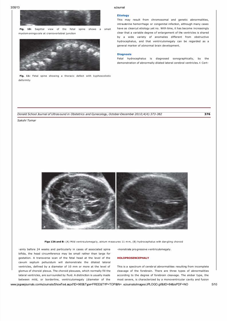
7282019 Ref Lecture eJournal
httpslidepdfcomreaderfullref-lecture-ejournal 510
7282019 Ref Lecture eJournal
httpslidepdfcomreaderfullref-lecture-ejournal 610
33013 eJournal
wwwjaypeejournalscomeJournalsShowTextaspxID=965ampType=FREEampTYP=TOPampIN=_eJournalsimagesJPLOGOgifampIID=84ampisPDF=NO 6
posterior horn 10-15 mm) and overt ventriculomegaly or
hydrocephalus (diameter greater than 15 mm)7 (Figs 13A and B)
Prognosis
Fetal or perinatal death and neurodevelopment in survivors are
strongly related to the presence of other malformations and
chromosomal defects Although mild also referred to as borderline
ventriculomegaly is generally associated with a good prognosis
affected fetuses form the group with the highest incidence of
chromosomal abnormalities (often trisomy 21) In addition in a few
cases with apparently isolated mild ventriculomegaly there may be an
underlying cerebral maldevelopment (such as lissencephaly) or
destructive lesion (such as periventricular leukomalacia) Recent
evidence suggests that in about 10 of cases there is mild to
moderate neurodevelopmental delay
Fetal Therapy
There is some experimental evidence that in utero cerebro spinal fluid
diversion may be beneficial However attempts in the 1980s to treat
hydrocephalic fetuses by ventriculoamniotic shunting have now been
abandoned because of poor results mainly because of inappropriate
selection of patients It is possible that intrauterine drainage may be
beneficial if all intraand extracerebral malformations and
chromosomal defects are excluded and if serial ultrasound scans de-
of the thalami In the semilobar type there is partial segmentation of
the ventricles and cerebral hemispheres posteriorly with incomplete
fusion of the thalami In lobar holoprosencephaly there is normal
separation of the ventricles and thalami but absence of the septum
pellucidum The first two types are often accompanied by
microcephaly and facial abnormalities8
Prevalence
Holoprosencephaly is found in about 1 per 10000 births
Etiology
Although in many cases the cause is a chromosomal abnormality
(usually trisomy 13) or a genetic disorder with an autosomal dominant
or recessive mode of transmission The etiology is unknown in many
cases The risk of recurrence for sporadic nonchromosomal
holoprosencephaly the empirical recurrence risk is 6
Diagnosis
In the standard transverse view of the fetal head for measurement of
the biparietal diameter there is a single dilated midline ventricle
replacing the two lateral ventricles or partial segmentation of the
ventricles The alobar and semilobar types are often associated with
facial defects such as hypotelorism or cyclopia facial cleft and nasal
hypoplasia or proboscis9 (Fig 14)
377 JAYPEE
Ultrasonographic Evaluation of the Fetal Neural Axis
Fig 14 In this fe tus at 11 weeks the absence of midline and presence
of fused choroid plexus are diagnostic of alobar holoprosencephaly
Prognosis
Alobar and semilobar holoprosencephaly are lethal Lobar
holoprosencephaly is associated with mental retardation
AGENESIS OF CORPUS CALLOSUM
The corpus callosum is a bundle of fibers that connects the two
cerebral hemispheres It develops at 12 to 18 weeks of gestation
Agenesis of the corpus callosum may be either complete or partial
(usually a ffecting the posterior part)
Prevalence
Agenesis of the corpus callosum is found in about 5 per 1000 births
Etiology
Agenesis of the corpus callosum may be due to maldevelopment or
secondary to a destructive lesion It is commonly associated with
chromosomal abnormalities usuall trisomies 18 13 and 8 and more
-tion of the lateral ventricles (enlargement of the posterior horns)10
Agenesis of the corpus callosum is demonstrated in the mid-coronal
and mid-sagittal views which may require vaginal sonography (Fig
15)
Prognosis
This depends on the underlying cause In about 90 of those with
apparently isolated agenesis of the corpus callosum development isnormal
DANDY-WALKER COMPLEX
The Dandy-Walker complex refers to a spectrum of abnormalities of
the cerebellar vermis cystic dilation of the fourth ventricle and
enlargement of the cisterna magna The condition is classified into (a)
Dandy-Walker malformation (complete or partial agenesis of the
cerebellar vermis and enlarged posterior fossa) (b) Dandy-Walker
variant (partial agenesis of the cerebellar vermis without enlargement
of the posterior fossa) and (c) mega-cisterna magna (normal vermis
and fourth ventricle)11
Prevalence
Dandy-Walker ma lformation is found in about 1 per 30000 births
Etiology
The Dandy-Walker complex is a non-specific endpoint of chromosomal
abnormalities (usually trisomy 18 or 13 and triploidy) more than 50
genetic syndromes congenital infection or teratogens such as
warfarin but it can also be an isolated finding
Diagnosis
Ultrasonographically the contents of the posterior fossa are visualized
through a transverse suboccipitobregmatic section of the fetal head In
the Dand -Walker malformation there is c stic dilatation of the fourth
7282019 Ref Lecture eJournal
httpslidepdfcomreaderfullref-lecture-ejournal 710
33013 eJournal
wwwjaypeejournalscomeJournalsShowTextaspxID=965ampType=FREEampTYP=TOPampIN=_eJournalsimagesJPLOGOgifampIID=84ampisPDF=NO 7
than 100 genetic syndromes
Diagnosis
The corpus callosum is not visible in the standard transverse views of
the brain but agenesis of the corpus callosum may be suspected by
the absence of cavum septum pellucidum and the teardrop configura-
Fig 15 Agenesis of corpus callosum
ventricle with partial or complete agenesis of the vermis (Figs 16A and
B) In more than 50 of the cases there is associated hydrocephalus
and other extracranial defects Enlarged cisterna magna is diagnosed
if the vertical distance from the vermis to the inner border of the skull
is more than 10 mm12
Figs 16A and B (A) Classic Dandy-Walker Malformation A large defect
of the cerebellar vermis connects the 4th ventricle with a cystic cisterna
magna (B) Megacisterna Magna
Donald School Journal of Ultrasound in Obstetrics and Gynecology October-December 20104(4)373-382 378
Sakshi Tomar Prenatal diagnosis of isolated partial agenesis of the vermis is difficult
and a false diagnosis can be made prior to 18 weeks gestation when
the formation of the vermis is incomplete and anytime in gestation if
the angle of insonation is too steep
Prognosis
Dandy-Walker malformation is associated with a high postnatal
mortality (about 20) and a high incidence (more than 50) of
impaired intellectual and neurological development Experience with
apparently isolated partial agenesis of the vermis or enlarged cisterna
magna is limited and the prognosis for these conditions is uncertain
DESTRUCTIVE CEREBRAL LESIONS
These lesions include hydranencephaly porencephaly and
schizencephaly In hydranencephaly there is absence of the cerebral
hemispheres with preservation of the midbrain and cerebellum In
porencephaly there are cystic cavities within the brain that usually
communicate with the ventricular system the subarachnoid space or
both Schizencephaly is associated with clefts in the fetal brain
connecting the lateral ventricles with the subarachnoid space
Prevalence
Destructive cerebral lesions are found in about 1 per 10000 births
Etiology
Hydranencephaly is a sporadic abnormality that may result from
widespread vascular occlusion in the distribution of internal carotid
arteries prolonged severe hydrocephalus or an overwhelming
infection such as toxoplasmosis and cytomegalovirus Porencephaly
may be caused by infarction of the cerebral arteries or hemorrhage
into the brain parenchyma Schizencephaly may be a primary disorder
of brain development or it may be due to bilateral occlusion of the
middle cerebral arteries
Diagnosis
Fig 17 Bilateral clefts typical of s chizencephaly are se en
Prognosis
Hydranencephaly is usually incompatible with survival beyond early
infancy The prognosis in porencephaly is related to the size and
location of the lesion and although there is increased r isk of impaired
neurodevelopment in some cases development is normal
Schizencephaly is associated with severe neurodevelopmental delay
and seizures
DISORDERS OF NERVE CELL PROLIFERATION
Microcephaly Microcephaly means small head and brain
Prevalence
Microcephaly is found in about 1 per 1000 births
Etiology
This may result from chromosomal and genetic abnormalities fetal
hypoxia congenital infection and exposure to radiation or other
teratogens such maternal anticoagulation with warfarin It is
7282019 Ref Lecture eJournal
httpslidepdfcomreaderfullref-lecture-ejournal 810
33013 eJournal
wwwjaypeejournalscomeJournalsShowTextaspxID=965ampType=FREEampTYP=TOPampIN=_eJournalsimagesJPLOGOgifampIID=84ampisPDF=NO 8
omp e e a sence o ec oes rom e an er or an m e ossa
distinguishes hydranencephaly from severe hydrocephalus in which a
thin rim of remaining cortex and the midline echo can always be
identified In porencephaly there is one or more cystic area in the
cerebral cortex which usually communicates with the ventricle The
differential diagnosis is from intracranial cysts (arachnoid
glioependymal) that are usually found either within the scissures or in
the midline and compress the brain In schizencephaly there are
bilateral clefts extending from the lateral ventricles to the
subarachnoid space and is usually associated with absence of the
cavum septum pellucidum13 (Fig 17)
commony oun n e presence o o er ra n a norma es suc as
cephalocele and holoprosencephaly
Diagnosis
The diagnosis is made by the demonstration of brain abnormalities
such as holoprosencephaly In cases with apparently isolated
microcephaly it is necessary to demonstrate progressive decrease in
the head to abdomen circumference ra tio to below the 1st centile with
advancing gestation14 Such diagnosis may not be apparent before
the third trimester In microcephaly there is a typical disproportion
between the size of the skull and face The brain is small with the cer- 379 JAYPEE
Ultrasonographic Evaluation of the Fetal Neural Axis
Fig 18 Fetal microcephaly Note the sloping of the forehead
-ebral hemispheres affected to a greater extent than the midbrain and
posterior fossa Demonstration of a sloping forehead also increases
the index of suspicion (Fig 18)
Prognosis
This depends on the underlying cause but in more than 50 of cases
there is severe mental retardation
B Megalencephaly Megalencephaly means large head and brain
PrevalenceMegalencephaly is a very rare abnormality
Etiology
This is usually familial with no adverse consequence However it may
also be the consequence of genetic syndromes such as Beckwith-
Wiedemann syndrome achondroplasia neurofibromatosis and
tuberous sclerosis Unilateral megalencephaly is a sporadic condition
Diagnosis
The diagnosis is made by the demonstration of a head to abdomen
circumference ratio above the 99th centile without the evidence of hydrocephalus or intracranial masses Unilateral megalencephaly is
characterized by macrocrania a shift in the midline echo borderline
enlargement of the lateral ventricle and a typical gyri of the affected
hemisphere15
Prognosis
Isolated megalencephaly is usually an asymptomatic condition
Unilateral megalencephaly is associated with severe mental
retardation and untreatable seizures
INTRACRANIAL HEMORRHAGE IN UTERO
Hemorrha e ma be seen in a t ical sube end mal or
junction
Perinatal asphyxia known to induce fluctuation in blood
pressure16
ARTERIOVENOUS MALFORMATION
VEIN OF GALEN ANEURYSM
This is a midline aneurysmal dilation of the vein of Galen due to an
arteriovenous malformation with major hemodynamic disturbances
Prevalence
Vein of Galen aneurysm is a rare abnormality
Etiology
Vein of Galen aneurysm is a sporadic abnormality
Diagnosis
The diagnosis is made by the demonstration of a supratentorial
midline translucent elongated cyst Color Doppler demonstrates active
arteriovenous flow within the cyst There may be associated evidence
of high-output heart failure17 (Fig 19)
Prognosis
In the neonatal period about 50 of the infants present with heart
failure and the rest are asymptomatic In later life hydrocephalus and
intracranial hemorrhage may develop Good results can be achieved
by catheterization and embolization of the malformation
CHOROID PLEXUS CYSTS
These cysts usually bilateral are in the choroid plexuses of the lateral
cerebral ventricles
PrevalenceChoro id plexus cysts are found in about 2 of fetuses at 20 weeks o f
gestation but in more than 90 of cases they resolve by 26 weeks
Etiology
The choroid plexus is easily visualized from 10 weeks of gestation
when it occupies almost the entire hemisphere Thereafter and until 26
weeks there is a rapid decrease in both the size of the choroid plexus
and of the lateral cerebral ventricle in relation to the hemisphere
Choro id plexus cysts contain cerebrospinal fluid and cellular debris
Diagnosis
Single or multiple cystic areas (greater than 2 mm in diameter) in one
or both choroid plexuses18 (Fig 20)
7282019 Ref Lecture eJournal
httpslidepdfcomreaderfullref-lecture-ejournal 910
33013 eJournal
wwwjaypeejournalscomeJournalsShowTextaspxID=965ampType=FREEampTYP=TOPampIN=_eJournalsimagesJPLOGOgifampIID=84ampisPDF=NO 9
intraparenchymal location
Etiology
Sudden changes in the cerebral blood pressure leading to
ruptured capillaries in the germinal matrix or capillary venous
Prognosis
They are usually of no pathological significance but they are
associated with an increased risk for trisomy 18 and possibly trisomy
21 In the absence of other markers of trisomy 18 the maternal age-
related risk is increased by a factor of 15
Donald School Journal of Ultrasound in Obstetrics and Gynecology October-December 20104(4)373-382 380
Sakshi Tomar
Fig 19 Vein of Galen aneurysm Color Doppler flow image of the fetal
head demonstrating a vein of galen aneurysm (arrow) supplied by the
feed ing vesse ls from the circle of willis
Fig 20 Bilateral choroid plexus cysts seen in a transverse axial
sonogram
ARACHNOID CYSTS
Arachnoid cysts are fluid-filled cyst contained within the arachnoid
space
Prevalence
Arachnoid cysts are extremely rare
Etiology
Unknown infectious process has been hypothesized but it is unlikely
that this may explain the congenital cysts
Diagnosis
Arachnoid cys ts appear on antenatal ultrasound as sonolucent lesions
Fig 21 Arachnoid Cyst Corresponding MRI T2W image is seen
difficult Interhemispheric cysts associated with agenesis of the corpus
callosum most likely are not arachnoid cysts but rather glioependymal
cysts
Prognosis
Large cysts may cause intracranial hypertension and require
neurosurgical treatment However a normal intellectual development
in the range of 80 to 90 is reported by most series Spontaneous
remission has been described both in the postnatal as well as in the
antenatal per iod Glioependymal cyst that should be suspected in the
cases with associated agenesis of corpus callosum probably re flects a
greater degree of derangement in the development of the brain and
this may be reflected in worse outcome
CONCLUSION
Modern ultrasound equipments yield a unique potential for evaluation
of normal and abnormal fetal CNS very early in pregnancy It should
be stressed however that some cerebral anomalies are the
consequence of disruptions or are characterized by intrauterine
development Counseling the parents and deciding on a sensible
obstetric management are frequently difficult
REFERENCES
Monteagudo A Fetal neurosonography Should it be routine
Should it be detailed [Editorial] Ultrasound Obstet Gynecol
1998121
Callen Ultrasonography in Obstetrics and Gynecology (4th ed)
277-304
Bronshtein M Ornoy A Acrania Anencephaly resulting from
secondary degeneration of a closed neural tube Two cases in
the same family J Clin Ultrasound 199119230
Levi S Hijazi Y Schapps J et al Sensitivity and specificity of
routine antenatal screening for congenital anomalies by
ultrasound The Belgian Multicentric Study Ultrasound Obstet
Gynecol 19911102
Goldstein RB LaPidus AS Filly RA Fetal cephaloceles Diagnosis
7282019 Ref Lecture eJournal
httpslidepdfcomreaderfullref-lecture-ejournal 1010
33013 eJournal
j j l J l Sh T t ID 965ampT FREEampTYP TOPampIN J l i JPLOGO ifampIID 84ampi PDF NO 10
with a thin regular outline and containing no blood flow do not
communicate with the lateral ventricles and anyhow are not associated
with the loss of brain tissue (Fig 21) They occur most frequently in
the area of cerebral fissure and midline Large cyst may cause
significant mass effect and the distinction from porencephaly may be
with US Radiology 1991180803
Filly RA Goldstein RB Callen PW Fetal ventricle Importance in
381 JAYPEE
Ultrasonographic Evaluation of the Fetal Neural Axis routine obstetric sonography Radiology 19911811
Cardoza JD Goldstein RB Filly RA Exclusion of fetal
ventriculomegaly with a single measurement The width of lateral
ventricular atrium Radiology 1988169711
DeMyer W Holoprosencephaly vol 30 Amsterdam Elsevier
1977
Bronshtein M Weiner Z Early transvaginal sonographic diagnosis
of a lobar holoprosencephaly Prenat Diagn 199111459
Bennett GL Bromley B Benacerraf BR Agenesis of corpus
callosum Prenatal detection usually is not possible before 22
week s o f ge station Ra diology 1996199447
Benda C The Dandy-Walker syndrome or the so called atresia of
the foramen Magendie J Neuropathol Exp Neurol 19541314
Pilu G Goldstein I Reece E et al Sonography of fetal Dandy
Walker Malformations A reappraisal Ultrasound Obstet Gynecol
19922151
Pilu G falco P Perolo A Differential diagnosis and outcome of
fetal intracranial hypoechoic lesions Report of 21 cases
Ultrasound Obstet Gynecol 19979229
Chervenak FA Jeanty P Cantraine F et al The diagnosis of
fetal microcephaly Am J Obstet Gynecol 1984149512
Bignami A Palladini G Zappella M Unilateral Megalencephaly
with nerve cell hypertrophy An anatomical and quantitative
histochemical study Brain Res 19689103
Vergani P Strobelt N Locatelli A et al Clinical significance of
fetal intracranial haemorrhage Am J Obstet Gynecol
1996175536
Comstock CH Kirk JS Arteriovenous malformations Location
and evolution in the fetal brain J Ultrasound Med 199110361
Snijders R Shawa L Nicolaides K Fetal choroid cysts a nd trisomy
18 Assessment of risk based on ultrasound findings and
maternal age Prenat Diagnosis 199414119
382
JAYPEE
copy Jaypee Brothers Medical Publishers (P) Ltd
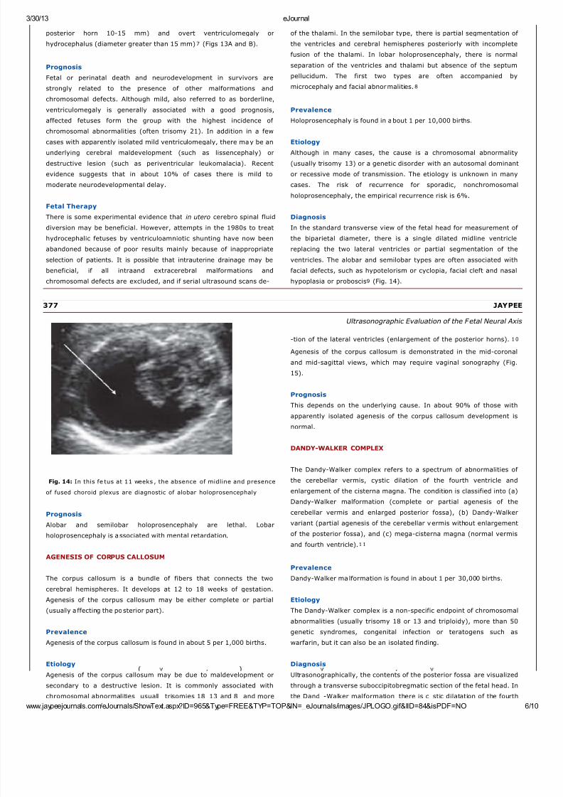
7282019 Ref Lecture eJournal
httpslidepdfcomreaderfullref-lecture-ejournal 610
33013 eJournal
wwwjaypeejournalscomeJournalsShowTextaspxID=965ampType=FREEampTYP=TOPampIN=_eJournalsimagesJPLOGOgifampIID=84ampisPDF=NO 6
posterior horn 10-15 mm) and overt ventriculomegaly or
hydrocephalus (diameter greater than 15 mm)7 (Figs 13A and B)
Prognosis
Fetal or perinatal death and neurodevelopment in survivors are
strongly related to the presence of other malformations and
chromosomal defects Although mild also referred to as borderline
ventriculomegaly is generally associated with a good prognosis
affected fetuses form the group with the highest incidence of
chromosomal abnormalities (often trisomy 21) In addition in a few
cases with apparently isolated mild ventriculomegaly there may be an
underlying cerebral maldevelopment (such as lissencephaly) or
destructive lesion (such as periventricular leukomalacia) Recent
evidence suggests that in about 10 of cases there is mild to
moderate neurodevelopmental delay
Fetal Therapy
There is some experimental evidence that in utero cerebro spinal fluid
diversion may be beneficial However attempts in the 1980s to treat
hydrocephalic fetuses by ventriculoamniotic shunting have now been
abandoned because of poor results mainly because of inappropriate
selection of patients It is possible that intrauterine drainage may be
beneficial if all intraand extracerebral malformations and
chromosomal defects are excluded and if serial ultrasound scans de-
of the thalami In the semilobar type there is partial segmentation of
the ventricles and cerebral hemispheres posteriorly with incomplete
fusion of the thalami In lobar holoprosencephaly there is normal
separation of the ventricles and thalami but absence of the septum
pellucidum The first two types are often accompanied by
microcephaly and facial abnormalities8
Prevalence
Holoprosencephaly is found in about 1 per 10000 births
Etiology
Although in many cases the cause is a chromosomal abnormality
(usually trisomy 13) or a genetic disorder with an autosomal dominant
or recessive mode of transmission The etiology is unknown in many
cases The risk of recurrence for sporadic nonchromosomal
holoprosencephaly the empirical recurrence risk is 6
Diagnosis
In the standard transverse view of the fetal head for measurement of
the biparietal diameter there is a single dilated midline ventricle
replacing the two lateral ventricles or partial segmentation of the
ventricles The alobar and semilobar types are often associated with
facial defects such as hypotelorism or cyclopia facial cleft and nasal
hypoplasia or proboscis9 (Fig 14)
377 JAYPEE
Ultrasonographic Evaluation of the Fetal Neural Axis
Fig 14 In this fe tus at 11 weeks the absence of midline and presence
of fused choroid plexus are diagnostic of alobar holoprosencephaly
Prognosis
Alobar and semilobar holoprosencephaly are lethal Lobar
holoprosencephaly is associated with mental retardation
AGENESIS OF CORPUS CALLOSUM
The corpus callosum is a bundle of fibers that connects the two
cerebral hemispheres It develops at 12 to 18 weeks of gestation
Agenesis of the corpus callosum may be either complete or partial
(usually a ffecting the posterior part)
Prevalence
Agenesis of the corpus callosum is found in about 5 per 1000 births
Etiology
Agenesis of the corpus callosum may be due to maldevelopment or
secondary to a destructive lesion It is commonly associated with
chromosomal abnormalities usuall trisomies 18 13 and 8 and more
-tion of the lateral ventricles (enlargement of the posterior horns)10
Agenesis of the corpus callosum is demonstrated in the mid-coronal
and mid-sagittal views which may require vaginal sonography (Fig
15)
Prognosis
This depends on the underlying cause In about 90 of those with
apparently isolated agenesis of the corpus callosum development isnormal
DANDY-WALKER COMPLEX
The Dandy-Walker complex refers to a spectrum of abnormalities of
the cerebellar vermis cystic dilation of the fourth ventricle and
enlargement of the cisterna magna The condition is classified into (a)
Dandy-Walker malformation (complete or partial agenesis of the
cerebellar vermis and enlarged posterior fossa) (b) Dandy-Walker
variant (partial agenesis of the cerebellar vermis without enlargement
of the posterior fossa) and (c) mega-cisterna magna (normal vermis
and fourth ventricle)11
Prevalence
Dandy-Walker ma lformation is found in about 1 per 30000 births
Etiology
The Dandy-Walker complex is a non-specific endpoint of chromosomal
abnormalities (usually trisomy 18 or 13 and triploidy) more than 50
genetic syndromes congenital infection or teratogens such as
warfarin but it can also be an isolated finding
Diagnosis
Ultrasonographically the contents of the posterior fossa are visualized
through a transverse suboccipitobregmatic section of the fetal head In
the Dand -Walker malformation there is c stic dilatation of the fourth
7282019 Ref Lecture eJournal
httpslidepdfcomreaderfullref-lecture-ejournal 710
33013 eJournal
wwwjaypeejournalscomeJournalsShowTextaspxID=965ampType=FREEampTYP=TOPampIN=_eJournalsimagesJPLOGOgifampIID=84ampisPDF=NO 7
than 100 genetic syndromes
Diagnosis
The corpus callosum is not visible in the standard transverse views of
the brain but agenesis of the corpus callosum may be suspected by
the absence of cavum septum pellucidum and the teardrop configura-
Fig 15 Agenesis of corpus callosum
ventricle with partial or complete agenesis of the vermis (Figs 16A and
B) In more than 50 of the cases there is associated hydrocephalus
and other extracranial defects Enlarged cisterna magna is diagnosed
if the vertical distance from the vermis to the inner border of the skull
is more than 10 mm12
Figs 16A and B (A) Classic Dandy-Walker Malformation A large defect
of the cerebellar vermis connects the 4th ventricle with a cystic cisterna
magna (B) Megacisterna Magna
Donald School Journal of Ultrasound in Obstetrics and Gynecology October-December 20104(4)373-382 378
Sakshi Tomar Prenatal diagnosis of isolated partial agenesis of the vermis is difficult
and a false diagnosis can be made prior to 18 weeks gestation when
the formation of the vermis is incomplete and anytime in gestation if
the angle of insonation is too steep
Prognosis
Dandy-Walker malformation is associated with a high postnatal
mortality (about 20) and a high incidence (more than 50) of
impaired intellectual and neurological development Experience with
apparently isolated partial agenesis of the vermis or enlarged cisterna
magna is limited and the prognosis for these conditions is uncertain
DESTRUCTIVE CEREBRAL LESIONS
These lesions include hydranencephaly porencephaly and
schizencephaly In hydranencephaly there is absence of the cerebral
hemispheres with preservation of the midbrain and cerebellum In
porencephaly there are cystic cavities within the brain that usually
communicate with the ventricular system the subarachnoid space or
both Schizencephaly is associated with clefts in the fetal brain
connecting the lateral ventricles with the subarachnoid space
Prevalence
Destructive cerebral lesions are found in about 1 per 10000 births
Etiology
Hydranencephaly is a sporadic abnormality that may result from
widespread vascular occlusion in the distribution of internal carotid
arteries prolonged severe hydrocephalus or an overwhelming
infection such as toxoplasmosis and cytomegalovirus Porencephaly
may be caused by infarction of the cerebral arteries or hemorrhage
into the brain parenchyma Schizencephaly may be a primary disorder
of brain development or it may be due to bilateral occlusion of the
middle cerebral arteries
Diagnosis
Fig 17 Bilateral clefts typical of s chizencephaly are se en
Prognosis
Hydranencephaly is usually incompatible with survival beyond early
infancy The prognosis in porencephaly is related to the size and
location of the lesion and although there is increased r isk of impaired
neurodevelopment in some cases development is normal
Schizencephaly is associated with severe neurodevelopmental delay
and seizures
DISORDERS OF NERVE CELL PROLIFERATION
Microcephaly Microcephaly means small head and brain
Prevalence
Microcephaly is found in about 1 per 1000 births
Etiology
This may result from chromosomal and genetic abnormalities fetal
hypoxia congenital infection and exposure to radiation or other
teratogens such maternal anticoagulation with warfarin It is
7282019 Ref Lecture eJournal
httpslidepdfcomreaderfullref-lecture-ejournal 810
33013 eJournal
wwwjaypeejournalscomeJournalsShowTextaspxID=965ampType=FREEampTYP=TOPampIN=_eJournalsimagesJPLOGOgifampIID=84ampisPDF=NO 8
omp e e a sence o ec oes rom e an er or an m e ossa
distinguishes hydranencephaly from severe hydrocephalus in which a
thin rim of remaining cortex and the midline echo can always be
identified In porencephaly there is one or more cystic area in the
cerebral cortex which usually communicates with the ventricle The
differential diagnosis is from intracranial cysts (arachnoid
glioependymal) that are usually found either within the scissures or in
the midline and compress the brain In schizencephaly there are
bilateral clefts extending from the lateral ventricles to the
subarachnoid space and is usually associated with absence of the
cavum septum pellucidum13 (Fig 17)
commony oun n e presence o o er ra n a norma es suc as
cephalocele and holoprosencephaly
Diagnosis
The diagnosis is made by the demonstration of brain abnormalities
such as holoprosencephaly In cases with apparently isolated
microcephaly it is necessary to demonstrate progressive decrease in
the head to abdomen circumference ra tio to below the 1st centile with
advancing gestation14 Such diagnosis may not be apparent before
the third trimester In microcephaly there is a typical disproportion
between the size of the skull and face The brain is small with the cer- 379 JAYPEE
Ultrasonographic Evaluation of the Fetal Neural Axis
Fig 18 Fetal microcephaly Note the sloping of the forehead
-ebral hemispheres affected to a greater extent than the midbrain and
posterior fossa Demonstration of a sloping forehead also increases
the index of suspicion (Fig 18)
Prognosis
This depends on the underlying cause but in more than 50 of cases
there is severe mental retardation
B Megalencephaly Megalencephaly means large head and brain
PrevalenceMegalencephaly is a very rare abnormality
Etiology
This is usually familial with no adverse consequence However it may
also be the consequence of genetic syndromes such as Beckwith-
Wiedemann syndrome achondroplasia neurofibromatosis and
tuberous sclerosis Unilateral megalencephaly is a sporadic condition
Diagnosis
The diagnosis is made by the demonstration of a head to abdomen
circumference ratio above the 99th centile without the evidence of hydrocephalus or intracranial masses Unilateral megalencephaly is
characterized by macrocrania a shift in the midline echo borderline
enlargement of the lateral ventricle and a typical gyri of the affected
hemisphere15
Prognosis
Isolated megalencephaly is usually an asymptomatic condition
Unilateral megalencephaly is associated with severe mental
retardation and untreatable seizures
INTRACRANIAL HEMORRHAGE IN UTERO
Hemorrha e ma be seen in a t ical sube end mal or
junction
Perinatal asphyxia known to induce fluctuation in blood
pressure16
ARTERIOVENOUS MALFORMATION
VEIN OF GALEN ANEURYSM
This is a midline aneurysmal dilation of the vein of Galen due to an
arteriovenous malformation with major hemodynamic disturbances
Prevalence
Vein of Galen aneurysm is a rare abnormality
Etiology
Vein of Galen aneurysm is a sporadic abnormality
Diagnosis
The diagnosis is made by the demonstration of a supratentorial
midline translucent elongated cyst Color Doppler demonstrates active
arteriovenous flow within the cyst There may be associated evidence
of high-output heart failure17 (Fig 19)
Prognosis
In the neonatal period about 50 of the infants present with heart
failure and the rest are asymptomatic In later life hydrocephalus and
intracranial hemorrhage may develop Good results can be achieved
by catheterization and embolization of the malformation
CHOROID PLEXUS CYSTS
These cysts usually bilateral are in the choroid plexuses of the lateral
cerebral ventricles
PrevalenceChoro id plexus cysts are found in about 2 of fetuses at 20 weeks o f
gestation but in more than 90 of cases they resolve by 26 weeks
Etiology
The choroid plexus is easily visualized from 10 weeks of gestation
when it occupies almost the entire hemisphere Thereafter and until 26
weeks there is a rapid decrease in both the size of the choroid plexus
and of the lateral cerebral ventricle in relation to the hemisphere
Choro id plexus cysts contain cerebrospinal fluid and cellular debris
Diagnosis
Single or multiple cystic areas (greater than 2 mm in diameter) in one
or both choroid plexuses18 (Fig 20)
7282019 Ref Lecture eJournal
httpslidepdfcomreaderfullref-lecture-ejournal 910
33013 eJournal
wwwjaypeejournalscomeJournalsShowTextaspxID=965ampType=FREEampTYP=TOPampIN=_eJournalsimagesJPLOGOgifampIID=84ampisPDF=NO 9
intraparenchymal location
Etiology
Sudden changes in the cerebral blood pressure leading to
ruptured capillaries in the germinal matrix or capillary venous
Prognosis
They are usually of no pathological significance but they are
associated with an increased risk for trisomy 18 and possibly trisomy
21 In the absence of other markers of trisomy 18 the maternal age-
related risk is increased by a factor of 15
Donald School Journal of Ultrasound in Obstetrics and Gynecology October-December 20104(4)373-382 380
Sakshi Tomar
Fig 19 Vein of Galen aneurysm Color Doppler flow image of the fetal
head demonstrating a vein of galen aneurysm (arrow) supplied by the
feed ing vesse ls from the circle of willis
Fig 20 Bilateral choroid plexus cysts seen in a transverse axial
sonogram
ARACHNOID CYSTS
Arachnoid cysts are fluid-filled cyst contained within the arachnoid
space
Prevalence
Arachnoid cysts are extremely rare
Etiology
Unknown infectious process has been hypothesized but it is unlikely
that this may explain the congenital cysts
Diagnosis
Arachnoid cys ts appear on antenatal ultrasound as sonolucent lesions
Fig 21 Arachnoid Cyst Corresponding MRI T2W image is seen
difficult Interhemispheric cysts associated with agenesis of the corpus
callosum most likely are not arachnoid cysts but rather glioependymal
cysts
Prognosis
Large cysts may cause intracranial hypertension and require
neurosurgical treatment However a normal intellectual development
in the range of 80 to 90 is reported by most series Spontaneous
remission has been described both in the postnatal as well as in the
antenatal per iod Glioependymal cyst that should be suspected in the
cases with associated agenesis of corpus callosum probably re flects a
greater degree of derangement in the development of the brain and
this may be reflected in worse outcome
CONCLUSION
Modern ultrasound equipments yield a unique potential for evaluation
of normal and abnormal fetal CNS very early in pregnancy It should
be stressed however that some cerebral anomalies are the
consequence of disruptions or are characterized by intrauterine
development Counseling the parents and deciding on a sensible
obstetric management are frequently difficult
REFERENCES
Monteagudo A Fetal neurosonography Should it be routine
Should it be detailed [Editorial] Ultrasound Obstet Gynecol
1998121
Callen Ultrasonography in Obstetrics and Gynecology (4th ed)
277-304
Bronshtein M Ornoy A Acrania Anencephaly resulting from
secondary degeneration of a closed neural tube Two cases in
the same family J Clin Ultrasound 199119230
Levi S Hijazi Y Schapps J et al Sensitivity and specificity of
routine antenatal screening for congenital anomalies by
ultrasound The Belgian Multicentric Study Ultrasound Obstet
Gynecol 19911102
Goldstein RB LaPidus AS Filly RA Fetal cephaloceles Diagnosis
7282019 Ref Lecture eJournal
httpslidepdfcomreaderfullref-lecture-ejournal 1010
33013 eJournal
j j l J l Sh T t ID 965ampT FREEampTYP TOPampIN J l i JPLOGO ifampIID 84ampi PDF NO 10
with a thin regular outline and containing no blood flow do not
communicate with the lateral ventricles and anyhow are not associated
with the loss of brain tissue (Fig 21) They occur most frequently in
the area of cerebral fissure and midline Large cyst may cause
significant mass effect and the distinction from porencephaly may be
with US Radiology 1991180803
Filly RA Goldstein RB Callen PW Fetal ventricle Importance in
381 JAYPEE
Ultrasonographic Evaluation of the Fetal Neural Axis routine obstetric sonography Radiology 19911811
Cardoza JD Goldstein RB Filly RA Exclusion of fetal
ventriculomegaly with a single measurement The width of lateral
ventricular atrium Radiology 1988169711
DeMyer W Holoprosencephaly vol 30 Amsterdam Elsevier
1977
Bronshtein M Weiner Z Early transvaginal sonographic diagnosis
of a lobar holoprosencephaly Prenat Diagn 199111459
Bennett GL Bromley B Benacerraf BR Agenesis of corpus
callosum Prenatal detection usually is not possible before 22
week s o f ge station Ra diology 1996199447
Benda C The Dandy-Walker syndrome or the so called atresia of
the foramen Magendie J Neuropathol Exp Neurol 19541314
Pilu G Goldstein I Reece E et al Sonography of fetal Dandy
Walker Malformations A reappraisal Ultrasound Obstet Gynecol
19922151
Pilu G falco P Perolo A Differential diagnosis and outcome of
fetal intracranial hypoechoic lesions Report of 21 cases
Ultrasound Obstet Gynecol 19979229
Chervenak FA Jeanty P Cantraine F et al The diagnosis of
fetal microcephaly Am J Obstet Gynecol 1984149512
Bignami A Palladini G Zappella M Unilateral Megalencephaly
with nerve cell hypertrophy An anatomical and quantitative
histochemical study Brain Res 19689103
Vergani P Strobelt N Locatelli A et al Clinical significance of
fetal intracranial haemorrhage Am J Obstet Gynecol
1996175536
Comstock CH Kirk JS Arteriovenous malformations Location
and evolution in the fetal brain J Ultrasound Med 199110361
Snijders R Shawa L Nicolaides K Fetal choroid cysts a nd trisomy
18 Assessment of risk based on ultrasound findings and
maternal age Prenat Diagnosis 199414119
382
JAYPEE
copy Jaypee Brothers Medical Publishers (P) Ltd
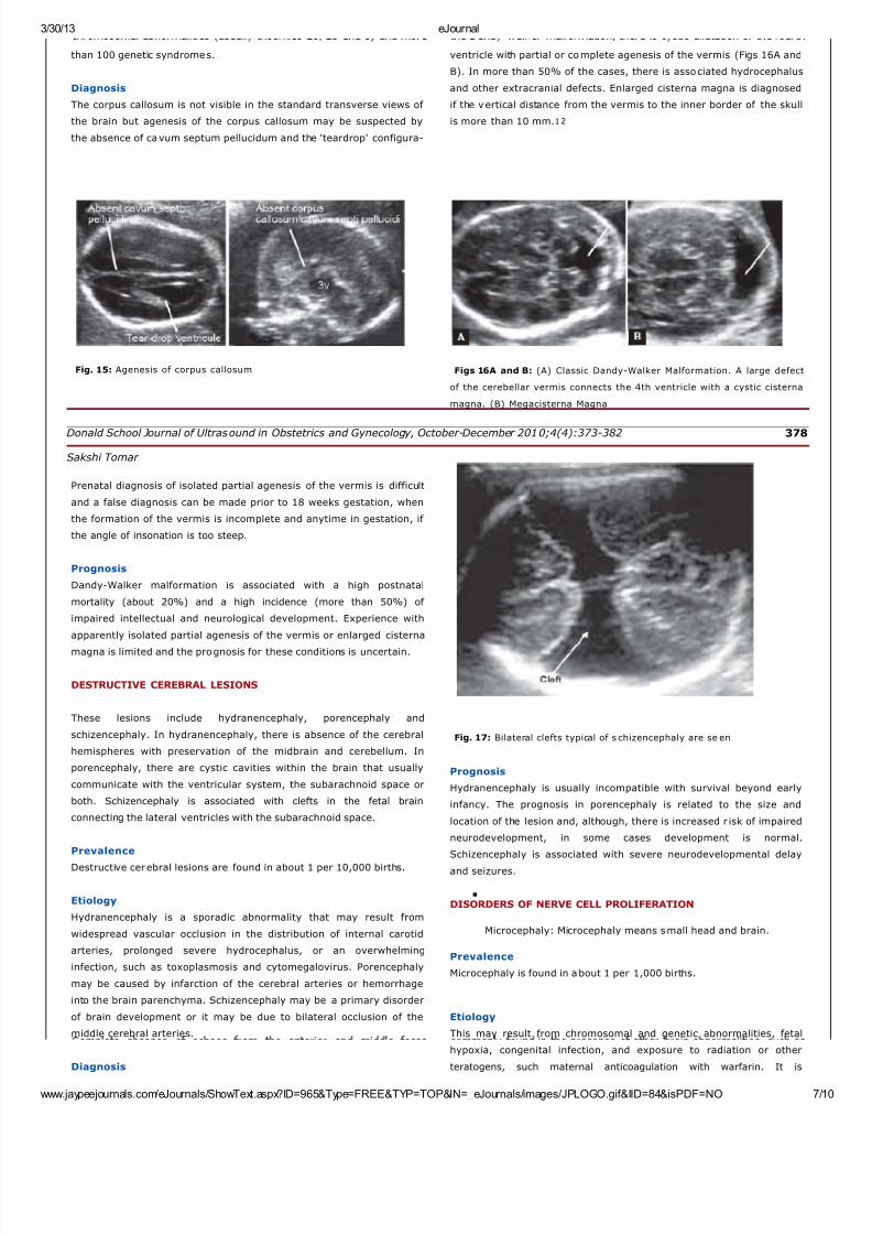
7282019 Ref Lecture eJournal
httpslidepdfcomreaderfullref-lecture-ejournal 710
33013 eJournal
wwwjaypeejournalscomeJournalsShowTextaspxID=965ampType=FREEampTYP=TOPampIN=_eJournalsimagesJPLOGOgifampIID=84ampisPDF=NO 7
than 100 genetic syndromes
Diagnosis
The corpus callosum is not visible in the standard transverse views of
the brain but agenesis of the corpus callosum may be suspected by
the absence of cavum septum pellucidum and the teardrop configura-
Fig 15 Agenesis of corpus callosum
ventricle with partial or complete agenesis of the vermis (Figs 16A and
B) In more than 50 of the cases there is associated hydrocephalus
and other extracranial defects Enlarged cisterna magna is diagnosed
if the vertical distance from the vermis to the inner border of the skull
is more than 10 mm12
Figs 16A and B (A) Classic Dandy-Walker Malformation A large defect
of the cerebellar vermis connects the 4th ventricle with a cystic cisterna
magna (B) Megacisterna Magna
Donald School Journal of Ultrasound in Obstetrics and Gynecology October-December 20104(4)373-382 378
Sakshi Tomar Prenatal diagnosis of isolated partial agenesis of the vermis is difficult
and a false diagnosis can be made prior to 18 weeks gestation when
the formation of the vermis is incomplete and anytime in gestation if
the angle of insonation is too steep
Prognosis
Dandy-Walker malformation is associated with a high postnatal
mortality (about 20) and a high incidence (more than 50) of
impaired intellectual and neurological development Experience with
apparently isolated partial agenesis of the vermis or enlarged cisterna
magna is limited and the prognosis for these conditions is uncertain
DESTRUCTIVE CEREBRAL LESIONS
These lesions include hydranencephaly porencephaly and
schizencephaly In hydranencephaly there is absence of the cerebral
hemispheres with preservation of the midbrain and cerebellum In
porencephaly there are cystic cavities within the brain that usually
communicate with the ventricular system the subarachnoid space or
both Schizencephaly is associated with clefts in the fetal brain
connecting the lateral ventricles with the subarachnoid space
Prevalence
Destructive cerebral lesions are found in about 1 per 10000 births
Etiology
Hydranencephaly is a sporadic abnormality that may result from
widespread vascular occlusion in the distribution of internal carotid
arteries prolonged severe hydrocephalus or an overwhelming
infection such as toxoplasmosis and cytomegalovirus Porencephaly
may be caused by infarction of the cerebral arteries or hemorrhage
into the brain parenchyma Schizencephaly may be a primary disorder
of brain development or it may be due to bilateral occlusion of the
middle cerebral arteries
Diagnosis
Fig 17 Bilateral clefts typical of s chizencephaly are se en
Prognosis
Hydranencephaly is usually incompatible with survival beyond early
infancy The prognosis in porencephaly is related to the size and
location of the lesion and although there is increased r isk of impaired
neurodevelopment in some cases development is normal
Schizencephaly is associated with severe neurodevelopmental delay
and seizures
DISORDERS OF NERVE CELL PROLIFERATION
Microcephaly Microcephaly means small head and brain
Prevalence
Microcephaly is found in about 1 per 1000 births
Etiology
This may result from chromosomal and genetic abnormalities fetal
hypoxia congenital infection and exposure to radiation or other
teratogens such maternal anticoagulation with warfarin It is
7282019 Ref Lecture eJournal
httpslidepdfcomreaderfullref-lecture-ejournal 810
33013 eJournal
wwwjaypeejournalscomeJournalsShowTextaspxID=965ampType=FREEampTYP=TOPampIN=_eJournalsimagesJPLOGOgifampIID=84ampisPDF=NO 8
omp e e a sence o ec oes rom e an er or an m e ossa
distinguishes hydranencephaly from severe hydrocephalus in which a
thin rim of remaining cortex and the midline echo can always be
identified In porencephaly there is one or more cystic area in the
cerebral cortex which usually communicates with the ventricle The
differential diagnosis is from intracranial cysts (arachnoid
glioependymal) that are usually found either within the scissures or in
the midline and compress the brain In schizencephaly there are
bilateral clefts extending from the lateral ventricles to the
subarachnoid space and is usually associated with absence of the
cavum septum pellucidum13 (Fig 17)
commony oun n e presence o o er ra n a norma es suc as
cephalocele and holoprosencephaly
Diagnosis
The diagnosis is made by the demonstration of brain abnormalities
such as holoprosencephaly In cases with apparently isolated
microcephaly it is necessary to demonstrate progressive decrease in
the head to abdomen circumference ra tio to below the 1st centile with
advancing gestation14 Such diagnosis may not be apparent before
the third trimester In microcephaly there is a typical disproportion
between the size of the skull and face The brain is small with the cer- 379 JAYPEE
Ultrasonographic Evaluation of the Fetal Neural Axis
Fig 18 Fetal microcephaly Note the sloping of the forehead
-ebral hemispheres affected to a greater extent than the midbrain and
posterior fossa Demonstration of a sloping forehead also increases
the index of suspicion (Fig 18)
Prognosis
This depends on the underlying cause but in more than 50 of cases
there is severe mental retardation
B Megalencephaly Megalencephaly means large head and brain
PrevalenceMegalencephaly is a very rare abnormality
Etiology
This is usually familial with no adverse consequence However it may
also be the consequence of genetic syndromes such as Beckwith-
Wiedemann syndrome achondroplasia neurofibromatosis and
tuberous sclerosis Unilateral megalencephaly is a sporadic condition
Diagnosis
The diagnosis is made by the demonstration of a head to abdomen
circumference ratio above the 99th centile without the evidence of hydrocephalus or intracranial masses Unilateral megalencephaly is
characterized by macrocrania a shift in the midline echo borderline
enlargement of the lateral ventricle and a typical gyri of the affected
hemisphere15
Prognosis
Isolated megalencephaly is usually an asymptomatic condition
Unilateral megalencephaly is associated with severe mental
retardation and untreatable seizures
INTRACRANIAL HEMORRHAGE IN UTERO
Hemorrha e ma be seen in a t ical sube end mal or
junction
Perinatal asphyxia known to induce fluctuation in blood
pressure16
ARTERIOVENOUS MALFORMATION
VEIN OF GALEN ANEURYSM
This is a midline aneurysmal dilation of the vein of Galen due to an
arteriovenous malformation with major hemodynamic disturbances
Prevalence
Vein of Galen aneurysm is a rare abnormality
Etiology
Vein of Galen aneurysm is a sporadic abnormality
Diagnosis
The diagnosis is made by the demonstration of a supratentorial
midline translucent elongated cyst Color Doppler demonstrates active
arteriovenous flow within the cyst There may be associated evidence
of high-output heart failure17 (Fig 19)
Prognosis
In the neonatal period about 50 of the infants present with heart
failure and the rest are asymptomatic In later life hydrocephalus and
intracranial hemorrhage may develop Good results can be achieved
by catheterization and embolization of the malformation
CHOROID PLEXUS CYSTS
These cysts usually bilateral are in the choroid plexuses of the lateral
cerebral ventricles
PrevalenceChoro id plexus cysts are found in about 2 of fetuses at 20 weeks o f
gestation but in more than 90 of cases they resolve by 26 weeks
Etiology
The choroid plexus is easily visualized from 10 weeks of gestation
when it occupies almost the entire hemisphere Thereafter and until 26
weeks there is a rapid decrease in both the size of the choroid plexus
and of the lateral cerebral ventricle in relation to the hemisphere
Choro id plexus cysts contain cerebrospinal fluid and cellular debris
Diagnosis
Single or multiple cystic areas (greater than 2 mm in diameter) in one
or both choroid plexuses18 (Fig 20)
7282019 Ref Lecture eJournal
httpslidepdfcomreaderfullref-lecture-ejournal 910
33013 eJournal
wwwjaypeejournalscomeJournalsShowTextaspxID=965ampType=FREEampTYP=TOPampIN=_eJournalsimagesJPLOGOgifampIID=84ampisPDF=NO 9
intraparenchymal location
Etiology
Sudden changes in the cerebral blood pressure leading to
ruptured capillaries in the germinal matrix or capillary venous
Prognosis
They are usually of no pathological significance but they are
associated with an increased risk for trisomy 18 and possibly trisomy
21 In the absence of other markers of trisomy 18 the maternal age-
related risk is increased by a factor of 15
Donald School Journal of Ultrasound in Obstetrics and Gynecology October-December 20104(4)373-382 380
Sakshi Tomar
Fig 19 Vein of Galen aneurysm Color Doppler flow image of the fetal
head demonstrating a vein of galen aneurysm (arrow) supplied by the
feed ing vesse ls from the circle of willis
Fig 20 Bilateral choroid plexus cysts seen in a transverse axial
sonogram
ARACHNOID CYSTS
Arachnoid cysts are fluid-filled cyst contained within the arachnoid
space
Prevalence
Arachnoid cysts are extremely rare
Etiology
Unknown infectious process has been hypothesized but it is unlikely
that this may explain the congenital cysts
Diagnosis
Arachnoid cys ts appear on antenatal ultrasound as sonolucent lesions
Fig 21 Arachnoid Cyst Corresponding MRI T2W image is seen
difficult Interhemispheric cysts associated with agenesis of the corpus
callosum most likely are not arachnoid cysts but rather glioependymal
cysts
Prognosis
Large cysts may cause intracranial hypertension and require
neurosurgical treatment However a normal intellectual development
in the range of 80 to 90 is reported by most series Spontaneous
remission has been described both in the postnatal as well as in the
antenatal per iod Glioependymal cyst that should be suspected in the
cases with associated agenesis of corpus callosum probably re flects a
greater degree of derangement in the development of the brain and
this may be reflected in worse outcome
CONCLUSION
Modern ultrasound equipments yield a unique potential for evaluation
of normal and abnormal fetal CNS very early in pregnancy It should
be stressed however that some cerebral anomalies are the
consequence of disruptions or are characterized by intrauterine
development Counseling the parents and deciding on a sensible
obstetric management are frequently difficult
REFERENCES
Monteagudo A Fetal neurosonography Should it be routine
Should it be detailed [Editorial] Ultrasound Obstet Gynecol
1998121
Callen Ultrasonography in Obstetrics and Gynecology (4th ed)
277-304
Bronshtein M Ornoy A Acrania Anencephaly resulting from
secondary degeneration of a closed neural tube Two cases in
the same family J Clin Ultrasound 199119230
Levi S Hijazi Y Schapps J et al Sensitivity and specificity of
routine antenatal screening for congenital anomalies by
ultrasound The Belgian Multicentric Study Ultrasound Obstet
Gynecol 19911102
Goldstein RB LaPidus AS Filly RA Fetal cephaloceles Diagnosis
7282019 Ref Lecture eJournal
httpslidepdfcomreaderfullref-lecture-ejournal 1010
33013 eJournal
j j l J l Sh T t ID 965ampT FREEampTYP TOPampIN J l i JPLOGO ifampIID 84ampi PDF NO 10
with a thin regular outline and containing no blood flow do not
communicate with the lateral ventricles and anyhow are not associated
with the loss of brain tissue (Fig 21) They occur most frequently in
the area of cerebral fissure and midline Large cyst may cause
significant mass effect and the distinction from porencephaly may be
with US Radiology 1991180803
Filly RA Goldstein RB Callen PW Fetal ventricle Importance in
381 JAYPEE
Ultrasonographic Evaluation of the Fetal Neural Axis routine obstetric sonography Radiology 19911811
Cardoza JD Goldstein RB Filly RA Exclusion of fetal
ventriculomegaly with a single measurement The width of lateral
ventricular atrium Radiology 1988169711
DeMyer W Holoprosencephaly vol 30 Amsterdam Elsevier
1977
Bronshtein M Weiner Z Early transvaginal sonographic diagnosis
of a lobar holoprosencephaly Prenat Diagn 199111459
Bennett GL Bromley B Benacerraf BR Agenesis of corpus
callosum Prenatal detection usually is not possible before 22
week s o f ge station Ra diology 1996199447
Benda C The Dandy-Walker syndrome or the so called atresia of
the foramen Magendie J Neuropathol Exp Neurol 19541314
Pilu G Goldstein I Reece E et al Sonography of fetal Dandy
Walker Malformations A reappraisal Ultrasound Obstet Gynecol
19922151
Pilu G falco P Perolo A Differential diagnosis and outcome of
fetal intracranial hypoechoic lesions Report of 21 cases
Ultrasound Obstet Gynecol 19979229
Chervenak FA Jeanty P Cantraine F et al The diagnosis of
fetal microcephaly Am J Obstet Gynecol 1984149512
Bignami A Palladini G Zappella M Unilateral Megalencephaly
with nerve cell hypertrophy An anatomical and quantitative
histochemical study Brain Res 19689103
Vergani P Strobelt N Locatelli A et al Clinical significance of
fetal intracranial haemorrhage Am J Obstet Gynecol
1996175536
Comstock CH Kirk JS Arteriovenous malformations Location
and evolution in the fetal brain J Ultrasound Med 199110361
Snijders R Shawa L Nicolaides K Fetal choroid cysts a nd trisomy
18 Assessment of risk based on ultrasound findings and
maternal age Prenat Diagnosis 199414119
382
JAYPEE
copy Jaypee Brothers Medical Publishers (P) Ltd
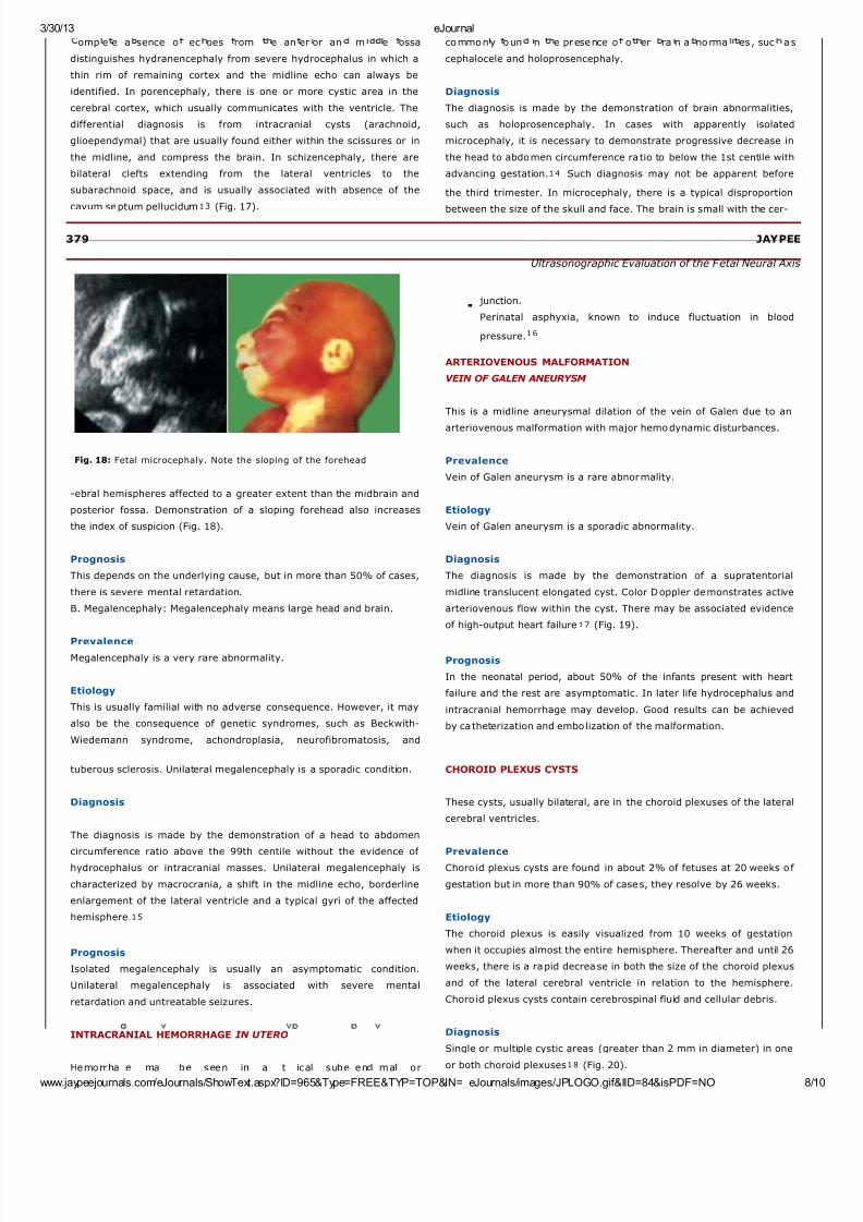
7282019 Ref Lecture eJournal
httpslidepdfcomreaderfullref-lecture-ejournal 810
33013 eJournal
wwwjaypeejournalscomeJournalsShowTextaspxID=965ampType=FREEampTYP=TOPampIN=_eJournalsimagesJPLOGOgifampIID=84ampisPDF=NO 8
omp e e a sence o ec oes rom e an er or an m e ossa
distinguishes hydranencephaly from severe hydrocephalus in which a
thin rim of remaining cortex and the midline echo can always be
identified In porencephaly there is one or more cystic area in the
cerebral cortex which usually communicates with the ventricle The
differential diagnosis is from intracranial cysts (arachnoid
glioependymal) that are usually found either within the scissures or in
the midline and compress the brain In schizencephaly there are
bilateral clefts extending from the lateral ventricles to the
subarachnoid space and is usually associated with absence of the
cavum septum pellucidum13 (Fig 17)
commony oun n e presence o o er ra n a norma es suc as
cephalocele and holoprosencephaly
Diagnosis
The diagnosis is made by the demonstration of brain abnormalities
such as holoprosencephaly In cases with apparently isolated
microcephaly it is necessary to demonstrate progressive decrease in
the head to abdomen circumference ra tio to below the 1st centile with
advancing gestation14 Such diagnosis may not be apparent before
the third trimester In microcephaly there is a typical disproportion
between the size of the skull and face The brain is small with the cer- 379 JAYPEE
Ultrasonographic Evaluation of the Fetal Neural Axis
Fig 18 Fetal microcephaly Note the sloping of the forehead
-ebral hemispheres affected to a greater extent than the midbrain and
posterior fossa Demonstration of a sloping forehead also increases
the index of suspicion (Fig 18)
Prognosis
This depends on the underlying cause but in more than 50 of cases
there is severe mental retardation
B Megalencephaly Megalencephaly means large head and brain
PrevalenceMegalencephaly is a very rare abnormality
Etiology
This is usually familial with no adverse consequence However it may
also be the consequence of genetic syndromes such as Beckwith-
Wiedemann syndrome achondroplasia neurofibromatosis and
tuberous sclerosis Unilateral megalencephaly is a sporadic condition
Diagnosis
The diagnosis is made by the demonstration of a head to abdomen
circumference ratio above the 99th centile without the evidence of hydrocephalus or intracranial masses Unilateral megalencephaly is
characterized by macrocrania a shift in the midline echo borderline
enlargement of the lateral ventricle and a typical gyri of the affected
hemisphere15
Prognosis
Isolated megalencephaly is usually an asymptomatic condition
Unilateral megalencephaly is associated with severe mental
retardation and untreatable seizures
INTRACRANIAL HEMORRHAGE IN UTERO
Hemorrha e ma be seen in a t ical sube end mal or
junction
Perinatal asphyxia known to induce fluctuation in blood
pressure16
ARTERIOVENOUS MALFORMATION
VEIN OF GALEN ANEURYSM
This is a midline aneurysmal dilation of the vein of Galen due to an
arteriovenous malformation with major hemodynamic disturbances
Prevalence
Vein of Galen aneurysm is a rare abnormality
Etiology
Vein of Galen aneurysm is a sporadic abnormality
Diagnosis
The diagnosis is made by the demonstration of a supratentorial
midline translucent elongated cyst Color Doppler demonstrates active
arteriovenous flow within the cyst There may be associated evidence
of high-output heart failure17 (Fig 19)
Prognosis
In the neonatal period about 50 of the infants present with heart
failure and the rest are asymptomatic In later life hydrocephalus and
intracranial hemorrhage may develop Good results can be achieved
by catheterization and embolization of the malformation
CHOROID PLEXUS CYSTS
These cysts usually bilateral are in the choroid plexuses of the lateral
cerebral ventricles
PrevalenceChoro id plexus cysts are found in about 2 of fetuses at 20 weeks o f
gestation but in more than 90 of cases they resolve by 26 weeks
Etiology
The choroid plexus is easily visualized from 10 weeks of gestation
when it occupies almost the entire hemisphere Thereafter and until 26
weeks there is a rapid decrease in both the size of the choroid plexus
and of the lateral cerebral ventricle in relation to the hemisphere
Choro id plexus cysts contain cerebrospinal fluid and cellular debris
Diagnosis
Single or multiple cystic areas (greater than 2 mm in diameter) in one
or both choroid plexuses18 (Fig 20)
7282019 Ref Lecture eJournal
httpslidepdfcomreaderfullref-lecture-ejournal 910
33013 eJournal
wwwjaypeejournalscomeJournalsShowTextaspxID=965ampType=FREEampTYP=TOPampIN=_eJournalsimagesJPLOGOgifampIID=84ampisPDF=NO 9
intraparenchymal location
Etiology
Sudden changes in the cerebral blood pressure leading to
ruptured capillaries in the germinal matrix or capillary venous
Prognosis
They are usually of no pathological significance but they are
associated with an increased risk for trisomy 18 and possibly trisomy
21 In the absence of other markers of trisomy 18 the maternal age-
related risk is increased by a factor of 15
Donald School Journal of Ultrasound in Obstetrics and Gynecology October-December 20104(4)373-382 380
Sakshi Tomar
Fig 19 Vein of Galen aneurysm Color Doppler flow image of the fetal
head demonstrating a vein of galen aneurysm (arrow) supplied by the
feed ing vesse ls from the circle of willis
Fig 20 Bilateral choroid plexus cysts seen in a transverse axial
sonogram
ARACHNOID CYSTS
Arachnoid cysts are fluid-filled cyst contained within the arachnoid
space
Prevalence
Arachnoid cysts are extremely rare
Etiology
Unknown infectious process has been hypothesized but it is unlikely
that this may explain the congenital cysts
Diagnosis
Arachnoid cys ts appear on antenatal ultrasound as sonolucent lesions
Fig 21 Arachnoid Cyst Corresponding MRI T2W image is seen
difficult Interhemispheric cysts associated with agenesis of the corpus
callosum most likely are not arachnoid cysts but rather glioependymal
cysts
Prognosis
Large cysts may cause intracranial hypertension and require
neurosurgical treatment However a normal intellectual development
in the range of 80 to 90 is reported by most series Spontaneous
remission has been described both in the postnatal as well as in the
antenatal per iod Glioependymal cyst that should be suspected in the
cases with associated agenesis of corpus callosum probably re flects a
greater degree of derangement in the development of the brain and
this may be reflected in worse outcome
CONCLUSION
Modern ultrasound equipments yield a unique potential for evaluation
of normal and abnormal fetal CNS very early in pregnancy It should
be stressed however that some cerebral anomalies are the
consequence of disruptions or are characterized by intrauterine
development Counseling the parents and deciding on a sensible
obstetric management are frequently difficult
REFERENCES
Monteagudo A Fetal neurosonography Should it be routine
Should it be detailed [Editorial] Ultrasound Obstet Gynecol
1998121
Callen Ultrasonography in Obstetrics and Gynecology (4th ed)
277-304
Bronshtein M Ornoy A Acrania Anencephaly resulting from
secondary degeneration of a closed neural tube Two cases in
the same family J Clin Ultrasound 199119230
Levi S Hijazi Y Schapps J et al Sensitivity and specificity of
routine antenatal screening for congenital anomalies by
ultrasound The Belgian Multicentric Study Ultrasound Obstet
Gynecol 19911102
Goldstein RB LaPidus AS Filly RA Fetal cephaloceles Diagnosis
7282019 Ref Lecture eJournal
httpslidepdfcomreaderfullref-lecture-ejournal 1010
33013 eJournal
j j l J l Sh T t ID 965ampT FREEampTYP TOPampIN J l i JPLOGO ifampIID 84ampi PDF NO 10
with a thin regular outline and containing no blood flow do not
communicate with the lateral ventricles and anyhow are not associated
with the loss of brain tissue (Fig 21) They occur most frequently in
the area of cerebral fissure and midline Large cyst may cause
significant mass effect and the distinction from porencephaly may be
with US Radiology 1991180803
Filly RA Goldstein RB Callen PW Fetal ventricle Importance in
381 JAYPEE
Ultrasonographic Evaluation of the Fetal Neural Axis routine obstetric sonography Radiology 19911811
Cardoza JD Goldstein RB Filly RA Exclusion of fetal
ventriculomegaly with a single measurement The width of lateral
ventricular atrium Radiology 1988169711
DeMyer W Holoprosencephaly vol 30 Amsterdam Elsevier
1977
Bronshtein M Weiner Z Early transvaginal sonographic diagnosis
of a lobar holoprosencephaly Prenat Diagn 199111459
Bennett GL Bromley B Benacerraf BR Agenesis of corpus
callosum Prenatal detection usually is not possible before 22
week s o f ge station Ra diology 1996199447
Benda C The Dandy-Walker syndrome or the so called atresia of
the foramen Magendie J Neuropathol Exp Neurol 19541314
Pilu G Goldstein I Reece E et al Sonography of fetal Dandy
Walker Malformations A reappraisal Ultrasound Obstet Gynecol
19922151
Pilu G falco P Perolo A Differential diagnosis and outcome of
fetal intracranial hypoechoic lesions Report of 21 cases
Ultrasound Obstet Gynecol 19979229
Chervenak FA Jeanty P Cantraine F et al The diagnosis of
fetal microcephaly Am J Obstet Gynecol 1984149512
Bignami A Palladini G Zappella M Unilateral Megalencephaly
with nerve cell hypertrophy An anatomical and quantitative
histochemical study Brain Res 19689103
Vergani P Strobelt N Locatelli A et al Clinical significance of
fetal intracranial haemorrhage Am J Obstet Gynecol
1996175536
Comstock CH Kirk JS Arteriovenous malformations Location
and evolution in the fetal brain J Ultrasound Med 199110361
Snijders R Shawa L Nicolaides K Fetal choroid cysts a nd trisomy
18 Assessment of risk based on ultrasound findings and
maternal age Prenat Diagnosis 199414119
382
JAYPEE
copy Jaypee Brothers Medical Publishers (P) Ltd
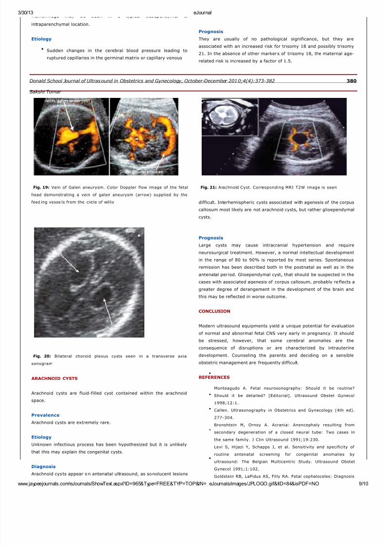
7282019 Ref Lecture eJournal
httpslidepdfcomreaderfullref-lecture-ejournal 910
33013 eJournal
wwwjaypeejournalscomeJournalsShowTextaspxID=965ampType=FREEampTYP=TOPampIN=_eJournalsimagesJPLOGOgifampIID=84ampisPDF=NO 9
intraparenchymal location
Etiology
Sudden changes in the cerebral blood pressure leading to
ruptured capillaries in the germinal matrix or capillary venous
Prognosis
They are usually of no pathological significance but they are
associated with an increased risk for trisomy 18 and possibly trisomy
21 In the absence of other markers of trisomy 18 the maternal age-
related risk is increased by a factor of 15
Donald School Journal of Ultrasound in Obstetrics and Gynecology October-December 20104(4)373-382 380
Sakshi Tomar
Fig 19 Vein of Galen aneurysm Color Doppler flow image of the fetal
head demonstrating a vein of galen aneurysm (arrow) supplied by the
feed ing vesse ls from the circle of willis
Fig 20 Bilateral choroid plexus cysts seen in a transverse axial
sonogram
ARACHNOID CYSTS
Arachnoid cysts are fluid-filled cyst contained within the arachnoid
space
Prevalence
Arachnoid cysts are extremely rare
Etiology
Unknown infectious process has been hypothesized but it is unlikely
that this may explain the congenital cysts
Diagnosis
Arachnoid cys ts appear on antenatal ultrasound as sonolucent lesions
Fig 21 Arachnoid Cyst Corresponding MRI T2W image is seen
difficult Interhemispheric cysts associated with agenesis of the corpus
callosum most likely are not arachnoid cysts but rather glioependymal
cysts
Prognosis
Large cysts may cause intracranial hypertension and require
neurosurgical treatment However a normal intellectual development
in the range of 80 to 90 is reported by most series Spontaneous
remission has been described both in the postnatal as well as in the
antenatal per iod Glioependymal cyst that should be suspected in the
cases with associated agenesis of corpus callosum probably re flects a
greater degree of derangement in the development of the brain and
this may be reflected in worse outcome
CONCLUSION
Modern ultrasound equipments yield a unique potential for evaluation
of normal and abnormal fetal CNS very early in pregnancy It should
be stressed however that some cerebral anomalies are the
consequence of disruptions or are characterized by intrauterine
development Counseling the parents and deciding on a sensible
obstetric management are frequently difficult
REFERENCES
Monteagudo A Fetal neurosonography Should it be routine
Should it be detailed [Editorial] Ultrasound Obstet Gynecol
1998121
Callen Ultrasonography in Obstetrics and Gynecology (4th ed)
277-304
Bronshtein M Ornoy A Acrania Anencephaly resulting from
secondary degeneration of a closed neural tube Two cases in
the same family J Clin Ultrasound 199119230
Levi S Hijazi Y Schapps J et al Sensitivity and specificity of
routine antenatal screening for congenital anomalies by
ultrasound The Belgian Multicentric Study Ultrasound Obstet
Gynecol 19911102
Goldstein RB LaPidus AS Filly RA Fetal cephaloceles Diagnosis
7282019 Ref Lecture eJournal
httpslidepdfcomreaderfullref-lecture-ejournal 1010
33013 eJournal
j j l J l Sh T t ID 965ampT FREEampTYP TOPampIN J l i JPLOGO ifampIID 84ampi PDF NO 10
with a thin regular outline and containing no blood flow do not
communicate with the lateral ventricles and anyhow are not associated
with the loss of brain tissue (Fig 21) They occur most frequently in
the area of cerebral fissure and midline Large cyst may cause
significant mass effect and the distinction from porencephaly may be
with US Radiology 1991180803
Filly RA Goldstein RB Callen PW Fetal ventricle Importance in
381 JAYPEE
Ultrasonographic Evaluation of the Fetal Neural Axis routine obstetric sonography Radiology 19911811
Cardoza JD Goldstein RB Filly RA Exclusion of fetal
ventriculomegaly with a single measurement The width of lateral
ventricular atrium Radiology 1988169711
DeMyer W Holoprosencephaly vol 30 Amsterdam Elsevier
1977
Bronshtein M Weiner Z Early transvaginal sonographic diagnosis
of a lobar holoprosencephaly Prenat Diagn 199111459
Bennett GL Bromley B Benacerraf BR Agenesis of corpus
callosum Prenatal detection usually is not possible before 22
week s o f ge station Ra diology 1996199447
Benda C The Dandy-Walker syndrome or the so called atresia of
the foramen Magendie J Neuropathol Exp Neurol 19541314
Pilu G Goldstein I Reece E et al Sonography of fetal Dandy
Walker Malformations A reappraisal Ultrasound Obstet Gynecol
19922151
Pilu G falco P Perolo A Differential diagnosis and outcome of
fetal intracranial hypoechoic lesions Report of 21 cases
Ultrasound Obstet Gynecol 19979229
Chervenak FA Jeanty P Cantraine F et al The diagnosis of
fetal microcephaly Am J Obstet Gynecol 1984149512
Bignami A Palladini G Zappella M Unilateral Megalencephaly
with nerve cell hypertrophy An anatomical and quantitative
histochemical study Brain Res 19689103
Vergani P Strobelt N Locatelli A et al Clinical significance of
fetal intracranial haemorrhage Am J Obstet Gynecol
1996175536
Comstock CH Kirk JS Arteriovenous malformations Location
and evolution in the fetal brain J Ultrasound Med 199110361
Snijders R Shawa L Nicolaides K Fetal choroid cysts a nd trisomy
18 Assessment of risk based on ultrasound findings and
maternal age Prenat Diagnosis 199414119
382
JAYPEE
copy Jaypee Brothers Medical Publishers (P) Ltd
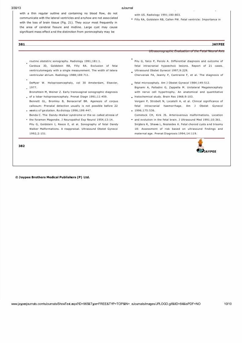
7282019 Ref Lecture eJournal
httpslidepdfcomreaderfullref-lecture-ejournal 1010
33013 eJournal
j j l J l Sh T t ID 965ampT FREEampTYP TOPampIN J l i JPLOGO ifampIID 84ampi PDF NO 10
with a thin regular outline and containing no blood flow do not
communicate with the lateral ventricles and anyhow are not associated
with the loss of brain tissue (Fig 21) They occur most frequently in
the area of cerebral fissure and midline Large cyst may cause
significant mass effect and the distinction from porencephaly may be
with US Radiology 1991180803
Filly RA Goldstein RB Callen PW Fetal ventricle Importance in
381 JAYPEE
Ultrasonographic Evaluation of the Fetal Neural Axis routine obstetric sonography Radiology 19911811
Cardoza JD Goldstein RB Filly RA Exclusion of fetal
ventriculomegaly with a single measurement The width of lateral
ventricular atrium Radiology 1988169711
DeMyer W Holoprosencephaly vol 30 Amsterdam Elsevier
1977
Bronshtein M Weiner Z Early transvaginal sonographic diagnosis
of a lobar holoprosencephaly Prenat Diagn 199111459
Bennett GL Bromley B Benacerraf BR Agenesis of corpus
callosum Prenatal detection usually is not possible before 22
week s o f ge station Ra diology 1996199447
Benda C The Dandy-Walker syndrome or the so called atresia of
the foramen Magendie J Neuropathol Exp Neurol 19541314
Pilu G Goldstein I Reece E et al Sonography of fetal Dandy
Walker Malformations A reappraisal Ultrasound Obstet Gynecol
19922151
Pilu G falco P Perolo A Differential diagnosis and outcome of
fetal intracranial hypoechoic lesions Report of 21 cases
Ultrasound Obstet Gynecol 19979229
Chervenak FA Jeanty P Cantraine F et al The diagnosis of
fetal microcephaly Am J Obstet Gynecol 1984149512
Bignami A Palladini G Zappella M Unilateral Megalencephaly
with nerve cell hypertrophy An anatomical and quantitative
histochemical study Brain Res 19689103
Vergani P Strobelt N Locatelli A et al Clinical significance of
fetal intracranial haemorrhage Am J Obstet Gynecol
1996175536
Comstock CH Kirk JS Arteriovenous malformations Location
and evolution in the fetal brain J Ultrasound Med 199110361
Snijders R Shawa L Nicolaides K Fetal choroid cysts a nd trisomy
18 Assessment of risk based on ultrasound findings and
maternal age Prenat Diagnosis 199414119
382
JAYPEE
copy Jaypee Brothers Medical Publishers (P) Ltd