regents biology monday, 12/8 today’s agenda: 1. turn in hw (cell craft) 2. go over limits to cell...
TRANSCRIPT
Regents Biology
Monday, 12/8Today’s Agenda:1. Turn in HW (Cell Craft)
2. Go over Limits To Cell Size lab
3. Lecture on viruses and bacteria
4. Time to work on HW or project
Bell Ringer (Answer in your notes WITHOUT looking back at them!):5. What are the 3 main jobs of organelles? Which
organelles carry out these functions?

Regents Biology
Cells have 3 main jobs make energy
need food + O2
cellular respiration & photosynthesis need to remove wastes
make proteins need instructions from DNA need to chain together amino acids & “finish”
& “ship” the protein make more cells
need to copy DNA & divide it up to daughter cells
Cell Summary
Our organellesdo all those
jobs!
Regents Biology
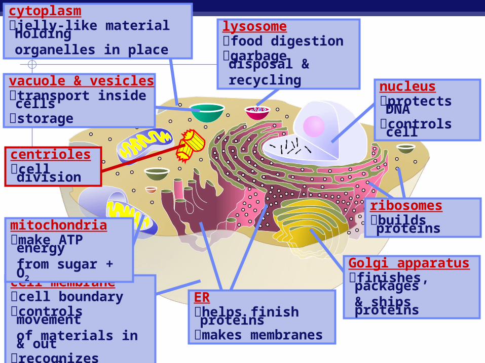
cell membranecell boundarycontrols movementof materials in & out
recognizes signals
cytoplasmjelly-like material holding organelles in place
mitochondriamake ATP energy
from sugar + O2
nucleusprotects DNAcontrols cell
ribosomesbuilds proteins
ERhelps finish proteinsmakes membranes
Golgi apparatusfinishes, packages
& ships proteins
lysosomefood digestiongarbage disposal &recycling
vacuole & vesiclestransport inside cellsstorage
centriolescell division

Regents Biology
Limit To Cell Size Lab5 questions were graded: 1, 4, 5, 7, and 8
1. As the cube increases in size the increase in volume is greater than the increase in surface area, therefore the SA:V ratio decreases.
4. The greater the SA:V ratio, the less time it takes for diffusion to occur.
5. As cells get larger they become less efficient at transporting essential nutrients/molecules to the center and have difficulty eliminating waste products and may essentially starve or become too toxic to survive.

Regents Biology
7. Oddly worded question – 2 points depending on your response
8. Discuss
Regents Biology
Ch. 19 - Viruses
Overview: A Borrowed Life Viruses called bacteriophages can infect and
set in motion a genetic takeover of bacteria, such as Escherichia coli
Viruses lead “a kind of borrowed life” between life-forms and chemicals
The origins of molecular biology lie in early studies of viruses that infect bacteria
© 2011 Pearson Education, Inc.
Regents Biology
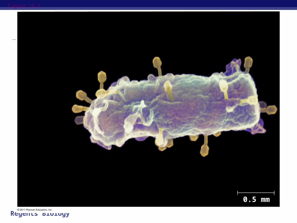
Figure 19.1
0.5 mm

Regents Biology
Concept 19.1: A virus consists of a nucleic acid surrounded by a protein coat
Structure of Viruses Viruses are not cells A virus is a very small infectious particle
consisting of nucleic acid enclosed in a protein coat and, in some cases, a membranous envelope
© 2011 Pearson Education, Inc.
Regents Biology
Viral Genomes
Viral genomes may consist of either Double- or single-stranded DNA, or Double- or single-stranded RNA
Depending on its type of nucleic acid, a virus is called a DNA virus or an RNA virus
© 2011 Pearson Education, Inc.

Regents Biology
Capsids and Envelopes
A capsid is the protein shell that encloses the viral genome
Capsids are built from protein subunits called capsomeres
A capsid can have various structures
© 2011 Pearson Education, Inc.

Regents Biology
Figure 19.3
Capsomereof capsid
RNA CapsomereDNA
Glycoprotein Glycoproteins
Membranousenvelope RNA
CapsidHead
DNA
Tailsheath
Tailfiber
18 250 nm 80 225 nm70–90 nm (diameter) 80–200 nm (diameter)
20 nm 50 nm 50 nm 50 nm(a) Tobacco
mosaic virus(b) Adenoviruses (c) Influenza viruses (d) Bacteriophage T4
Regents Biology
Some viruses have membranous envelopes that help them infect hosts
These viral envelopes surround the capsids of influenza viruses and many other viruses found in animals
Viral envelopes, which are derived from the host cell’s membrane, contain a combination of viral and host cell molecules
© 2011 Pearson Education, Inc.

Regents Biology
Bacteriophages, also called phages, are viruses that infect bacteria
They have the most complex capsids found among viruses
Phages have an elongated capsid head that encloses their DNA
A protein tail piece attaches the phage to the host and injects the phage DNA inside
© 2011 Pearson Education, Inc.
Regents Biology
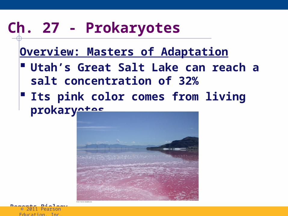
Overview: Masters of Adaptation Utah’s Great Salt Lake can reach a salt
concentration of 32% Its pink color comes from living prokaryotes
Ch. 27 - Prokaryotes
© 2011 Pearson Education, Inc.

Regents Biology
Thrive almost everywhere, including places that are too: Acidic Salty Cold/Hot
Most are microscopic, but what they lack in size they make up for in #s
There are more in a handful of fertile soil than the number of people who have ever lived
Prokaryotes are divided into two domains: Bacteria Archaea
© 2011 Pearson Education, Inc.
Prokaryotes

Regents Biology
Concept 27.1: Structural and functional adaptations contribute to prokaryotic success Most likely 1st organisms on Earth Most are unicellular, although some species
form colonies Sizes are usually 0.5–5 µm
Eykaryotic cells are usually 10–100 µm Come in a variety of shapes, the 3 most
common shapes are: spheres (cocci) rods (bacilli) spirals
© 2011 Pearson Education, Inc.

Regents Biology
Cell-Surface Structures
Cell wall: Maintains cell shape Protects the cell Prevents cell from bursting in a hypotonic
environment A eukaryotic cell wall is made of cellulose or
chitin Bacterial cell walls contain peptidoglycan
A network of sugar polymers cross-linked by polypeptides
© 2011 Pearson Education, Inc.

Regents Biology
Archaea contain polysaccharides and proteins but lack peptidoglycan
Scientists use the Gram stain to classify bacteria by cell wall composition Gram-positive bacteria have simpler walls with
a large amount of peptidoglycan Gram-negative bacteria have less
peptidoglycan and an outer membrane that can be toxic
© 2011 Pearson Education, Inc.

Regents Biology
Figure 27.3
(a) Gram-positive bacteria: peptidoglycan traps crystal violet.
Gram-positivebacteria
Peptido-glycanlayer
Cellwall
Plasmamembrane
10 m
Gram-negativebacteria
Outermembrane
Peptido-glycanlayer
Plasma membrane
Cellwall
Carbohydrate portionof lipopolysaccharide
(b) Gram-negative bacteria: crystal violet is easily rinsed away, revealing red dye.

Regents Biology
Many antibiotics target peptidoglycan and damage bacterial cell walls
Gram-negative bacteria are more likely to be antibiotic resistant
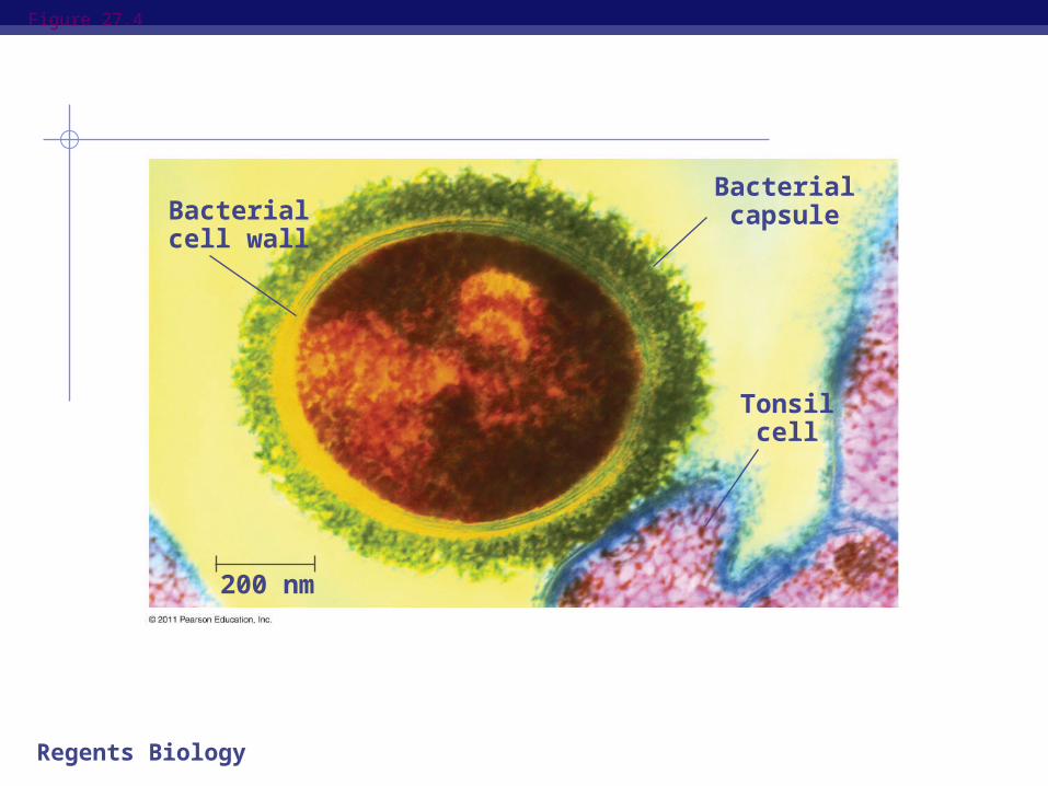
A polysaccharide or protein layer called a capsule covers many prokaryotes
© 2011 Pearson Education, Inc.
Regents Biology
Figure 27.4
Bacterialcell wall
Bacterialcapsule
Tonsilcell
200 nm

Regents Biology
Some prokaryotes have fimbriae, which allow them to stick to their substrate or other individuals in a colony
Pili (or sex pili) are longer than fimbriae and allow prokaryotes to exchange DNA
© 2011 Pearson Education, Inc.

Regents Biology
Figure 27.5
Fimbriae
1 m

Regents Biology
Motility
In a heterogeneous environment, many bacteria exhibit taxis: the ability to move toward or away from a
stimulus Chemotaxis is the movement toward or away
from a chemical stimulus Towards nutrients Away from toxins
© 2011 Pearson Education, Inc.

Regents Biology
Most motile bacteria propel themselves by flagella scattered about the surface or concentrated at one or both ends
Flagella of bacteria, archaea, and eukaryotes are composed of different proteins and likely evolved independently
© 2011 Pearson Education, Inc.

Regents Biology
Figure 27.6
Flagellum
Hook
Motor
Filament
RodPeptidoglycan
layerPlasma
membrane
Cell wall
20 nm
Regents Biology
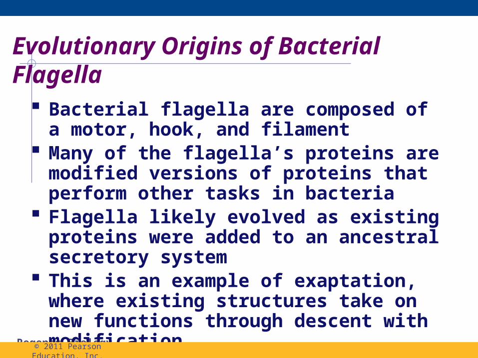
Evolutionary Origins of Bacterial Flagella
Bacterial flagella are composed of a motor, hook, and filament
Many of the flagella’s proteins are modified versions of proteins that perform other tasks in bacteria
Flagella likely evolved as existing proteins were added to an ancestral secretory system
This is an example of exaptation, where existing structures take on new functions through descent with modification
© 2011 Pearson Education, Inc.
Regents Biology
Internal Organization and DNA
Prokaryotic cells usually lack complex compartmentalization
Some prokaryotes do have specialized membranes that perform metabolic functions
These are usually infoldings of the plasma membrane
© 2011 Pearson Education, Inc.

Regents Biology
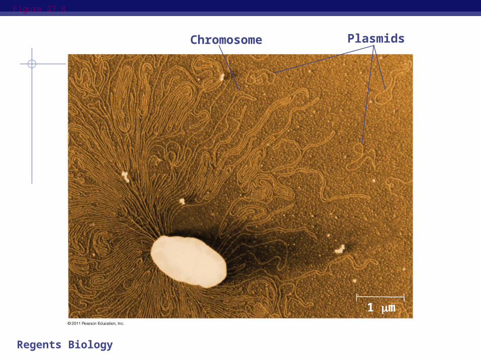
The prokaryotic genome has less DNA than the eukaryotic genome
Most of the genome consists of a circular chromosome
The chromosome is not surrounded by a membrane; it is located in the nucleoid region
Some species of bacteria also have smaller rings of DNA called plasmids
© 2011 Pearson Education, Inc.
Regents Biology
Figure 27.8
Chromosome Plasmids
1 m

Regents Biology
There are some differences between prokaryotes and eukaryotes in DNA replication, transcription, and translation
These allow people to use some antibiotics to inhibit bacterial growth without harming themselves
© 2011 Pearson Education, Inc.

Regents Biology
Reproduction and Adaptation
Prokaryotes reproduce quickly by binary fission and can divide every 1–3 hours
Key features of prokaryotic reproduction: They are small They reproduce by binary fission They have short generation times
© 2011 Pearson Education, Inc.

Regents Biology
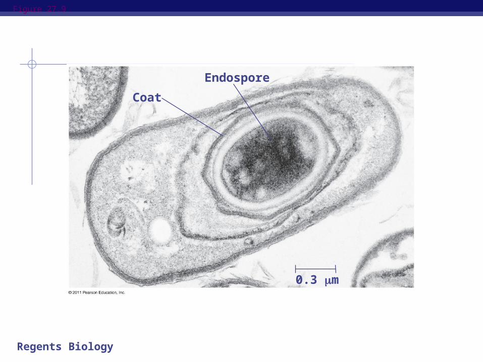
Many prokaryotes form metabolically inactive endospores, which can remain viable in harsh conditions for centuries
© 2011 Pearson Education, Inc.
Regents Biology
Figure 27.9
Coat
Endospore
0.3 m
Regents Biology
Their short generation time allows prokaryotes to evolve quickly
For example, adaptive evolution in a bacterial colony was documented in a lab over 8 years
Prokaryotes are not “primitive” but are highly evolved
© 2011 Pearson Education, Inc.