regional mapping of the batten disease locus (cln3) to human
TRANSCRIPT
Am. J. Hum. Genet. 49:1372-1377, 1991
Regional Mapping of the Batten Disease Locus (CLN3) toHuman Chromosome 16p12D. F. Callen,* E. Baker,* S. Lane,* J. Nancarrow,* A. Thompson,* S. A. Whitmore,*D. H. MacLennan,t R. Berger,$ D. Cherif, 1. jarvela,II L. Peltonen,II G. R. Sutherland,*and R. M. Gardiner§
*Department of Cytogenetics and Molecular Genetics, Adelaide Children's Hospital, North Adelaide; tDepartment of Medical Research, CharlesH. Best Institute, University of Toronto, Toronto; tINSERM Unite 301, Institut de G6netique Mol6culaire, Paris; §Department of Paediatrics,University College and Middlesex School of Medicine, Rayne Institute, London; and IILaboratory of Molecular Genetics, National Public Health Institute,Helsinki
Summary
The gene for Batten disease (CLN3) has been mapped to human chromosome 16 by demonstration of linkageto the haptoglobin locus, and its localization has been further refined using a panel of DNA markers. Theaim of this work was to refine the genetic and physical mapping of this disease locus. Genetic linkage analysiswas carried out in a larger group of families by using markers for five linked loci. Multipoint analysisindicated a most likely location for CLN3 in the interval between D16S67 and D16S148 (Z = 12.5). Physicalmapping of linked markers was carried out using somatic cell hybrid analysis and in situ hybridization. Amouse/human hybrid cell panel containing various segments of chromosome 16 has been constructed. Therelative order and physical location of breakpoints in the proximal portion of 16p were determined. Physicalmapping in this panel of the markers for the loci flanking CLN3 positioned them to the bands 16pl2.1-"16pl2.3. Fluorescent in situ hybridization of metaphase chromosomes by using these markers positionedthem to the region 16pll.2-16pl2.1. These results localize CLN3 to an interval of about 2 cM in the region16pl2.
Introduction
Batten disease, the juvenile-onset subtype of neuronalceroid lipofuscinosis, is an autosomal recessive neuro-degenerative disease characterized by the accumula-tion of autofluorescent lipopigment in neurones andother tissues. Genetic linkage studies using "classical"protein markers identified linkage to the haptoglobinlocus, allowing assignment of this locus (CLN3) tohuman chromosome 16 (Eiberg et al. 1989). Furtherstudies, using DNA markers in 42 families, indicatedthat the most likely map location for CLN3 was in theinterval between D16S148 and D16S150 (Gardiner etal. 1990).
Received May 16, 1991; revision received August 8, 1991.Address for correspondence and reprints: R. M. Gardiner, De-
partment of Paediatrics, Rayne Institute, University Street, LondonWC1E 6JJ, England.i 1991 by The American Society of Human Genetics. All rights reserved.0002-9297/91 /4906-0025$02.00
The aim of the present work was to further refine thegenetic and physical localization of CLN3. Linkageanalysis was carried out in a total of 58 families byusing an additional DNA marker (CRI-0391, identi-fying D16S67).
Physical mapping of linked markers was carried outusing a mouse-human hybrid cell panel containingvarious segments of human chromosome 16. This hasbeen constructed from constitutional chromosome 16rearrangements ascertained in patients by cytogeneticstudies (Callen 1986; Callen et al., in press). The rela-tive order and physical location of these breakpointshave been determined by cytogenetic analysis and bymapping genes or anonymous DNA probes by South-ern analysis and PCR (Callen et al. 1988, 1989, andin press; Richards et al., in press). In the present study,breakpoints in the proximal portion of the short armof chromosome 16 were utilized to physically localizethe markers which genetically flank CLN3. Fluores-cent in situ hybridization provided evidence for local-
1372
Batten Disease Maps to Chromosome l6pl2
ization to an overlapping region extending more prox-imally. This combination of genetic and physicalmapping has allowed the CLN3 locus to be mappedto chromosome 16pl2.
Subjects and Methods
Families
The families used in the present study were (a) thetwo-generation kindreds analyzed elsewhere (Gardi-ner et al. 1990) and (b) an additional 16 families. Intotal, 97 affected individuals and 70 unaffected sib-lings were studied. There were three affected siblingsin each of five pedigrees, two affected siblings in eachof 30 pedigrees, and one affected sibling with one tofive unaffected sibling(s) in each of 23 pedigrees. Diag-nostic criteria were as described elsewhere (Gardineret al. 1990).
Genetic Mapping
DNA marker phenotyping was carried out by South-ern hybridization using standard methods describedelsewhere (Gardiner et al. 1990). Members of thesekindreds were typed with four of the markers used intwo previous studies: Julier et al. (1990) detected lociD16S159 (by pCJ52-94T1), D16S148 (by pCJ52-95M1), D16S150 (by CJ52-161), and D16S151 (bypCJ52-209M1), and Keith et al. (1990) detectedD16S67 (by CRI-0391). The multipoint method wasused for linkage analysis (Lathrop et al. 1985). Thelod scores were calculated by dividing the locationscore by 4.6. The map of five marker loci was assumedto be fixed, and the location of CLN3 was allowedto vary over a range large enough to encompass freerecombination on either side of the marker cluster.The sex-averaged genetic distance between markerloci was calculated from previously published dataand information from the CEPH data base.
Physical Mapping
Construction and characterization of the hybrid celllines have been described elsewhere (Callen 1986; Callenet al., in press). For the hybrid cell lines CY12, CY165,CY15, and CY13 the portion of chromosome 16 re-tained in each hybrid consisted of the region from thebreakpoint on the short arm to the end of the longarm. The cell line CY160 contained an interstitial de-letion of the region 16pll.2- pl2.1.
The marker probes used for linkage analysis wereexamined, in addition to the following: the gene
ATP2A, detected by a cDNA probe encoding the hu-man fast-twitch muscle Ca-ATPase (MacLennan etal. 1987), and the gene IL4R, detected by a 670-bpPstI-EcoRI fragment of the mouse IL4R clone B4cDNA. DNA samples isolated from cell hybrids, hu-man leukocytes, and the mouse cell line A9 were ana-lyzed by Southern hybridization according to amethod described elsewhere (Hyland et al. 1988).Some probes contained repetitive sequences, and pre-association with an excess of sonicated human DNAwas necessary (Sealey et al. 1985). The CR3A genewas mapped by PCR using DNA primer sequencesconstructed from the known gene sequences (Richardset al., in press). The mouse line A9, the hybrid mouseparent, and CY18, a mouse human hybrid which con-tains a chromosome 16 as the only human chromo-some, provided appropriate controls.The fragile site FRA16E was expressed in lympho-
blastoid cell lines by addition of Hoechst 33258 24 hbefore harvest (Hori et al. 1988). Location of gene oranonymous DNA probes with respect to this fragilesite was carried out by in situ hybridization usingtritium-labeled probes according to a method de-scribed elsewhere (Pritchard et al., in press).
Competitive in situ hybridization experiments wereperformed according to the method of Cherif et al.(1990). Metaphase chromosomes from phytohemag-glutinin-stimulated blood cells of healthy males werecultured according to the methotrexate-5-bromeode-oxyuridine (BrdU) method. Cosmid probes were la-beled by nick translation with bio-1 1 dUTP and werehybridized. The hybridization signal was revealedwith avidin FITC and was amplified according to amethod described by Pinkel et al. (1986). The hybrid-ized probes appeared as yellow-green spots on redR-banded chromosomes.
Results
Linkage Analysis
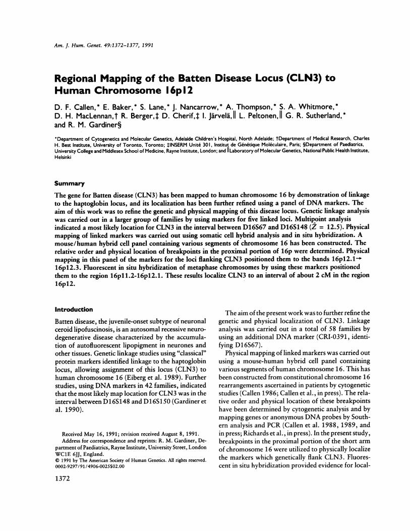
A map summarizing the lod scores calculated forCLN3 at various positions by using multipoint analy-sis and a fixed map of the five marker loci is shown infigure 1. The analysis suggested that the CLN3 geneis most likely located between D16S148 and D16S67(maximum lod score 12.50), with 5.0 x 10⁶:1 oddsfavoring this location over the second-best location.
Somatic Cell Hybrid Analysis
Somatic cell hybrid analysis allowed (a) the probesto be physically mapped to six intervals within the
1373
Callen et al.
LOO SCORE
* t
-61
MAP DISTANCE
11-2
11*111-1
Figure I Location map summarizing lod scores calculatedfor CLN3 at various map positions on fixed marker map ofchromo-some 16. The loci used were D16S159 (A), D16S67 (B), D16S148(C), D16S150 (D) and D16S151 (E). D16S67 (CRI-0391) was arbi-trarily placed at 0. The sex-combined recombination fractions usedwere D16S159-.058-D16S67-.023-D16S148-.096-D16S15O-.122-D16S151. Map distance in centimorgans was calculated usingHaldane's formula.
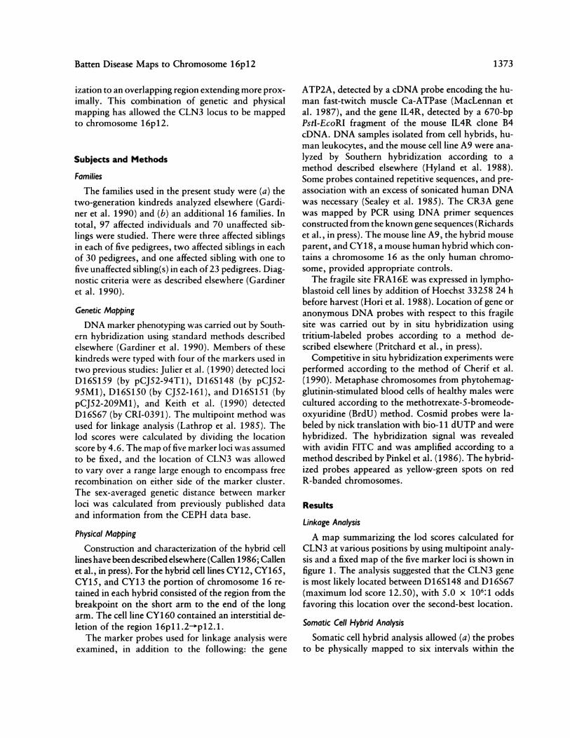
proximal portion ofchromosome 16p (see table 1) and(b) their order to be confirmed. The hybrid CY160contains an interstitial deletion of the short arm ofchromosome 16, a deletion which encompasses thegenes ATP2A, IL4R, and CR3A, with a distal break-point between ATP2A and D16S148. The D16S148locus is distal to this breakpoint, since it is distalto the CY12 breakpoint. These data enable six dif-ferent intervals to be distinguished in the region16pl2.3- pll .1, as ilustrated in figure 2. The FRA16E
D16S159 CY13
CY156OATP2A
-C10
FRA16E
IL4R
CY12
\CCR3A\ ~~CY16OP
Figure 2 Ideogram ofhuman chromosome 16p, showing ap-
proximate location of breakpoints and physical location of markerloci. The interstitial deletion of 16p is indicated by the distalbreakpoint (CY160D) and the proximal breakpoint (CY160P).
site provides an accurate anchor point for this physicalmap to the chromosome. Therefore, the two probesD16S148 and D16S67, which genetically flank CLN3,are clearly located in the region 16pl2.1-p1 2.3. Thehybrid CY165 possesses a breakpoint which is be-tween these two probes. The other two probes,D16S150 and D16S151, which have been shown to
be genetically linked to CLN3, map to the proximal halfof the long arm of chromosome 16 (data not shown).The gene ATP2A has been shown by MacLennan
et al. (1987) to be localized on chromosome 16. The
Table I
Results of Somatic Cell Hybrid DNA Analysis: Physical Mapping of Probes toProximal Portion of Chromosome 16p
STATUS ATa
Locus (probe) CY13 CY15 CY16S FRA16E CY12 CY160
D16S159 (pCJS2-94T1 .... + - - - +D16S67 (pCRI-0391) ....... + + - - +D16S148 (pCJ52-95MI) + + + - +ATP2A ................. + + + - - -IL4R ................. + + + + - -CR3A ................. + + + + -
a A plus sign ( + ) indicates either that the probe hybridized to the hybrid DNA or that a positive resultwas obtained using PCR; a minus sign (-) indicates a negative result by either method. For FRA16E,investigated by in situ hybridization, the plus sign indicates hybridization proximal to FRA16E, and theminus sign indicates hybridization distal to this fragile site.
1374
4
E
Batten Disease Maps to Chromosome l6pl2
a RS~ /i_
ate~~~~ ~~~~lia E" l! :: :.U::;-:1.",! ! In 5 - s > OX<toss:,P At, , Nu iTjw.i
b~~~
E~~~~~~~~~~~~~~~~~~~~~!T~trmna
Pj~ ...:!:.:. .......
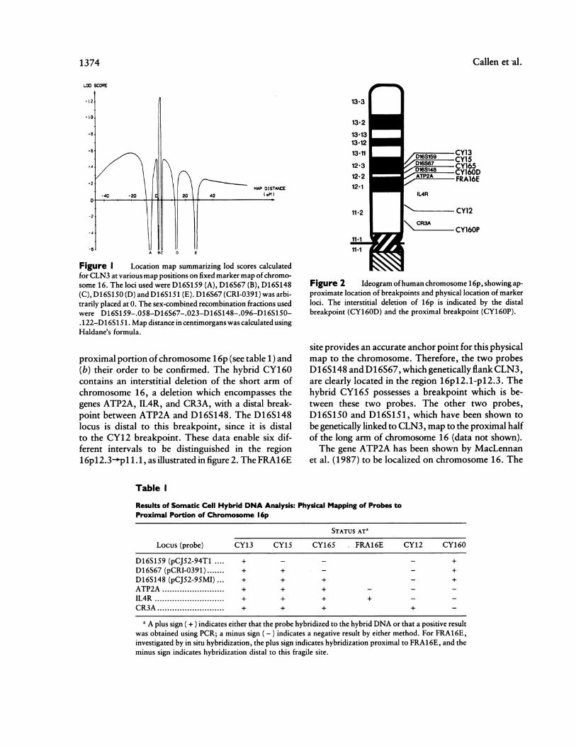
R -~! RXM: .. .. 's~~~~~~~~~~~~~~~~~~~~~~~~~~~~~~~~~~~~~~~~~~~~~~~.;.....;..'.,.!.Figure3 Insituhybridization of tritiated probes to FRA16E: partial metaphases showing expression of thefragilesiteFRA16Eat~~~~~~~~~~~~~~~~~~~~~~~~~~~~~~~~~~~~~~~~~~~~~~.........
16pl.1. , I sit hyridiatin wih pobe etetingIL4 locs, howig grinsproxmalto beakointof ragie ste. he L4R ocuis proximaltoFRA16E.b,In situ hybridization with probe detecting ATP2A locus, showing grains distal to breakpointoffragilesite.The~~~~~~~~~~~~~~~~~~~~~~~~~~~~~~~~~~~~~~~~~........
ATP2Alocus is distal to FRA16E.~~~~~~~~~~IR!un -L
present report allows the localization ofATP2A to berefined to the short arm of chromosome 16, in bandpl2.1-pl2.2 just distal to the fragile site FRA16E (fig.3a). The localization of the genes IL4R (fig. 3b) andCR3A is consistent with published data (Pritchard et
al., in press; Richards et al., in press).
In Situ Hybridization
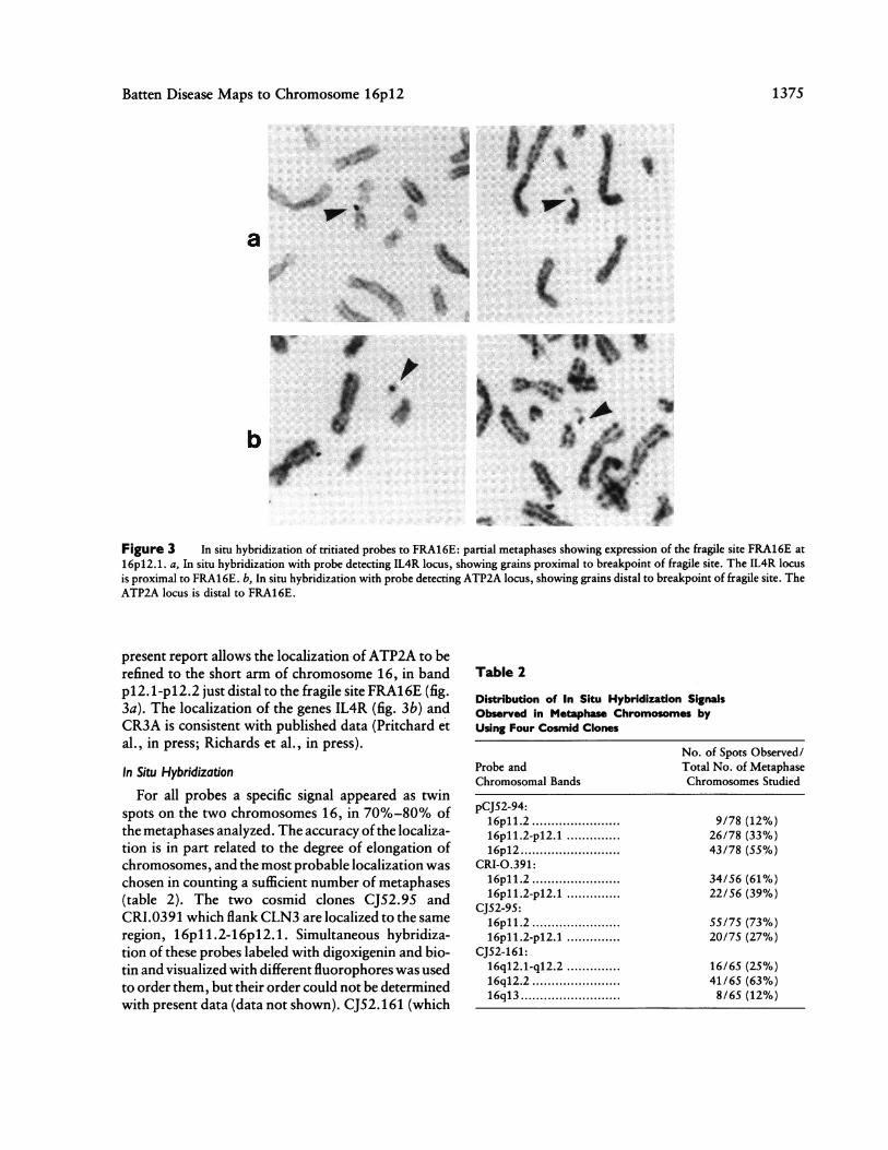
For all probes a specific signal appeared as twinspots on the two chromosomes 16, in 70%-80% ofthe metaphases analyzed. The accuracy ofthe localiza-tion is in part related to the degree of elongation ofchromosomes, and the most probable localization waschosen in counting a sufficient number of metaphases(table 2). The two cosmid clones CJ52.95 andCRI.0391 which flank CLN3 are localized to the sameregion, 16pll.2-16pl12.l. Simultaneous hybridiza-tion of these probes labeled with digoxigenin and bio-tin and visualized with different fluorophores was usedto order them, but their order could not be determinedwith present data (data not shown). CJ52.161 (which
Table 2
Distribution of In Situ Hybridization SignalsObserved in Metaphase Chromosomes byUsing Four Cosmid Clones
No. of Spots Observed/Probe and Total No. of MetaphaseChromosomal Bands Chromosomes Studied
pCJ52-94:16pll.2 ................ 9/78 (12%)16pll.2-pl2 ..............l 26/78 (33%)16pl2 ................ 43/78 (55%)
CRI-0.391:16pl1.2 ................ 34/56 (61%)16pll.2-pl2.1 .............. 22/56 (39%)
CJ52-95:16pll.2 ................ 55/75 (73%)16pll.2-pl2 ..............l 20/75 (27%)
CJ52-161:16q12.1-q12.2 .............. 16/65 (25%)16q12.2 ................ 41/65 (63%)16q13 ................ 8/65 (12%)
137S
1376 Callen et al.
detects locus D16S150) mapped to the long arm inband 16q12.2.
Discussion
A combination of genetic and physical mapping hasallowed CLN3 to be mapped to human chromosome16p12. Linkage to the haptoglobin locus on the longarm of human chromosome 16 allowed initial local-ization of CLN3 to this chromosome (Eiberg et al.1989), and additional analysis using markers for oneofthe published genetic maps ofchromosome 16 (Gar-diner et al. 1990) indicated that this disease locus wasat a considerable genetic distance from HP. The physi-cal localization of these markers was, however, un-known.
Genetic data have been further refined using addi-tional families and markers from the other availablechromosome 16 genetic map (Keith et al. 1990). Thedata presented here indicate that the most likely loca-tion of CLN3 is in the interval between D16S67 andD16S148. The physical location of these markers inthe proximal region of the short arm of human chro-mosome 16 has now been established using somaticcell hybrid analysis and in situ hybridization.The data summarized in table 1 enable six different
regions to be distinguished in the region p1.1.p12.3. It is generally difficult to unequivocally locatebreakpoints of chromosome translocations, becauseof the limitations in interpreting the G-banding pat-tern at the translocation breakpoint. However, theFRA16E does provide an accurate anchor point forthe chromosome, since the position of fragile sites canbe determined precisely. These data therefore clearlylocalize, to bands pl21-3pl2.3, the two probes CRI-0391 (D16S67) and pCJ5295-Ml (D16S148), whichflank CLN3.
In situ hybridization using these probes providesresults consistent with these observations. Variationsin the degree of elongation of chromosomes is, ofcourse, one factor limiting resolution. The distribu-tion of signals observed suggested a more proximallocation but encompassed the region to which theseprobes map by somatic cell hybrid analysis. Togetherwith the somatic cell hybrid data, inability to separatepCJ52-95 and CRI-0391 during simultaneous hybrid-ization suggests that these DNA sequences are locatedwithin a region of approximately 3 Mb.Work in progress is using additional markers
mapped to this region, as well as newly available fami-lies, to further narrow the genetic interval encom-
passing CLN3, as a prelude to constitution of a physi-cal map of the CLN3 region. The availability both ofYAC libraries of the human genome and of a cosmidcontig map covering half of the euchromatin contentof chromosome 16 (Stallings et al. 1990) should facili-tate work directed toward cloning a region ofgenomicDNA encompassing CLN3.
AcknowledgmentsWe are grateful to the National Fund for Research into
Crippling Diseases, to the Medical Research Council (UK),to the Child Brain Disease Foundation, to the Research Trustfor Metabolic Diseases in Childhood and to the Academy ofFinland, the Rinnekati Research Foundation, for financialsupport. The work at the Adelaide Children's Hospital wassupported by U.S. Department of Energy, grant DE-FG02-89ER60863. The support of French Ministere de la Recher-che et de la Technologie grant 89C0878 is acknowledged.The study would not have been possible without the gener-ous cooperation of the patients, their families, and collab-orating physicians. Andrew Sandford and Mary Deadmanprovided excellent technical assistance.
ReferencesCallen DF (1986) A mouse-human hybrid cell panel for map-
ping human chromosome 16. Ann Genet 29:235-239Callen DF, Baker E, Lane S. An expanded mouse-human
hybrid cell panel for mapping human chromosome 16.Ann Genet (in press)
Callen DF, Hyland VJ, Barker EG, Fratini A, Gedeon AK,Mulley JC, Fernandez KEW, et al (1989) Mapping theshort arm of human chromosome 16. Genomics 4:348-354
Callen DF, Hyland VJ, Baker EG, Fratini A, Simmers RN,Mulley JC, Sutherland GR (1988) Fine mapping of geneprobes and anonymous DNA fragments to the long armof chromosome 16. Genomics 2:144-153
Cherif D, Julier C, Delattre 0, DerreJ, Lathrop GM, BergerR (1990) Simultaneous localization of cosmids and chro-mosome R-banding by fluorescence microscopy -appli-cation to regional mapping of human chromosome 11.Proc Natl Acad Sci USA 87:6639-6643
Eiberg H, Gardiner RM, Mohr J (1989) Batten disease(Spielmeyer-Sjogren disease) and haptoglobins (HP): indi-cation of linkage and assignment to chromosome 16. ClinGenet 36:217-218
Gardiner RM, Sandford A, Deadman M, Poulton J, Cook-son W, Reeders S, Jokiaho I, et al (1990) Batten disease(Spielmeyer-Vogt disease, juvenile onset neuronal ceroid-lipofuscinosis) gene (CLN3) maps to human chromosome6. Genomics 8:387-390
Hori T, Takahashi E, Ishihara T, Minamihisamatsu M,
Batten Disease Maps to Chromosome 16p12 1377
Kaneko Y, Murata M (1988) Distamycin A-induciblefragile sites and cancer proneness. Cancer Genet Cyto-genet 34:177-187
Hyland VJ, Grist S, Callen DF, Sutherland GR (1988) Anon-ymous DNA probes to human chromosome 16 derivedfrom a flow-purified library. Am J Hum Genet 42:373-379
Julier C, Nakamura Y, Lathrop M, O'Connell P, LeppertM, Mohandas T, Lalouel J-M, et al (1990) A primarymap of 24 loci on human chromosome 16. Genomics 6:419-427
Keith TP, Green P, Reeders ST, Brown VA, Phipps P,Bricker A, Falls K, et al (1990) Genetic linkage map of 46DNA markers on human chromosome 16. Proc Natl AcadSci USA 87:5754-5758
Lathrop GM, Lalouel JM, Julier C, OttJ (1985) Multilocuslinkage analysis in humans: detection of linkage and esti-mation of recombination. Am J Hum Genet 37:482-498
MacLennan DH, Brandl CJ, Champanieria S, Holland PC,Powers VE, Willard H (1987) Fast-twitch and slow-twitch/cardiac Ca2 + ATPase genes map to human chro-mosomes 16 and 12. Somatic Cell Mol Genet 13:441-446
Pinkel D, Straume T, Gray JW (1986) Cytogenetic analysisusing quantitative, high-sensitivity, fluorescence hybrid-ization. Proc Natl Acad Sci USA 83:2934-2938
Pritchard MA, Baker EG, Whitmore SA, Sutherland GR,Idzerola RL, Park LS, Jenkins NA, et al. The interleukin-4receptor gene (IL4R) maps to 16p 1 .2-16pl 2.1 in humanand to the distal region ofchromosome 7 in mouse. Geno-mics (in press)
Richards RI, Holman K, Lane S, Sutherland GR, CallenDF. Human chromosome 16 physical map: mapping ofsomatic cell hybrids using multiplex PCR deletion analysisof sequence tagged sites. Genomics (in press)
Sealey PG, Whittaker PA, Southern EM (1985) Removal ofrepeated sequences from hybridization probes. NucleicAcids Res 13:1905-1922
Stallings RL, Torney DC, Hildebrand CE, Longmire JL,Deaven LL, Jett JH, Doggett NA, et al (1990) Physicalmapping of human chromosomes by repetitive sequencefingerprinting. Proc Natl Acad Sci USA 87:6218-6223