registration of free-breathing 3d+t abdominal perfusion ct …cohen/mypapers/prevostmiccai20… ·...
TRANSCRIPT

Registration of Free-Breathing 3D+t AbdominalPerfusion CT Images via Co-Segmentation
R. Prevost1,2, B. Romain1,3, R. Cuingnet1, B. Mory1,L. Rouet1, O. Lucidarme4, L.D. Cohen2, and R. Ardon1
1 Philips Research Medisys, Suresnes, France2 CEREMADE UMR 7534, CNRS, Universite Paris Dauphine, Paris, France
4 MAS, Ecole Centrale Paris, Chatenay Malabry, France3 Hopital La Pitie-Salpetriere, AP-HP, Paris, France
Abstract. Dynamic contrast-enhanced computed tomography (DCE-CT) is a valuable imaging modality to assess tissues properties, partic-ularly in tumours, by estimating pharmacokinetic parameters from theevolution of pixels intensities in 3D+t acquisitions. However, this requiresa registration of the whole sequence of volumes, which is challenging es-pecially when the patient breathes freely. In this paper, we propose ageneric, fast and automatic method to address this problem. As standardiconic registration methods are not robust to contrast intake, we ratherrely on the segmentation of the organ of interest. This segmentation isperformed jointly with the registration of the sequence within a novelco-segmentation framework. Our approach is based on implicit templatedeformation, that we extend to a co-segmentation algorithm which pro-vides as outputs both a segmentation of the organ of interest in everyimage and stabilising transformations for the whole sequence. The pro-posed method is validated on 15 datasets acquired from patients withrenal lesions and shows improvement in terms of registration and esti-mation of pharmacokinetic parameters over the state-of-the-art method.
1 Introduction
1.1 Clinical context
Dynamic contrast-enhanced (DCE) or perfusion imaging consists in acquiringa time sequence of images after a contrast agent injection. Parametric imagesare then generated by fitting at each voxel a pharmacokinetic model to its time-intensity curve. This technique is particularly used for oncologic applications,such as renal tumours follow-up, as the estimated parameters yield valuableinformation on healthy tissues and lesions [1].
Perfusion images can be acquired by MRI or CT systems. In this paper, wefocus on DCE-CT sequences as it presents several advantages over DCE-MRI [1].First, there is a linear relation between contrast agent concentration and imageintensities (Hounsfield units), which simplifies pharmacokinetic models fitting.Second, CT is a cheaper and more widespread modality than MRI. However,

2 R. Prevost et al.
... ......
Kidney SegmentationRegistering Transformations
OutputsInitial model
φ0
Input image Ii Input image Ij
L GGi Gj
Fig. 1. Tracking via co-segmentation of the kidney in a sequence of volumes (Ii)i.The segmentation is performed by template deformation (φ0 L G) using the wholesequence, while stabilising transformations (Gi)i are simultaneously estimated.
this modality generates ionizing radiations that may harm the patient. In or-der to limit those risks, such acquisitions are performed in a very limited fieldof view with a reduced dose and a low framerate. This results in small, low-resoluted volumes (both in spatially and temporally) that are noisier than staticCT acquisitions (see Figure 1).
To capture the full dynamics of the contrast diffusion, a perfusion proto-col lasts several minutes. Because of the patient’s breathing, voxels correspondto different anatomical locations across the sequence. The major challenge inparameter estimation is therefore to design a robust registration method, thatcannot use temporal consistency because of the low framerate.
1.2 Related work and contributions
Several methods have already been proposed to register DCE sequences. Non-rigid iconic registration methods [2–4] are computationally demanding and relyon their similarity criterion. Standard choices such as mutual information arenot effective in DCE-CT sequences [5]. In [6], Bhushan et al. used the pharma-cokinetic model fitting error as registration criterion for DCE-MR, thus couplingthe two tasks of sequence stabilisation and parameter estimation. However, thelatter is a highly non-convex problem: including additional unknowns (namelythe pharmacokinetic parameters) increases the risk of falling in local minima.
Some methods rather use a segmentation of the organ of interest to guidethe registration [7–9]. Because of contrast diffusion, edge information is indeedmore robust than region-based terms. In [7–9], segmentation and registrationprocesses are performed sequentially. Yet they are inter-dependent (and equallychallenging in DCE-CT images). In this paper, we propose a method to addressboth tasks simultaneously.
To do so, we extend the model-based segmentation algorithm proposed byMory et al. [10] (that already proved effective kidney segmentation in CT im-ages [11]) to a co-segmentation method that uses multiples images. This exten-
Registration of 3D+t Perfusion CT Images via Co-Segmentation 3
sion was inspired by a paper of Yezzi et al. [12], in which they segment a pairof CT/MR images with a single shape. However their work was based on ac-tive contours, while we adapt this approach to a more elaborate segmentationmethod and generalize it towards a novel model-based tracking method.
Concerning free-breathing DCE-CT registration, previous work is limited - tothe best of our knowledge - to [5] in which Romain et al. proposed a registrationby block-matching with a modified entropy-based similarity measure. This shallbe considered as the baseline method.
Section 2 describes the segmentation framework and extends it to a trackingalgorithm. In Section 3, we present results in terms of registration accuracy andparametric images and compare our approach to the baseline method [5]. Finally,Section 4 provides some discussion and concludes the paper.
2 Registration via kidney co-segmentation
As in [7–9], the proposed registration relies on a kidney segmentation. Ourmethod is based on implicit template deformation [10], that we extend to ageneric tracking method that simultaneously estimates the shape of the kidneyand its pose in every frame of the sequence.
2.1 Template-based kidney segmentation
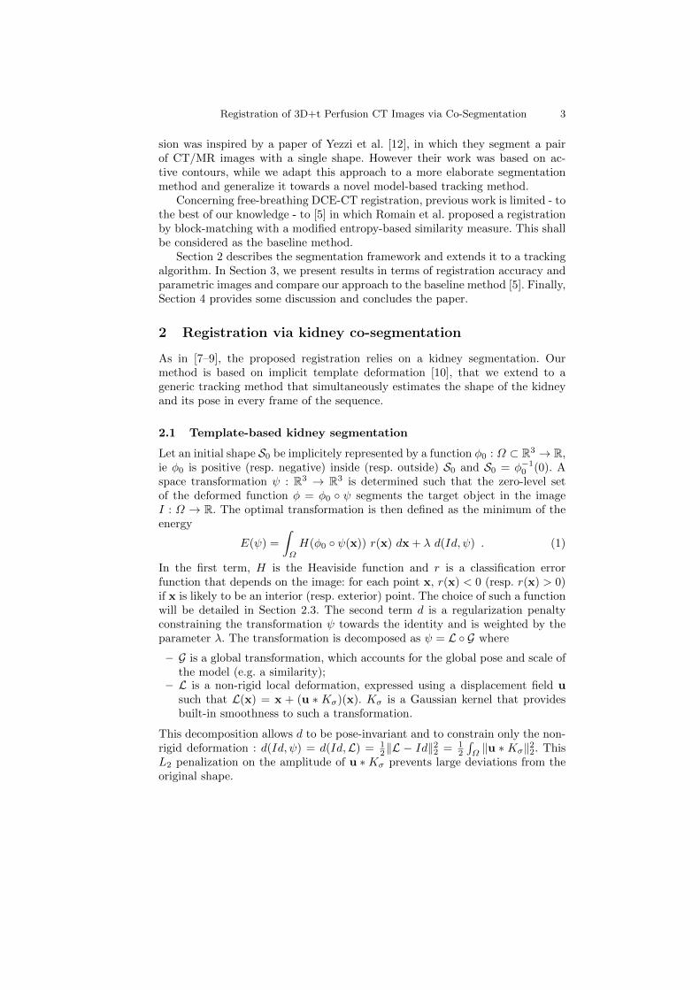
Let an initial shape S0 be implicitely represented by a function φ0 : Ω ⊂ R3 → R,ie φ0 is positive (resp. negative) inside (resp. outside) S0 and S0 = φ−10 (0). Aspace transformation ψ : R3 → R3 is determined such that the zero-level setof the deformed function φ = φ0 ψ segments the target object in the imageI : Ω → R. The optimal transformation is then defined as the minimum of theenergy
E(ψ) =
∫Ω
H(φ0 ψ(x)) r(x) dx + λ d(Id, ψ) . (1)
In the first term, H is the Heaviside function and r is a classification errorfunction that depends on the image: for each point x, r(x) < 0 (resp. r(x) > 0)if x is likely to be an interior (resp. exterior) point. The choice of such a functionwill be detailed in Section 2.3. The second term d is a regularization penaltyconstraining the transformation ψ towards the identity and is weighted by theparameter λ. The transformation is decomposed as ψ = L G where
– G is a global transformation, which accounts for the global pose and scale ofthe model (e.g. a similarity);
– L is a non-rigid local deformation, expressed using a displacement field usuch that L(x) = x + (u ∗ Kσ)(x). Kσ is a Gaussian kernel that providesbuilt-in smoothness to such a transformation.
This decomposition allows d to be pose-invariant and to constrain only the non-rigid deformation : d(Id, ψ) = d(Id,L) = 1
2‖L − Id‖22 = 1
2
∫Ω‖u ∗ Kσ‖22. This
L2 penalization on the amplitude of u ∗Kσ prevents large deviations from theoriginal shape.

4 R. Prevost et al.
2.2 Tracking and stabilisation by co-segmentation
The previously described approach can be generalized to segment a given ob-ject in a collection of N images (Ii)i=1..N . Following a similar approach to [12], asingle shape shall segment all images. We introduce for every image Ii a transfor-mation Gi that we call the pose of the object in this image. The segmentation ofthe object in the i-th image is then the zero level-set of the function φ0LGGi.In the object’s neighborhood, the transformations (Gi)i=1..N act as stabilisationtransformations from any image to a common reference (see Figure 1). EnforcingG1 = Id sets this reference as the first image and resolves any possible ambiguity.Here we assume every Gi to be a global rigid transformation. Indeed the kid-ney is a rigid organ whose motion across the sequence is mainly due to patientbreathing. Note however that the proposed framework can be easily extendedto any kind of stabilising transformations. We finally define the cosegmentationenergy as a function that now depends on transformations (Gi)i :
E(L,G, (Gi)i) =
N∑i=1
∫Ω
H(φ0 L G Gi) . ri +λ
2‖L − Id‖22 (2)
where ri is the i-th image-based classification error function. This energy isminimized with a gradient descent, simultaneously performed on the deformationfield u and the parameters of the transformations G, as well as the whole set(Gi)i. At the end of the process, we obtain both the kidney shape as the zerolevel-set of φ0LG and the transformations (Gi)i that allow a global registrationof the images.
2.3 Choice of the image-based term
The choice of the image-based term r is paramount for the segmentation. A
common choice is r(x) = log Pext(I(x))Pint(I(x))
if intensities distributions are known
inside (Pint) and outside (Pext) the target object. However image intensities varyalong the sequence because of contrast agent injection and kidney’s appearancemay change even between two successive acquisitions. It is therefore not robust touse such intensity models. We rather rely on edge information by only assumingthat in every image Ii of the sequence, the kidney is brighter than its surrounding.This assumption is based on the fact that kidneys are highly vascularized organswhose contrast uptake is very early. The selected criterion to be minimized isthe image gradient flux through the segmentation surface in image Ii denotedSi = (φ0 L G Gi)−1(0) :∫
Si−⟨~∇Ii(x) , ~n(x)
⟩dS(x) =
∫inside Si
−∆Ii(x) dV (x) . (3)
where ∆ denotes the Laplacian operator. The right-hand term in this equationis obtained by application of divergence theorem. In practice, we only take intoaccount relevant edges by applying a Gaussian filter on each image before com-puting its derivatives. The proposed image-based term falls into the described

Registration of 3D+t Perfusion CT Images via Co-Segmentation 5
framework by setting for each image, ri(x) = −∆(Kσim ∗ Ii)(x). The segmenta-tions thus included tumors (as they do respond to the contrast) and excludedcysts (that do not). In both cases however, this does not hinder the registrationsince for a given sequence, the same structure is segmented in all frames.
3 Experiments and Results
3.1 Material
The experiments are based on 15 3D+t sequences coming from six differentpatients with renal tumours, enrolled in a longitudinal study. The data wereacquired on a Brilliance iCT 256 Philips scanner. For each patient, a dynamicCT protocol of perfusion was used. 66 volumes were acquired per sequence (48volumes every 2.5 seconds then 18 volumes every 10 seconds). The patientswere asked to breathe normally during the whole exam. Typical image size was512 × 512 × 22 voxels with a spatial resolution of 0.68 × 0.68 × 2.5 mm. Themodel φ0 was set to an ellipsoid inside the kidney (with one click but we believethis could be easily automated) in the first frame and all transformations (Gi)iwere initialized to the identity. Our algorithm, implemented in C++, processesa whole 3D+t sequence in 30 seconds on a standard computer (Intel Core i5 2.67GHz with 4GB RAM). Such a small computational time is possible because theregistration is driven by the segmentation and thus only requires computationson the boundary of the segmented organ.
Fig. 2. Crops of coro-nal and axial slices be-tween an original se-quence (top) and thesame sequence regis-tered with our method(bottom) along the ac-quisition times. Notethe stabilisation of thesmall blood vessel andthe lesion (arrows).
3.2 Evaluation of registration
An example of a sequence before and after registration is given in Figure 2. Wefirst assess the quality of the registration by computing error measures on land-marks. In our application, we are particularly interested in the region near therenal lesion: the selected landmark was therefore this lesion. For every sequence,the lesion has been manually segmented in each frame. If the registration wereperfect, the segmentation (after compensation by the motion estimated via the

6 R. Prevost et al.
Sequence number
Dic
e co
effic
ient
1 2 3 4 5 6 7 8 9 10 11 12 13 14 150
0.2
0.4
0.6
0.8
1
No Registration
Block-matching [5]
Proposed
Fig. 3. Boxplots of lesions Dice coefficients from original sequences (red), sequences reg-istered with entropy-based block matching [5] (blue) and the proposed method (green).
kidney) would be stable along the frames. We thus evaluated our registrationby computing the Dice coefficient between the lesion in each frame and the le-sion in the reference frame. Figures 3 shows this score, in comparison with theoriginal sequence and the sequence registered by the block-matching method of[5]. Our method globally provides more precise and robust registration in thearea of the tumour as the obtained Dice coefficients have both a higher meanand a lower variance. It outperforms [5] in every sequence but one, in which thelesion was extremely large (bigger than the kidney). Furthermore, the motionof center of the lesion, which was of 6.6mm (median over the datasets) in theoriginal sequences, was reduced to 1.6mm after our stabilisation, which is belowthe resolution in z.
3.3 Parametric images
Our method was further evaluated by comparing parametric images estimatedfrom the registered sequence. In each voxel, parameters (θ1, θ2) were obtainedby fitting the time-intensity curve I(t) - which is proportional to the contrastconcentration Ctissue(t) - to the solution of a Tofts model [13] :
dCtissue(t)
dt= θ1 Caorta(t)− θ2 Ctissue(t) (4)
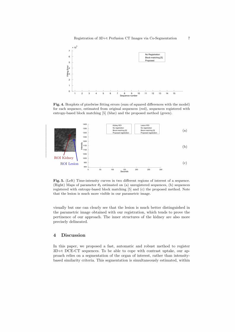
where Caorta, which denotes the contrast concentration in the aorta, is modelledas the sum of two sigmoids and a Gaussian function. The sum of squared errorswas minimized using a Levenberg-Marquardt method. In each voxel, we computethe residual fitting error at convergence, which quantifies how much the time-intensity curve deviates from the model. These are reported in Figure 4, whichshows an improvement over baseline method in every tested sequence but one.This is illustrated by the example given in Figure 5. Our method provides muchsmoother curves, which improves the reliability of the subsequent parameterestimation. As the true parameters are unknown, we can only assess their value
Registration of 3D+t Perfusion CT Images via Co-Segmentation 7
Sequence number
Fitt
ing
Err
or
1 2 3 4 5 6 7 8 9 10 11 12 13 14 150
1
2
3
4
5
6
7
× 105
No Registration
Block-matching [5]
Proposed
Fig. 4. Boxplots of pixelwise fitting errors (sum of squared differences with the model)for each sequence, estimated from original sequences (red), sequences registered withentropy-based block matching [5] (blue) and the proposed method (green).
Seconds
Inte
nsity
0 50 100 150 200 250 300
900
950
1000
1050
1100
1150
1200
1250
1300
1350
1400Lesion ROI
No registration
Block-matching [5]
Proposed registration
Kidney ROI
No registration
Block-matching [5]
Proposed registration(a)
(b)
(c)
ROI Kidney
ROI Lesion
Fig. 5. (Left) Time-intensity curves in two different regions of interest of a sequence.(Right) Maps of parameter θ2 estimated on (a) unregistered sequences, (b) sequencesregistered with entropy-based block matching [5] and (c) the proposed method. Notethat the lesion is much more visible in our parametric image.
visually but one can clearly see that the lesion is much better distinguished inthe parametric image obtained with our registration, which tends to prove thepertinence of our approach. The inner structures of the kidney are also moreprecisely delineated.
4 Discussion
In this paper, we proposed a fast, automatic and robust method to register3D+t DCE-CT sequences. To be able to cope with contrast uptake, our ap-proach relies on a segmentation of the organ of interest, rather than intensity-based similarity criteria. This segmentation is simultaneously estimated, within
8 R. Prevost et al.
a co-segmentation framework. Experiments showed that it provides better re-sults than the state-of-the-art both quantitatively in terms of registration andqualitatively in terms of pharmacokinetic parameters estimation.
The proposed approach is generic and can be extended to other organs. Forthe kidney, a rigid transformation was enough to capture the movement of theregion of interest. Other organs may undergo a different kind of movement, suchas affine or even deformations. This can be taken into account within our frame-work by adapting (Gi)i transformations. The co-segmentation could also havebeen directly applied to the tumour instead of the organ, but the definition ofthe image-based classification terms (ri)i would have been challenging. In ourexperiments, the acquisitions frequency was so low (min 2.5 seconds) that notemporal coherence was enforced. For other applications, temporal consistencycan however be useful. This could be achieved by adding extra terms in the en-ergy to constrain the transformations (Gi)i and is currently under investigation.
References
1. Kambadakone, A., Sahani, D.: Body perfusion CT: technique, clinical applications,and advances. Radiologic clinics of North America 47(1) (2009) 161–178
2. Sance, R., et al.: Alignment of 3D DCE-MRI abdominal series for optimal quan-tification of kidney function. In: Proceedings of IEEE ISPA 2007. (2007) 413–17
3. Linguraru, M., et al.: Renal tumor quantification and classification in contrast-enhanced abdominal CT. Pattern recognition 42(6) (2009) 1149–61
4. Zollner, F., et al.: Assessment of 3D DCE-MRI of the kidneys using non-rigidimage registration and segmentation of voxel time courses. Computerized MedicalImaging and Graphics 33(3) (2009) 171–181
5. Romain, B., Letort, V., Lucidarme, O., d’Alche Buc, F., Rouet, L.: Registrationof free-breathing abdominal 3D contrast-enhanced CT. In: Abdominal Imaging.Computational and Clinical Applications. Volume 7601. Springer (2012) 274–282
6. Bhushan, M., et al.: Motion correction and parameter estimation in DCE-MRIsequences: application to colorectal cancer. In: MICCAI. Volume 6891 of LNCS.Springer (2011) 476–83
7. Sun, Y., et al.: Contrast-invariant registration of cardiac and renal MR perfusionimages. In: MICCAI. Volume 3216 of LNCS. Springer (2004) 903–10
8. Song, T., et al.: Integrated four dimensional registration and segmentation ofdynamic renal MR images. In: MICCAI. Volume 4191 of LNCS. Springer (2006)758–65
9. El-Baz, A., et al.: A new CAD system for the evaluation of kidney diseases usingDCE-MRI. In: MICCAI. Volume 4191 of LNCS. Springer (2006) 446–53
10. Mory, B., et al.: Real-time 3D image segmentation by user-constrained templatedeformation. In: MICCAI. Volume 7510 of LNCS. Springer (2012) 561–8
11. Cuingnet, R., et al.: Automatic detection and segmentation of kidneys in 3D CTimages using random forests. In: MICCAI. Volume 7512 of LNCS. Springer (2012)66–74
12. Yezzi, A., et al.: A variational framework for integrating segmentation and regis-tration through active contours. MedIA 7(2) (2003) 171–85
13. Tofts, P., et al.: Estimating kinetic parameters from dynamic contrast-enhancedT1-weighted MRI of a diffusable tracer: standardized quantities and symbols. Jour-nal of Magnetic Resonance Imaging 10(3) (1999) 223–232