regulation of adult neurogenesis by excitatory input and nmda
TRANSCRIPT

The Journal of Neuroscience, June 1995, 75(6): 46874692
Regulation of Adult Neurogenesis by Excitatory Input and NMDA Receptor Activation in the Dentate Gyrus
Heather A. Cameron, Bruce S. McEwen, and Elizabeth Gould
Laboratory of Neuroendocrinology, The Rockefeller University, New York, New York 10021
The effects of afferent input and Kmethyl-D-aspartate (NMDA) receptor activation on neurogenesis were exam- ined in an intact system, the rat dentate gyrus, where neu- rons are naturally born in the adult. In the adult dentate gyrus, activation of NMDA receptors rapidly decreased the number of cells synthesizing DNA, whereas blockade of NMDA receptors rapidly increased the number of cells in the S phase identified with 3H-thymidine. Acute treatment with NMDA receptor antagonists increased the birth of neu- rons and increased the overall density of neurons in the granule cell layer. Lesion of the entorhinal cortex, the main excitatory afferent population to the granule neurons, also increased the birth of cells in the dentate gyrus. These re- sults suggest that adult neurogenesis in the dentate gyrus of the rat is altered by afferent input, via NMDA receptors, and may be regulated naturally by endogenous excitatory amino acids.
[Key words: hippocampus, entorhinal cortex, granule cell, neurogenesis, cell division, glutamate, NMDA recep- tors]
Once development is complete, the vast majority of neuronal precursors in the mammalian brain undergo terminal differenti- ation and become unable to divide. In contrast, granule neurons of the rat dentate gyrus continue to be produced from immature precursors into adulthood. Cells that are born in the adult dentate gyrus migrate to the granule cell layer (Cameron et al., 1993), receive synaptic input (Kaplan and Hinds, 1977), extend axons into the mossy fiber layer (Stanfield and Trite, 1988), express a neuronal marker (Cameron et al., 1993), and survive for at least 1 month (Cameron et al., 1993). The unusually extended period of neurogenesis makes the dentate gyrus an interesting and use- ful system in which to study the factors that mediate neuroge- nesis.
A recent study has shown that axonal contact stimulates pro- gression through the cell cycle in Drosophila neuronal precur- sors (Selleck et al., 1992), suggesting that afferent input may regulate neurogenesis. Studies performed in several in vitro sys- tems have indicated that excitation regulates important cellular processes, including neuronal birth (Stillwell et al., 1973; Cone and Cone, 1976) and survival (Brenneman and Eiden, 1986;
Received Sept. 23, 1994; revised Dec. 5, 1994; accepted Dec. 9, 1994.
We thank Fernando Nottebohm and Mary E. Hatten for helpful comments on the manuscript. This work was supported by MH 52423, a grant from the American Paralysis Association, and a NARSAD Young Investigator Award (E.G.). H.A.C. was supported by NRSA Training Grant GM07524.
Correspondence should be addressed to Heather A. Cameron, The Rocke- feller University, 1230 York Avenue, New York, NY 10021.
Copyright 0 1995 Society for Neuroscience 0270.6474/95/154687-06$05.00/O
Lipton, 1986). The NMDA subtype of glutamate receptors, in particular, has been shown to play an important role in devel- opmental processes; blockade of these receptors inhibits migra- tion (Komuro and Rakic, 1993) and survival (Balazs et al., 1988, 1990) of developing cerebellar granule neurons in vitro and in- creases cell birth and cell death in the developing subependymal layer and dentate gyrus (Gould et al., 1994). Although granule neurons in the dentate gyrus receive their major excitatory input from the entorhinal cortex via NMDA receptors (Collingridge, 1989), no previous studies have explored the possibility that excitatory input and NMDA receptor activation control neuronal birth in the adult. In order to test this hypothesis, we examined the numbers of newly born neurons using ‘H-thymidine auto- radiography and immunohistochemistry for the neuronal marker neuron-specific enolase (NSE) following treatment with drugs which either activate or block NMDA receptors or lesion of the entorhinal cortex.
Materials and Methods
Animal treatments. Adult (>3 months old) male Sprague-Dawley rats were used in all experiments (n = 5 for each group). In the first ex- periment, animals were injected with NMDA (30 mg/kg, i.p.) in saline or saline alone. They were given an injection of )H-thymidine (5.0 mCi ‘H-thymidinelkg, i.p.; New England Nuclear, specific activity 80 Ci/ mmol; this dose was used in all four experiments) 1 hr after treatment and were perfused 1 hr after ‘H-thymidine injection. This survival time allows for the incorporation of 3H-thymidine by cells synthesizing DNA, but not for the completion of mitosis (Lewis, 1978; Nowakowski et al., 1989) or migration (Cameron et al., 1993). In the second exper- iment, animals were injected with MK-801 (1.0 mg/kg in saline, i.p.; gift of Merck, Sharpe and Dohme), a specific noncompetitive NMDA antagonist (Vezzani et al., 1989), CGP 37849 in saline (5.0 mg/kg in saline, i.p.; gift of Ciba Geigy), a specific competitive NMDA receptor antagonist (Schmutz et al., 1990), or saline alone. These doses are suf- ficient to block NMDA receptors (Vezzani et al., 1989; Schmutz et al., 1990). The rats were injected with 3H-thymidine 1 hr after treatment and were perfused 1 hr after 3H-thymidine injection. In the third ex- periment, animals were injected with the same doses of MK-801, CGP 37849, and saline as in the second experiment and were injected with 3H-thymidine 1 hr later, but they were perfused following a 4 week survival interval.
In the fourth experiment, animals were anesthetized with Nembutal and Metofane, and unilateral stereotaxic lesions of the medial entorhinal cortex were made with 0.5 p,l of a 1% w:v solution of ibotenic acid in saline using stereotaxic coordinates AP -5.2, LM 6.2, DV 6.1 (from Paxinos and Watson, 1982). Ibotenic acid is an excitotoxin that kills cells whose somata are located at the site of the lesion while sparing fibers of passage, and has been used extensively to produce discrete lesions in the forebrain (Zimmer et al., 1989). Two days after the lesion, when synaptic degeneration in the dentate gyms is maximal (Matthews et al., 1976a; Cotman and Nadler, 1978), the rats were injected with )H-thymidine and perfused 1 hr later. Since virtually all connections from the entorhinal cortex to the granule neurons are ipsilateral (Stew- ard, 1976), comparisons between lesion and control were made within brains.
4688 Cameron et al. . NMDA Receptors Regulate Adult Neurogenesis
Histological procedures. Following treatments, rats were anesthetized with Metofane and transcardially perfused with 4% paraformaldehyde. The brains were postfixed overnight in 4% paraformaldehyde and cut in the coronal plane with a Vibratome (40 brn) into a bath of phosphate- buffered saline (PBS). For the first, second, and fourth experiments, ?H- thymidine autoradiography followed by Nissl staining with cresyl violet was performed on brain sections from all animals according tb a pre- viously published protocol (Gould et al., 1994). Briefly, the sections were mounted onto gelatinized slides, dried, rinsed in distilled water, dried, dipped in photographic emulsion (NTB-2, Kodak), and stored in the dark at 4°C for 4 weeks. The slides were then develooed in Dektol (Kodak), rinsed in distilled water, fixed in Ektaflo (Kodak): rinsed again, stained for Nissl using cresyl violet, and coverslipped under Permount.
For the third experiment, combined NSE immunohistochemistry, ‘H- thymidine autoradiography, and Nissl staining were performed on brain sections as previously described (Cameron et al., 1993). The sections were rinsed in PBS and incubated for 24 hr in a solution containing polyclonal antisera to NSE (Polysciences, diluted I:2000 in PBS). Pre- vious studies have shown that in the brain (Schmechel et al., 1980; Marangos and Schmechel, 1987), and specifically in the adult dentate gyms (Cameron et al., 1993), NSE is expressed exclusively by neurons. The sections were rinsed in PBS and incubated for 2 hr in a solution of biotinylated anti-rabbit secondary antibodies in PBS. Following this, the sections were rinsed again and incubated for 2 hr in a solution of avidin-biotin-HRP in PBS. The sections were rinsed again and reacted with diaminobenzidine and hydrogen peroxide in PBS for 15 min. The sections were rinsed again, mounted onto gelatinized slides, dried, rinsed in distilled water, dried, dipped in photographic emulsion (NTB- 2, Kodak), and stored in the dark at 4°C for 4 weeks. The slides were then developed in Dektol (Kodak), rinsed in distilled water, fixed in Ektaflo (Kodak), rinsed again, stained for Nissl using cresyl violet, and coverslipped under Permount. Control sections were processed as de- scribed above with omission of the primary antisera and revealed no nonspecific staining of secondary antibodies.
Data analysis. Data analysis involved counting the number of ‘H- thymidine-labeled cells (cells with 25 silver grains over the nucleus; this value is >20 times the background level) in the granule cell layer and hilus on neuroanatomically matched sections from the middle den- tate gyrus. In the fourth experiment, labeled cells were counted at three different levels of the dentate gyrus: rostra1 (=AP -2.6), middle (ZAP -4.2), and temporal (=AP -5.8) according to Paxinos and Watson (1982). For the third experiment, a distinction was made between NSE- immunoreactive and NSE-nonimmunoreactive iH-thymidine-labeled cells. In addition, the density of granule neurons in the granule cell layer was determined in the third experiment by counting (at 400X) the number of NSE-immunoreactive granule cells within an oval of known area placed under the camera lucida drawing tube positioned in the center of the suprapyramidal blade. The cross-sectional area of each region was determined with a Zeiss Interactive Digitizing Analysis Sys- tem and the data were expressed as number of cells per 10h pm?. No ?H-thymidine-labeled cells were observed in other neuronal regions, nor were any NSE-immunoreactive cells observed in any other area in the long survival experiment, indicating that ?H-thymidine labeling is evi- dence of DNA synthesis during cell division and not of random DNA repair.
Results
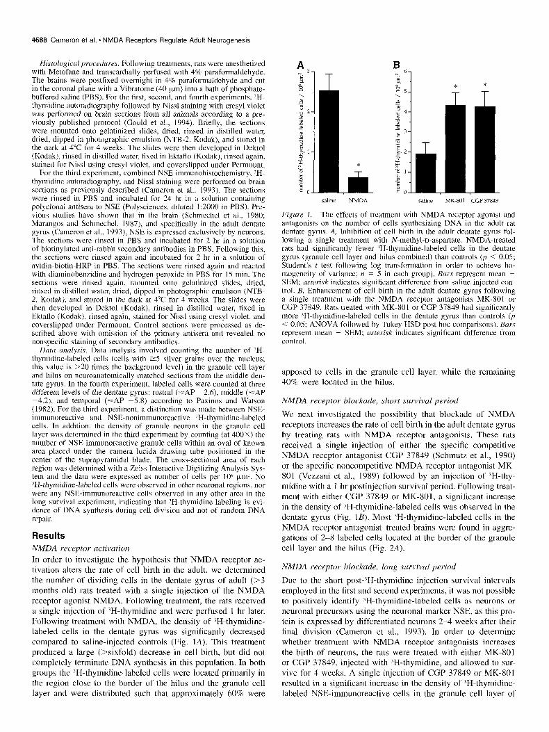
NMDA receptor activation In order to investigate the hypothesis that NMDA receptor ac- tivation alters the rate of cell birth in the adult, we determined the number of dividing cells in the dentate gyrus of adult (>3 months old) rats treated with a single injection of the NMDA receptor agonist NMDA. Following treatment, the rats received a single injection of ‘H-thymidine and were perfused 1 hr later. Following treatment with NMDA, the density of 3H-thymidine- labeled cells in the dentate gyrus was significantly decreased compared to saline-injected controls (Fig. IA). This treatment produced a large (>sixfold) decrease in cell birth, but did not completely terminate DNA synthesis in this population. In both groups the ?H-thymidine-labeled cells were located primarily in the region close to the border of the hilus and the granule cell layer and were distributed such that approximately 60% were
saline NMDA
* *
T T
Iu saline MK-801 CGP 37849
Figure I. The effects of treatment with NMDA receptor agonist and antagonists on the number of cells synthesizing DNA in the adult rat dentate gyrus. A, Inhibition of cell birth in the adult dentate gyrus fol- lowing a single treatment with N-methyl-D-aspartate. NMDA-treated rats had significantly fewer lH-thymidine-labeled cells in the dentate gyrus (granule cell layer and hilus combined) than controls (p < 0.05; Student’s t test following log transformation in order to achieve ho- mogeneity of variance; n = 5 in each group). Burs represent mean + SEM; asterisk indicates significant difference from saline-injected con- trol. B, Enhancement of cell birth in the adult dentate gyrus following a single treatment with the NMDA receptor antagonists MK-801 or CGP 37849. Rats treated with MK-801 or CGP 37849 had significantly more ?H-thymidine-labeled cells in the dentate gyrus than controls (p < 0.05; ANOVA followed by Tukey HSD post hoc comparisons). Bars represent mean + SEM; asterisk indicates significant difference from control.
apposed to cells in the granule cell layer, while the remaining 40% were located in the hilus.
NMDA receptor blockade, short survival period
We next investigated the possibility that blockade of NMDA receptors increases the rate of cell birth in the adult dentate gyrus by treating rats with NMDA receptor antagonists. These rats received a single injection of either the specific competitive NMDA receptor antagonist CGP 37849 (Schmutz et al., 1990) or the specific noncompetitive NMDA receptor antagonist MK- 801 (Vezzani et al., 1989) followed by an injection of ‘H-thy- midine with a 1 hr postinjection survival period. Following treat- ment with either CGP 37849 or MK-801, a significant increase in the density of ‘H-thymidine-labeled cells was observed in the dentate gyrus (Fig. 1B). Most ‘H-thymidine-labeled cells in the NMDA receptor antagonist-treated brains were found in aggre- gations of 2-8 labeled cells located at the border of the granule cell layer and the hilus (Fig. 2A).
NMDA receptor blockade, long survivul period
Due to the short post-‘H-thymidine injection survival intervals employed in the first and second experiments, it was not possible to positively identify ‘H-thymidine-labeled cells as neurons or neuronal precursors using the neuronal marker NSE, as this pro- tein is expressed by differentiated neurons 2-4 weeks after their final division (Cameron et al., 1993). In order to determine whether treatment with NMDA receptor antagonists increases the birth of neurons, the rats were treated with either MK-801 or CGP 37849, injected with ‘H-thymidine, and allowed to sur- vive for 4 weeks. A single injection of CGP 37849 or MK-801 resulted in a significant increase in the density of ‘H-thymidine- labeled NSE-immunoreactive cells in the granule cell layer of

The Journal of Neuroscience, June 1995, 15(6) 4689
h
h
Fi,g~e 2. Photomicrographs of ZH-thymidine-labeled cells in the den- tate gyrus. A, Dividing cells (u~~ocv,s) labeled by Wthymidine autora- diography and Nissl staining, located in the hilus (h) of the dentate gyms near the border of the granule cell layer (gel). B, Neuron (arrow) born in the adult, labeled with JH-thymidine and NSE immunohisto- chemistry, in the granule cell layer of the dentate gyms. After 4 weeks, newly generated neurons are morphologically indistinguishable from surrounding cells in the granule cell layer. Scale bar in B equals 20 km and applies to both A and B.
the dentate gyrus (Fig. 3) suggesting that neurogenesis is stim- ulated by NMDA receptor blockade. Quantitative analysis of granule cell layer cross-sectional area showed no significant changes following treatment with MK-801 or CGP 37849, in- dicating that changes in cell density actually reflect changes in the number of cells. In treated animals, as in controls, these newly born neurons were located in the granule cell layer and were morphologically identical to the surrounding granule neu- rons (Fig. 2B). In contrast, the number of ‘H-thymidine-labeled cells that were not immunoreactive for NSE did not increase following treatment with NMDA receptor antagonists (Fig. 3). A single treatment with CGP 37849 resulted, after 4 weeks, in a 24% increase in the density of neurons in the granule cell layer (Fig. 4), suggesting that the increased rate of neurogenesis re- sults in a net increase in the number of granule neurons. The number of neurons in the granule cell layer following treatment with MK-801 was between that of the control group and the CGP 37849 group (Fig. 4).
We next investigated the possibility that input from the entorhi- nal cortex, the chief excitatory afferent population to the granule neurons (Steward, 1976), which involves NMDA receptor acti- vation (Collingridge, 1989) mediates the rate of cell birth. Uni- lateral lesion of the medial entorhinal cortex resulted in a sig- nificant increase in the number of ‘II-thymidine-labeled cells in the dentate gyrus on the side of the lesion compared to the con- trol side (Fig. 5A). The increase in ?H-thymidine-labeled cells was observed at both middle and temporal, but not rostral, levels of the dentate gyrus (Fig. 5A). Quantitative analysis of the cross-
NSE+ cells NSE- cells
Figure 3. Stimulation of neurogenesis following a single treatment with the NMDA receptor antagonists MK-801 or CGP 37849. The den- sity of NSE-immunoreactive (NSE+) ‘H-thymidine-labeled neuron\ in the granule cell layer is increased compared to saline-injected values (open hur.\) following treatment with NMDA receptor antagonists MK- 801 (gray bars) or CGP 37849 (h/a& burr) (77 < 0.0.5; ANOVA fo- lowed by Tukey HSD post hoc comparisons). NSE-nommmunoreactive (NSE-) ‘H-thymidine-labeled cell denstty in the granule cell layer was not altered by NMDA receptor antagonist treatment. White bars repre- sent saline-injected control rats (mean + SEM), gray bars represent MK-801 -treated rats, black harr represent CGP 37849-treated rats. A>- terisk Indicates significant difference from control.
sectional areas of the granule cell layer and hilus showed no significant changes following entorhinal cortex lesion. Exami- nation of the entorhinal cortex revealed areas of high pyknotic cell density and low density of healthy cells in the medial en- torhinal cortex at the site of the lesion (Fig. 5B).
Discussion
The results of this study show that activation of NMDA recep- tors results in a significant reduction in the rate of cell birth in
saline MK-801 CGP 37849
Figure 4. Increased density of neurons m the granule cell layer fol- lowing treatment with NMDA receptor antagonists, Treatment with CGP 37849 resulted in significantly more NSE-tmmunoreactive cells, that is, granule neurons, in the granule cell layer compared to controls (p < 0.05; ANOVA followed by Tukey HSD post hoc comparisons). Bars represent mean + SEM; n.cterrsk Indicates sigrnticant difference from control.

4690 Cameron et al. 0 NMDA Receptors Regulate Adult Neurogenesis
rostra1 middle dentate gyrus level
Figure 5. Stimulation of cell birth following removal of afferent input. A, Increased density of 3H-thymidine-labeled cells in the dentate gyms granule cell layer following partial destruction of the entorhinal cortex. Lesion of medial entorhinal cortex resulted in significantly more ‘H- thymidine cells on the side ipsilateral to the lesion (black bars) com- pared to the contralateral side (open bars) in middle and temporal regions of the dentate gyms 1 hr after ‘H-thymidine injection (p < 0.05; two-tailed paired Student’s t test). The rostra1 level corresponds to =AP -2.6, middle corresponds to =AP -4.2, and temporal corresponds to =AP -5.8, according to Paxinos and Watson (1982). Asterisk indicates significant difference from control side. B, Schematic drawings showing rostral-caudal extent of the entorhinal cortex lesions. Black line indi- cates site of lesion; stippling indicates areas of degeneration character- ized by high density of pyknotic cells and low density of healthy cells compared to the nonlesioned side. d, dentate gyms; h, hilus; ec, entorhi- nal cortex.
the dentate gyms, whereas deactivation of NMDA receptors, through removal of input or pharmacological blockade, results in a significant increase in the rate of cell birth. Many of these newly born cells become neurons that survive for at least 4 weeks and add to the total population of granule cells.
Methodological considerations
The finding that MK-801 produced a slightly greater increase in the number of ‘H-thymidine-labeled cells compared to CGP 37849 in the short- and long-term studies while having a smaller effect than CGP 37849 on total cell density in the long-term study may be explained by the more rapid action and elimination of MK-801. MK-801 reaches its maximum brain concentration lo-30 min after intraperitoneal injection and is rapidly elimi- nated (Vezzani et al., 1989), whereas CGP 37849 is maximally
effective 2 hr after intraperitoneal injection and is longer acting (Schmutz et al., 1990; Massieu et al., 1993).
The increase in granule cell density observed following NMDA receptor antagonist treatment appears relatively large compared to the increase observed in ?H-thymidine-labeled NSE-immunoreactive cells. However, it is likely that the ob- served increase in ‘H-thymidine-labeled cells underestimated the actual changes in cell birth produced by NMDA receptor antag- onist treatment for several reasons. First, the autoradiographic emulsion can detect ‘H only in the top 3 pm of the 40 pm section (Feinendegen, 1967), so the number of 3H-thymidine- labeled cells observed underestimates the number of “H-thymi- dine-incorporating cells in the section. For the total density counts, however, granule neurons throughout the entire thickness of the section were counted. Second, the maximal effect of NMDA blockade on cell birth may not have occurred during the time of ‘H-thymidine availability. For example, CGP 37849 is maximally effective at blocking NMDA receptors 2 hr after in- traperitoneal injection (Schmutz et al., 1990; Massieu et al., 1993), the time at which the rats in the short survival study were perfused, so the observed effect of CGP 37849 on cell birth is likely to underestimate the actual increase in cell birth produced by this drug. Third, CGP 37849 is available in the brain for several hours (Schmutz et al., 1990; Massieu et al., 1993) while 3H-thymidine is available for only 1 hr. Fourth, effects on cell birth initiated by NMDA receptor blockade may persist beyond the time when the drug is present in the brain, resulting in in- creases in cell division through several cell cycles.
Following lesion of the entorhinal cortex, the increase in 3H- thymidine-labeled cells was observed at both middle and tem- poral, but not rostral, levels of the dentate gyms. The placement of these lesions within the entorhinal cortex may account for the regional differences in effects; the lesion was made in the medial entorhinal cortex, which projects primarily to middle and tem- poral levels of the dentate gyrus (Steward, 1976). In contrast, the rostra1 level of the dentate gyms receives projections pri- marily from the lateral entorhinal cortex (Steward, 1976) which was spared in all lesioned animals of this study. Following en- torhinal cortex lesion, the number of degenerating terminals in the dentate gyrus is maximal at 2 d following surgery (Matthews et al., 1976a), the survival time used in this study. At later time points after entorhinal cortex lesion, the number of excitatory synapses begins to rise, presumably due to collateral sprouting, and eventually reaches normal, unlesioned levels (Matthews et al., 1976b). Although our results indicate that cell birth is stim- ulated when the number of excitatory synapses is low, it is pres- ently unknown whether regenerative synapses, that is, those which form at postlesion survival times greater than 2 d, are capable of inhibiting cell birth in this region. Future postlesion time course studies will seek to determine whether the number of excitatory synapses are negatively correlated with the rate of cell birth in this region.
Mechanisms underlying inhibition of neurogenesis
The results of our lesion study indicate that excitatory afferents from the entorhinal cortex normally inhibit cell birth in the den- tate gyrus. This pathway may mediate neurogenesis in this sys- tem through two, not mutually exclusive, mechanisms. First, ax- onal contact may inhibit the birth of granule neurons. A previous study performed in the CNS of Drosophila has shown that axons selectively approach neuronal precursors and that contact with these axons stimulates neuroblasts to undergo their final division

The Journal of Neuroscience, June 1995, 15(6) 4691
(Selleck et al., 1992). If this scenario occurs in the rat dentate gyrus, then removal of axons could induce proliferation, whereas innervation by peiforant path axons could stimulate a final di- vision. Second, excitation may inhibit neurogenesis in the den- tate gyrus. In this regard, it is relevant that activation of gluta- mate receptors has been shown to decrease the birth of astroglia (Condorelli et al., 1989). Our results showing that NMDA in- hibits cell birth and MK-801 or CGP 37849 stimulates cell birth strongly suggest that NMDA receptor-mediated excitation plays a role in regulating neuronal birth.
Although the results of this study indicate that NMDA recep- tor activation inhibits neurogenesis, there is currently no evi- dence to indicate that this inhibition occurs through synapses located on granule neuron precursors. NMDA receptors have very high agonist affinity (Blanton and Kriegstein, 1992) and are present early in development (Riva et al., 1994), making them ideally suited to receive nonsynaptic input and thus regu- late developmental processes, such as neuronal birth, prior to synaptogenesis. Previous studies in other systems have shown that NMDA receptors can be activated prior to synapse forma- tion (Blanton et al., 1990; Blanton and Kriegstein, 1992). During development, NMDA receptor channels can be activated by glu- tamate released by neighboring cells, even after action potential blockade, to produce a background current (Blanton and Krieg- stein, 1992). These findings raise the possibility that NMDA receptor regulation of neuronal birth is not mediated synaptical- ly. It is possible that glutamate released from neighboring astro- cytes (Parpura et al., 1994) is responsible for the NMDA recep- tor-mediated inhibition of neurogenesis in the dentate gyrus. The observation that axonal growth cones release glutamate that can activate NMDA receptors in the absence of mature synapses (Young and Poo, 1983) presents another nonsynaptic method for NMDA receptor-mediated inhibition. However, the finding that lesion of the entorhinal cortex has an effect on neuronal birth that is virtually identical to NMDA receptor blockade suggests that the perforant pathway is at least indirectly involved in sup- pression of neurogenesis.
Effects of excitatory input on cell survival Studies performed in several other systems have shown that ex- citatory input and/or NMDA receptors regulate cell death (Bal- azs et al., 1988, 1990). We have shown that NMDA receptor activation regulates cell birth and cell death in the developing dentate gyrus; blockade of NMDA receptors during the first postnatal week substantially increases the number of dividing and degenerating cells. The increase in cell death outweighs the increase in cell birth such that a decrease in the total number of granule neurons results (Gould et al., 1994). However, the pres- ent study showed no evidence of increased cell death in the dentate gyrus with NMDA receptor antagonist treatment in adulthood. Since only two time points after drug treatment (2 hr and 4 weeks) were examined in this study, it is possible that granule neurons die but that degenerating cells were indetectable at the time points examined. However, the observation that the number of granule neurons actually increases with CGP 37849 treatment indicates that the predominant action of NMDA re- ceptor blockade on the adult granule neuron population is to increase cell birth.
The role of excitatory input in dentate gyrus development and maintenance Several lines of evidence indicate that excitatory input naturally regulates the birth of cells in the rat dentate gyrus throughout
life. First, the timing of development of excitatory input and granule cell genesis is consistent with this possibility. Excitatory synapses are immature (Cotman et al., 1973; Crain et al., 1973) and the density of NMDA receptors is still rising (Tremblay et al., 1988) when granule cell birth begins and peaks (Schlessinger et al., 1975). Perforant path axons enter the dentate gyrus (Loy et al., 1977), excitatory synapses are formed (Crain et al., 1973), and NMDA receptor densities reach a maximum (Tremblay et al., 1988) at the same time granule cell birth begins to diminish (Schlessinger et al., 1975). Second, we have shown that block- ade of NMDA receptors on postnatal day 5, the time of maximal neurogenesis, further increases the birth of cells in the dentate gyrus (Gould et al., 1994), suggesting that NMDA receptor ac- tivation normally slows neuronal birth during the postnatal pe- riod. Third, the results from these experiments collectively sug- gest that neurogenesis in the adult dentate gyrus is normally suppressed by afferent input via NMDA receptor activation. However, inhibition of neurogenesis is not complete in the adult dentate gyrus under normal conditions, since neurons continue to be born at a slow rate (Kaplan and Hinds, 1977; Cameron et al., 1993). This slow rate of neuronal birth, which can be sup- pressed by NMDA receptor activation and enhanced by NMDA receptor inactivation, may provide a means whereby natural al- terations in the degree of excitatory input control the number of new granule neurons.
Possible consequences of alterations in granule cell number
Many of the cells that are born in the dentate gyrus following treatment with NMDA receptor antagonists become neurons that reside in the granule cell layer, appear morphologically indistin- guishable from the surrounding granule neurons, and express NSE, which is a reliable marker of functional neuronal metabolic activity (Rosenstein, 1993). These new granule cells survive for at least 4 weeks, and add to the total population of granule neurons. It is likely that the increased number of granule neurons which results from treatment with NMDA receptor antagonists has functional consequences for the dentate gyrus.
Because newly born granule neurons take 34 weeks to ma- ture (Cameron et al., 1993), functional effects of increased gran- ule cell number are likely to be observed only over a relatively long time course. The dentate gyrus is believed to be involved in spatial memory (see Jarrard, 1993, for review). Studies com- paring different strains of mice have suggested a link between the number of hippocampal neurons and the ability to learn spa- tial tasks (Wimer et al., 1978, 1980; Symons et al., 1988). In addition, NMDA receptor activation has been shown to play a role in spatial learning (Butelman, 1989; McLamb et al., 1990; Bischoff and Teidtke, 1992; Davis et al., 1992). The current findings present the possibility that treatment with NMDA re- ceptor antagonists would also have delayed, yet long-lasting, effects on the learning ability of rats. NMDA receptor regulation of neurogenesis may provide a mechanism for stabilizing the granule neuron population during periods of active learning, while increasing storage capacity by adding new neurons during periods of inactivity.
References Balazs R, Jorgensen OS, Hack N (1988) N-methyl-D-aspartate promotes
the survival of cerebellar granule cells in culture. Neuroscienct 27: 437-451.
Balazs R, Hack N, Jorgensen OS (1990) Interactive effects involving different classes of excitatory amino acid receptors and the survival of cerebellar granule cells in culture. Int J Dev Neurosci 8:347-360.

4692 Cameron et al. * NMDA Receptors Regulate Adult Neurogenesis
Bischoff C, Teidtke PI (1992) Competitive and non-competitive NMDA receptor antagonists in spatial learning tasks. Em J Pharmacol 213: 269-273.
Blanton MD, Kriegstein AR (1992) Properties of amino acid neuro- transmitter receptors of embryonic cortical neurons when activated by exogenous and endogenous agonists. J Neurophysiol 67: 1185- 1200.
Blanton MD, Lo Turco JJ, Kriegstein AR (1990) Endogenous neuro- transmitter activates N-methyl&aspartate receptors on differentiating neurons in embrvonic cortex. Proc Nat1 Acad Sci USA 87:8027- 8030.
Brenneman DE, Eiden LE (1986) Vasoactive intestinal peptide and electrical activitv influence neuronal survival. Proc Nat1 Acad Sci USA 83:1159-li62.
Butelman ER (1989) A novel NMDA antagonist, MK-801, impairs per- formance in a hippocampal-dependent spatial learning task. Phar- macol Biochem Behav 34:13-16.
Cameron HA, Woolley CS, McEwen BS, Gould E (1993) Differentia- tion of newly born neurons and glia in the dentate gyrus of the adult rat. Neuroscience 56:337-344.
Collingridge GL (1989) Synaptic function of N-methyl-o-aspartate re- ceptors in the hippocampus. In: The hippocampus: new vistas, Vol 52 (Chan-Palay V, Kohler C, eds), pp 329-346. New York: Liss.
Condorelli DE Ingrao F, Magri G, Bruno V, Nicoletti F, Avola R (1989) Activation of excitatory amino acid receptors reduces thymidine in- corporation and cell proliferation rate in primary cultures of astro- cytes. Glia 2:67-69.
Cone C, Cone C (1976) Mitosis in mature neurons in central nervous system by sustained depolarization. Science 192: 155-157.
Cotman CW, Nadler JV (1978) Reactive synaptogenesis in the hippo- campus. In: Neuronal plasticity (Cotman CW, ed), pp 227-271. New York: Raven.
Cotman C, Taylor D, Lynch G (1973) Ultrastructural changes in syn- apses in the dentate gyrus of the rat during development. Brain Res 63:205-213.
Crain B, Cotman C, Taplor D, Lynch G (1973) A quantitative electron microscopic study of synaptogenesis in the dentate gyms of the rat. Brain Res 63: 195-204.
Davis S, Butcher SP Morris RG (1992) The NMDA receptor antagonist D-2-amino-5-phosphonopentanoate (D-APS) impairs spatial learning and LTP in viva at intracerebral concentrations comparable to those that block LTP in vitro. J Neurosci 12:21-34.
Feinendegen LE (1967) Tritium-labeled molecules in biology and med- icine. New York: Academic.
Gould E, Cameron HA, McEwen BS (1994) Blockade of NMDA re- ceptors increases cell death and birth in the developing rat dentate gyms. J Comp Neurol 340:551-565.
Jarrard LE (1993) On the role of the hippocampus in learning and memory in the rat. Behav Neural Biol 60:9-26.
Kaplan MS, Hinds JW (1977) Neurogenesis in the adult rat: electron microscopic analysis of light radioautographs. Science 197:1092- 1094.
Komuro H, Rakic P (1993) Modulation of neuronal migration by NMDA receptors. Science 260:95-97.
Lewis PD (1978) Kinetics of cell proliferation in the postnatal rat den- tate gyms. Neuropathol Appl Neurobiol 4: 191-195.
Lipton SA (1986) Blockade of electrical activity promotes the death of mammalian retinal ganglion cells in culture. Proc Nat1 Acad Sci USA 83:9774-9778.
Loy R, Lynch G, Cotman CW (1977) Development of afferent lami- nation in the fascia dentata of the rat. Brain Res 121:229-243.
Marangos PJ, Schmechel DE (1987) Neuron specific enolase, a clini- cally useful marker for neurons and neuroendocrine cells. Annu Rev Neurosci 10:269-295.
Massieu L, Thedinga KH, McVey M, Fagg GE (1993) A comparative analysis of the neuroprotective properties of competitive and uncom- petitive N-methyl-o-aspartate receptor antagonists in viva: implica- tions for the process of excitotoxic degeneration and its therapy. Neu- roscience 55:883-892.
Matthews DA, Cotman CW, Lynch G (1976a) An electron microscopic study of lesion induced synaptogenesis in the dentate gyrus of the adult rat. I. Magnitude and time course on degeneration. Brain Res 115:1-21.
Matthews DA, Cotman CW, Lynch G (1976b) An electron microscopic study of lesion induced synaptogenesis in the dentate gyrus of the adult rat. II. Reappearance of morphologically normal synaptic con- tacts. Brain Res 115:23-41.
McLamb RL, Williams LR, Nanry KP, Wilson WA, Tilson HA (1990) MK-801 impedes the acquisition of a spatial memory task in rats. Pharmacol Biochem Behav 37:4145.
Nowakowski RS, Lewin SB, Miller MW (1989) Bromodeoxyuridine immunohistochemical determination of the lengths of the cell cycle and the DNA-synthetic phase for an anatomically defined population. J Neurocytol 18:311-318.
Parpura V, Basarsky TA, Liu F (1994) Glutamate-mediated astrocyte- neuron signalling. Nature 369:744-747.
Paxinos G, Watson C (1982) The rat brain in stereotaxic coordinates. Sydney: Academic.
Riva MA, Tascedda R, Molteni R, Racagni G (1994) Regulation of NMDA receptor subunit mRNA expression in the rat brain during postnatal development. Brain Res 25:209-216.
Rosenstein JM (1993) Developmental expression of neuron-specific enolase immunoreactivity and cytochrome oxidase activity in neu- ronal transplants. Exp Neural 124:208-2 18.
Schlessinger AR, Cowan WM, Gottlieb DI (1975) An autoradiographic study of the time of origin and the pattern of granule cell migration in the dentate gyms of the rat. J Comp Neurol 159:159-176.
Schmechel DE, Brightman MW, Marangos PJ (1980) Neurons switch from non-neuronal enolase to neuron-specific enolase during differ- entiation. Brain Res 190: 195-214.
Schmutz J, Portet C, Jeker A, Klebs K, Vassout A, Allgeier H, Heck- endorn R, Fagg GE, Olpe HR, van Riezen H (1990) The competitive NMDA receptor antagonists CGP 37849 and CGP 39551 are potent, orally-active anticonvulsants in rodents. Naunyn-Schmiedeberg’s Arch Pharmacol 342:61-66.
Selleck SB, Gonzalez C, Glover DM, White K (1992) Regulation of the Gl-S transition in postembryonic neuronal precursors by axon ingrowth. Nature 355:253-255.
Stanfield BB, Trite JE (1988) Evidence that granule cells generated in the dentate gyms of adult rats extend axonal projections. Exp Brain Res 72:399406.
Steward 0 (1976) Topographic organization of the projections from the entorhinal area to the hippocampal formation of the rat. J Comp Neu- rol 167:285-314.
Stillwell EE Cone CF, Cone CD (1973) Stimulation of DNA synthesis in CNS neurones by sustained depolarisation. Nature New Biol 246: 110-111.
Symons JP, Davis RE, Marriott JG (1988) Water-maze learning and effects of cholinergic drugs in mouse strains with high and low hip- pocampal pyramidal cell counts. Life Sci 42:375-383.
Tremblay E, Roisin MP, Represa A, Charriaut-Marlangue C, Ben-Ari Y (1988) Transient increased density of NMDA binding sites in devel- oping rat hippocampus. Brain Res 461:393-396.
Vezzani A, Serafini R, Stasi MA, Caccia S, Conti I, Tridico RV, Sa- manin R (1989) Kinetics of MK-801 and its effect on quinolinic acid-induced seizures and neurotoxicity in rats. J Pharmacol Exp Ther 249:278-283.
Wimer RE, Wimer CC, Vaughn JE, Barber RP, Balvanz BA, Chernow CR (1978) The genetic organization of neuron number in the granule cell layer of the-area den&a in house mice. Brain Res 157:105-122.
Wimer RE. Wimer CC. Chernow CR, Balvanz, BA (1980) The genetic organization of neuron number in the pyramidal‘cell layer of hip- pocampal region superior in house mice. Brain Res 196:59-77.
Young SH, Poo M (1983) Spontaneous release of transmitter from growth cones of embryonic neurons. Nature 305:634-637.
Zimmer J, Tonder NM, Sorensen T (1989) Hippocampus and fascia dentata transplants: anatomical organization and connections. In: The hippocampus: new vistas, Vol 52 (Chan-Palay V, Kohler C, eds), pp 257-286. New York: Liss.