regulation of gene expression in eukaryotes -...
TRANSCRIPT
12
Regulation of Gene Expression in Eukaryotes WORKING WITH THE FIGURES 1. In Figure 12-4, certain mutations decrease the relative transcription rate of the
-globin gene. Where are these mutations located, and how do they exert their effects on transcription?
Answer: The mutations that decrease transcription all fall within the promotor-
proximal and promotor elements, which are located upstream of the coding region and serve as binding sites for RNA PolII and general transcription factors. Mutations in these regions hinder protein-DNA interactions and thereby reduce transcription. The figure shows that this reduction can be substantial when even a single nucleotide is changed.
2. Based on the information in Figure 12-6, how does Gal4 regulate four different
GAL genes at the same time? Contrast this mechanism with how the Lac repressor controls the expression of three genes.
Answer: Gal4 has a DNA binding domain that targets a specific UAS sequence.
This sequence is present in the enhancer region of four different GAL genes. Gal4 is able to activate transcription from the four separate GAL promoters through its capacity to bind with all four UASs. In prokaryotes, the Lac repressor controls expression of three genes by blocking or unblocking access of RNA Polymerase to a single promotor region that is common to all three genes. These two regulatory systems are similar in that genes involved in metabolic pathways for a specific sugar are coordinately induced by the presence of the sugar.
3. In any experiment, controls are essential in order to determine the specific effect
of changing some parameter. In Figure 12-7, which constructs are the �“controls�” that serve to establish the principle that activation domains are modular and interchangeable?

Chapter Twelve 326
Answer: Controls lack certain experimental treatments, to establish a baseline
for resolution of treatment effects. In this figure, one control (we could call this a positive control) is illustrated in panel a, which demonstrates normal expression of the lacZ reporter when the Gal4 DNA binding domain and activation domain are both present. The other set of controls (negative controls) consists of DNA binding domains from LexA and Gal4 that lack activation domains. They show that in the absence of an activation domain, the reporter gene lacZ is not expressed. Together, these controls allow a robust interpretation of panel d, where a LexA-Gal4 hybdrid is used, which is that the match between the activation domain and the promotor region activates transcription independently of the DNA binding domain.
4. Contrast the role of the MCM1 protein in different yeast cell types shown in
Figure 12-10. How are the a-specific genes controlled differently in different cell types?
Answer: In a cells, MCM1 protein acts alone to activate a-specific genes. - specific genes are not activated because the necessary helper proteins are not present in a cells. In cells, 1 protein binds with MCM1 to activate -specific genes. Another protein, 2 binds with MCM1 to prevent activation of a- specific genes. In a/ hybrids, neither the a-specific nor the -specific genes are activated because MCM1 is prevented from activating a-specific genes by the presence of 2 protein, and 1, which is necessary for activation of - specific genes, is repressed by an a1/ 2 complex. In a cells, a-specific genes are activated by MCM1 alone. In other cells, the activation of a-specific genes is prevented by 2/MCM1.
5. In Figure 12-11b, in what chromosomal region are you likely to find the most
H1 histone protein? Answer: Figure 12-11c illustrates variation in the density of condensation along
a chromosome. H1 histone is evenly distributed along an uncondensed chromatin fiber (shown in 12-11a), and the density increases with the increasing condensation of the chromosome. The most H1 histone would be found where the chromosome is most condensed, in the heterochromatin (depicted in red in the figure).
6. What is the conceptual connection between Figures 12-13 and 12-19? Answer: Figure 12-13 depicts a general model for how chromatin remodeling
can uncover previously hidden regions along the chromosome. Figure 12-19 shows the details of how SWI-SNF participates in chromatin remodeling to expose the TATA box of -interferon, allowing binding of the TATA binding
Chapter Twelve 327
protein and initiation of transcription. Both illustrate the same basic process, but 12-13 is a general model and 12-19 shows the details for a specific gene.
7. In Figure 12-19, where is the TATA box located before the enhanceosome
forms at the top of the figure? Answer: Before the enhanceosome forms, the TATA box is located within the
region of the chromosome bound to the core histone proteins. It is inaccessible to the TATA binding protein so transcription cannot be initiated. Formation of the enhanceosome uncovers the TATA box, allowing transcription to be initiated.
8. Let�’s say that you have incredible skill and can isolate the white and red patches
of tissue from the Drosophila eyes shown in Figure 12-24 in order to isolate mRNA from each tissue preparation. Using your knowledge of DNA techniques from Chapter 10, design an experiment that would allow you to determine whether RNA is transcribed from the white gene in the red tissue or the white tissue or both. If you need it, you have access to radioactive white-gene DNA.
Answer: If sufficient RNA could be isolated from the different tissue patches, a
Northern blot could be done, in which RNA from each tissue patch is fractionated by gel electrophoresis, then probed with radioactively labeled white-gene DNA. Presence of a radioactive band would indicate mRNA from the white gene, and would be expected in the red tissue. Lack of a radioactive band would indicate no white-gene mRNA and would be expected in the white tissue. Control lanes on the gel would consist of RNA from white gene wildtypes, which would have all red eyes, and white gene mutants, which would have all white eyes. The more likely case is that very little RNA would be present. In this event, cDNA could be synthesized from the RNA using reverse transcriptase. The cDNA could then be amplified with PCR using primers unique to the white gene. Amplification of white-gene DNA would indicate the presence of a white gene transcript, and would be expected from red tissue. Lack of amplification would mean the white gene transcript was absent and would be expected in the white tissue. In this experiment, radioactive white- gene DNA would not be needed because detection of the white-gene mRNA would be based on whether or not the DNA amplified.
9. In Figure 12-26, provide a biochemical mechanism for why HP-1 can bind to
the DNA only on the left side of the barrier insulator. Similarly, why can HMTase bind only to the DNA on the left of the barrier insulator?
Answer: The barrier insulator recruits HAT, which hyperacetylates nearby
histones. This inhibits binding of HMTase, and prevents methylation of histones. Without methylation, HP-1 cannot bind and drive the formation of
Chapter Twelve 328
heterochromatin. In this way, heterochromatin-promoting proteins are prevented from binding near or to the right of the barrier insulator.
10. In reference to Figure 12-28, draw the outcome if there is a mutation in gene B. Answer: If a mutant B gene is inherited from the father, the phenotypic outcome
will be unaffected because that gene is already silenced by imprinting. If the mutant B gene is inherited from the mother, the mutant phenotype will be expressed because the paternal allele has been silenced and cannot mask the mutation. The mutation will appear to be dominant even though it is not.
BASIC PROBLEMS 11. What analogies can you draw between transcriptional trans-acting factors that
activate gene expression in eukaryotes and the corresponding factors in bacteria? Give an example.
Answer: Activation of gene expression by trans-acting factors occurs in both
prokaryotes and eukaryotes. In both cases, the trans-acting factors interact with specific DNA sequences that control expression of cis genes.
In prokaryotes, proteins bind to specific DNA sequences, which in turn regulate
one or more downstream genes.

Chapter Twelve 329
In eukaryotes, highly conserved sequences such as CCAAT and various
enhancers in conjunction with trans-acting binding proteins increase transcription controlled by the downstream TATA box promoter. Several proteins have been found that bind to the CCAAT sequence, upstream GC boxes, and the TATA sequence in Drosophila, yeast, and other organisms. Specifically, the Sp1 protein recognizes the upstream GC boxes of the SV40 promoter and many other genes; GCN4 and GAL4 proteins recognize upstream sequences in yeast; and many hormone receptors bind to specific sites on the DNA (e.g., estrogen complexed to its receptor binding to a sequence upstream of the ovalbumin gene in chicken oviduct cells). Additionally, the structure of some of these trans-acting DNA-binding proteins is quite similar to the structure of binding proteins seen in prokaryotes. Further, protein-protein interactions are important in both prokaryotes and eukaryotes. For the above reasons, eukaryotic regulation is now thought to be very close to the model for regulation of the bacterial ara operon.
12. Contrast the states of genes in bacteria and eukaryotes with respect to gene
activation. Answer: In general, the ground state of a bacterial gene is �“on.�” Thus,
transcription initiation is prevented or reduced if the binding of RNA polymerase is blocked. In contrast, the ground state in eukaryotes is �“off.�” Thus, the transcriptional machinery (including RNA polymerase II and associated general transcription factors) cannot bind to the promoter in the absence of other regulatory proteins.
13. Predict and explain the effect on GAL1 transcription, in the presence of
galactose alone, of the following mutations: a. Deletion of one Gal4-binding site in the GAL1 UAS element. b. Deletion of all four Gal4-binding sites in the GAL1 UAS element. c. Deletion of the Mig1-binding site upstream of GAL1. d. Deletion of the Gal4 activation domain. e. Deletion of the GAL80 gene. f. Deletion of the GAL1 promoter. g. Deletion of the GAL3 gene. Answer: a. Generally, transcription factors bind to multiple sites in a cooperative
manner and their effect on transcription is synergistic. The GAL1 UAS element has four binding sites for the Gal4 protein. The UAS elements near the GAL2 and GAL7 genes each have two Gal4 binding sites. Loss of one Gal4 binding site from the GAL1 UAS element may have a small
Chapter Twelve 330
effect on GAL1 transcription, but you would still expect to see Gal4-specific transcriptional activation due to the three remaining binding sites.
b. The deletion of all four Gal4-binding sites from the UAS would prevent
transcriptional activation of the GAL1 gene. Experiments have shown that Gal4-DNA binding at the UAS is necessary for its function.
c. The binding of Mig1 at its binding site is necessary for the repression of
GAL1 in the presence of glucose. In this case, the absence of the Mig1 binding site will have no effect on GAL1 transcription in the presence of galactose and the absence of glucose. It would effect GAL1 transcription if both sugars were present as transcription would still be activated.
d. If the Gal4 protein is missing its activation domain, it would still bind at
the UAS but it would not promote transcription. The activation domain helps recruit the transcriptional machinery to the promoter.
e. The product of the GAL80 gene inhibits Gal4 function by binding to a
region within the Gal4 activation domain. Therefore, the deletion of GAL80 will have no effect on the transcription of GAL1 in the presence of galactose.
f. The deletion of the GAL1 promoter would prevent transcription of the
gene. The promoter is required for the binding of the TATA-binding protein and then by extension, the binding of RNA polymerase II.
g. The product of the GAL3 gene is both a sensor and inducer. It binds
galactose (and ATP) causing an allosteric change that allows Gal3 to bind Gal80, which in turn releases Gal4 from Gal80. When Gal80 is bound to Gal4, Gal4 cannot activate transcription so its release by Gal80 is necessary for gene activation. In the absence of Gal3 protein, Gal4 will remain bound to Gal80 and will not activate GAL1 transcription.
14. How is the activation of the GAL1 gene prevented in the presence of galactose
and glucose? Answer: In the presence of both galactose and glucose, the activation of the
GAL1 gene is prevented by the Mig1 protein. Mig1 is a sequence-specific DNA-binding protein that binds to a site between the UAS element and the promoter of the GAL1 gene. Mig1 recruits a protein complex called Tup1 that contains a histone deacetylase and represses gene transcription.
15. What are the roles of histone deacetylation and histone acetylation in gene
regulation, respectively?

Chapter Twelve 331
Answer: The acetylation of histones is one of several types of covalent modifications that are part of what is called a histone code. Histones associated with nucleosomes of active genes are hyperacetylated, whereas they are hypoacetylated in nucleosomes associated with inactive genes. Given this, histone deacetylation plays a key role in gene repression while histone acetylation plays a key role in gene activation.
16. An strain of yeast that cannot switch mating type is isolated. What mutations
might it carry that would explain this phenotype? Answer: Among the mutations that would prevent an strain of yeast from
switching mating type would be mutations in the HO and HMRa genes. The HO gene encodes an endonuclease that cuts the DNA to initiate switching, and the HMRa locus contains the �“cassette�” of unexpressed genetic information for the MATa mating type.
17. What genes are regulated by the 1 and 2 proteins in an a cell? Answer: In a haploid cell, the 1 protein, in a complex with MCM1 protein,
binds to specific DNA sites and activates the transcription of -specific genes. The 2 protein, in a complex with MCM1 protein, binds to specific DNA sites and represses a-specific genes.
18. What are Sir proteins? How do mutations in SIR genes affect the expression of
mating-type cassettes? Answer: In yeast, the Sir proteins (silent information regulators) are necessary
to keep the genetic information in the HMR and HML cassettes silent. Their functions facilitate the condensation of chromatin and help lock up HMR and HML in chromatin domains that are inaccessible to transcriptional activators. Mutations in SIR genes are sterile since both a and information is expressed in the absence of this specific gene silencing.
19. What is meant by the term epigenetic inheritance? What are two examples of
such inheritance? Answer: The term epigenetic inheritance is used to describe heritable alterations
in which the DNA sequence itself is not changed. It can be defined operationally as the inheritance of chromatin states from one cell generation to next. Genomic imprinting, X-chromosome inactivation, and position-effect variegation are several such examples.
Chapter Twelve 332
20. What is an enhanceosome? Why could a mutation in any one of the enhanceosome proteins severely reduce the transcription rate?
Answer: An enhancersome is a large protein complex that acts synergistically to
activate transcription. Because this is a case where the whole is greater than the sum of its parts, loss of any one of the required proteins may severely impact the required synergy.
21. Why are mutations in imprinted genes usually dominant? Answer: Imprinted genes are functionally hemizygous. Maternally imprinted
genes are inactive when inherited from the mother, and paternally imprinted genes are inactive when inherited from the father. A mutation in one of these genes is dominant when an offspring inherits a mutant allele from one parent and a �“normal�” but inactivated allele from the other parent.
22. What has to happen for the expression of two different genes on two different
chromosomes to be regulated by the same miRNA? Answer: Complementarity between the seed region of the miRNA and the
3 UTR of the mRNA is the basis for regulation by miRNA. For one miRNA to coordinately regulate two genes, the seed region would have to be complementary to both 3 UTRs. This is probably a common occurrence because many miRNAs have seed regions complementary to the 3 UTR of several genes. It would not matter whether the genes in question were on the same chromosome or not because miRNA is a trans-acting agent targeting the mRNA during translation.
23. What mechanisms are thought to be responsible for the inheritance of epigenetic
information? Answer: The inheritance of chromatin structure is thought to be responsible for
the inheritance of epigenetic information. This is due to the inheritance of the histone code and may also include inheritance of DNA methylation patterns.
24. What is the fundamental difference in how bacterial and eukaryotic genes are
regulated? Answer: Many DNA-protein interactions are shared by prokaryotes and
eukaryotes, but the mechanisms by which proteins bound to DNA at great distances from the start of transcription affect that transcription is unique to eukaryotes. Also, mechanisms of gene regulation based on chromatin structure are distinctly eukaryotic.
Chapter Twelve 333
25. Why is it said that transcriptional regulation in eukaryotes is characterized by
combinatorial interactions? Answer: Transcription factors can be broadly divided into two classes:
recruitment and activation of the transcriptional apparatus, and chromatin remodeling or modifying enzymes. Importantly, many of the transcriptional complexes that form share many subunits and appear to be assembled in a modular fashion. The many different combinatorial interactions that are possible can result in distinct patterns of gene expression.
26. The following diagram represents the structure of a gene in Drosophila
melanogaster; blue segments are exons, and yellow segments are introns.
a. Which segments of the gene will be represented in the initial RNA
transcript? b. Which segments of the gene will be removed by RNA splicing? c. Which segments would most likely bind proteins that interact with RNA
polymerase? Answer: a. D through J �— the primary transcript will include all exons and introns. b. E, G, I �— all introns will be removed. c. A, C, L �— the promoter and enhancer regions will bind various
transcription factors that may interact with RNA polymerase. CHALLENGING PROBLEMS 27. The transcription of a gene called YFG (your favorite gene) is activated when
three transcription factors (TFA, TFB, TFC) interact to recruit the co-activator CRX. TFA, TFB, TFC, and CRX and their respective binding sites constitute an enhanceosome located 10 kb from the transcription start site. Draw a diagram showing how you think the enhanceosome functions to recruit RNA polymerase to the promoter of YFG.
Answer: There are numerous possibilities. As one example:
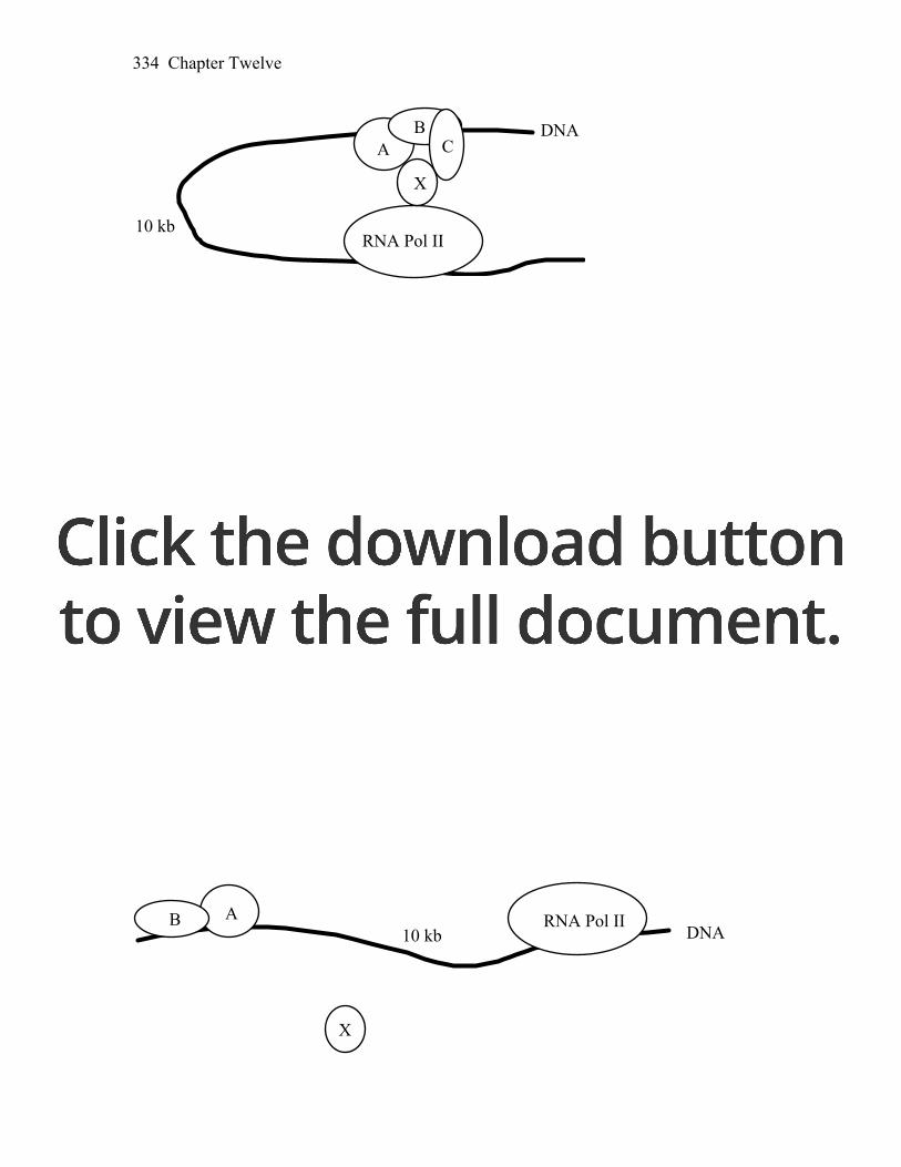
Chapter Twelve 334
DNA
RNA Pol II
AB
C
X
10 kb
28. A single mutation in one of the transcription factors in Problem 27 results in a
drastic reduction in YFG transcription. Diagram what this mutant interaction might look like.
Answer: The following represents a cell that is mutant for TFC. In this case,
CRX does not bind and the activation of transcription does not occur.
X
DNARNA Pol II
10 kbAB
29. Diagram the effect of a mutation in the binding site for one of the transcription
factors in Problem 27. Answer: The same as 18 but now the binding site for TFC is mutant.
C
X
DNARNA Pol II
10 kbAB

Chapter Twelve 335
30. How does an epigenetically silenced gene differ from a mutant gene (a null
allele of the same gene)? Answer: A gene not expressed due to alteration of its DNA sequence will never
be expressed and the inactive form will be inherited generation to generation. An epigenetically inactivated gene may still be regulated. Chromatin structure can change in the course of the cell cycle, for example, when transcription factors modify the histone code. Also, unlike mutational inactivation, epigenetic inactivation may change from generation to generation.
31. What are epigenetic marks? Which are associated with heterochromatin? How
are epigenetic marks thought to be interpreted into chromatin structure? Answer: Epigenetic marks include both DNA methylation and histone
modifications. Heritable epigenetic marks on histones and DNA have profound effects on chromatin structure and gene expression. Heterochromatin is associated with nucleosomes containing methylated histone H3. Proteins, such as HP-1 (heterochromatin protein-1) interact with specific epigenetic marks and are required in some way to produce or maintain heterochromatin. Other proteins act in concert with specific DNA sequences to insulate regions of euchromatin from regions of heterochromatin. Since epigenetic marks are inherited, chromatin structure is also inherited. Marked histones from nucleosomes of the parental strands are mixed with new, unmarked histones so that both daughter strands become associated with nucleosomes that contain both marked and unmarked histones. The code carried by the old histones most likely guides the modification of the new histones, ensuring that the chromatin state is remembered and passed on in future divisions.
32. You receive four strains of yeast in the mail and the accompanying instructions
state that each strain contains a single copy of transgene A. You grow the four strains and determine that only three strains express the protein product of transgene A. Further analysis reveals that transgene A is located at a different position in the yeast genome in each of the four strains. Provide a hypothesis to explain this result.
Answer: Chromatin structure has a profound effect on gene expression.
Transgenes inserted into regions of euchromatin would more likely be capable of expression than those inserted into regions of heterochromatin.
33. In Neurospora, all mutants affecting the enzymes carbamyl phosphate
synthetase and aspartate transcarbamylase map at the pyr-3 locus. If you induce pyr-3 mutations by ICR-170 (a chemical mutagen), you find that either both enzyme functions are lacking or only the transcarbamylase function is lacking;

Chapter Twelve 336
in no case is the synthetase activity lacking when the transcarbamylase activity is present. (ICR-170 is assumed to induce frameshifts.) Interpret these results in regard to a possible operon.
Answer: If there is an operon governing both genes, then a frameshift mutation
could cause the stop codon separating the two genes to be read as a sense codon. Therefore, the second gene product will be incorrect for almost all amino acids. However, there are no known polycistronic messages in eukaryotes. The alternative, and better, explanation is that both enzymatic functions are performed by the same gene product. Here, a frameshift mutation beyond the first function, carbamyl phosphate synthetase, will result in the second half of the protein molecule being nonfunctional.
34. You wish to find the cis-acting regulatory DNA elements responsible for the
transcriptional responses of two genes, c-fos and globin. Transcription of the c-fos gene is activated in response to fibroblast growth factor (FGF), but it is inhibited by cortisol (Cort). On the other hand, transcription of the globin gene is not affected by either FGF or cortisol, but it is stimulated by the hormone erythropoietin (EP). To find the cis-acting regulatory DNA elements responsible for these transcriptional responses, you use the following clones of the c-fos and globin genes, as well as two �“hybrid�” combinations (fusion genes), as shown in the diagram below. The letter A represents the intact c-fos gene, D represents the intact globin gene, and B and C represent the c-fos–globin gene fusions. The c-fos and globin exons (E) and introns (I) are numbered. For example, E3(f) is the third exon of the c-fos gene and I2(g) is the second intron of the globin gene. (These labels are provided to help you make your answer clear.) The transcription start sites (black arrows) and polyadenylation sites (red arrows) are indicated.
You introduce all four of these clones simultaneously into tissue-culture cells
and then stimulate individual aliquots of these cells with one of the three factors. Gel analysis of the RNA isolated from the cells gives the following results:

Chapter Twelve 337
The levels of transcripts produced from the introduced genes in response to
various treatments are shown; the intensity of these bands is proportional to the amount of transcript made from a particular clone. (The failure of a band to appear indicates that the level of transcript is undetectable.)
a. Where is the DNA element that permits activation by FGF? b. Where is the DNA element that permits repression by Cort? c. Where is the DNA element that permits induction by EP? Explain your
answer. Answer: a. Clone A is activated by FGF, but B, C, and D are not. This indicates that
the DNA binding site for this activation is located somewhere between the 3´ end of exon 1 [E1(f)] and the 5´ end of exon 3 [E3(f)]. This is the region of DNA found only in clone A.
b. Cortisol represses transcription of clone A and B, but not C. (You do not
expect any effect on D, the intact globin gene.) Comparing these clones indicates that the DNA site involved in this repression must be located in the 3´ region of E3(f) or the 3´ flanking sequences of this gene.
c. Activation by EP is seen in clones C and D, but not B. (Again, you do not
expect A, the intact c-fos gene to respond to EP.) This indicates that the DNA site involved in this activation must be localized to the 3´ side of E3(g) or the 3´ flanking regions of the globin gene.