regulation of human cerebral cortical ... - walsh lab · joseph g. gleeson, md3 and christopher a....
TRANSCRIPT
Regulation of human cerebral cortical development byEXOC7 and EXOC8, components of the exocyst complex, and
roles in neural progenitor cell proliferation and survivalMichael E. Coulter, MD, PhD1,2, Damir Musaev, BSc3, Ellen M. DeGennaro, BA1,4,
Xiaochang Zhang, PhD1,5, Katrin Henke, PhD6, Kiely N. James, PhD3, Richard S. Smith, PhD1,R. Sean Hill, PhD1, Jennifer N. Partlow, MS1, Muna Al-Saffar, MBChB, MSc1,7, A. Stacy Kamumbu, BA1,Nicole Hatem, BA1, A. James Barkovich, MD8, Jacqueline Aziza, MD9, Nicolas Chassaing, MD, PhD10,11,Maha S. Zaki, MD, PhD12, Tipu Sultan, MD13, Lydie Burglen, MD, PhD14,15, Anna Rajab, MD, PhD16,Lihadh Al-Gazali, MBChB, MSc7, Ganeshwaran H. Mochida, MD, MMSc1,17, Matthew P. Harris, PhD6,
Joseph G. Gleeson, MD3 and Christopher A. Walsh, MD, PhD 1
Purpose: The exocyst complex is a conserved protein complex thatmediates fusion of intracellular vesicles to the plasma membraneand is implicated in processes including cell polarity, cell migration,ciliogenesis, cytokinesis, autophagy, and fusion of secretory vesicles.The essential role of these genes in human genetic disorders,however, is unknown.
Methods: We performed homozygosity mapping and exomesequencing of consanguineous families with recessively inheritedbrain development disorders. We modeled an EXOC7 splice variantin vitro and examined EXOC7 messenger RNA (mRNA) expressionin developing mouse and human cortex. We modeled exoc7 loss-of-function in a zebrafish knockout.
Results: We report variants in exocyst complex members, EXOC7and EXOC8, in a novel disorder of cerebral cortex development. InEXOC7, we identified four independent partial loss-of-function
(LOF) variants in a recessively inherited disorder characterized bybrain atrophy, seizures, and developmental delay, and in severecases, microcephaly and infantile death. In EXOC8, we found ahomozygous truncating variant in a family with a similar clinicaldisorder. We modeled exoc7 deficiency in zebrafish and found theabsence of exoc7 causes microcephaly.
Conclusion: Our results highlight the essential role of the exocystpathway in normal cortical development and how its perturbationcauses complex brain disorders.
Genetics in Medicine (2020) https://doi.org/10.1038/s41436-020-0758-9
Keywords: exocyst; EXOC7; EXOC8; microcephaly; develop-mental delay
INTRODUCTIONEight genes in the human genome, EXOC1–EXOC8, encodethe exocyst complex, a multimeric, evolutionarily conservedcomplex that traffics vesicles within the cell to the plasmamembrane for fusion. The exocyst complex has been shown toplay a role in several cellular processes, including cell polarity,cell migration, ciliogenesis, cytokinesis, autophagy, and fusionof secretory vesicles,1 but human disorders associated with
definitive loss-of-function variants in any of these compo-nents have not yet been reported. Although a missense variantin EXOC82 was reported in a single case of Joubert syndrome(MIM 213300), and a missense variant in EXOC43 wasreported in a case of Meckel–Gruber syndrome (MIM249000), the pathogenicity of these two variants has not yetbeen confirmed. As such, the essential role of individualproteins of the exocyst complex remains unclear.
Submitted 12 September 2019; revised 16 January 2020; accepted: 27 January 2020
1Division of Genetics and Genomics and Howard Hughes Medical Institute, Boston Children’s Hospital, Departments of Pediatrics and Neurology, Harvard Medical School, Boston,MA, USA; 2Program in Neuroscience and Harvard/MIT MD-PHD Program, Harvard Medical School, Boston, MA, USA; 3Department of Neurosciences and Howard HughesMedical Institute, University of San Diego, La Jolla, CA, USA; 4Harvard–MIT Division of Health Sciences and Technology, Massachusetts Institute of Technology, Cambridge, MA,USA; 5Department of Human Genetics, University of Chicago, Chicago, IL, USA; 6Division of Orthopedic Research, Boston Children’s Hospital, Department of Genetics, HarvardMedical School, Boston, MA, USA; 7Department of Paediatrics, College of Medicine and Health Sciences, United Arab Emirates University, Al Ain, United Arab Emirates; 8BenioffChildren’s Hospital, Departments of Radiology, Pediatrics, Neurology, and Neurological Surgery, University of California San Francisco, San Francisco, CA, USA; 9Département dePathologie, Institut Universitaire du Cancer de Toulouse–Oncopole–CHU Toulouse, Toulouse, France; 10Service de Génétique Médicale, CHU Toulouse, Toulouse, France;11UDEAR; UMR 1056 Inserm–Université de Toulouse, Toulouse, France; 12Clinical Genetics Department, Human Genetics and Genome Research Division, National ResearchCentre, Cairo, Egypt; 13Department of Pediatric Neurology, Institute of Child Health & The Children’s Hospital, Lahore, Pakistan; 14Centre de référence des malformations etmaladies congénitales du cervelet, Département de génétique, AP-HP.Sorbonne Université, Paris, France; 15Hôpital Trousseau and Developmental Brain Disorders Laboratory,Imagine Institute, INSERM UMR 1163, Paris, France; 16National Genetics Center, Directorate General of Health Affairs, Ministry of Health, Muscat, Oman; 17Department ofNeurology, Massachusetts General Hospital, Boston, MA, USA. Correspondence: Joseph G. Gleeson ([email protected]) or Christopher A. Walsh ([email protected])
ARTICLE
GENETICS in MEDICINE | Volume 0 | Number 0 | Month 1

Several of the reported functions of the exocyst complex, cellpolarity and migration, cytokinesis, and ciliogenesis,1,4–6 areintegral processes during cerebral cortical development and somotivated us to test the hypothesis that variants in exocystencoding genes cause brain development disorders. First,establishing cell polarity and cell migration are essential forcortical development. Variants in radial glial cell progenitors(RGC) polarity genes, such as Pals1 and Par3, disrupt corticaldevelopment through massive cell death or premature cell cycleexit, respectively.7,8 In addition, variants in genes required forneuronal migration can cause one of several cortical malforma-tions, including lissencephaly (MIM 607432, PAFAH1B1/LIS19), double cortex syndrome (MIM 300067, DCX10,11), andcortical dysplasia (MIM 610031, TUBB3,12 TUBB5,13 KIF5C,14
KIF2A14). Second, robust and rapid cell division of corticalprogenitors is essential for cortical development and severalgenetic causes of microcephaly (MIM 251200) exhibit disruptedcytokinesis as a result of supernumerary (CDK5RAP2,15,16
KATNB117,18), or missing spindle poles (ASPM,19 WDR6220)stemming from dysfunctional centrosomes.21 Finally, defects incilia formation lead to ciliopathy syndromes (MIM 209900),complex syndromes with disrupted brain development(INPP5E,22 C2CD3,23 BBS124).Here we provide a systematic analysis of variants in two
exocyst components, by defining several variants in EXOC7and EXOC8. We identify four independent, partial loss-of-function variants in EXOC7, associated with developmentalbrain disorders of variable severity characterized by develop-mental delay, seizures, brain atrophy, microcephaly, andinfantile death. We also describe one loss-of-function variantin EXOC8 similarly associated with severe developmentaldelay, seizures, brain atrophy, microcephaly, and prematuredeath. We further provide a zebrafish genetic model ofEXOC7 loss-of-function and offer genetic evidence thatEXOC7 is required for neuron survival.
MATERIALS AND METHODSHuman subjectsThis study was conducted with the approval of institutionalreview boards and according to the ethical standards of theparticipating institutions: Boston Children’s Hospital; Uni-versity of California–San Diego; the Faculty of Medicine,United Arab Emirates University; and AP-HP SorbonneUniversité. Informed consent was received from all partici-pants. Permission was received to publish patient photographs.
IACUC approval of zebrafish housing and experimentsA complete description of the husbandry and environmentalconditions in housing for the fish used in these experiments isavailable as a collection in protocols.io (https://doi.org/10.17504/protocols.io.mrjc54n). All animals were cared forhumanely and all experiments were approved by BostonChildren’s Hospital Institutional Animal Care and UseCommittee (IACUC).
Exome sequencingDNA was extracted from whole blood and exome sequencingwas performed (See Supplement). We filtered out variantswith allele frequency >10% in the Middle Easternpopulation.25
Sanger sequencingPrimers surrounding the reported variant in each family wereused for polymerase chain reaction (PCR) and subsequentSanger sequencing to confirm genotype from exome sequen-cing and determine segregation within the family.
Minigene cloning and expressionA ~5-kb section of human EXOC7 locus was amplified withprimers (F: AAGGACTGAAGGAGCATTTC, R: CAGGGAGTCGAAGGTCTTCT) from a BAC and cloned into pCAGexpression vector. The splice acceptor variant from family Iwas introduced with site-directed mutagenesis. Wild-type(WT) or splice variant containing vector was transfected intomouse N2A cells and after 48 hours RNA was isolated andretrotranscribed into complementary DNA (cDNA). N2Acells were cultured at 37 °C and 5% CO2 in high-glucoseDMEM (GIBCO) supplemented with 10% fetal bovine serumand 1% penicillin–streptomycin.
HAP1 mutant cell lineHAP1 human cell line was cultured at 37 °C and 5% CO2 inhigh-glucose DMEM (GIBCO) supplemented with 10% fetalbovine serum and 1% penicillin–streptomycin. The spliceacceptor variant from family I was introduced into a HAP1cell line as a hemizygous variant using CRISPR/Cas9mutagenesis (Supplemental Methods)26 (Horizon Discovery).Immunoblot was performed using human EXOC7 antibody(Abcam, ab118792).
Exoc7 alternative splicing in developing cortexAlternative splicing analysis of EXOC7 in developing human(GW13–16) and mouse (E14.5) cortex was performed asdescribed previously.27 Aligned BAM files from RNAsequencing data sets were analyzed with the MISO pipeline(version 0.4.6) to determine the inclusion frequency ofalternatively spliced exons.
Generation of exoc7 mutant zebrafish linesExoc7 mutant zebrafish were generated by CRISPR/Cas9mutagenesis. Cas9 messenger RNA (mRNA) (250 ng/μl)and exoc7 targeting guide RNA (target: CCGTCCTCATCCTGGACGCC, 80 ng/μl) were injected into 1-cellembryos. Embryos developed to adulthood and then Sangersequencing was used to identify potential heterozygousexoc7 mutant carriers in F1 progeny. A 1-bp frameshiftdeletion in exon 5 was identified and this fish wasbackcrossed to WT to generate heterozygous carriers. Thisallele is mh111.
ARTICLE COULTER et al
1234
5678
90():,;
2 Volume 0 | Number 0 | Month | GENETICS in MEDICINE
Toluidine blue staining of zebrafishFive days postfertilization (dpf) embryos were fixed in 4%PFA overnight at 4 °C and then embedded in JB-4 resinaccording to manufacturer’s protocol (Polysciences Inc). Fishwere sectioned at 2 μm, and then matching sections werestained with toluidine blue and imaged with a bright-fieldmicroscope.
Immunostaining of zebrafish progenitor cellsFive dpf embryos were fixed in 4% PFA overnight at 4 °C,embedded in OCT, and sectioned coronally at 20 μm on acryostat. Matched sections were stained with a primaryantibody against Sox2 (Abcam, ab97959). Tissue waspermeabilized and blocked in 3% BSA, 0.3% Triton X-100,0.3% sodium azide in PBS. Primary antibodies were incubatedovernight at 4 °C. Sections were stained with Alexa secondaryantibodies and Hoechst. Imaging was done on Zeiss 510confocal microscope. Sox2-positive nuclei in telencephalonwere counted.
TUNEL staining in developing zebrafishFive dpf embryos were fixed in 4% PFA overnight at 4 °C,embedded in OCT, and cryosectioned coronally. Apoptoticcells in matched sections were labeled with TUNEL stainingusing the Apoptag kit (Millipore) according to the manu-facturer’s instructions. Imaging was done on Zeiss 510confocal microscope. TUNEL positive cells in telencephalonwere counted.
RNAscopeRNAscope on human fetal brain tissue was performedaccording to manufacturer’s protocol (ACDBio). Tissue wasfixed in 4% PFA, frozen, and sectioned at 20 μm on a cryostat.
Quantification and statistical analysisIn all analyses, mean values are presented for pooled data anderrors bars are SEM. For all quantifications, statisticalsignificance was determined using a two-tailed, unpairedt test (GraphPad Prism).
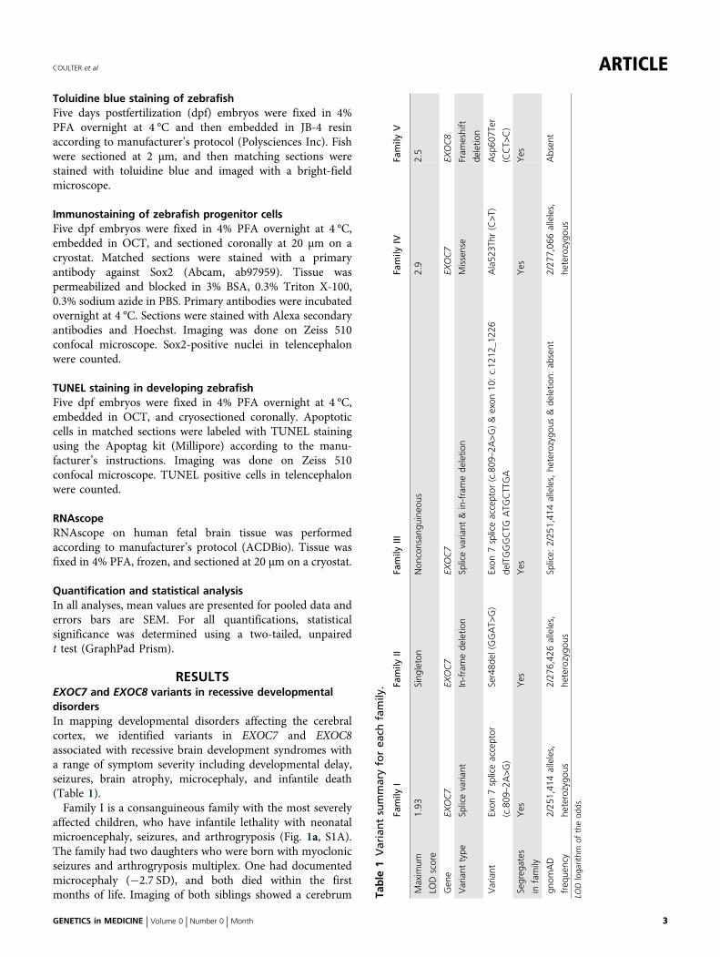
RESULTSEXOC7 and EXOC8 variants in recessive developmentaldisordersIn mapping developmental disorders affecting the cerebralcortex, we identified variants in EXOC7 and EXOC8associated with recessive brain development syndromes witha range of symptom severity including developmental delay,seizures, brain atrophy, microcephaly, and infantile death(Table 1).Family I is a consanguineous family with the most severely
affected children, who have infantile lethality with neonatalmicroencephaly, seizures, and arthrogryposis (Fig. 1a, S1A).The family had two daughters who were born with myoclonicseizures and arthrogryposis multiplex. One had documentedmicrocephaly (−2.7 SD), and both died within the firstmonths of life. Imaging of both siblings showed a cerebrum Ta
ble
1Variantsummaryforea
chfamily
.
Family
IFa
mily
IIFa
mily
IIIFa
mily
IVFa
mily
V
Maxim
um
LOD
score
1.93
Sing
leton
Non
consan
guineo
us2.9
2.5
Gen
eEX
OC7
EXOC7
EXOC7
EXOC7
EXOC8
Variant
type
Splicevaria
ntIn-framede
letio
nSp
licevaria
nt&in-framede
letio
nMissense
Fram
eshift
deletio
n
Variant
Exon
7spliceacceptor
(c.809
–2A
>G)
Ser48d
el(GGAT>
G)
Exon
7spliceacceptor
(c.809
–2A
>G)&exon
10:c.12
12_1
226
delTGGGCTG
ATG
CTTGA
Ala52
3Thr
(C>T)
Asp60
7Ter
(CCT>
C)
Segreg
ates
infamily
Yes
Yes
Yes
Yes
Yes
gnom
AD
freq
uency
2/25
1,41
4alleles,
heterozygo
us
2/27
6,42
6alleles,
heterozygo
us
Splice:
2/25
1,41
4alleles,he
terozygo
us&de
letio
n:ab
sent
2/27
7,06
6alleles,
heterozygo
us
Absen
t
LODloga
rithm
oftheod
ds.
COULTER et al ARTICLE
GENETICS in MEDICINE | Volume 0 | Number 0 | Month 3

a
I-03
G G A C GA A
EXOC7 Intron 6 Exon 7
I-04
I-01
I-05
I-02I-02 I-01 I-05
I-03I-04
Control I-01 I-05
IV-06
IV-02IV-01
IV-05IV-04IV-03 IV-07 IV-08 IV-09
Family I
Family IVd
EXOC7 Exon 3 Family IIb
II-01 II-02
II-03 II-04
3
C T G A G C G C C A T CEXOC7 Exon 15
II-01
II-02
II-03
II-04
IV-01
IV-02
IV-05
IV-07
IV-09
Leu Ser Ala Ile PheHumanLeu Ser Ala Ile PheRhesusLeu Ser Ala Ile PheMouseLeu Ser Ala Ile PheDogLeu Ser Ala Ile PheElephantLeu Ser Ala Ile PheChickenLeu Lys Ala Ile PheX tropicalisLeu Arg Ala Ile PheZebrafish
521 522 523 524 525
EXOC8EXOC7
Family IVLeu Ser Ala Ile PheLeu Ser Thr Ile Phe
Reference
Family IILeu Ser SerLeu Ser
Reference
V-04 V-03
V-01 V-02
eFamily V
V-03
V-04
V-01
V-02
EXOC8 Exon 1
V-05 V-06
V-02V-01
V-06
V-06
6071
7251ReferenceEXOC8
proteinFamily V Asp607Ter
Ser48
PhePhe
48
Family IIIc
III-01 III-02
III-03 III-04
G G A C GA A
EXOC7 Intron 6 Exon 7
III-01
III-02
III-03
III-04
III-01, 03, 04
III-02
EXOC7 Exon 10
15 bp deletion
Family III heterozygous deletion
1220
1230 12401220
ARTICLE COULTER et al
4 Volume 0 | Number 0 | Month | GENETICS in MEDICINE
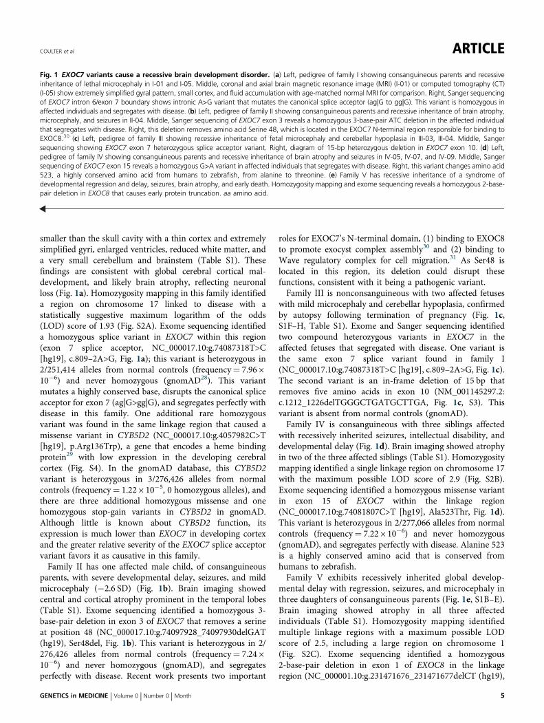
smaller than the skull cavity with a thin cortex and extremelysimplified gyri, enlarged ventricles, reduced white matter, anda very small cerebellum and brainstem (Table S1). Thesefindings are consistent with global cerebral cortical mal-development, and likely brain atrophy, reflecting neuronalloss (Fig. 1a). Homozygosity mapping in this family identifieda region on chromosome 17 linked to disease with astatistically suggestive maximum logarithm of the odds(LOD) score of 1.93 (Fig. S2A). Exome sequencing identifieda homozygous splice variant in EXOC7 within this region(exon 7 splice acceptor, NC_000017.10:g.74087318T>C[hg19], c.809–2A>G, Fig. 1a); this variant is heterozygous in2/251,414 alleles from normal controls (frequency= 7.96 ×10⁻⁶) and never homozygous (gnomAD28). This variantmutates a highly conserved base, disrupts the canonical spliceacceptor for exon 7 (ag|G>gg|G), and segregates perfectly withdisease in this family. One additional rare homozygousvariant was found in the same linkage region that caused amissense variant in CYB5D2 (NC_000017.10:g.4057982C>T[hg19], p.Arg136Trp), a gene that encodes a heme bindingprotein29 with low expression in the developing cerebralcortex (Fig. S4). In the gnomAD database, this CYB5D2variant is heterozygous in 3/276,426 alleles from normalcontrols (frequency= 1.22 × 10⁻⁵, 0 homozygous alleles), andthere are three additional homozygous missense and onehomozygous stop-gain variants in CYB5D2 in gnomAD.Although little is known about CYB5D2 function, itsexpression is much lower than EXOC7 in developing cortexand the greater relative severity of the EXOC7 splice acceptorvariant favors it as causative in this family.Family II has one affected male child, of consanguineous
parents, with severe developmental delay, seizures, and mildmicrocephaly (−2.6 SD) (Fig. 1b). Brain imaging showedcentral and cortical atrophy prominent in the temporal lobes(Table S1). Exome sequencing identified a homozygous 3-base-pair deletion in exon 3 of EXOC7 that removes a serineat position 48 (NC_000017.10:g.74097928_74097930delGAT(hg19), Ser48del, Fig. 1b). This variant is heterozygous in 2/276,426 alleles from normal controls (frequency= 7.24 ×10⁻⁶) and never homozygous (gnomAD), and segregatesperfectly with disease. Recent work presents two important
roles for EXOC7’s N-terminal domain, (1) binding to EXOC8to promote exocyst complex assembly30 and (2) binding toWave regulatory complex for cell migration.31 As Ser48 islocated in this region, its deletion could disrupt thesefunctions, consistent with it being a pathogenic variant.Family III is nonconsanguineous with two affected fetuses
with mild microcephaly and cerebellar hypoplasia, confirmedby autopsy following termination of pregnancy (Fig. 1c,S1F–H, Table S1). Exome and Sanger sequencing identifiedtwo compound heterozygous variants in EXOC7 in theaffected fetuses that segregated with disease. One variant isthe same exon 7 splice variant found in family I(NC_000017.10:g.74087318T>C [hg19], c.809–2A>G, Fig. 1c).The second variant is an in-frame deletion of 15 bp thatremoves five amino acids in exon 10 (NM_001145297.2:c.1212_1226delTGGGCTGATGCTTGA, Fig. 1c, S3). Thisvariant is absent from normal controls (gnomAD).Family IV is consanguineous with three siblings affected
with recessively inherited seizures, intellectual disability, anddevelopmental delay (Fig. 1d). Brain imaging showed atrophyin two of the three affected siblings (Table S1). Homozygositymapping identified a single linkage region on chromosome 17with the maximum possible LOD score of 2.9 (Fig. S2B).Exome sequencing identified a homozygous missense variantin exon 15 of EXOC7 within the linkage region(NC_000017.10:g.74081807C>T [hg19], Ala523Thr, Fig. 1d).This variant is heterozygous in 2/277,066 alleles from normalcontrols (frequency= 7.22 × 10⁻⁶) and never homozygous(gnomAD), and segregates perfectly with disease. Alanine 523is a highly conserved amino acid that is conserved fromhumans to zebrafish.Family V exhibits recessively inherited global develop-
mental delay with regression, seizures, and microcephaly inthree daughters of consanguineous parents (Fig. 1e, S1B–E).Brain imaging showed atrophy in all three affectedindividuals (Table S1). Homozygosity mapping identifiedmultiple linkage regions with a maximum possible LODscore of 2.5, including a large region on chromosome 1(Fig. S2C). Exome sequencing identified a homozygous2-base-pair deletion in exon 1 of EXOC8 in the linkageregion (NC_000001.10:g.231471676_231471677delCT (hg19),
Fig. 1 EXOC7 variants cause a recessive brain development disorder. (a) Left, pedigree of family I showing consanguineous parents and recessiveinheritance of lethal microcephaly in I-01 and I-05. Middle, coronal and axial brain magnetic resonance image (MRI) (I-01) or computed tomography (CT)(I-05) show extremely simplified gyral pattern, small cortex, and fluid accumulation with age-matched normal MRI for comparison. Right, Sanger sequencingof EXOC7 intron 6/exon 7 boundary shows intronic A>G variant that mutates the canonical splice acceptor (ag|G to gg|G). This variant is homozygous inaffected individuals and segregates with disease. (b) Left, pedigree of family II showing consanguineous parents and recessive inheritance of brain atrophy,microcephaly, and seizures in II-04. Middle, Sanger sequencing of EXOC7 exon 3 reveals a homozygous 3-base-pair ATC deletion in the affected individualthat segregates with disease. Right, this deletion removes amino acid Serine 48, which is located in the EXOC7 N-terminal region responsible for binding toEXOC8.30 (c) Left, pedigree of family III showing recessive inheritance of fetal microcephaly and cerebellar hypoplasia in III-03, III-04. Middle, Sangersequencing showing EXOC7 exon 7 heterozygous splice acceptor variant. Right, diagram of 15-bp heterozygous deletion in EXOC7 exon 10. (d) Left,pedigree of family IV showing consanguineous parents and recessive inheritance of brain atrophy and seizures in IV-05, IV-07, and IV-09. Middle, Sangersequencing of EXOC7 exon 15 reveals a homozygous G>A variant in affected individuals that segregates with disease. Right, this variant changes amino acid523, a highly conserved amino acid from humans to zebrafish, from alanine to threonine. (e) Family V has recessive inheritance of a syndrome ofdevelopmental regression and delay, seizures, brain atrophy, and early death. Homozygosity mapping and exome sequencing reveals a homozygous 2-base-pair deletion in EXOC8 that causes early protein truncation. aa amino acid.
COULTER et al ARTICLE
GENETICS in MEDICINE | Volume 0 | Number 0 | Month 5

Asp607Ter, Fig. 1e), which is absent from normal controls(gnomAD). This frameshifting deletion creates a prematurestop codon at amino acid 607, short of the full-length protein(725 aa), and segregates perfectly with disease in this family.One additional homozygous variant was detected in thefamily (stop-gain in RP1L1). In gnomAD, this variant ispresent as a homozygote in one individual and RP1L1 isassociated with adult-onset retinitis pigmentosum (OMIM613587), so this variant is unlikely to contribute tomicrocephaly in this family.In total, we report four novel missense and splice site
variants in EXOC7 and one novel truncating variant inEXOC8 in families with a recessive syndrome of brainatrophy, seizures, and developmental delay, and in moresevere cases microcephaly and infantile death (Table 1). Thepresence of cerebral atrophy in all families indicatesneurodegeneration and suggests EXOC7 and EXOC8 arerequired for neuronal survival. Loss-of-function variants inEXOC7 have not been previously linked to human disease,and we have recently reported one homozygous missensevariant in EXOC8 in a single case of Joubert syndrome.2
EXOC7 splice variant disrupts mRNA splicing patterns andreduces protein expressionEXOC7 is an alternatively spliced gene with 5 verifiedtranscripts;32 three of which include exon 7. We generated aminigene assay to model the exon 7 splice acceptor variantfound in both families I and III and found this variantdisrupts splicing and decreases EXOC7 protein level. Theminigene was constructed using 5 kb of genomic DNA fromthe human EXOC7 locus spanning exon 6 to exon 9 (Fig. 2a).Reverse transcription PCR (RT-PCR) of mRNA transcribedfrom the minigene plasmid expressed in mouse N2A cellsrevealed three splicing disruptions caused by the humanvariant (Fig. 2a). First, cDNA encoding a high-abundancetranscript (including exons 6, 7, and 9) was isolated fromwild-type minigene but completely absent from the variant(Fig. 2a). A low-abundance larger product that could not besubcloned for sequencing was found in wild-type and likelyencodes a transcript including exons 6, 7, 8, and 9 (*, Fig. 2a).Second, two novel out-of-frame splice forms that arepredicted to encode early truncations were found exclusivelyin variant minigene cDNA (Fig. 2a). Form A splices in the last
a
b c
WT
Variant
WT Variant
6841
6531
2861
2901
6531
WT Variant
EXOC7R
elat
ive
prot
ein
WT
Variant
1
Band 1
1.5
1.0
0.5
0.0Band 2
p = 0.088
p = 0.045
6 7 8 9
6 7 8 9
6 7 8 9
6 7 8 9
6 7 8 9
cDNA
cDNA
Protein
Protein
cDNA
cDNA
Protein
Protein
Protein
cDNA
GAPDH
212
*
Minigene assay
7 8* A>G EXOC7
6 9
*
Form
A
Variant form A Variant form B
Form B
Intron 6 Exon 7
37 bases spliced-in
Frameshift
WT
Variant
14 bases spliced-out
Frameshift
Intron 6 Exon 7
WT
Variant
Fig. 2 EXOC7 exon 7 splice acceptor variant disrupts splicing. (a) Top, diagram of EXOC7 human minigene construct with splice acceptor variant. Bluearrows mark reverse transcription polymerase chain reaction (RT-PCR) primers used to generate complementary DNA (cDNA) products shown in gel image.Arrows from each band in gel point to splicing diagram determined by Sanger sequencing of the cDNA. Wild-type (WT) minigene generated two in-frameisoforms (left), whereas variant minigene generated one in-frame isoform and two novel out-of-frame isoforms (right). Two variant isoforms encode novelstop codons that lead to premature protein truncation. Asterisk (*) indicates low-abundance product that could not be subcloned for sequencing. (b)Immunoblot of EXOC7 protein in WT and variant HAP1 cells showing reduction of two EXOC7 isoforms. GAPDH is a protein loading control. (c)Quantification of (b) showed significant 50% reduction of larger EXOC7 band (band 1, p= 0.045), while the lower weight band (band 2) was notsignificantly reduced. P values calculated by two-tailed t test. Error bars represent SEM.
ARTICLE COULTER et al
6 Volume 0 | Number 0 | Month | GENETICS in MEDICINE

37 bases of intron 6 causing a frameshift that is predicted tocreate a premature stop codon at amino acid 286, well short ofthe full-length protein of 684 amino acids. Form B splices outthe first 14 bases of exon 7 causing a frameshift, predicted tocreate a premature stop codon at amino acid 290. An isoformthat skips exon 7 and includes exons 6 and 9 was observed inboth wild-type and variant minigenes. These minigene resultsshow that the variant disrupts EXOC7 mRNA splicing. Toassess the variant’s effect on EXOC7 protein we mutated aHAP1 cell line to encode the patient variant (hemizygous).Immunoblot of EXOC7 protein showed reduced expression invariant HAP1 cells (Fig. 2b, c). We detected two isoforms: (1)a larger isoform was significantly reduced by 50% (two-tailedt test, p= 0.045) and (2) a smaller isoform showed a trendtowards reduction (two-tailed t test, p= 0.088). Together,these results support the pathogenicity of this variant.
EXOC7 is highly expressed in developing cerebral cortexEXOC7 is highly expressed in the developing human cortex,consistent with the affected individuals’ phenotypes. In situhybridization using RNAscope in human fetal cortexshowed abundant EXOC7 expression in the ventricularzone (adjacent to the ventricle and coexpressing VIM), theouter subventricular zone (coexpressing TBR2) and in thecortical plate (adjacent to the pial surface) (Fig. 3a, b).Similarly, Exoc7 is expressed in developing mouse cortex(MGI). RNA sequencing analysis of human fetal cortex33
confirms this expression pattern with expression in theventricular zone and the inner and outer subventricular
zones at a similar level as ASPM, a canonical microcephalygene, and in the cortical plate (Fig. 3c). EXOC7 expressionin both progenitors and postmitotic neurons suggests it hasimportant roles in both cell types during cortical develop-ment. Notably, exon 7 of EXOC7, which contains a spliceacceptor variant in families I and III, is differentially splicedin cortical progenitors compared with postmitotic corticalneurons. Using a previously published method,27 weidentified differentially spliced exons in the developingcortex with RNA sequencing of separated cortical progeni-tors and postmitotic neurons isolated from both developingmouse and human brain tissue. During fetal human corticaldevelopment (GW15), exon 7 is included in 93% oftranscripts from the cortical plate and 47% oftranscripts from ventricular zone (Fig. 3d). Similarly, duringcortical development in mice (E14.5), exon 7 is included in87% of transcripts isolated from the cortical plate and 35%of transcripts from the ventricular zone (Fig. 3d). Therole of this differential splicing in the regulation ofEXOC7 function in development is unknown, but ourevidence that the splicing variant identified in families I andIII alters this differential expression, by eliminatingexpression of the exon 7 included isoform (Fig. 2), suggeststhis variant is pathogenic by disrupting corticaldevelopment.
Exoc7 is essential for vertebrate embryonic developmentEXOC7 protein is highly conserved among vertebrates(Fig. 4a). To further characterize the function of EXOC7 in
EXOC7
Human EXOC7
Mouse Exoc7
db
E6 E7 E8 E9 E10 E11
E6 E7 E8 E9 E10 E11
A>G
c
CP
VZ
OSVZ
*
a
93%
47%
87%
35%
VZ
OSVZ
CP
VZa
SVZa
OSVZa
CP
50
40
30
20
10
0
FP
KM
EXOC7 ASPM
SVZ
RNAsequencing
Splicing analysis(MISO)
Isoformidentification
EXOC7
TBR2
VIM
EXOC7
MergeEXOC7
EXOC7 Merge
CP
VZ
CP
VZ50 um
Fig. 3 EXOC7 is highly expressed in developing cortex. (a) Diagram of cortical section of human fetal cortex indicating locations of RNAscope imagingin (b) and RNA sequencing in (c). (b) RNAscope imaging of fetal human cortex shows EXOC7 expression in VZ and OSVZ, two progenitor zones, and in CP,the location of postmitotic neurons. (c) EXOC7 is highly expressed in developing human cortex and shown in comparison with ASPM. Expression levelsmeasured based on RNA sequencing.33 (d) RNA sequencing data from developing human fetal cortex (GW15) and mouse cortex (E14.5) showingdifferential inclusion of exon 7 in CP vs. VZ. FPKM fragments per kilobase of transcript per million mapped reads.
COULTER et al ARTICLE
GENETICS in MEDICINE | Volume 0 | Number 0 | Month 7
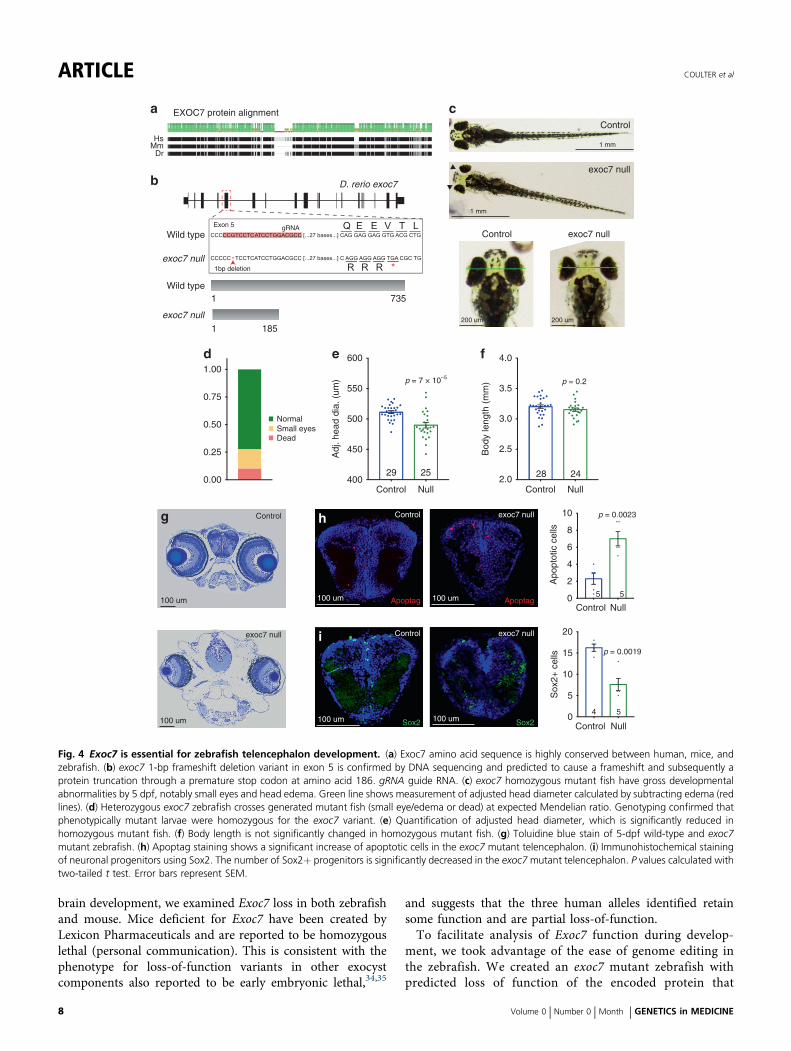
brain development, we examined Exoc7 loss in both zebrafishand mouse. Mice deficient for Exoc7 have been created byLexicon Pharmaceuticals and are reported to be homozygouslethal (personal communication). This is consistent with thephenotype for loss-of-function variants in other exocystcomponents also reported to be early embryonic lethal,34,35
and suggests that the three human alleles identified retainsome function and are partial loss-of-function.To facilitate analysis of Exoc7 function during develop-
ment, we took advantage of the ease of genome editing inthe zebrafish. We created an exoc7 mutant zebrafish withpredicted loss of function of the encoded protein that
c
b
d
Wild type
exoc7 null
Control
1 mm
1 mm
200 um 200 um
exoc7 null
Wild type
4.06001.00
0.75
0.50
0.25
0.00
550
500Normal
DeadSmall eyes
450
400
3.5
3.0
2.5
2.0
10
20
15
10
5
0
8
6
4
2
0
735
D. rerio exoc7
1
1851
exoc7 null
e f
28
Adj
. hea
d di
a. (
um)
Bod
y le
ngth
(m
m)p = 7 × 10⁻⁵
p = 0.2
Control exoc7 null
Control
Sox
2+ c
ells
4
p = 0.0019
Apo
ptot
ic c
ells
Control
5 5
p = 0.0023h
i
Control exoc7 null
Control exoc7 null
Apoptag Apoptag
Sox2 Sox2
Control
exoc7 null
g
Exon 5 gRNA
1bp deletion
HsMm
Dr
EXOC7 protein alignmenta
24
Null
29
Control
25
Null
Null
Control Null
5
100 um 100 um
100 um100 um
100 um
100 um
Fig. 4 Exoc7 is essential for zebrafish telencephalon development. (a) Exoc7 amino acid sequence is highly conserved between human, mice, andzebrafish. (b) exoc7 1-bp frameshift deletion variant in exon 5 is confirmed by DNA sequencing and predicted to cause a frameshift and subsequently aprotein truncation through a premature stop codon at amino acid 186. gRNA guide RNA. (c) exoc7 homozygous mutant fish have gross developmentalabnormalities by 5 dpf, notably small eyes and head edema. Green line shows measurement of adjusted head diameter calculated by subtracting edema (redlines). (d) Heterozygous exoc7 zebrafish crosses generated mutant fish (small eye/edema or dead) at expected Mendelian ratio. Genotyping confirmed thatphenotypically mutant larvae were homozygous for the exoc7 variant. (e) Quantification of adjusted head diameter, which is significantly reduced inhomozygous mutant fish. (f) Body length is not significantly changed in homozygous mutant fish. (g) Toluidine blue stain of 5-dpf wild-type and exoc7mutant zebrafish. (h) Apoptag staining shows a significant increase of apoptotic cells in the exoc7 mutant telencephalon. (i) Immunohistochemical stainingof neuronal progenitors using Sox2. The number of Sox2+ progenitors is significantly decreased in the exoc7mutant telencephalon. P values calculated withtwo-tailed t test. Error bars represent SEM.
ARTICLE COULTER et al
8 Volume 0 | Number 0 | Month | GENETICS in MEDICINE

allowed us to examine the function of exoc7 in development.We used CRISPR/Cas9 mutagenesis to create a 1-bpdeletion in exon 5 of exoc7 that generates a predictedframeshift and subsequent nonsense variant. This variant ispredicted to lead to a prematurely truncated protein 185amino acids long, well short of the full-length peptide at 735(Fig. 4b).We found that exoc7 is essential for zebrafish development.
At 5 dpf, approximately 25% of progeny from a heterozygousincross showed head edema and small eyes, consistent withMendelian inheritance (Fig. 4c, d, g). Genotyping confirmedthat these phenotypes were associated with loss of exoc7 anddemonstrated that the phenotype was highly penetrant(Fig. S5A). The mutant fish from the incross (edema andsmall eyes) die shortly after day 5, showing exoc7 is essentialfor early zebrafish survival.Quantification of the small eye phenotype revealed that the
eye area was reduced by 26% in mutant fish (two-tailed t test,3.6 × 10−24, Fig. S5B). Broader characterization of thephenotype in exoc7 mutant fish revealed general defects inhead size although this was partially masked with the presentedema. To account for the changes caused by the edema, wemeasured the distance from the outside of one eye to the otherand then subtracted the regions of edema (Fig. 4c). Even withthis conservative measure, head diameter was 4% smaller inexoc7 null fish compared with clutch controls (two-tailedt test, p= 6.8 × 10⁻⁵, Fig. 4e). We detected no difference inbody length in mutant fish, suggesting that these defects arespecifically caused by the loss of exoc7 and not simply bydevelopmental delay or allometric changes (two-tailed t test,p= 0.21, Fig. 4f).
Cellular defects in exoc7 null developing telencephalonTo identify cellular mechanisms underlying microphthalmiaand microcephaly in exoc7 mutant zebrafish (Fig. 4g), wemeasured apoptosis and counted progenitor cells in thedeveloping telencephalon. At 5 dpf, we found a threefoldincrease in the number of apoptotic cells (TUNEL stain) inthe telencephalon of exoc7 null zebrafish (control n= 5,exoc7 knockout [KO] n= 5, two-tailed t test, p= 0.0023,Fig. 4h). At the same age, we also found a 53% decrease inthe number of Sox2-positive telencephalon progenitor cells(control n= 4, exoc7 KO n= 5, two-tailed t test, p= 0.0019,Fig. 4i). The number of Sox2-positive neuroprogenitors wasalso decreased in the retina of exoc7 null fish, with a 76%decrease compared with controls (control n= 11, exoc7 KOn= 10, two-tailed t test, p= 3.0 × 10⁻⁶, Fig. S5C). Weexamined Hoechst-stained mitotic figures in developingtelencephalon and did not find a detectable increase inabnormal mitoses in exoc7 null fish, suggesting normalcytokinesis (Fig. S5E). Together, these results highlightspecific cellular defects that drive microcephaly in zebrafishin the absence of exoc7, and further suggest that the atrophyand microcephaly observed in humans with EXOC7 variantsreflect loss of proliferating progenitor cells and postmitoticneurons.
DISCUSSIONWe identified four independent presumably hypomorphicvariants in EXOC7 and one predicted loss-of-function variantin EXOC8 causing a recessive human brain developmentdisorder characterized by brain atrophy, seizures, anddevelopmental delay and in more severe cases, microcephalyand infantile death. We show that EXOC7, a member of themammalian exocyst complex, is highly expressed in develop-ing human cortex. In addition, a zebrafish model of Exoc7deficiency recapitulates the human disorder with increasedapoptosis and decreased progenitor cells during telencephalondevelopment, suggesting that the brain atrophy in humancases reflects neuronal degeneration. These findings providekey inroads into understanding the role of the exocystcomplex in normal cortical development and complexneurodegenerative disorders.
Exocyst variants cause a range of brain developmentdisordersOur work represents the first systematic genetic analysis ofthe role of exocyst components in human genetic disease. Thefour distinct alleles that we describe in EXOC7 show a rangeof severity consistent with the degree to which they likelydamage the protein, with the splicing variant being mostsevere, a 5–amino acid deletion having similar severity, a1–amino acid deletion being less severe, and an amino acidsubstitution being the mildest. However, all families sharecentral nervous system (CNS) disease and specifically corticalatrophy.We also report a null variant in EXOC8 associated with
severe phenotypes within this spectrum. Interestingly, wepreviously reported that a single affected individual withJoubert syndrome had a homozygous missense variant(E265G) in EXOC8.2 This variant occurred at a highlyconserved amino acid and was predicted to be damaging toprotein function. Careful clinical review of the affectedindividuals here confirms that they do not have classic signsof Joubert syndrome. It is possible that these two alleles,E265G and Asp607Ter, lead to different clinical syndromesbased on differing variant severity, where a hypomorphicmissense variant causes Joubert syndrome and a null variantcauses cortical atrophy and microcephaly. Consistent withthis idea, homozygous null Exoc8 mice are reported to haveearly embryonic lethality (MGI, Mouse Phenotyping Con-sortium). Here we report that loss-of-function variants ineither EXOC7 or EXOC8 lead to highly overlapping clinicalfeatures, suggesting perhaps that disruption of the exocystcomplex broadly impairs normal cortical development. Thisis supported by previous work reporting an individual withMeckel–Gruber syndrome and microcephaly had a homo-zygous missense variant (Gln578Arg) in another exocystcomponent, EXOC4.3 The exact mechanism for exocystdysfunction causing microcephaly and cortical atrophy isunknown, but previous work and our current results suggestthe exocyst may be essential for multiple molecularprocesses during cortical development. Joubert syndrome
COULTER et al ARTICLE
GENETICS in MEDICINE | Volume 0 | Number 0 | Month 9
and Meckel–Gruber syndrome both have features ofciliopathies, and the exocyst is reported to localize to theprimary cilium where some members are required fornormal ciliogenesis (EXOC536). In developing zebrafish, aciliopathy phenotype of abdominal or cardiac edema,upward tail curvature, and small eyes has been observedwith loss of exoc5,37 and knockdown of Joubert syndromegene arl13b38 (OMIM 612291). We find that exoc7 mutantzebrafish have a phenotype with some ciliopathy featuresincluding small eyes with edema but missing other featuressuch as abdominal edema or upward tail curvature(Fig. S5D). Further investigation will be required todetermine if loss of exoc7 causes mild cilia dysfunction.Interestingly, the patients we report with EXOC7 hypo-morphic variants do not have classic ciliopathy features. Wefind that loss of EXOC7 leads to apoptosis, cell loss, andatrophy in the developing brain and further studies willdetermine which cellular processes (or combination thereof)are disrupted including RGC polarity and cilia function.
Exocyst complex in cortical developmentOur results agree with previous studies of Exoc7 function inneurons and add new details for its role in brain development.Previous work reported that expression of dominant negativeExoc7 in developing mouse cortex impaired neuron migra-tion39 and in vitro Exoc7 knockdown in cultured neuronsdisrupted polarization and prevented process outgrowth.4,5 Aconditional mouse knockout of Arp2/3, an actin bindingprotein that interacts with Exoc7, in neuroprogenitors showedcortical disorganization characterized by radial glia processtruncation, impaired neuron migration, and abundantapoptosis.40 Here we find exoc7 deficiency in the zebrafishdeveloping telencephalon is also associated with abundantapoptosis.We report the first identification of human variants in an
exocyst member, EXOC7, and show these variants and a nullvariant in another exocyst member, EXOC8, cause aneurodevelopmental syndrome of brain atrophy, seizures,and developmental delay with microcephaly and infantiledeath. This study exposes key, shared properties of twoexocyst components in brain development. Our exoc7 loss-of-function zebrafish model provides a new tool that can shedlight on the mechanisms of the exocyst complex in thedeveloping brain supporting the role of EXOC7 as the cause ofthis complex neurological disorder.
SUPPLEMENTARY INFORMATIONThe online version of this article (https://doi.org/10.1038/s41436-020-0758-9) contains supplementary material, which is availableto authorized users.
ACKNOWLEDGEMENTSWe thank the families for their invaluable participation in ourstudy. M.E.C. was supported by F30 MH102909, Howard HughesMedical Institute Medical Student Fellowship, and Nancy LurieMarks Family Foundation Medical Student Fellowship. C.A.W.
was supported by R01 NS35129 and R01NS032457 from theNational Institute of Neurological Disorders and Stroke (NINDS),U01MH106883 from the National Institute of Mental Health(NIMH), and the Allen Discovery Center program through ThePaul G. Allen Frontiers Group. C.A.W. and J.G.G. are Investigatorsof the Howard Hughes Medical Institute. X.Z. was supported byK01MH109747 from the NIMH. K.H. and M.P.H. were supportedin part through funding from Children’s Hospital OrthopaedicSurgery Foundation. This work was also supported by the BroadCenter for Mendelian Genomics (UM1 HG008900) funded by theNational Human Genome Research Institute (NHGRI). E.M.D. wassupported by the National Institute of Biomedical Imaging andBioengineering (NIBIB) under award 5T32EB1680. R.S.S. wassupported by NINDS (F32NS100033801, K99NS112604). J.A., N.C., and L.B. were supported by the French Health Ministry(PNMR2-PNMR3).
DISCLOSUREThe authors declare no conflicts of interest.
Publisher’s note Springer Nature remains neutral with regard tojurisdictional claims in published maps and institutionalaffiliations.
REFERENCES1. Polgar N, Fogelgren B. Regulation of cell polarity by exocyst-mediated
trafficking. Cold Spring Harb Perspect Biol. 2018;10:a031401.2. Dixon-Salazar TJ, Silhavy JL, Udpa N, et al. Exome sequencing can
improve diagnosis and alter patient management. Sci Transl Med.2012;4:138ra178.
3. Shaheen R, Faqeih E, Alshammari MJ, et al. Genomic analysis ofMeckel–Gruber syndrome in Arabs reveals marked genetic heterogeneityand novel candidate genes. Eur J Hum Genet. 2013;21:762–768.
4. Dupraz S, Grassi D, Bernis ME, et al. The TC10-Exo70 complex is essentialfor membrane expansion and axonal specification in developing neurons.J Neurosci. 2009;29:13292–13301.
5. Fujita A, Koinuma S, Yasuda S, et al. GTP hydrolysis of TC10 promotesneurite outgrowth through exocytic fusion of Rab11- and L1-containingvesicles by releasing exocyst component Exo70. PLoS ONE. 2013;8:e79689.
6. Liu J, Zhao Y, Sun Y, et al. Exo70 stimulates the Arp2/3 complex forlamellipodia formation and directional cell migration. Curr Biol.2012;22:1510–1515.
7. Kim S, Lehtinen MK, Sessa A, et al. The apical complex couples cell fateand cell survival to cerebral cortical development. Neuron.2010;66:69–84.
8. Costa MR, Wen G, Lepier A, Schroeder T, Gotz M. Par-complex proteinspromote proliferative progenitor divisions in the developing mousecerebral cortex. Development. 2008;135:11–22.
9. Reiner O, Carrozzo R, Shen Y, et al. Isolation of a Miller-Diekerlissencephaly gene containing G protein beta-subunit-like repeats.Nature. 1993;364:717–721.
10. Gleeson JG, Allen KM, Fox JW, et al. Doublecortin, a brain-specific genemutated in human X-linked lissencephaly and double cortex syndrome,encodes a putative signaling protein. Cell. 1998;92:63–72.
11. des Portes V, Pinard JM, Billuart P, et al. A novel CNS gene required forneuronal migration and involved in X-linked subcortical laminarheterotopia and lissencephaly syndrome. Cell. 1998;92:51–61.
12. Poirier K, Saillour Y, Bahi-Buisson N, et al. Mutations in the neuronal ss-tubulin subunit TUBB3 result in malformation of cortical developmentand neuronal migration defects. Hum Mol Genet. 2010;19:4462–4473.
13. Breuss M, Heng JI, Poirier K, et al. Mutations in the beta-tubulin geneTUBB5 cause microcephaly with structural brain abnormalities. Cell Rep.2012;2:1554–1562.
ARTICLE COULTER et al
10 Volume 0 | Number 0 | Month | GENETICS in MEDICINE

14. Poirier K, Lebrun N, Broix L, et al. Mutations in TUBG1, DYNC1H1, KIF5Cand KIF2A cause malformations of cortical development andmicrocephaly. Nat Genet. 2013;45:639–647.
15. Lizarraga SB, Margossian SP, Harris MH, et al. Cdk5rap2 regulatescentrosome function and chromosome segregation in neuronalprogenitors. Development. 2010;137:1907–1917.
16. Pagnamenta AT, Murray JE, Yoon G, et al. A novel nonsense CDK5RAP2mutation in a Somali child with primary microcephaly and sensorineuralhearing loss. Am J Med Genet A. 2012;158A:2577–2582.
17. Hu WF, Pomp O, Ben-Omran T, et al. Katanin p80 regulates humancortical development by limiting centriole and cilia number. Neuron.2014;84:1240–1257.
18. Mishra-Gorur K, Caglayan AO, Schaffer AE, et al. Mutations in KATNB1cause complex cerebral malformations by disrupting asymmetricallydividing neural progenitors. Neuron. 2014;84:1226–1239.
19. Bond J, Roberts E, Mochida GH, et al. ASPM is a major determinant ofcerebral cortical size. Nat Genet. 2002;32:316–320.
20. Yu TW, Mochida GH, Tischfield DJ, et al. Mutations in WDR62,encoding a centrosome-associated protein, cause microcephaly withsimplified gyri and abnormal cortical architecture. Nat Genet.2010;42:1015–1020.
21. O’Neill RS, Schoborg TA, Rusan NM. Same but different: pleiotropy incentrosome-related microcephaly. Mol Biol Cell. 2018;29:241–246.
22. Bielas SL, Silhavy JL, Brancati F, et al. Mutations in INPP5E, encodinginositol polyphosphate-5-phosphatase E, link phosphatidyl inositolsignaling to the ciliopathies. Nat Genet. 2009;41:1032–1036.
23. Thauvin-Robinet C, Lee JS, Lopez E, et al. The oral-facial-digital syndromegene C2CD3 encodes a positive regulator of centriole elongation. NatGenet. 2014;46:905–911.
24. Muller J, Stoetzel C, Vincent MC, et al. Identification of 28 novel mutationsin the Bardet-Biedl syndrome genes: the burden of private mutations in anextensively heterogeneous disease. Hum Genet. 2010;127:583–593.
25. Scott EM, Halees A, Itan Y, et al. Characterization of Greater MiddleEastern genetic variation for enhanced disease gene discovery. NatGenet. 2016;48:1071–1076.
26. Essletzbichler P, Konopka T, Santoro F, et al. Megabase-scale deletionusing CRISPR/Cas9 to generate a fully haploid human cell line. GenomeRes. 2014;24:2059–2065.
27. Zhang X, Chen MH, Wu X, et al. Cell-type-specific alternative splicinggoverns cell fate in the developing cerebral cortex. Cell.2016;166:1147–62 e1115.
28. Lek M, Karczewski KJ, Minikel EV, et al. Analysis of protein-codinggenetic variation in 60,706 humans. Nature. 2016;536:285–291.
29. Bruce A, Rybak AP. CYB5D2 requires heme-binding to regulate HeLa cellgrowth and confer survival from chemotherapeutic agents. PLoS ONE.2014;9:e86435.
30. Mei K, Li Y, Wang S, et al. Cryo-EM structure of the exocyst complex. NatStruct Mol Biol. 2018;25:139–146.
31. Biondini M, Sadou-Dubourgnoux A, Paul-Gilloteaux P, et al. Directinteraction between exocyst and Wave complexes promotes cellprotrusions and motility. J Cell Sci. 2016;129:3756–3769.
32. Lu H, Liu J, Liu S, et al. Exo70 isoform switching upon epithelial-mesenchymal transition mediates cancer cell invasion. Dev Cell.2013;27:560–573.
33. Fietz SA, Lachmann R, Brandl H, et al. Transcriptomes of germinal zonesof human and mouse fetal neocortex suggest a role of extracellularmatrix in progenitor self-renewal. Proc Natl Acad Sci USA.2012;109:11836–11841.
34. Mizuno S, Takami K, Daitoku Y, et al. Peri-implantation lethality in micecarrying megabase-scale deletion on 5qc3.3 is caused by Exoc1 nullmutation. Sci Rep. 2015;5:13632.
35. Friedrich GA, Hildebrand JD, Soriano P. The secretory protein Sec8 isrequired for paraxial mesoderm formation in the mouse. Dev Biol.1997;192:364–374.
36. Zuo X, Guo W, Lipschutz JH. The exocyst protein Sec10 is necessary forprimary ciliogenesis and cystogenesis in vitro. Mol Biol Cell.2009;20:2522–2529.
37. Lobo GP, Fulmer D, Guo L, et al. The exocyst is required forphotoreceptor ciliogenesis and retinal development. J Biol Chem.2017;292:14814–14826.
38. Seixas C, Choi SY, Polgar N, et al. Arl13b and the exocyst interactsynergistically in ciliogenesis. Mol Biol Cell. 2016;27:308–320.
39. Letinic K, Sebastian R, Toomre D, Rakic P. Exocyst is involved in polarizedcell migration and cerebral cortical development. Proc Natl Acad Sci USA.2009;106:11342–11347.
40. Wang PS, Chou FS, Ramachandran S, et al. Crucial roles of the Arp2/3complex during mammalian corticogenesis. Development. 2016;143:2741–2752.
Open Access This article is licensed under a Creative CommonsAttribution-NonCommercial-NoDerivatives 4.0 International
License, which permits any non-commercial use, sharing, distribution andreproduction in any medium or format, as long as you give appropriate credit tothe original author(s) and the source, and provide a link to the CreativeCommons license. You do not have permission under this license to shareadaptedmaterial derived from this article or parts of it. The images or other thirdparty material in this article are included in the article’s Creative Commonslicense, unless indicated otherwise in a credit line to thematerial. Ifmaterial is notincluded in the article’s Creative Commons license and your intended use is notpermitted by statutory regulation or exceeds the permitted use, you will need toobtain permission directly from the copyright holder. To view a copy of thislicense, visit http://creativecommons.org/licenses/by-nc-nd/4.0/.
© The Author(s) 2020
COULTER et al ARTICLE
GENETICS in MEDICINE | Volume 0 | Number 0 | Month 11