regulation of v-atpase reassembly by the … of v-atpase reassembly by the glycolysis flow ......
TRANSCRIPT
1
Regulation of V-ATPase Reassembly by the Glycolysis Flow in PFK-1 Deficient Yeast Cells
Chun-Yuan Chan1,2, Dennis Dominguez1, and Karlett J. Parra1§
From the 1Department of Biochemistry and Molecular Biology of the School of Medicine, University of New
Mexico Health Sciences Center, Albuquerque, New Mexico 87131
*Running title: Yeast V-ATPase regulation by glycolysis §To whom correspondence should be addressed: Karlett J. Parra, Department of Biochemistry and Molecular Biology, School of Medicine, University of New Mexico, MSC08 4670, Albuquerque, NM 87131. Tel: (505) 272-1633; Fax: (505) 272-6587; Email: [email protected] Keywords: vacuolar ATPase, V-ATPase, V1Vo-ATPase, yeast, Saccharomyces cerevisiae, vacuole, vacuolar pH; glucose, glycolysis, 6-phosphofructo-1-kinase.
ABSTRACT Yeast 6-phosphofructo-1-kinase (PFK-1) has two subunits, Pfk1p and Pfk2p. Deletion of Pfk2p alters glucose-dependent V-ATPase reassembly and vacuolar acidification (Chan CY and Parra KJ, J. Biol. Chem. 2014 289(28): 19448-57). This study capitalized on the mechanisms suppressing V-ATPase in pfk2Δ to gain new knowledge of the mechanisms underlying glucose-dependent V-ATPase regulation. Since V-ATPase is fully assembled in pfk2Δ and glycolysis partially suppressed at steady state, we manipulated glycolysis and assessed its direct involvement on V-ATPase function. At steady state, the ratio of proton transport to ATP hydrolysis increased 24% after increasing the glucose concentration from 2% to 4% to enhance the glycolysis flow in pfk2Δ . Tighter coupling restored vacuolar pH when glucose was abundant and glycolysis operated below capacity. After re-addition of glucose to glucose-deprived cells, glucose-dependent V1Vo reassembly was proportional to the glycolysis flow. Re-addition of 2% glucose to pfk2Δ cells, which restored 62% of ethanol concentration led to equivalent 60% V1Vo reassembly levels. Steady state level of assembly (100% reassembly) was reached at 4% glucose when glycolysis reached a threshold in pfk2Δ (≥ 40% the wild-type flow). At 4% glucose, the level of Pfk1p co-immunoprecipitated with V-ATPase decreased 58% in pfk2Δ , suggesting that Pfk1p binding to V-ATPase may be inhibitory in the mutant. We concluded that V-ATPase activity, at steady state, and V-ATPase reassembly, after re-
addition of glucose to glucose-deprived cells, are controlled by the glycolysis flow. We propose a new mechanism by which glucose regulates V-ATPase catalytic activity that occurs at steady state without changing V1Vo assembly. Vacuolar H+-ATPase (V-ATPase) is a highly conserved ATP-driven proton pump distributed throughout the endomembrane system. Intracellular V-ATPase generates and maintains the acidic organelle luminal pH necessary for membrane trafficking, protein sorting and processing, and zymogen activation (1-5). V-ATPase is present also at the plasma membrane of cells specialized for active proton transport. The kidney intercalated cells (6), bone osteoclasts (7), and epididymis clear cells (6) express V-ATPases at the plasma membrane that are necessary for the systemic acid-base balance, bone resorption, and sperm maturation, respectively. V-ATPase consists of two multisubunit domains, V1 and Vo that are responsible for ATP hydrolysis and proton transport, respectively (8,9). Peripheral subunits form V1, the catalytic domain attached to the cytosolic side of the membrane. V1 is bound to Vo, the integral-membrane domain that forms the path for proton transport. During catalysis, ATP hydrolysis within V1 drives active transport of protons from the cytosol to the other side of the membrane via rotation of a proteolipid ring structure in Vo. Detachment of V1 from Vo is an important mechanism that inhibits V-ATPase proton transport (3,10,11). Disassembly and reassembly of V1Vo has been observed in yeast, insects, and mammalian cells (3,11-13). However, the cellular mechanisms governing V-ATPase
http://www.jbc.org/cgi/doi/10.1074/jbc.M116.717488The latest version is at JBC Papers in Press. Published on May 23, 2016 as Manuscript M116.717488
Copyright 2016 by The American Society for Biochemistry and Molecular Biology, Inc.
by guest on June 20, 2018http://w
ww
.jbc.org/D
ownloaded from

2
regulation by reversible disassembly are not well understood. In yeast, V-ATPase reversible disassembly is intertwined with cytosol pH changes (14,15) the RAS/cAMP/PKA pathway (16), and glycolysis (3). Disassembly of the yeast V1Vo complex occurs when glucose is not available. Upon glucose depletion, the V1 subunit-C is released into the cytoplasm causing the rest of the V1 domain to dissociate from Vo (10,17,18). Disassembly completely inactivates V-ATPase pumps, because V1 without Vo cannot hydrolyze ATP and Vo without V1 cannot transport protons (2). Consequently, protons cannot not be redistributed from the cytosol into the vacuole, which alkalinizes the vacuolar lumen and acidifies the cytosol (19,20). Disassembly is reversed by re-addition of glucose to yeast cells (10,21). After reassembly, V1Vo resumes ATP-driven proton transport that restores vacuolar and cytosol pH homeostasis (20). Complete glucose depletion causes about 75% of the V1Vo complexes to disassemble (10,21), the remaining 25% of the pumps maintain the V-ATPase activity necessary to sustain basal cellular functions. Presumably yeast V1Vo disasssembly preserves energy when glucose, the main energy source, is limited. This hypothesis is supported indirectly by observations that rapidly fermentable carbon sources like glucose, mannose and fructose trigger reassembly (21), while less efficiently fermentable carbon sources like raffinose and galactose, or non-fermentable carbon sources like glycerol and ethanol cannot trigger reassembly (10,21). These studies indirectly linked the V1Vo assembly levels with glycolysis. Additional evidence suggesting that reassembly correlates with glycolysis comes from studies using the nonmetabolizable glucose analogue 2-deoxyglucose that cannot substitute for glucose in triggering reassembly (21). Also, from studies using a phosphoglucose isomerase (pgi1) glycolytic mutant in which fructose, but not glucose, induces V1Vo reassembly (21). The glycolytic enzyme aldolase binds to and stabilizes V1Vo complexes (22,23). Other glycolytic enzymes, such as 6-phosphofructo-1-kinase (PFK-1) (24,25) and glyceraldehyde-3-phosphate dehydrogenase (23) co-immunoprecipitate with V-ATPase, although a direct regulatory role on V-ATPase assembly has not been assigned to these enzymes. PFK-1 may be necessary for human kidney V-ATPase function, because V-ATPase mutations
that cause distal renal tubular acidosis prevent binding of PFK-1 to V-ATPase (25). Yeast PFK-1 is a hetero-octameric complex (α4β4) (26) that co-purifies with vacuolar membranes and co-immunoprecipitates with V-ATPase subunits (24). The genes PFK1 and PFK2 encode the subunits α (Pfk1p) and β (Pfk2p), respectively (26). Because each subunit has catalytic and regulatory functions, yeast cells can form partially active homomeric complexes upon deletion of one PFK-1 structural gene (27). The PFK-1 single deletion mutants, pfk1Δ and pfk2Δ, metabolize glucose (27), although at a more reduced rate than wild-type cells. Glycolysis is more severely reduced in pfk2Δ than pfk1Δ cells (27), indicating that the subunits do not contribute equally to the PFK-1 complex activity. In pfk2Δ cells, the concentration of fructose 1,6-biphosphate, the PFK-1 reaction product, is 5- to 7.5-fold lower than is found in pfk1Δ cells. Accordingly, pfk2Δ exhibits reduced downstream glycolytic intermediates, glucose consumption, and ethanol production (27-29). The fact that pfk2Δ does not have detectable glycolytic oscillations (30), indicates that glycolysis self-regulatory functions are defective in this strain. V-ATPase function is defective in both PFK-1 deletion mutants, but it is more severely impaired in pfk2Δ (24). Glucose-dependent V1Vo reassembly is normal in pfk1Δ, but it is reduced by 40-50% in pfk2Δ. In addition, V-ATPase proton transport is partially suppressed at steady state. The cells have alkalinized vacuoles and display pH- and calcium growth sensitivity that are characteristic of yeast cells with partially defective V-ATPase activity. The mechanisms by which V-ATPase is suppressed in pfk2Δ cells are elusive. Subunit Pfk1p may regulate V-ATPase through its interaction with V1Vo in the pfk2Δ mutant. In addition, a reduction in PFK-1 function and the glycolysis flow may inhibit V-ATPase in pfk2Δ cells. We have now investigated the interrelation between glycolysis and V-ATPase function to gain new knowledge of the mechanisms underlying glucose-dependent V-ATPase regulation. After re-addition of glucose to glucose-deprived cells, V1Vo reassembly is proportional to the glycolysis flow until pfk2Δ reaches a metabolic threshold necessary to complete V1Vo reassembly. At steady state, V-ATPase also communicates with the glycolysis flow. This communication modulates proton transport to restore pH homeostasis when glucose is abundant in pfk2Δ.
by guest on June 20, 2018http://w
ww
.jbc.org/D
ownloaded from
3
This is a new mechanism by which glucose regulates V-ATPase. It occurs at steady state and does not involve disassembly/reassembly. RESULTS Stimulation of glycolysis rescues V-ATPase function in pfk2Δ at steady state. To test the hypothesis that V-ATPase function is adjusted in response to changes in the glycolysis flow, we manipulated glycolysis in pfk2Δ cells and measured V-ATPase functions. First, we measured PFK-1 activity in pfk2Δ cells to determine the extent of PFK-1 inhibition. As shown in Figure 1A, pfk2Δ cells only express subunit Pfk1p. The enzymatic activity of PFK-1 was 42% lower in pfk2Δ than wild-type cells. This result is consistent with previous reports indicating that PFK-1 homomeric complexes consisting of subunit α (pfk2Δ cells) are less active than wild-type native PFK-1 hetero-octameric complexes (α4β4) (27). In yeast, the final product of glycolysis, pyruvate, is fermented into ethanol, the levels of which were measured in both strains at steady state (Fig. 1B). In the presence of 2% glucose, which is the standard glucose concentration in yeast growth media, the ethanol concentration was observed to be 6.6 ± 0.9 mM and 3.8 ± 0.2 mM in the wild-type and pfk2Δ strains, respectively. This result indicates that glycolysis was defective in pfk2Δ. The 42% ethanol concentration reduction is consistent with the 42% reduction of PFK-1 activity measured in pfk2Δ cells, and with independent studies that reported reduced fructose-1,6-biphosphate, the 6-phosphofructo-1-kinase reaction product (27-29). Previous characterizations of glycolysis (27) and V-ATPase pumps (24) in pfk2Δ cells were conducted in 2% glucose. While 2% glucose is optimal for wild-type cells, it might not be optimal for PFK-1 mutants. We reasoned that the pfk2Δ glycolytic mutant would require larger concentrations of glucose to maintain a substantial glycolytic flow. The pfk2Δ cells were grown in media containing 2% or 4% glucose and the ethanol concentration was compared to wild-type cells (Fig. 1B). While lower ethanol levels were detected in 2% glucose, the ethanol concentration increased in 4% glucose in pfk2Δ cells. It reached 83% of the ethanol concentration in the wild type cells, indicating that glycolysis was stimulated.
Since the vacuolar pH is regulated by V-ATPase, vacuolar alkalinization is a direct consequence of inhibiting V-ATPase activity. The effects of stimulating glycolysis on V-ATPase proton transport were determined by measuring the vacuolar pH in vivo. We used the pH sensitive fluorophore BCECF that accumulates in yeast vacuoles. The cells were loaded with BCECF-AM and the pH was measured fluorometrically according to standard calibration curves (20,24,31). The vacuolar pH was measured in the wild-type cells and pfk2Δ after growing the cells in 2% versus 4% glucose. The vma3Δ cells were used as control because vma3Δ completely lacks V-ATPase function (32). In wild-type cells, where the V-ATPase pumps are fully active, the vacuolar lumen was acidic (pH = 5.2 - 5.9) at both glucose concentrations (Fig. 2A). In 2% glucose, the pfk2Δ vacuoles were alkalinized (pH 6.6) similar to the inactive V-ATPase mutant vma3Δ (pH 6.8). In 4% glucose, the acidic pH of the vacuoles was rescued. The pfk2Δ vacuolar lumen pH in 4% glucose (pH 5.8) was almost identical to the wild-type vacuolar lumen pH in 2% glucose (pH 5.9). These results show that stimulation of glycolysis with 4% glucose stimulated V-ATPase proton transport in pfk2Δ cells at steady state. Thus, they indicate that V-ATPase proton transport is coupled to the flow of glycolysis at steady state. Sensitivity to elevated pH and calcium at all temperatures is a signature of mutants that lack all V-ATPase activity (vacuolar and Golgi V-ATPase). It is known as the Vma- (vacuolar membrane ATPase) growth phenotype (33), and is only shown when over ~70% of the V-ATPase function is compromised (34-36). The vma mutants grow at pH 5.0, but growth is reduced or abolished at neutral pH in the presence of high concentration of calcium (32). The physiological basis of the Vma- growth is not fully understood, because the downstream consequences of inhibiting V-ATPase are extensive (33). Vacuolar alkalinization alone is not sufficient to cause the Vma- phenotype (37). However, pfk2Δ growth was fairly reduced with 2% glucose at the non-permissive growth conditions at 37 oC (Fig. 2B) (24). This modest growth defect was indicative of defective vacuolar, but not Golgi, V-ATPase function in pfk2Δ (37). To determine whether the Vma- growth is rescued after stimulation of glycolysis, the pfk2Δ cells were plated on media containing 100 mM CaCl2 buffered to pH 7.5 (Fig. 2B) that was supplemented with 2%
by guest on June 20, 2018http://w
ww
.jbc.org/D
ownloaded from
4
glucose (left panel) versus 4% glucose (right panel). The Vma- growth defect was rescued in 4% glucose, indicating that the vacuolar V-ATPase function was restored. As expected, the wild-type strain grew at all the conditions and vma3Δ, which completely lacks V-ATPase function (32), failed to grow at the non-permissive condition. The pfk1Δ mutant strain was examined for reference. The pfk1Δ strain lacks the PFK-1 subunit Pfk1p but has greater glycolysis flow than the pfk2Δ strain (27). The pfk1Δ cells closely mimicked the wild type cells, as reported before (24). A normal pfk1Δ growth was consistent with the concept that vacuolar V-ATPase function is coupled to the glycolysis flow. Metabolic reactivation is defective in pfk2Δ cells. Re-addition of glucose to cells deprived of glucose rapidly re-activates glycolysis (38-40). We measured ethanol under V-ATPase disassembly (glucose depletion) and reassembly (glucose re-addition) conditions to determine whether the glycolysis flow dictates the V-ATPase reassembly level. As described below, glucose-dependent metabolic reactivation was defective in pfk2Δ, which cannot sufficiently reassemble V1Vo after re-addition of glucose. Under disassembly conditions (10 minutes after glucose removal) the ethanol concentration dropped to about 2.5 ± 0.2 mM in the wild-type and pfk2Δ strains (Fig. 3A). As a control, the ethanol concentration was determined in cells treated with 2-deoxyglucose after glucose was removed. Due to its inability to activate glycolysis beyond 2-deoxyglucose-6-phosphate formation (41), the observed ethanol concentration of 2.5 mM was comparable to that of the glucose-deprived wild type. Since 2-deoxyglucose does not support ethanol production, this indicates that glycolysis had stalled upon removal of glucose. Under reassembly conditions (10 minutes after glucose re-addition), the concentration of ethanol increased, indicating that glycolysis resumed. Although addition of 2% glucose increased ethanol concentrations to 4.1 ± 0.2 mM and 2.9 ± 0.2 mM in the wild-type and pfk2Δ strains, steady state levels were not reached. These results also showed that 2% glucose resumed glycolysis in the two strains, but to a lesser extent in pfk2Δ. The pfk2Δ mutant when 2% glucose was re-added mimicked the wild-type cells deprived of glucose, suggesting that insufficient
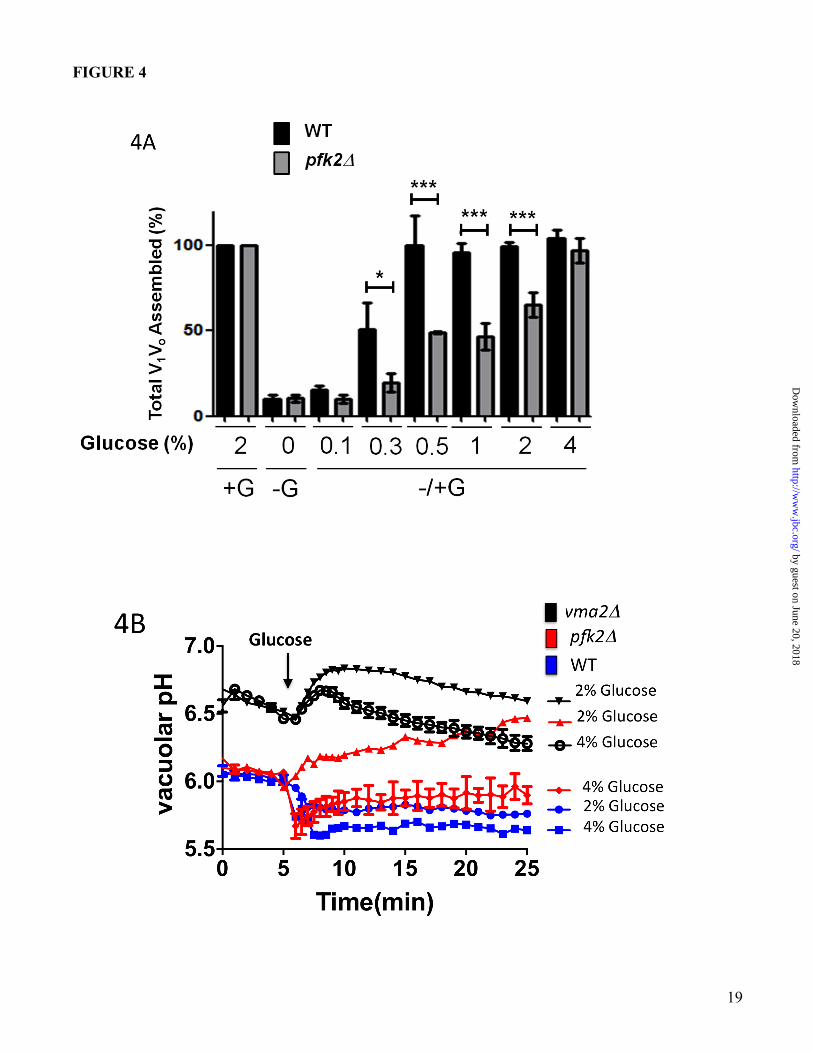
glycolytic flow upon reactivation is responsible for the V1Vo reassembly defects in pfk2Δ. Addition of glucose after nutrient limitation is known to trigger a rapid increase of NADH/NAD+ that inhibits the glycolytic enzyme triose phosphate dehydrogenase (39), and results in a transient peak in NADH levels (42). To monitor the rate of metabolic reactivation when glucose was re-added, NADH was measured by autofluorescence. Re-addition of 2% glucose stimulated NADH synthesis in the wild-type and pfk2Δ cells (Fig. 3B). However, the NADH synthesis rate was reduced by 70% in the pfk2Δ cells. These results are consistent with the ethanol measurements, indicating that glucose-mediated metabolic reactivation was defective in pfk2Δ. We measured metabolic reactivation after adding 4% glucose. Figure 3A and B shows the level of ethanol and NADH formation that resulted from the addition of 4% glucose to pfk2Δ after a brief glucose depletion period. The concentration of ethanol reached a steady state level of 6 mM ± 0.6 mM and 3.8 ± 0.7 mM for the wild type and pfk2Δ, respectively. The rate of NADH synthesis in wild-type and pfk2Δ cells increased by 15%. It reached up to 46% in pfk2Δ cells, which was a larger increase than that observed upon addition of 2% glucose. Thus, doubling the concentration of glucose from 2% to 4% stimulated glucose-dependent metabolic re-activation by about 50% in pfk2Δ, as indicated by ethanol levels and the NADH formation rate. Next, we exposed pfk2Δ to 2% and 4% glucose to manipulate glycolysis and directly establish the role of glycolysis in V-ATPase assembly and activity. 4% glucose rescues glucose-dependent reassembly and vacuolar acidification. If V1Vo reassembly is governed by the glycolysis flow, we anticipated reassembly levels to increase after stimulating glycolysis in pfk2Δ cells. Figure 4A shows the extent of V1Vo reassembly in pfk2Δ, after addition of 4% glucose. Reassembly levels were measured using biosynthetically 35S-radiolabeled cells in pulse-chase experiments as described in Experimental Procedures. The radiolabeled cells were chased in YEP media containing 2% glucose for 20 minutes (steady state condition, + G), YEP media without glucose for 10 minutes (disassembly condition, - G), and after re-addition of varied concentrations of glucose (0.1% - 4%) for an additional 10 minutes (reassembly conditions, ± G). After the chases, the V-ATPase complexes were immunoprecipitated under
by guest on June 20, 2018http://w
ww
.jbc.org/D
ownloaded from

5
nondenaturing conditions with anti-V1 subunit-B to immunoprecipitate V1 and V1Vo, versus anti-Vo subunit-a that can only immunoprecipitate Vo when it is disassembled from V1. About 80% of the V1Vo complexes disassembled upon glucose depletion (Fig. 4A). After glucose re-addition, reassembly was proportional to the concentration of glucose added. However, pfk2Δ required greater concentrations of glucose to reach wild-type levels of reassembly. In the wild-type cells, 50% reassembly was detected after addition of 0.3% glucose; 100% reassembly was reached with 0.5% glucose. In contrast, addition of 0.3% glucose did not trigger significant reassembly in pfk2Δ cells, and 2% glucose led to only 60% reassembly. Approaching steady state levels of assembly (100%) required 4% glucose. Re-addition of 2% glucose, which restored 62% of the steady state ethanol concentration, triggered comparable levels of V1Vo reassembly (60%). Re-addition of 4% glucose, which restored steady state ethanol levels led to steady state reassembly levels (100%). Collectively, these results showed that V1Vo reassembly was proportional to the glycolysis flow in pfk2Δ until the cells reached ≥ 40% of the steady state wild-type ethanol concentration and NADH formation rate. The V1Vo complexes reactivate after reassembly, which acidifies the vacuolar lumen and restores membrane gradients and secondary transport systems (20). To determine whether reassembly restored V-ATPase proton transport, we measured the vacuolar pH after re-addition of 4% glucose to pfk2Δ. Vacuolar acidification was detected within 1-2 minutes of glucose re-addition to the wild-type cells (Fig. 4B), which is within the time-scale when reassembly occurs (3,21). The V1Vo reassembly level was somewhat higher in 4% glucose (Fig. 4A) and the wild-type vacuole pH slightly more acidic. Glucose-induced acidification was defective in pfk2Δ in response to 2% glucose, even though 60% of the V1Vo complexes reassembled in the glycolytic mutant (Fig. 4A). However, V-ATPase proton transport resumed after 4% glucose re-addition. Reactivation of V-ATPase proton transport was indicated by the rapid acidification of the vacuolar lumen. The vma2Δ strain that cannot assemble V1Vo complexes (43) was used as negative control, because vma2Δ completely lacks V-ATPase activity and vacuolar buffering capacity (2). As expected, the net
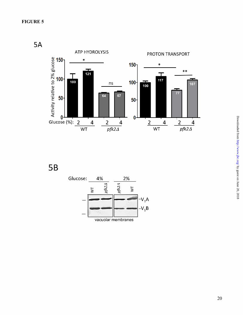
vacuole pH was considerably more alkaline in vma2Δ than wild-type and pfk2Δ cells. A gradual pH drop was detected in vma2Δ at 3 minutes after V-ATPase reactivation (acidification) was complete in the wild-type and pfk2Δ cells (Fig. 4B). This observation further indicated that glucose mediated vacuolar acidification was V-ATPase dependent in the pfk2Δ cells. Proton transport is increased at vacuolar membranes. The rates of proton transport and ATP hydrolysis were measured in purified vacuolar membranes from pfk2Δ cells. The V-ATPase activity was determined in the presence and absence of the specific V-ATPase inhibitor concanamycin A, and the ratio of proton pumping to ATP hydrolysis used as a means of estimating the coupling efficiency of the enzyme when glycolysis was stimulated in pfk2Δ cells. In wild type cells cultured in 4% glucose, ATP hydrolysis and proton transport activities increased by 21% and 17%, respectively, compared to those grown in 2% glucose (Fig. 5A). In contrast, ATP hydrolysis was unchanged in pfk2Δ membranes from cells cultured in 2% versus 4% glucose, which was 64% - 67% of the wild-type ATP hydrolysis. However, proton transport significantly increased (by 30%) when the pfk2Δ cells were grown in 4% glucose from that observed in 2% glucose. As a result, the proton transport/ATP hydrolysis ratio increased 24% in the pfk2Δ mutant (from 0.63 to 0.83). Western blot analyses of vacuolar membrane fractions showed comparable levels of V1 subunit-A and subunit-B in 2% glucose and 4% glucose (Fig. 5B). Based on these results, we concluded that the V-ATPase pumps fine-tuned their catalytic activity in 4% glucose, which enabled pfk2Δ cells to restore vacuolar pH homeostasis. Pfk1p subunit binding to V-ATPase decreases in 4% glucose. The individual PFK-1 subunits expressed in pfk1Δ (subunit Pfk2p) and pfk2Δ (subunit Pfk1p) cells co-immunoprecipitate with V-ATPase and co-purify with vacuolar membrane fractions (24). Thus, deletion of one PFK-1 subunit does not prevent interaction of the other subunit with V-ATPase. We asked if binding of the subunit Pfk1p to V-ATPase was affected in 4% glucose in the pfk2Δ mutant. The level of Pfk1p subunit co-immunoprecipitated with the anti-V1 subunit-A monoclonal antibodies that recognize V1 and V1Vo
by guest on June 20, 2018http://w
ww
.jbc.org/D
ownloaded from
6
complexes (21) was analyzed. About 60% fewer Pfk1p co-immunoprecipiated with V1 subunits in pfk2Δ cells in 4% glucose (Fig. 6). This decrease in Pfk1p-V-ATPase binding was not caused by change in expression of Pfk1p and/or V-ATPase subunits, since these proteins expression levels were comparable in whole cell lysates from 2% and 4% glucose (input, Fig. 6). Rather, Pfk1p-V-ATPase binding was decreased in 4% glucose in pfk2Δ, which was suggestive of lower Pfk1p-V-ATPase binding affinity in this mutant. DISCUSSION This study capitalized on the cellular mechanisms suppressing V-ATPase function in pfk2Δ to gain new knowledge of the mechanisms underlying glucose-dependent V-ATPase regulation. We took advantage of the fact that V-ATPase is fully assembled in pfk2Δ cells at steady state and glycolysis partially suppressed to manipulate the glycolysis flow and assess its direct involvement on V-ATPase function. At steady state, the ratio of proton transport to ATP hydrolysis increases in response to high glucose levels in pfk2Δ. Enhanced V-ATPase proton transport restores vacuolar pH homeostasis. It likely allows cells to preserve energy when glycolysis is suboptimal and glucose abundant (4% glucose). One importance of these findings is that they revealed V-ATPase elasticity of coupling as a new mechanism how glucose regulates V-ATPase pumps without changing the V1Vo assembly state. This study also showed that under V1Vo reassembly conditions the level of glucose-induced reassembly directly corresponds to the glycolysis flow in pfk2Δ cells. V1Vo reassembly is complete after the rate of glycolysis reaches a threshold (40 - 46% of the wild-type rate in 2% glucose) once metabolism resumes after glucose is re-added to glucose-deprived cells. The glycolysis flow communicates with V-ATPase and regulates its activity at steady state. At steady state, in 2% glucose, the ethanol concentration is significantly reduced (by 42%) in the pfk2Δ cells. This level of glycolytic reduction mimics the level of PFK-1 activity reduction (42%) (Fig. 1A) as expected, since PFK-1 catalyzes the first limiting step reaction of the pathway. V-ATPase proton transport is suppressed in 2% glucose in pfk2Δ. Consequently, the vacuolar lumen is alkalinized (Fig. 2A). This alteration in V-ATPase function is not due to V1Vo disassembly nor catalytic
defects, because pfk2Δ cells cultured in 2% glucose display wild-type levels of V1Vo complexes at the membrane and express catalytically competent wild-type V-ATPase pumps (24). Rather vacuolar V-ATPase function is inhibited in vivo in pfk2Δ cells cultured in 2% glucose. This study indicates that V-ATPase activity is suppressed in pfk2Δ, because glycolysis is suppressed. Regulation of V-ATPase activity at steady state by the glycolysis flow has not been reported before. V-ATPase activity is remarkably sensitive to changes in the glycolysis flow. Addition of 4% glucose restores 83% of wild-type ethanol concentration at steady state (Fig. 1B). This significant stimulation of glycolysis also restores the vacuolar acidic pH (Fig. 2A). This result is indicative of a causal correlation between the glycolysis flow and V-ATPase activity. It is evidence that glucose regulates V-ATPase activity at steady state. Accordingly, the PFK-1 subunit deletion mutant, pfk1Δ, that has milder glycolysis defects than pfk2Δ (27) has milder V-ATPase alterations (24). Thus, V-ATPase proton transport is adjusted in response to the glycolytic flow at steady state. V-ATPase adjusts catalytic activity in response to glucose when glycolysis is suboptimal. V-ATPase catalytic coupling is tighter in pfk2Δ cells in 4% glucose (Fig. 5A), when glycolysis operates below capacity at steady state. The proton transport/ATPase ratio increases by 24%, indicating that V-ATPase is more efficient (transports more protons per ATP hydrolyzed) probably to help cells to preserve energy. Importantly, these findings indicate that ATP hydrolysis and proton transport by V-ATPase pumps may not always be optimally coupled in vivo. Using the pfk2Δ cells this study showed that glucose can control V-ATPase activity without changing the V-ATPase assembly state. Until now, glucose-dependent regulation of yeast V-ATPase has been via V1Vo disassembly and reassembly in response to glucose depletion and re-addition, respectively (3). Although the ability of V-ATPase to change coupling of ATP hydrolysis and proton pumping has been described before (44), the involvement of glucose and glycolysis has not been reported. Increased efficiency of coupling was observed in some genetic mutants (45) and when V-ATPase pumps were exposed to low ATP concentrations in vitro (46). In yeast, the N-end domain of Vo subunit-a (47,48) and a non-homologous region of the V1 subunit-A (45) have been implicated in this type of regulation.
by guest on June 20, 2018http://w
ww
.jbc.org/D
ownloaded from
7
Changes in the proton transport to ATPase activity ratio may be linked to ‘loose’ conformations required for disassembly and reassembly, because during catalysis, relative rotation of V1 and Vo subunits requires stable V1Vo assembly. Accordingly, catalytic slip does not occur in the molecular motors that do not reversibly disassemble, such as the F1Fo ATP synthase. One possibility is that in 4% glucose, V1Vo intermediary structure(s) that slip during catalysis are stabilized in pfk2Δ to restore the membrane pH gradients necessary for an array of other transporters when glucose concentration is high (20). Complete V1Vo reassembly after glucose re-addition requires the glycolysis flow to reach a threshold. Glucose depletion, which inhibits glycolysis, leads to V1 dissociation from Vo; and its re-addition, which resumes glycolysis, causes V1 to re-associate with Vo (3). Thus, yeast V-ATPase assembly is intimately linked to the glycolysis flow. We reasoned that if a metabolic input drives V1Vo reassembly, it would reach a certain threshold within the first minutes after glucose re-addition to glucose-deprived cells. Below this threshold, reassembly is defective. We found the threshold to be 40 - 46% of the wild-type glycolytic capacity. We have chosen to monitor the glycolytic intermediate NADH to establish the metabolic threshold for reassembly after glucose re-addition. The NADH formation rate corresponds to the rate of glycolysis. In addition, NADH auto-fluorescence is measureable in intact cells at real-time. The wild-type NADH formation rate in 2% glucose is used as reference for these studies, because 2% is the standard glucose concentration in yeast growth media. When 2% glucose is re-added, the NADH formation rate is only 31% of the wild-type rate. Under these conditions the pfk2Δ cells cannot sufficiently reassemble V1Vo. Addition of 4% glucose that stimulates the rate of glycolysis to 46% (the proposed NADH threshold), prompts wild-type levels of V1Vo reassembly in pfk2Δ (Fig. 4A). Likewise, V1Vo reassembly is complete when the cells produce ≥40% the wild-type ethanol levels (Fig. 3A). With reassembly, V1Vo proton transport resumes and the vacuoles are acidified after 1-2 minute of 4% glucose re-addition to pfk2Δ. The finding that V1Vo remains silenced in pfk2Δ after re-addition of 2% glucose, even though 60% of the V-ATPase complexes are reassembled is intriguing.
It can be explained if inhibitory MgADP is trapped in the catalytic sites (49), since glycolysis is significantly suppressed. Alternatively, a regulatory subunit such as V1 subunit-H that silences V1 after disassembly (49) and activates V1Vo complexes at the membrane (50), could have retained an inhibitory conformation preventing proton transport in 2% glucose (51,52). Although glycolytic ATP formation is an attractive possibility to signal reassembly and/or reactivation in 4% glucose, the levels of ATP during glucose deprivation and re-addition do not appear to correlate to the V-ATPase assembly state (21). Wild-type yeast cells recover steady state ATP concentrations in the absence of glucose (21). How does PFK-1 regulates V-ATPase activity? The finding that V-ATPase function is fully rescued in 4% glucose in pfk2Δ cells that lack the PFK2 gene, argues against direct regulation of V-ATPase through its physical interaction with the PFK-1 subunit Pfk2p. However, the PFK-1 subunit Pfk1p may play an inhibitory role in pfk2Δ cells. The level of Pfk1p that co-immunoprecipitates with V-ATPase subunits is significantly reduced in pfk2Δ cells in 4% glucose (Fig. 6). This decrease can be explained if Pfk1p is recruited to support greater glycolytic demands in 4% glucose, after which V-ATPase proton transport is enhanced. Whether binding of subunit Pfk1p to V-ATPase is inhibitory in an interesting possibility that will be addressed in future studies. This study showed that at least one mechanism how PFK-1 modulates V-ATPase is via the glycolysis flow. The RAS/cAMP/PKA signaling pathway, which has been linked to glucose-dependent V1Vo disassembly and reassembly, also is intertwined with PFK-1. Activation of the RAS/cAMP/PKA pathway enhances the glycolysis flow, because PKA stimulates formation of fructose-2,6-bisphosphate, the most potent activator of PFK-1. Thus, RAS/cAMP/PKA could control V-ATPase assembly via glycolysis (16). We cannot eliminate the possibility that glucose-dependent pH changes also could regulate V-ATPase. Cytosol alkalinization is glucose concentration dependent and correlates to the level of V1Vo assembly (14,53). A larger ΔpH between the cytosolic and the luminal sides of the vacuolar membrane may stimulate V1Vo activity. Similar to the activating effect that ΔpH has on the Fo subunit-a of the evolutionary related F-ATP synthase (54),
by guest on June 20, 2018http://w
ww
.jbc.org/D
ownloaded from

8
binding of protons to the membrane-bound Vo subunit-a of the V-ATPase could prime V1Vo proton transport. In Arabidopsis, the V-ATPase coupling ratios are sensitive to the cytosol and vacuolar pH (55). In the lemon fruit, variable coupling and pH-dependent slip regulate the V-ATPase pump (56). Clearly, the scope of glucose-dependent V-ATPase regulation is more complex than initially anticipated. It extends beyond V1Vo disassembly and/or reassembly and is intimately linked to the metabolic state of a cell, specifically the glycolysis flow. The finding that V-ATPase changes catalytic efficiency when the cellular demands for membrane transport increase (4% glucose) and glycolytic ATP production operates below capacity, can have implications in human health. Particularly, for distal renal tubular acidosis (25), viral infections (57) and the metabolic switch in cancers (58,59) where glucose-dependent regulation of V-ATPase is conserved. EXPERIMENTAL PROCEDURES Materials and strains. Tran[35S]-label was from MP Biomedicals (Santa Ana, CA). The antibodies anti-Myc, anti-phosphoglycerate kinase-1 (22C5D8), anti-V-ATPase (10D7 and 13D11) were from Invitrogen (Carlsbad, CA). Alkaline phosphatase-conjugated secondary antibodies were from Promega (Madison, WI) and horseradish peroxidase secondary antibodies from Invitrogen (Carlsbad, CA). Zymolase 100T was purchased from Seikagaku (Tokyo, Japan), concanamycin A from Wako Biochemicals (Richmond, VA), and ficoll and carbonyl cyanide m-chlorophenyl hydrazone (CCCP) from United Stated Biologicals (Swampscott, MA). The ATP assay kit K354-100 was purchased from Biovision (Milpitas, CA) and ethanol assay kit ab65343 from Abcam (Cambridge, MA). All other reagents were from Sigma (St. Louis, MO). The Saccharomyces cerevisiae strains referred to throughout are BY4742 wild-type (MATα; his3Δ1; leu2 Δ; lys2Δ; ura3Δ), BY4742 pfk1 (pfk1Δ:KanMX6), BY4742 pfk2Δ (pfk2Δ:KanMX6), and BY4742 vma3Δ (vm3Δ:KanMX6). The mutant strains pfk2Δ and pfk1Δ were verified by PCR using primers to amplify the PFK2 and PFK1 genes with 5’-XhoI and 3’-XmaI cutting sites. Growth phenotype. Cells were grown overnight to 0.6 – 1.0 Abs600/mL in YEPD medium buffered to pH 5.0 with 50 mM succinic acid/50 mM sodium phosphate. Cultures were washed twice with sterile ddH2O and an optical density of 2.5 Abs600 cells
resuspended into 1 mL sterile ddH2O. Ten-fold serial dilutions were stamped onto YEP containing 2% glucose and 4% glucose. Plates were buffered to pH 5.0 with 50 mM succinic acid/50 mM sodium phosphate, pH 7.5 with 50 mM MES/50 mM MOPS, or pH 7.5 with 100 mM calcium chloride added. The plates were incubated for 72 hours at 30 oC and 37 oC. Vacuolar pH. Vacuolar pH was measured using the ratiometric fluorescent dye BCECF-AM (31,60). Cells were grown to an optical density of 0.6 – 1.0 Abs600/mL and stained with 50µM BCECF-AM for 30 minutes at 30oC. Calibration curves were generated as described before (20) in calibration buffers pH 5.5, 5.75, 6.0, 6.25, 6.5, 7 consisting of 50 mM MES, 50 mM HEPES, 50 mM KCl, 50 mM NaCl, 200 mM ammonium acetate, 10 mM sodium azide, 10 mM 2-deoxyglucose, 50 µM CCCP. For steady state analyses of the vacuolar pH, the BCECF stained cells were transferred to 1 mM HEPE/MES pH 5.0 buffer with 2% or 4% glucose (final concentration) and the BCECF fluorescence (ratio of 490nm/450 nm; 525 excitation) measured for 10 minutes at 30oC in a FluoroMax 4 spectrofluorometer (Horiba Jobin Yvon Inc., NJ). Data collected between 3 minutes and 8 minutes was averaged. For V-ATPase disassembly reassembly conditions, BCECF stained cells deprived of glucose on ice for 10 min and the vacuolar pH was monitored 20 min after 2% or 4% glucose re-addition (final concentration). Immunoprecipitations. Pulse-chase experiments were conducted at the indicated glucose concentrations and times following protocols described before (21). The V-ATPase complexes were immunoprecipitated from whole cell lysates with the monoclonal antibodies 13D11 (anti-V1 subunit B) and 10D7 (anti-Vo subunit a, Vph1p isoform) and the protein separated by SDS-PAGE (13% acrylamide gels). The gels were dried, scanned in a Fuji Scanner FLA-5100, and analyzed using the Multi Gauge and GraphPad Prism 5 software as previously described (24). The proportion of Vo assembled into V1Vo, determined by comparing the amount of Vo subunit-a immunoprecipitated with 13D11 to the total amount of Vo subunit-a immunoprecipitated with both antibodies. For non-radiolabeled immunoprecipitations, the 13D11 antibody was used to immunoprecipate V-ATPase from whole cell lysates (61). Protein was separated by SDS-PAGE in 10% gels and analyzed by Western blots using the monoclonal antibodies 8B1 (anti-V1 subunit-A) and 13D77 (anti-V1 subunit-B) and yeast PFK-1 polyclonal antibodies. The nitrocellulose
by guest on June 20, 2018http://w
ww
.jbc.org/D
ownloaded from
9
membranes were blotted with horseradish peroxidase secondary antibody and scanned using a Bio-Rad ChemiDoc XRS+ and the intensity of protein bands quantified using the Multi Gauge and GraphPad Prism 5 software. NADH autofluorescence. NADH was monitored as described by Poulsen et al (42) with the following modifications. The cells were grown overnight to an optical density of 0.6 – 1.0 Abs600/mL in YEPD, harvested, and an optical density of 100 Abs600 resuspended in 50 mM potassium phosphate buffer pH 6.8 up to a density of 10% by weight. The cells were starved from glucose by incubation in the same phosphate buffer for 3 hours on a rotary shaker at 30oC and placed on ice for 10 minutes. The NADH fluorescence intensity (excitation at 366 nm; emission at 450 nm) was monitored without glucose for 60 seconds and then continuously recorded for an additional 90 seconds with re-addition of 2% glucose or 4% glucose at 30oC in a FluoroMax 4 spectrofluorometer (Horiba Jobin Yvon Inc., NJ). Ethanol concentration. For steady state analyses, the cells were cultured overnight to an optical density of 0.6 – 1.0 Abs600/mL in YEP pH 5.0 media (wild-type and pfk2Δ) containing 2% or 4% final glucose concentration. A total of 2.0 optical density Abs600/mL per condition were harvested and the cells converted to sheroplasts by zymolase treatment (35). The spheroplasts were incubated at 30 oC for 10 min in YEP adjusted to pH 5.0 with 50 mM succinic acid/50 mM sodium phosphate containing 2% or 4% glucose plus 1.2 M sorbitol. The ethanol concentration was measured using the Ethanol Assay Kit ab65343 (Abcam, Cambridge, MA, US) according to the manufacturer instructions. For glucose-depletion and re-addition analyses, the cells were grown in media containing 2% glucose
overnight, converted to spheroplasts as described above, and the spheroplasts incubated in YEP media with 2% or 4% glucose for 20 minutes, in YEP for 10 minutes, and in YEP for 10 minutes followed by addition of 2% glucose, 4% glucose or 2% 2-deoxyglucose for 10 minutes. PFK-1 enzymatic activity. Wild-type and pfk2Δ cells were cultured overnight to an optical density of 0.6 – 1.0 Abs600/mL in YEPD pH 5.0 media. Cells were converted to spheroplasts and lysed in 15 mM MES-Tris pH 6.9 containing 5% glycerol at a final concentration of 1.0 optical density Abs600/µL. PFK-1 activity was measured spectrophotometrically at 37 °C using the coupled enzyme assay of Lotscher et al. (62). Whole cell lysates (20.0 optical density Abs600) was added to the enzymatic assay mixture (25 mM Tris pH 6.9, 2 mM ATP, 5 mM MgCl2, 2 mM phosphoenol pyruvate, 30 Units/mL pyruvate kinase, 30 Units/mL L-lactic dehydrogenase, 0.5 mM NADH), and the reaction started by addition of 5 mM fructose-6-phospate. NADH oxidation was monitored spectrophotometrically at 340 nm for 5 min. One unit of PFK-1 activity is defined as 1 µmol F1,6BP formed /min in an optical density of 20 Abs600
Other methods. Vacuolar membranes fractions were purified by ficoll density gradient centrifugation as described before (35,48,61). Protein concentration was measured by the Bradford assay (63). ATP hydrolysis was measured by monitoring NADH oxidation spectrophotometrically (62) using 5µg of total vacuolar membrane protein in the presence and absence of 100 nM concanamycin-A. Proton transport was measured by monitoring 9-amino-6-chloro-2-methoxyacridin (ACMA) quenching after addition of MgATP (48).
Acknowledgments−We thank Dr. Jurgen Heinisch (Osnabrueck) for generously providing the PFK-1 polyclonal antibodies. We thank Dr. Wayne Frasch for the helpful discussions. Conflict of Interest: The authors declare that they have no conflicts of interest with the contents of this article. Authors Contribution: KJP conceived and coordinated the study, analyzed results, and wrote the paper. CYC designed, performed, and analyzed the experiments. DRD performed PFK1 activity measurements.
by guest on June 20, 2018http://w
ww
.jbc.org/D
ownloaded from
10
REFERENCES
1. Forgac, M. (2007) Vacuolar ATPases: rotary proton pumps in physiology and pathophysiology. Nature reviews. Molecular cell biology 8, 917-929
2. Kane, P. M. (2006) The where, when, and how of organelle acidification by the yeast vacuolar H+-ATPase. Microbiology and molecular biology reviews : MMBR 70, 177-191
3. Parra, K. J., Chan, C. Y., and Chen, J. (2014) Saccharomyces cerevisiae vacuolar H+-ATPase regulation by disassembly and reassembly: one structure and multiple signals. Eukaryotic cell 13, 706-714
4. Marshansky, V., Rubinstein, J. L., and Gruber, G. (2014) Eukaryotic V-ATPase: novel structural findings and functional insights. Biochimica et biophysica acta 1837, 857-879
5. Cotter, K., Stransky, L., McGuire, C., and Forgac, M. (2015) Recent Insights into the Structure, Regulation, and Function of the V-ATPases. Trends in biochemical sciences 40, 611-622
6. Breton, S., and Brown, D. (2013) Regulation of luminal acidification by the V-ATPase. Physiology (Bethesda) 28, 318-329
7. Hinton, A., Bond, S., and Forgac, M. (2009) V-ATPase functions in normal and disease processes. Pflugers Archiv : European journal of physiology 457, 589-598
8. Muench, S. P., Trinick, J., and Harrison, M. A. (2011) Structural divergence of the rotary ATPases. Quarterly reviews of biophysics 44, 311-356
9. Zhao, J., Benlekbir, S., and Rubinstein, J. L. (2015) Electron cryomicroscopy observation of rotational states in a eukaryotic V-ATPase. Nature 521, 241-245
10. Kane, P. M. (1995) Disassembly and reassembly of the yeast vacuolar H(+)-ATPase in vivo. The Journal of biological chemistry 270, 17025-17032
11. Sumner, J. P., Dow, J. A., Earley, F. G., Klein, U., Jager, D., and Wieczorek, H. (1995) Regulation of plasma membrane V-ATPase activity by dissociation of peripheral subunits. The Journal of biological chemistry 270, 5649-5653
12. Liberman, R., Bond, S., Shainheit, M. G., Stadecker, M. J., and Forgac, M. (2014) Regulated Assembly of Vacuolar ATPase Is Increased during Cluster Disruption-induced Maturation of Dendritic Cells through a Phosphatidylinositol 3-Kinase/mTOR-dependent Pathway. The Journal of biological chemistry 289, 1355-1363
13. Trombetta, E. S., Ebersold, M., Garrett, W., Pypaert, M., and Mellman, I. (2003) Activation of lysosomal function during dendritic cell maturation. Science 299, 1400-1403
14. Dechant, R., Binda, M., Lee, S. S., Pelet, S., Winderickx, J., and Peter, M. (2010) Cytosolic pH is a second messenger for glucose and regulates the PKA pathway through V-ATPase. The EMBO journal 29, 2515-2526
15. Dechant, R., Saad, S., Ibanez, A. J., and Peter, M. (2014) Cytosolic pH regulates cell growth through distinct GTPases, Arf1 and Gtr1, to promote Ras/PKA and TORC1 activity. Molecular cell 55, 409-421
16. Bond, S., and Forgac, M. (2008) The Ras/cAMP/protein kinase A pathway regulates glucose-dependent assembly of the vacuolar (H+)-ATPase in yeast. The Journal of biological chemistry 283, 36513-36521
17. Oot, R. A., Huang, L. S., Berry, E. A., and Wilkens, S. (2012) Crystal structure of the yeast vacuolar ATPase heterotrimeric EGC(head) peripheral stalk complex. Structure 20, 1881-1892
18. Tabke, K., Albertmelcher, A., Vitavska, O., Huss, M., Schmitz, H. P., and Wieczorek, H. (2014) Reversible disassembly of the yeast V-ATPase revisited under in vivo conditions. The Biochemical journal 462, 185-197
19. Kane, P. M. (2016) Proton Transport and pH Control in Fungi. Advances in experimental medicine and biology 892, 33-68
by guest on June 20, 2018http://w
ww
.jbc.org/D
ownloaded from

11
20. Martinez-Munoz, G. A., and Kane, P. (2008) Vacuolar and plasma membrane proton pumps collaborate to achieve cytosolic pH homeostasis in yeast. The Journal of biological chemistry 283, 20309-20319
21. Parra, K. J., and Kane, P. M. (1998) Reversible association between the V1 and V0 domains of yeast vacuolar H+-ATPase is an unconventional glucose-induced effect. Molecular and cellular biology 18, 7064-7074
22. Lu, M., Ammar, D., Ives, H., Albrecht, F., and Gluck, S. L. (2007) Physical interaction between aldolase and vacuolar H+-ATPase is essential for the assembly and activity of the proton pump. The Journal of biological chemistry 282, 24495-24503
23. Lu, M., Sautin, Y. Y., Holliday, L. S., and Gluck, S. L. (2004) The glycolytic enzyme aldolase mediates assembly, expression, and activity of vacuolar H+-ATPase. The Journal of biological chemistry 279, 8732-8739
24. Chan, C. Y., and Parra, K. J. (2014) Yeast phosphofructokinase-1 subunit Pfk2p is necessary for pH homeostasis and glucose-dependent vacuolar ATPase reassembly. The Journal of biological chemistry 289, 19448-19457
25. Su, Y., Blake-Palmer, K. G., Sorrell, S., Javid, B., Bowers, K., Zhou, A., Chang, S. H., Qamar, S., and Karet, F. E. (2008) Human H+ATPase a4 subunit mutations causing renal tubular acidosis reveal a role for interaction with phosphofructokinase-1. American journal of physiology. Renal physiology 295, F950-958
26. Banaszak, K., Mechin, I., Obmolova, G., Oldham, M., Chang, S. H., Ruiz, T., Radermacher, M., Kopperschlager, G., and Rypniewski, W. (2011) The crystal structures of eukaryotic phosphofructokinases from baker's yeast and rabbit skeletal muscle. Journal of molecular biology 407, 284-297
27. Heinisch, J. (1986) Construction and physiological characterization of mutants disrupted in the phosphofructokinase genes of Saccharomyces cerevisiae. Current genetics 11, 227-234
28. Arvanitidis, A., and Heinisch, J. J. (1994) Studies on the function of yeast phosphofructokinase subunits by in vitro mutagenesis. The Journal of biological chemistry 269, 8911-8918
29. Heinisch, J. (1986) Isolation and characterization of the two structural genes coding for phosphofructokinase in yeast. Molecular & general genetics : MGG 202, 75-82
30. Schroder, T. D., Ozalp, V. C., Lunding, A., Jernshoj, K. D., and Olsen, L. F. (2013) An experimental study of the regulation of glycolytic oscillations in yeast. The FEBS journal 280, 6033-6044
31. Brett, C. L., Tukaye, D. N., Mukherjee, S., and Rao, R. (2005) The yeast endosomal Na+K+/H+ exchanger Nhx1 regulates cellular pH to control vesicle trafficking. Molecular biology of the cell 16, 1396-1405
32. Ohya, Y., Umemoto, N., Tanida, I., Ohta, A., Iida, H., and Anraku, Y. (1991) Calcium-sensitive cls mutants of Saccharomyces cerevisiae showing a Pet- phenotype are ascribable to defects of vacuolar membrane H(+)-ATPase activity. The Journal of biological chemistry 266, 13971-13977
33. Kane, P. M. (2007) The long physiological reach of the yeast vacuolar H+-ATPase. Journal of bioenergetics and biomembranes 39, 415-421
34. Liu, J., and Kane, P. M. (1996) Mutational analysis of the catalytic subunit of the yeast vacuolar proton-translocating ATPase. Biochemistry 35, 10938-10948
35. Owegi, M. A., Carenbauer, A. L., Wick, N. M., Brown, J. F., Terhune, K. L., Bilbo, S. A., Weaver, R. S., Shircliff, R., Newcomb, N., and Parra-Belky, K. J. (2005) Mutational analysis of the stator subunit E of the yeast V-ATPase. The Journal of biological chemistry 280, 18393-18402
36. Curtis, K. K., Francis, S. A., Oluwatosin, Y., and Kane, P. M. (2002) Mutational analysis of the subunit C (Vma5p) of the yeast vacuolar H+-ATPase. The Journal of biological chemistry 277, 8979-8988
by guest on June 20, 2018http://w
ww
.jbc.org/D
ownloaded from

12
37. Smardon, A. M., and Kane, P. M. (2014) Loss of vacuolar H+-ATPase activity in organelles signals ubiquitination and endocytosis of the yeast plasma membrane proton pump Pma1p. The Journal of biological chemistry 289, 32316-32326
38. Smets, B., Ghillebert, R., De Snijder, P., Binda, M., Swinnen, E., De Virgilio, C., and Winderickx, J. (2010) Life in the midst of scarcity: adaptations to nutrient availability in Saccharomyces cerevisiae. Current genetics 56, 1-32
39. Kresnowati, M. T., van Winden, W. A., Almering, M. J., ten Pierick, A., Ras, C., Knijnenburg, T. A., Daran-Lapujade, P., Pronk, J. T., Heijnen, J. J., and Daran, J. M. (2006) When transcriptome meets metabolome: fast cellular responses of yeast to sudden relief of glucose limitation. Molecular systems biology 2, 49
40. Lenzen, S. (2014) A fresh view of glycolysis and glucokinase regulation: history and current status. The Journal of biological chemistry 289, 12189-12194
41. Ibanez, A. J., Fagerer, S. R., Schmidt, A. M., Urban, P. L., Jefimovs, K., Geiger, P., Dechant, R., Heinemann, M., and Zenobi, R. (2013) Mass spectrometry-based metabolomics of single yeast cells. Proceedings of the National Academy of Sciences of the United States of America 110, 8790-8794
42. Poulsen, A. K., Andersen, A. Z., Brasen, J. C., Scharff-Poulsen, A. M., and Olsen, L. F. (2008) Probing glycolytic and membrane potential oscillations in Saccharomyces cerevisiae. Biochemistry 47, 7477-7484
43. Doherty, R. D., and Kane, P. M. (1993) Partial assembly of the yeast vacuolar H(+)-ATPase in mutants lacking one subunit of the enzyme. The Journal of biological chemistry 268, 16845-16851
44. Nelson, N. (2003) A journey from mammals to yeast with vacuolar H+-ATPase (V-ATPase). Journal of bioenergetics and biomembranes 35, 281-289
45. Shao, E., Nishi, T., Kawasaki-Nishi, S., and Forgac, M. (2003) Mutational analysis of the non-homologous region of subunit A of the yeast V-ATPase. The Journal of biological chemistry 278, 12985-12991
46. Arai, H., Pink, S., and Forgac, M. (1989) Interaction of anions and ATP with the coated vesicle proton pump. Biochemistry 28, 3075-3082
47. Kawasaki-Nishi, S., Bowers, K., Nishi, T., Forgac, M., and Stevens, T. H. (2001) The amino-terminal domain of the vacuolar proton-translocating ATPase a subunit controls targeting and in vivo dissociation, and the carboxyl-terminal domain affects coupling of proton transport and ATP hydrolysis. The Journal of biological chemistry 276, 47411-47420
48. Chan, C. Y., Prudom, C., Raines, S. M., Charkhzarrin, S., Melman, S. D., De Haro, L. P., Allen, C., Lee, S. A., Sklar, L. A., and Parra, K. J. (2012) Inhibitors of V-ATPase proton transport reveal uncoupling functions of tether linking cytosolic and membrane domains of V0 subunit a (Vph1p). The Journal of biological chemistry 287, 10236-10250
49. Parra, K. J., Keenan, K. L., and Kane, P. M. (2000) The H subunit (Vma13p) of the yeast V-ATPase inhibits the ATPase activity of cytosolic V1 complexes. The Journal of biological chemistry 275, 21761-21767
50. Ho, M. N., Hirata, R., Umemoto, N., Ohya, Y., Takatsuki, A., Stevens, T. H., and Anraku, Y. (1993) VMA13 encodes a 54-kDa vacuolar H(+)-ATPase subunit required for activity but not assembly of the enzyme complex in Saccharomyces cerevisiae. The Journal of biological chemistry 268, 18286-18292
51. Diab, H., Ohira, M., Liu, M., Cobb, E., and Kane, P. M. (2009) Subunit interactions and requirements for inhibition of the yeast V1-ATPase. The Journal of biological chemistry 284, 13316-13325
52. Jefferies, K. C., and Forgac, M. (2008) Subunit H of the vacuolar (H+) ATPase inhibits ATP hydrolysis by the free V1 domain by interaction with the rotary subunit F. The Journal of biological chemistry 283, 4512-4519
by guest on June 20, 2018http://w
ww
.jbc.org/D
ownloaded from
13
53. Parra, K. J., and Kane, P. M. (1996) Wild-type and mutant vacuolar membranes support pH-dependent reassembly of the yeast vacuolar H+-ATPase in vitro. The Journal of biological chemistry 271, 19592-19598
54. Moore, K. J., and Fillingame, R. H. (2008) Structural interactions between transmembrane helices 4 and 5 of subunit a and the subunit c ring of Escherichia coli ATP synthase. The Journal of biological chemistry 283, 31726-31735
55. Rienmuller, F., Dreyer, I., Schonknecht, G., Schulz, A., Schumacher, K., Nagy, R., Martinoia, E., Marten, I., and Hedrich, R. (2012) Luminal and cytosolic pH feedback on proton pump activity and ATP affinity of V-type ATPase from Arabidopsis. The Journal of biological chemistry 287, 8986-8993
56. Muller, M. L., Jensen, M., and Taiz, L. (1999) The vacuolar H+-ATPase of lemon fruits is regulated by variable H+/ATP coupling and slip. The Journal of biological chemistry 274, 10706-10716
57. Kohio, H. P., and Adamson, A. L. (2013) Glycolytic control of vacuolar-type ATPase activity: a mechanism to regulate influenza viral infection. Virology 444, 301-309
58. Fogarty, F. M., O'Keeffe, J., Zhadanov, A., Papkovsky, D., Ayllon, V., and O'Connor, R. (2013) HRG-1 enhances cancer cell invasive potential and couples glucose metabolism to cytosolic/extracellular pH gradient regulation by the vacuolar-H ATPase. Oncogene
59. Sennoune, S. R., and Martinez-Zaguilan, R. (2012) Vacuolar H(+)-ATPase signaling pathway in cancer. Current protein & peptide science 13, 152-163
60. Ali, R., Brett, C. L., Mukherjee, S., and Rao, R. (2004) Inhibition of sodium/proton exchange by a Rab-GTPase-activating protein regulates endosomal traffic in yeast. The Journal of biological chemistry 279, 4498-4506
61. Ediger, B., Melman, S. D., Pappas, D. L., Jr., Finch, M., Applen, J., and Parra, K. J. (2009) The tether connecting cytosolic (N terminus) and membrane (C terminus) domains of yeast V-ATPase subunit a (Vph1) is required for assembly of V0 subunit d. The Journal of biological chemistry 284, 19522-19532
62. Lotscher, H. R., deJong, C., and Capaldi, R. A. (1984) Modification of the F0 portion of the H+-translocating adenosinetriphosphatase complex of Escherichia coli by the water-soluble carbodiimide 1-ethyl-3-[3-(dimethylamino)propyl]carbodiimide and effect on the proton channeling function. Biochemistry 23, 4128-4134
63. Bradford, M. M. (1976) A rapid and sensitive method for the quantitation of microgram quantities of protein utilizing the principle of protein-dye binding. Analytical biochemistry 72, 248-254
FOOTNOTES
*This work was supported by National Institutes of Health Grant R01GM086495 (to KJP), American Heart Association Grant 14PRE19020015 (to CYC). 1Department of Biochemistry and Molecular Biology, School of Medicine, University of New Mexico, Albuquerque, New Mexico, 87131 §To whom correspondence should be addressed: Karlett J. Parra, Department of Biochemistry and Molecular Biology, School of Medicine, University of New Mexico, MSC08 4670, Albuquerque, NM 87131. Tel: (505) 272-1633; Fax: (505) 272-6587; Email: [email protected] 2Current Address: Van Andel Institute, Grand Rapids, MI 49503 3Important abbreviations used are: V-ATPase, vacuolar proton-translocating ATPase; yeast, S. cerevisiae; vma, vacuolar membrane ATPase; PFK-1, 6-phosphofructo-1-kinase; Pfk2p, phosphofructokinase-1 subunit β; BCECF-AM, 2′,7′-Bis(2-carboxyethyl)-5(6)-carboxyfluorescein acetoxymethyl ester.
by guest on June 20, 2018http://w
ww
.jbc.org/D
ownloaded from
14
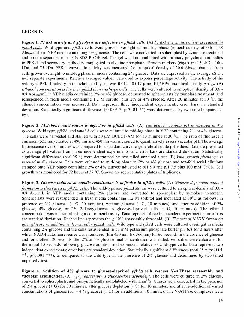
LEGENDS Figure 1. PFK-1 activity and glycolysis are defective in pfk2Δ cells. (A) PFK-1 enzymatic activity is reduced in pfk2Δ cells. Wild-type and pfk2Δ cells were grown overnight to mid-log phase (optical density of 0.6 - 0.8 Abs600/mL) in YEP media containing 2% glucose. The cells were converted to spheroplast by zymolase treatment and protein separated on a 10% SDS-PAGE gel. The gel was immunoblotted with primary polyclonal antibodies to PFK-1 and secondary antibodies conjugated to alkaline phosphate. Protein markers (right) are 150-kDa, 100-kDa, and 75-kDa. PFK-1 enzymatic activity was measured for an optical density of 20.0 Abs600 obtained from cells grown overnight to mid-log phase in media containing 2% glucose. Data are expressed as the average ±S.D.; n=3 separate experiments. Relative averaged values were used to express percentage activity. The activity of the wild-type PFK-1 activity in the whole cell lysate was 0.014 - 0.017 µmol F1,6BP/min/optical density Abs600. (B) Ethanol concentration is lower in pfk2Δ than wild-type cells. The cells were cultured to an optical density of 0.6 - 0.8 Abs600/mL in YEP media containing 2% or 4% glucose, converted to spheroplasts by zymolase treatment, and resuspended in fresh media containing 1.2 M sorbitol plus 2% or 4% glucose. After 20 minutes at 30 oC, the ethanol concentration was measured. Data represent three independent experiments; error bars are standard deviation. Statistically significant differences (p<0.05 *, p<0.01 **) were determined by two-tailed unpaired t-test. Figure 2. Metabolic reactivation is defective in pfk2Δ cells. (A) The acidic vacuolar pH is restored in 4% glucose. Wild type, pfk2Δ, and vma3Δ cells were cultured to mid-log phase in YEP containing 2% or 4% glucose. The cells were harvested and stained with 50 µM BCECF-AM for 30 minutes at 30 oC. The ratio of fluorescent emission (535 nm) excited at 490 nm and 450 nm was measured to quantitatively assess vacuolar pH. The average fluorescence over 6 minutes was compared to a standard curve to generate absolute pH values. Data are presented as average pH values from three independent experiments, and error bars are standard deviation. Statistically significant differences (p<0.05 *) were determined by two-tailed unpaired t-test. (B) Vma- growth phenotype is rescued in 4% glucose. Cells were cultured to mid-log phase in 2% or 4% glucose and ten-fold serial dilutions stamped onto YEP plates containing 2% or 4% glucose adjusted to pH 5.0 and pH 7.5 plus 100 mM CaCl2. Cell growth was monitored for 72 hours at 37 oC. Shown are representative plates of triplicates. Figure 3. Glucose-induced metabolic reactivation is defective in pfk2Δ cells. (A) Glucose-dependent ethanol formation is decreased in pfk2Δ cells. The wild-type and pfk2Δ strains were cultured to an optical density of 0.6 - 0.8 A600/mL in YEP media containing 2% glucose and converted to spheroplast by zymolase treatment. Spheroplasts were resuspended in fresh media containing 1.2 M sorbitol and incubated at 30oC as follows: in presence of 2% glucose (+ G, 20 minutes), without glucose (− G, 10 minutes), and after re-addition of 2% glucose, 4% glucose, or 2% 2-deoxyglucose to glucose-deprived cells (± G, 10 minutes). The ethanol concentration was measured using a colorimetric assay. Data represent three independent experiments; error bars are standard deviation. Dashed line represents the ≥ 40% reassembly threshold. (B) The rate of NADH formation after glucose re-addition is decreased in pfk2Δ cells. Wild type and pfk2Δ cells were cultured overnight in media containing 2% glucose and the cells resuspended in 50 mM potassium phosphate buffer pH 6.8 for 3 hours after which NADH autofluorescence was monitored (Em 450 nm; Ex 366 nm) for 60 seconds in the absence of glucose and for another 120 seconds after 2% or 4% glucose final concentration was added. Velocities were calculated for the initial 15 seconds following glucose addition and expressed relative to wild-type cells. Data represent two independent experiments; error bars are standard deviation. Statistically significant differences (p<0.05 *, p<0.01 **, p<0.001 ***), as compared to the wild type in the presence of 2% glucose and determined by two-tailed unpaired t-test. Figure 4. Addition of 4% glucose to glucose-deprived pfk2Δ cells rescues V-ATPase reassembly and vacuolar acidification. (A) V1Vo reassembly is glucose-dose dependent. The cells were cultured in 2% glucose, converted to spheroplasts, and biosynthetically radiolabeled with Tran35S. Chases were conducted in the presence of 2% glucose (+ G) for 20 minutes, after glucose depletion (- G) for 10 minutes, and after re-addition of varied concentrations of glucose (0.1 - 4 % glucose) (± G) for an additional 10 minutes. The V-ATPase complexes were
by guest on June 20, 2018http://w
ww
.jbc.org/D
ownloaded from
15
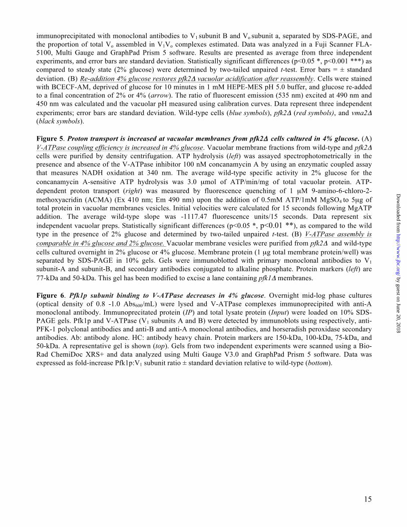
immunoprecipitated with monoclonal antibodies to V1 subunit B and Vo subunit a, separated by SDS-PAGE, and the proportion of total Vo assembled in V1Vo complexes estimated. Data was analyzed in a Fuji Scanner FLA-5100, Multi Gauge and GraphPad Prism 5 software. Results are presented as average from three independent experiments, and error bars are standard deviation. Statistically significant differences (p<0.05 *, p<0.001 ***) as compared to steady state (2% glucose) were determined by two-tailed unpaired t-test. Error bars = ± standard deviation. (B) Re-addition 4% glucose restores pfk2Δ vacuolar acidification after reassembly. Cells were stained with BCECF-AM, deprived of glucose for 10 minutes in 1 mM HEPE-MES pH 5.0 buffer, and glucose re-added to a final concentration of 2% or 4% (arrow). The ratio of fluorescent emission (535 nm) excited at 490 nm and 450 nm was calculated and the vacuolar pH measured using calibration curves. Data represent three independent experiments; error bars are standard deviation. Wild-type cells (blue symbols), pfk2Δ (red symbols), and vma2Δ (black symbols). Figure 5. Proton transport is increased at vacuolar membranes from pfk2Δ cells cultured in 4% glucose. (A) V-ATPase coupling efficiency is increased in 4% glucose. Vacuolar membrane fractions from wild-type and pfk2Δ cells were purified by density centrifugation. ATP hydrolysis (left) was assayed spectrophotometrically in the presence and absence of the V-ATPase inhibitor 100 nM concanamycin A by using an enzymatic coupled assay that measures NADH oxidation at 340 nm. The average wild-type specific activity in 2% glucose for the concanamycin A-sensitive ATP hydrolysis was 3.0 µmol of ATP/min/mg of total vacuolar protein. ATP-dependent proton transport (right) was measured by fluorescence quenching of 1 µM 9-amino-6-chloro-2-methoxyacridin (ACMA) (Ex 410 nm; Em 490 nm) upon the addition of 0.5mM ATP/1mM MgSO4 to 5µg of total protein in vacuolar membranes vesicles. Initial velocities were calculated for 15 seconds following MgATP addition. The average wild-type slope was -1117.47 fluorescence units/15 seconds. Data represent six independent vacuolar preps. Statistically significant differences (p<0.05 *, p<0.01 **), as compared to the wild type in the presence of 2% glucose and determined by two-tailed unpaired t-test. (B) V-ATPase assembly is comparable in 4% glucose and 2% glucose. Vacuolar membrane vesicles were purified from pfk2Δ and wild-type cells cultured overnight in 2% glucose or 4% glucose. Membrane protein (1 µg total membrane protein/well) was separated by SDS-PAGE in 10% gels. Gels were immunoblotted with primary monoclonal antibodies to V1 subunit-A and subunit-B, and secondary antibodies conjugated to alkaline phosphate. Protein markers (left) are 77-kDa and 50-kDa. This gel has been modified to excise a lane containing pfk1Δ membranes. Figure 6. Pfk1p subunit binding to V-ATPase decreases in 4% glucose. Overnight mid-log phase cultures (optical density of 0.8 -1.0 Abs600/mL) were lysed and V-ATPase complexes immunoprecipited with anti-A monoclonal antibody. Immunoprecitated protein (IP) and total lysate protein (Input) were loaded on 10% SDS-PAGE gels. Pfk1p and V-ATPase (V1 subunits A and B) were detected by immunoblots using respectively, anti-PFK-1 polyclonal antibodies and anti-B and anti-A monoclonal antibodies, and horseradish peroxidase secondary antibodies. Ab: antibody alone. HC: antibody heavy chain. Protein markers are 150-kDa, 100-kDa, 75-kDa, and 50-kDa. A representative gel is shown (top). Gels from two independent experiments were scanned using a Bio-Rad ChemiDoc XRS+ and data analyzed using Multi Gauge V3.0 and GraphPad Prism 5 software. Data was expressed as fold-increase Pfk1p:V1 subunit ratio ± standard deviation relative to wild-type (bottom).
by guest on June 20, 2018http://w
ww
.jbc.org/D
ownloaded from


Chun-Yuan Chan, Dennis R. Dominguez and Karlett J. ParraCells
Regulation of V-ATPase Reassembly by the Glycolysis Flow in PFK-1 Deficient Yeast
published online May 23, 2016J. Biol. Chem.
10.1074/jbc.M116.717488Access the most updated version of this article at doi:
Alerts:
When a correction for this article is posted•
When this article is cited•
to choose from all of JBC's e-mail alertsClick here
by guest on June 20, 2018http://w
ww
.jbc.org/D
ownloaded from