regulation parathyroid hormone release and cytosolic...
TRANSCRIPT

Regulation of Parathyroid Hormone Release and Cytosolic Calciumby Extracellular Calcium in Dispersed and Cultured Bovineand Pathological Human Parathyroid CellsMeryl S. LeBoff, Dolores Shoback, Edward M. Brown, Joseph Thatcher, Ronald Leombruno, Denise Beaudoin,Marguerite Henry, Richard Wilson, Johanna Pallotta, Samuel Marynick, John Stock, and George LeightEndocrine-Hypertension Unit and Departments of Medicine and Surgery, Brigham and Women's Hospital, Harvard Medical School,Boston, Massachusetts 02115; Division of Endocrinolqgy, Beth Israel Hospital (J.P.), Boston, Massachusetts 02115;Departments of Internal Medicine and Pathology, Baylor University Medical Center (S.M.), Dallas, Texas 75246;Division of Endocrinology, Memorial Hospital (J.S.), Worcester Massachusetts 01605; and Department of Surgery,Duke University Medical Center (G.L), Durham, North Carolina 27710
Abstract
Alterations in parathyroid glandular sensitivity to calcium maycontribute to the hypersecretion of PTH in hyperparathyroidism.Since the cytosolic calcium concentration may mediate theeffects of extracellular calcium on PTH release, we haveemployed the calcium-sensitive intracellular dye QUIN-2 toexamine the relationship between extracellular calcium, cytosoliccalcium, and PTH secretion in adult, neonatal, and culturedbovine as well as pathological human parathyroid cells. PTHrelease was measured using C- and N-terminal radioimmu-noassays. Neonatal bovine parathyroid cells showed a greaterset-point for secretion (the Ca" concentration causing half ofthe maximal inhibition of PTH release) than adult cells(1.27±0.11 vs. 1.06±0.11 mMextracellular calcium, P < 0.01)and a slightly higher extracellular calcium was necessary toraise the cytosolic calcium concentration to a given level inneonatal than in adult bovine parathyroid cells. In individualneonatal and adult cell preparations, there was a close corre-lation between the set-point for secretion and the "set-point"for cytosolic calcium (r = 0.832, P < 0.001). In cells from fivehuman parathyroid adenomas, which had an increase in set-point for secretion, the extracellular calcium concentrationnecessary to raise the cytosolic calcium concentration to agiven level was slightly greater than in the neonatal cells. Infour preparations of human parathyroid cells there was asignificant correlation between the set-points for secretion andcytosolic calcium (r = 0.856, P < 0.01). Because neonatal bovineand pathological human parathyroid glands show cellular hy-perplasia, we studied the temporal relationship between cellularproliferation and the regulation of PTH release and cytosoliccalcium concentration in cultured bovine parathyroid cells.Cellular proliferation, estimated by 3H-thymidine incorporation,
This paper was presented in part at the 5th Annual Meeting of theAmerican Society for Bone and Mineral Research, San Antonio, TX,1983, abstract A51, and at the National Meeting of the AmericanFederation for Clinical Research, Washington, DC, 1984, abstract269A.
Address reprint requests to Dr. LeBoff, Brigham & Women'sHospital.
Received for publication 19 April 1984 and in revised form 31August 1984.
increased significantly in culture from 104±10.1 counts/wellon day 1 (first 24 h in culture) to 588±188 and 6,156±649counts/well on days 2 and 4, respectively. In cultured cells onday 1, high Ca" (2-3 mM) inhibited maximal PTH releaseby 58.8±3.2%, which decreased significantly (P <0.001) to38.2±1.9 and 17.1±3.7% on days 2 and 4, respectively. Thecytosolic calcium observed at 3 mMcalcium on day 1 was701±43 nM, which declined to 466±60 and 314±14 nM ondays 2 and 4 (P < 0.05). There was a close correlation betweenthis progressive decrease in maximal inhibition of PTI4 releaseand the cytosolic calcium at high extracellular calcium incultured cells (r = 0.99, P < 0.001). Thus, during activeproliferation of cultured cells, there is an alteration in theregulation of cytosolic calcium and PTH release by extracellularcalcium. These results suggest that decreased sensitivity to theinhibitory effects of extracellular calcium on PTHrelease mayresult from a low cytosolic calcium at a given extracellularcalcium concentration, and changes in the regulation of PTHrelease and cytosolic calcium by extracellular calcium may berelated to enhanced cellular proliferation.
Introduction
The normal parathyroid cell shows an inverse, sigmoidalrelationship between parathyroid hormone (PTH)' secretionand the extracellular ionized calcium concentration (1-3). Ina variety of settings, there may be changes in the sensitivity or"set-point" (the calcium concentration half-maximally sup-pressing PTH release) of the parathyroid gland for calcium.For example, parathyroid tissue from hypercalcemic, neonatalcalves shows an increase in set-point for calcium bdth in vivo(4) and in vitro (2). In primary hyperparathyroidism, patho-logical parathyroid tissue shows an increase in set-point thatmay contribute to the hypercalcemia in this disorder (3).Parathyroid cells maintained in culture for 3-4 d also developreduced sensitivity to the suppressive effects of calcium onparathyroid hormone release (5).
The elevated set-point for calcium in primary hyperpara-thyroidism may be reduced toward normal with the divalentcation ionophore A23187 (6). Moreover, increased extracellularpotassium concentrations, which may decrease cytosolic cal-cium through an alteration in Na'-Ca+ exchange, promotean increase in set-point in bovine parathyroid cells (7). Thus,indirect evidence suggests that changes in set-point may be
1. Abbreviation used in this paper: PTH, parathyroid hormone.
Regulation of Parathyroid Hormone Release and Cytosolic Calcium 49
J. Clin. Invest.C The American Society for Clinical Investigation, Inc.0021-9738/85/01/0049/09 $ 1.00Volume 75, January 1985, 49-57

related to alterations in the regulation of the cytosolic calciumconcentration. The recent development of the calcium-sensitiveintracellular dye QUIN-2 has made it feasible to determinedirectly the cytosolic calcium concentration in dispersed para-thyroid cells (8). In the present studies, we have used QUIN-2 to examine whether alterations in parathyroid cellular sen-sitivity to calcium might be related to changes in the relationshipbetween the extracellular and cytosolic calcium concentrations.The results suggest that decreased sensitivity to the suppressiveeffects of extracellular calcium on PTH release may resultfrom an inappropriately low cytosolic calcium concentrationat a given extracellular calcium concentration, and that changesin the regulation of PTH secretion and cytosolic calcium byextracellular calcium may be related to enhanced cellularproliferation.
Methods
Dispersed bovine and human parathyroid cells were prepared bydigestion with collagenase and DNase as described previously (3, 9).Parathyroid cells prepared in this fashion maintain high ratios ofcellular potassium to cellular sodium (7) and have high levels of ATP(Brown, E. M., unpublished data). Moreover, PTH release is linear forseveral hours and is inhibited by up to 80% by calcium concentrationssimilar to those which inhibit PTH secretion in vivo (3). Dispersedparathyroid cells contained no visible fat cells by sudan staining or incytocentrifuge preparations, and showed >95% exclusion of trypanblue.
Dispersed bovine parathyroid cells were cultured for 4 d as recentlydescribed (5): cells were prepared under sterile conditions and platedonto fibronectin-coated cluster wells (2 or 4 cm2; Costar, Cambridge,MA) in DMEM-F12medium with 5 ug/ml insulin, 15% newborn calfserum, and antibiotics (5). Plating efficiency is =34% under theseconditions and the cells approach a confluent monolayer after 3-4 d.Cultured cells were removed from the plates with 0.25% trypsin (GibcoLaboratories, Grand Island, NY) in Hank's balanced salt solution with1.0 mMCaCl2 and 0.5 mMMgSO4.
For loading with QUIN-2, parathyroid cells (20 X 106/ml) wereincubated with the acetoxymethyl ester of QUIN-2 (10-20 MM) (Lan-caster Synthesis, Ltd., Lancaster, England) at 37°C for 20 min inEagle's minimal essential medium (Earle's salts with bicarbonate, Ca++and Mge' deleted) supplemented with 0.02 MHepes (adjusted to pH7.47 with NaOH), 0.2% bovine serum albumin (BSA), 1.0 mMCaCI2,and 0.5 mMMgSO4("standard medium") (8). The cellular suspensionwas then diluted 10-fold with standard medium and incubated for anadditional 20-60 min. The cellular pellet was washed three times witha saline solution that contained 0.025 MHepes (adjusted to pH 7.47with NaOH), 125 mMNaCl, 5 mMKCl, I mMCaC12, 0.5 mMMgSO4, 1 gm/L dextrose, I mMNa2HPO4, and 0.1% BSA ("saline").The washed cellular pellet was resuspended in 3 ml of the salinesolution with 0.1% BSA for determination of cellular fluorescence at acellular concentration of 7-10 X 106 cells/ml. Fluorescence was mon-itored in thermostatted cuvettes (37°C) in a Perkin-Elmer MPF3spectrofluorimeter (excitation 339 nm, emission 492 nM). Cellularsuspensions were mechanically stirred during all experiments, exceptwhen recording was interrupted for 10-20 s during the addition ofCaC12. Extracellular calcium was raised by the addition of 0.15 MCaC12; direct determination of calcium concentrations in these solutionsby atomic absorption spectrometry showed that they were within 3%of the calculated value. Fluorescence was monitored for 3-10 minafter the addition of CaC12, or until the signal was stable as specifiedbelow. The calibration of cellular fluorescence (F) at the end of astudy was achieved by cellular lysis with the detergent Triton X-100(0.06-0.12%, vol/vol) in 21 mMcalcium (F,,.), and after addition of10 mMEGTAand alkalinization with I M Tris base (free calcium
I nM, Farm) to render the pH > 8.3 (10). Cytosolic calcium was
then calculated from the equation: [Ca"+] = 115 nM (F -F,,)I(F.-F) (10).
The dissociation constant for QUIN-2 used in this calculation (1 15nM) assumes a cytosolic free Mge' which has been arbitrarily assigneda value of 1 mM(10). Corrections were made for any changes in theautofluorescence of unloaded parathyroid cells at the same cell concen-tration due to the additives used in each experiment.
PTH release was assessed by incubation of QUIN-2-loaded cells(1-2 X 10⁶ cells/ml) in 0.2-0.3 ml of saline at varying extracellularcalcium concentrations in parallel with studies of cellular fluorescencein 5 ml polypropylene scintillation vials (Sarstedt, Inc., Princeton, NJ)at 370C for 1-2 h. Supernatant samples for determination of PTHwere frozen after sedimentation of the cellular pellet at 250 g for 2min in a desk-top centrifuge (GLC-2B, Ivan Sorvall, Inc., Norwalk,CT). We previously showed that QUIN-2-loaded neonatal bovineparathyroid cells do not differ from unloaded cells with respect totrypan blue exclusion, cellular levels of ATP and K+, and calcium-regulated PTH release (8). In the present studies, maximal rates ofPTH release at low calcium concentrations in QUIN-2-loaded adultbovine, cultured bovine, and pathological human parathyroid cellswere 85% or more of those in unloaded cells, which suggests thatloading with QUIN-2 did not adversely affect cellular viability in thesecells as well.
Radioimmunoassay for PTH in samples from incubations withbovine parathyroid cells was performed in duplicate on triplicateincubation vials as previously described (9), using an anti-bovine PTHantiserum raised in a sheep, GW-l, which recognizes intact hormoneand C-terminal fragments of PTH. In studies with human parathyroidcells, PTH release was determined using an N-terminal antiserum(CK-13) raised in a chicken against hPTH(1-34) (generously donatedby Dr. G. V. Segre, Massachusetts General Hospital, Boston, MA).Samples or standards [hPTH(1-34) or bPTH(l-84)] were incubated at4°C for 24 h with the antiserum (final dilution 1:100,000) in 0.05 Mbarbital, pH 8.5, with 1:6 (vol/vol) outdated human plasma. '25bPTH(I-84) was then added and the incubation continued for another 3-5 dat 4°C. Tracer was purchased from Cambridge Nuclear Corp. (Billeria,MA) and was purified by high performance liquid chromatography(I 1) before use. Bound and free hormone were separated by precipitationwith polyethylene glycol (9). Supernatants from incubations withdispersed human parathyroid cells generally diluted in parallel withboth hPTH(1-34) and bPTH(1-84), and results were calculated interms of either peptide. In some assays, bPTH(1-84) displaced'25IbPTH(1-84) with a flatter slope than that for hPTH(1-34) or theincubation samples. In these cases, results were expressed in terms ofhPTH(1-34). The reasons for the variability of the bPTH(1-84) indisplacing tracer are unknown. bPTH(44-68) and bPTH(53-84) didnot displace 1251-bPTH(l-84) at concentrations as high as 100 ng/tube.
Cellular proliferation was determined using 3H-thymidine incor-poration into DNA(12). After washing the cells three times in standardmedium, the dispersed and cultured cells in cluster wells (2 cm2,Costar, Rochester, NY) were incubated with I MCi/ml of 3H-thymidine(New England Nuclear, Boston, MA) in standard medium at 37°C forI h. After sedimentation of the dispersed cells in centrifuge tubes, andaspiration of the medium from the cultured and dispersed cells, thecells were washed on ice with Dulbecco's phosphate-buffered saline(Gibco Laboratories, Grand Island, NY). The cells were then incubatedin 5% cold TCA for 15 min. After sedimentation of the dispersed cellsand aspiration of the TCA from the dispersed and cultured cells, thecells were dissolved in a total of 500 M1 of 1 N NaGHand the culturedcells were then removed from the culture plates with a rubber policemanand transferred to centrifuge tubes. After the addition of 375 ML of50% TCAand 60 Mul of BSA (6 mgBSA/ml sterile water) and vortexing,the precipitate was centrifuged for 5 min at 2,500 rpm in a desk-topcentrifuge. The resulting pellet was washed two times with 5% TCAand resuspended in 250 Ml of 0. 1 N NaOH. The incorporation of 3H-thymidine into TCA insoluble radioactivity was then determined usingliquid scintillation spectrometry.
Cellular levels of calmodulin were determined by radioimmunoassay
50 LeBoff el al.

(Amersham Corp., Arlington Heights, IL) after extraction of cells witha buffer containing 125 mMborate, I mMEGTA, and 75 mMsodiumchloride, pH 8.4. Purified porcine calmodulin was employed as thestandard in this assay because it has been previously characterized inour laboratory (13). Results obtained using this standard were similarto those obtained employing the standard provided with the radioim-munoassay kit, which is extracted from rat testes. Cellular protein wasquantified using the Biorad protein method (5).
Statistical evaluation. Means have been presented with the SEMas the index of dispersion. Statistical significance was determined withthe t test, or regression analysis and logarithmic transformation whereapplicable. The null hypothesis was rejected when a P value of <0.05was obtained.
Results
Effect of extracellular calcium on PTH release and cytosoliccalcium concentration in bovine parathyroid cells. Fig. 1 Ashows the effects of varying extracellular calcium concentrationson PTH release from QUIN-2-loaded parathyroid cells fromneonatal calves and adult cows. The data for cells fromneonatal calves are similar to those shown previously (8) andare included for comparison with those for cells from cowswhich were studied concurrently. Maximal suppression ofPTH release was comparable with adult and neonatal parathy-roid tissue (70 vs. 71%, respectively). Parathyroid cells fromneonatal calves showed a greater maximal secretory rate atlow calcium (0.5-1-mM concentrations) than those from cows(5.8±0.4 vs. 2.6±0.6 ng/105 cells/h, respectively, P < 0.001),as well as a higher set-point for calcium (1.27±0.1 1 vs.1.06±0.11 mMextracellular Ca", respectively, P < 0.01).
When QUIN-2-loaded parathyroid cells from neonatal andadult parathyroid tissue were resuspended in 0.5 mMcalcium
PTHRELEASE A Figure 1. (A) The effects ofvaried extracellular calcium
100- concentrations on PTH release3 \ \T in QUIN-2-loaded parathyroid
75- \ \cells from neonatal calves andadult cows. Incubation of para-
KS so- N \thyroid cells and measurementR +ofPTH release were carried
25 out as described in Methods.Each point represents the
0 mean±SEMfor 18-24 obser-800 CYTOSOL/C[CO*7 B vations from 6-8 experiments,
each assayed in duplicate. PTH600 - [OAdit(-6F / release was normalized to per-
I *NL t(N8) centage of maximal release; ab-:IZ400 wsolute secretory rates are given100 in Results. (B) The effects of
varied extracellular calcium200- concentrations on the cytosolic
calcium concentrations in neo-natal and adult bovine para-
0 0.5 1.0 1.5 2ao thyroid cells. Parathyroid cellsEXRCLUA]m
were loaded with the acetoxy-methylester of QUIN-2 (10-20 ,uM) as described in Methods. Thecells were washed, and cellular fluorescence of the parathyroid cellsuspension (7-10 X 106 cells/ml) was determined as extracellularcalcium was raised by 0.25-mM increments by the addition of 0.15MCaC12. Cellular fluorescence was calibrated at the end of theexperiments and cytosolic calcium was calculated as described inMethods. The results represent the mean±SEMfor 6-8 observationsfrom 3 to 4 experiments.
and exposed to progressively higher calcium concentrations,there was a stepwise increase in cellular fluorescence (notshown). When cytosolic free calcium concentrations werecalculated as described in Methods, there was a correspondingrise in cytosolic calcium concentrations with increasing levelsof extracellular calcium (Fig. 1 B). Cytosolic calcium increasedmore at lower levels of extracellular calcium in cells fromadult than in those from neonatal parathyroid tissue, althoughthis difference was not statistically significant by t testing.
The relationship between the effect of extracellular calciumon cytosolic calcium and PTH release in neonatal and adultparathyroid tissue. The results shown in Fig. 1 suggested thatchanges in the sensitivity of the secretory process to extracellularcalcium might be related to alterations in the relationshipbetween the extracellular and cytosolic calcium concentrations.To investigate this possibility further, the set-point for secretionwas plotted against the cytosolic calcium concentration at theset-point in a series of cell preparations from neonatal andadult bovine parathyroid tissue (Fig. 2 A). The cytosoliccalcium concentration associated with half-maximal inhibitionof secretion was independent of the set-point for PTH release,which suggests that inhibition of hormonal secretion occurredat similar cytosolic calcium concentrations regardless of theextracellular calcium concentration necessary to modify theseparameters. For each cell preparation, the set-point for secretionwas then plotted against the "set-point" for cytosolic calciumor the extracellular calcium concentration necessary to achievethe average cytosolic calcium concentration associated withthe secretory set-point for all the cell preparations (i.e., 308nM, see Fig. 2 A) (Fig. 2 B). There was a close correlation(r = 0.832, P < 0.001) between these parameters for cellpreparations from neonatal and adult bovine parathyroidtissue with set-points ranging from 0.93 to 1.48 mM.
Effect of varying extracellular calcium concentrations oncytosolic calcium concentration and PTHrelease in pathologicalhuman parathyroid tissue. Fig. 3 shows the relationship betweenthe extracellular and cytosolic calcium concentrations in dis-persed parathyroid cells from 12 adenomas and five glandsfrom four patients with uremic secondary hyperparathyroidism.In cells from these pathological human parathyroid glands, theextracellular calcium concentration necessary to raise thecytosolic calcium concentration to a given level was comparableto or greater than that for neonatal bovine parathyroid cells.
For studies on the effects of extracellular calcium on PTHrelease in human parathyroid tissue, an N-terminal antiserumwas employed to minimize the interpretational difficultiesinherent with the use of antisera recognizing inactive,C-terminal fragments of the hormone. The characteristics ofthis assay are described in Methods. Preliminary studies withbovine parathyroid cells showed that the set-point for secretionwas identical when measured with the N- and C-terminalassays (1.2+0.045 and 1.2±0.054 mM, respectively, n = 3),although maximal suppressibility was slightly greater with theN-assay (70±4.5 vs. 54±1.5%).
PTH release as a function of the extracellular calciumconcentration was measured in dispersed parathyroid cellsfrom five adenomas and three glands from patients withuremic secondary hyperparathyroidism. Set-points using theN-terminal assay for the adenomas and tissue showing second-ary hyperplasia were comparable to those we have foundpreviously using a C-terminal assay (1.33+0.15 vs. 1.26+0.13
Regulation of Parathyroid Hormone Release and Cytosolic Calcium 51

1200
0000 0
0
I
-,~600
(t3
j~4000)
L.Z0
- -- Adul Bovine
n-u Adnom(nz1216 2* HyWo°s(nW5)
)F
200[-
0 0.5 1.0 1.5 2.0 2.5EXTRACELWLAR(C?7.mM
0.9 1.0 1.1 1.2 1.3 1.4 1.5SET-PO/NTFORPTHSECRETIOAV, mMEC[Cotl
Figure 3. The relationship between the extracellular and cytosoliccalcium concentrations in human parathyroid cells from 12 adeno-mas and five glands from four patients with uremic secondary hyper-parathyroidism. Cellular loading with QUIN 2 and measurement andcalibration of fluorescence were carried out as described above.
0
0
0
0
0
R=Q832P<0.0o1
0 0.9 1.0 1.1 1.2 1.3 1.4 1.5i7 -PO/NTF()? CYTCJSL/C IC; r/, CfCO71
Figure 2. (A) The relationship between the set-point for secretion(calcium concentration causing half-maximal inhibition of PTH re-
lease) and the cytosolic calcium concentration at the secretory set-point in cell preparations from neonatal (.) and adult (o) bovineparathyroid tissue. The average cytosolic calcium associated with thesecretory set-point was 308 mM. (B) The relationship between theset-point for secretion and the extracellular calcium concentrationnecessary to achieve the average cytosolic calcium concentrationassociated with the secretory set-point (308 mM)("set-point forcytosolic calcium") in neonatal and adult bovine parathyroid cells.The secretory set-points ranged from 0.93 to 1.48 mM. There was a
close correlation between these parameters (r = 0.832, P < 0.001).
and 1.22±0.03 vs. 1.17±0.19, respectively) (14). Figs. 4 and 5show a comparison between the effects of extracellular calciumon cytosolic calcium and PTH secretion in cell preparationsfrom four adenomas. In three, changes in PTH release andcytosolic calcium occurred at similar levels of extracellularcalcium (Fig. 4). In one cell preparation, however, despiteextracellular calcium-induced increases in cytosolic calciumcomparable to those with adult bovine parathyroid tissue,there was markedly impaired suppression of PTH release(Fig. 5).
In four cell preparations in which there was >50% suppres-
sion of PTH secretion by 2-3 mMcalcium, the cytosoliccalcium concentration at which PTH release was half-maximally
suppressed averaged 304 nM, nearly identical to that forbovine parathyroid cells (308 nM; see above). In addition, as
with adult and neonatal bovine tissue, there was a significantcorrelation between the set-point for secretion and the extra-cellular calcium concentration necessary to raise the cytosoliccalcium concentration to the average value at which PTHrelease was half-maximally suppressed (r = 0.856, P < 0.01)(Fig. 6).
Effect of extracellular calcium on PTHsecretion and cyto-solic calcium concentration in cultured bovine parathyroid cellsas a function of the culture interval. We recently found thatbovine parathyroid cells proliferating in culture over 3-4 dlose normal suppressibility of PTH release by extracellularcalcium similar to the most severe abnormalities in parathyroidadenomas (5). Because this parathyroid cell culture systemmight provide a model for investigating the changes in para-
thyroid function in hyperparathyroidism, we examined therelationship between the regulation of PTH release and cytosolic
, A Figure 4. The relationshipS 6 between the effect of extra-
< cellular calcium on cyto-50 K4 solic calcium and PTH re-
oo-o200 lease in cells from three
I_cV parathyroid adenomas.*i1a E s , , i,s600i PTH release (mean±SEM)
be s xo was determined as de-
% f_]4004 scribed in Methods after in-50-S , cubation at 370C for 1-2 h.
~~~~~-ok Radioimmunoassay forPTH
was performed in du-
go i} OKO~t plicate on triplicate incuba-
loo tion vials using an N-termi-VC X -10o nal assay as described in
\z/
Methods. Cytosolic calcium50 _ was measured after the cellshI \40 were loaded with 10-20
I_ I__ ' G MQUIN-2 as described
0 0.5 1.0 1.5 zo'm in Fig. 1. Maximal rates of£XTRACELLLLAR[CC"*, mMM
PTH release were 5.2, 2.7,and 0.93 ng/bPTH (1-84) equivalent/105 cells per h in A, B, and C,respectively.
52 LeBoffet al.
700
t3500
o- 400
. 300
I-.
0
200-
1.I
i1.4
~1.1.2
kh 1.1
kk 1.0
ZosRl 9.0.
A

I
lb
bi.50k.zQ.
10
8(
it4'
EXTRACELLULAR[C47", mM
Figure 5. The relationship between the effect of extracellular calciumon cytosolic calcium and PTH release in one parathyroid adenoma.PTH release and cytosolic calcium were determined as describedabove. PTH release was inhibited by 29% of maximal and thecytosolic calcium concentration increased to 565 nM at 2 mMextra-cellular calcium. The maximal rate of PTH release was 183 pg hPTH(1-34) equivalent/105 cells per h.
calcium by extracellular calcium in cultured cells. Fig. 7 Ademonstrates the effect of extracellular calcium on PTH secre-tion in QUIN-2-loaded, cultured neonatal bovine parathyroidcells as a function of the culture interval, and in acutelydispersed, neonatal bovine parathyroid cells for comparison(8). The sequential changes start with day 1, or the first 24 hin culture. Similar to acutely dispersed cells, which display a50-75% inhibition of maximal PTH release at high extracellularcalcium (2-3 mM), cultured parathyroid cells on day 1 showeda 58.8±3.22% inhibition of maximal PTH release (n = 12)(Fig. 7 A). The set-point in cultured cells on day 1 was 1.35mMcalcium, slightly higher than the value of 1.30 mMcalcium observed with acutely dispersed cells. As culturedparathyroid cells proliferated in culture, however, the percentageof maximal inhibition of PTH release decreased to *38.2±1.86%(n = 14) on day 2 and then to *17.1±3.73% on day 4 as thecells approached confluency (*P < 0.001 compared with day
0
/V. I.N 1.1 I.C[ l.J 1.m 4SET-POI/r FORCY7CSO/CCin *1,nmlE'C I
Figure 6. The relationship between the set-point for secretion and theset-point for cytosolic calcium (the extracellular calcium concentra-tion necessary to raise the cytosolic calcium concentration to theaverage value at which PTH release was half-maximally suppressed)in four parathyroid adenomas. There was a close correlation(r = 0.856, P < 0.01) between these two parameters (solid line). Thedashed line indicates the relationship between these parameters forbovine parathyroid cells.
60C
200
- I
CY7mSC /CG7* B
0.5 1.0 1.5 20 2.5 30EMRAAELULAR[Co7**n,?
Figure 7. (A) The effect of extracellular calcium on maximal PTHrelease in dispersed and cultured neonatal bovine parathyroid cells asa function of the culture interval. PTH release (percentage of maxi-mal) was determined in QUIN-2-loaded parathyroid cells as de-scribed in Methods. Absolute secretory rates are given in Results.Each point represents the mean±SEMfrom 6 to 30 observationseach assayed in duplicate on triplicate incubation vials. (B) The effectof extracellular calcium on cytosolic calcium concentration in dis-persed and cultured bovine parathyroid cells as a function of theculture interval. Cytosolic calcium was measured as described inMethods. Each point represents the mean±SEMfrom 4 to 13 experi-ments.
1). While acutely dispersed, neonatal cells showed a maximalsecretory rate of 5.8±0.4 ng PTH/105 cells/h (n = 30), themaximal secretory rate in cultured cells removed from cultureplates with trypsin (see Methods) was 3.38±0.215, 2.60±0.279,and 4.16±0.474 ng PTH/105 cells per h on days 1, 2, and 4,respectively (n = 6-15). Because maximal hormonal secretionwas less than previously reported in cultured cells incubatedon the culture plates compared with those exposed to trypsin(9.3±0.8 vs. 4.16±0.474 ng PTH/105 cells/h on day 4) (5), westudied the effects of high calcium (2-3 mM)on the inhibitionof maximal PTH release in cells on the culture plates andthose exposed to trypsin. In cultured cells in culture wells vs.those trypsinized and incubated in scintillation vials, thepercentage of maximal suppression of PTH secretion at highcalcium was *55.5±2.06 vs. 58.8±3.22% on day 1, *38.6±4.45vs. 38.2±1.86% on day 2, and, as shown previously,*19.44±4.18 vs. 17.1±3.73% on day 4 (*P < 0.05, n = 12-24) (5). Thus, while trypsinization may have contributed tothe reduced maximal secretory rate observed here, it had noeffect on the sensitivity of the parathyroid cells to calcium.
Regulation of Parathyroid Hormone Release and Cytosolic Calcium 53
0-
0-
0-
0-
)L
0
F-R
-
-XC

To assess whether the progressive reduction in the sensitivityof PTH release to extracellular calcium in cultured cells wasdue to a change in the relationship between the extracellularand cytosolic calcium concentrations, we measured the cytosoliccalcium concentration in cultured cells. The effects of variedextracellular calcium concentrations on the cytosolic free cal-cium concentrations in cultured cells studied sequentially fromdays 1-4 are shown in Fig. 7 B. The cytosolic calciumconcentrations of dispersed parathyroid cells at varied extra-cellular calcium concentrations are also included in Fig. 7 Bfor comparison. On day 1, before the development of thereduced sensitivity of PTH secretion to extracellular calciumin cultured parathyroid cells, basal cytosolic calcium concen-tration decreased from 179±7.6 nM in dispersed to 106±8.5nM in cultured cells (P < 0.001), and there was a shift in therelationship between extracellular and cytosolic calcium con-centrations to the right, which indicated that a higher extra-cellular calcium concentration was necessary to raise thecytosolic calcium concentration to a level comparable to thatin the dispersed cells (Fig. 7 B). At 3 mMextracellular calcium,however, the cultured cells on day 1 achieved a cytosoliccalcium concentration of 701±43.1 nM (n = 4), which wasnot significantly different from the value of 646±68 nMobserved in dispersed cells at 2 mMextracellular calcium (P> 0.05). With the decline in calcium sensitivity of PTH releaseto extracellular calcium in cultured cells on day 2 and subse-quently on day 4, the cytosolic calcium concentration achievedat high extracellular calcium (3 mM) was *466±59.7 nM onday 2 and **314±13.5 nM on day 4 of the culture interval(*P < 0.05 and **P < 0.001, compared with day 1). Usinglinear regression analysis, the progressive decrease in thepercentage of maximal inhibition of PTH release in culturedcells correlated with the reduced rise in cytosolic calciumconcentration at 3 mMextracellular calcium from day 1 today 4 (r = 0.99, P < 0.001).
To exclude the possibility that the reduced rise in cytosolicfree calcium concentration was a consequence of a change inthe amount of the calcium-binding protein calmodulin, wemeasured the amount of calmodulin corrected for cellularprotein in cellular extracts of cultured cells from day 1 to day4. The amount of calmodulin in cultured cells on days 1, 2,and 4 was 4.6±1.2, 4.1±0.3, and 3.6±0.3 ug/mg of protein,respectively (n = 6-7, P > 0.05).
Cellular proliferation in dispersed and in cultured bovineparathyroid cells as a function of the culture interval. Toexamine the association between changes in cellular prolifera-tion and the regulation of PTH release and cytosolic calciumby extracellular calcium, we studied 3H-thymidine incorporationinto DNAin acutely dispersed and cultured bovine parathyroidcells as a function of the culture period. The 3H-thymidineincorporation into DNA in acutely dispersed cells was35.7±5.24 counts2 (n = 23). As demonstrated in Fig. 8, DNAsynthesis increased significantly in cultured bovine parathyroidcells, resulting in an increase in 3H-thymidine incorporationfrom *104±10.1 counts/well on day 1 to **588±188 counts/well on day 2 and *6,156±649 counts/well on day 4 (*P<0.001, and **P < 0.01, compared with acutely dispersed
2. 3H-Thymidine incorporation in acutely dispersed cells was measuredin 170,000 cells which corresponded to the number of viable cells perwell after 15-17 h in culture.
Figure 8. 3H-thymidine in-Bovine PbrothwodCOflS corporation into DNAin
6000 - 0 AcuelDsred acutely dispersed cells2 andCultured cultured bovine parathyroid
3000 / cells (counts [cpmj/well) as
a function of days in cul-ture. The cells were incu-bated with I MCi/ml of3H-thymidine in standardmedium at 37°C for 1 h.
R _W gThe incorporation of 3H-thymidine into TCA-insol-uble radioactivity was then
3Wc ~ / determined as described inMethods. Each point repre-
(>-12 3 4 sents the mean±SEMofDAYS 17-24 observations.
cells, n = 17-21). Wehave previously shown that viable cellnumber increased significantly from 289,000±63,000 cells/well on day I to 530,000±60,000 cells/well on day 4 (n = 6;P <0.02). This increment in cell number, when plottedsemilogarithmically against time, revealed a doubling time of-4 d. Total cellular protein also increased significantly from40.00±1.40 Ag/well on day 1 to 177.60±57.10 jig/well on day4 (n = 9; P < 0.035) (5). Thus, this 59-fold increase in3H-thymidine incorporation from day 1 until day 4 corre-sponded to an approximately twofold increment in cell numberand fourfold increase in cellular protein (5). Autoradiographicstudies on near confluent monolayers of cultured parathyroidcells (day 4) revealed uptake of [3H]thymidine predominantlyin the parathyroid cells, with only 6.73±1.26% of the labelednuclei representing spindle-shaped cells, which were presumablyfibroblasts (n = 4). There was a significant correlation betweenthe temporal changes in 3H-thymidine incorporation and bothmaximal suppression of PTH release (r = -0.997, P < 0.01)as well as cytosolic calcium concentration (r = -0.995,P < 0.01) at high extracellular calcium (2-3 mM).
Discussion
The mechanisms by which extracellular calcium regulates PTHrelease have not been established with certainty. We recentlyshowed a close correlation between the effects of extracellularcalcium on PTH release and cytosolic calcium in dispersedbovine parathyroid cells (8). Since the effects of elevatedextracellular calcium concentrations on PTH release can bemimicked by addition of the divalent cation ionophore iono-mycin at a fixed calcium concentration (15), the cytosoliccalcium concentration per se may be an important intracellularmediator of hormonal secretion. It is possible, therefore, thatalterations in the sensitivity of parathyroid tissue to extracellularcalcium result from changes in the relationship between theextracellular and cytosolic calcium concentrations. The presentresults suggest that there is a close relationship between theeffects of extracellular calcium on cytosolic calcium and PTHrelease in bovine and pathological human parathyroid tissuewith widely varying sensitivities to calcium.
Keaton et al. (4) have demonstrated in vivo that neonatalcalves, unlike adult cows, are hypercalcemic, and displayreduced sensitivity to the inhibitory effects of calcium on PTHrelease. Our in vitro studies have confirmed and extendedthese observations by showing that the set-point for secretion
54 LeBoffet al.
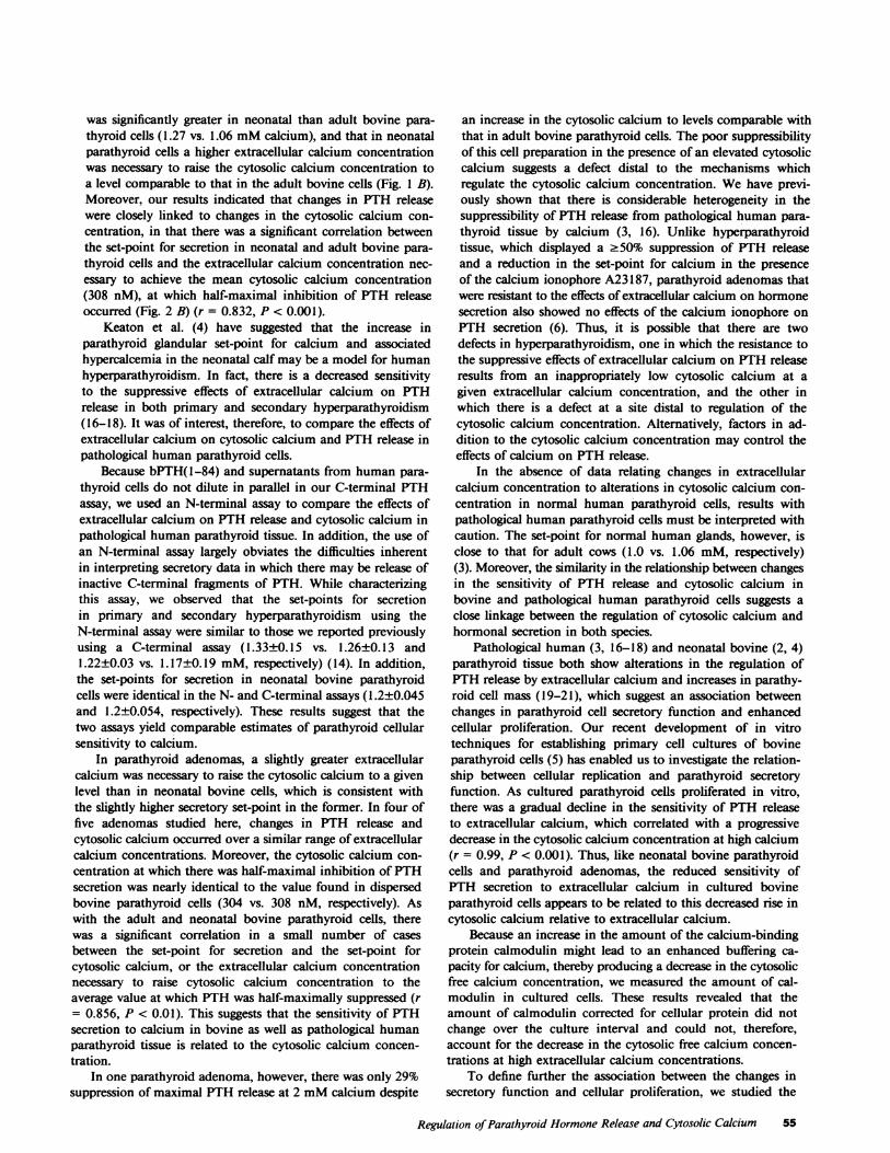
was significantly greater in neonatal than adult bovine para-thyroid cells (1.27 vs. 1.06 mMcalcium), and that in neonatalparathyroid cells a higher extracellular calcium concentrationwas necessary to raise the cytosolic calcium concentration toa level comparable to that in the adult bovine cells (Fig. 1 B).Moreover, our results indicated that changes in PTH releasewere closely linked to changes in the cytosolic calcium con-centration, in that there was a significant correlation betweenthe set-point for secretion in neonatal and adult bovine para-thyroid cells and the extracellular calcium concentration nec-essary to achieve the mean cytosolic calcium concentration(308 nM), at which half-maximal inhibition of PTH releaseoccurred (Fig. 2 B) (r = 0.832, P < 0.001).
Keaton et al. (4) have suggested that the increase inparathyroid glandular set-point for calcium and associatedhypercalcemia in the neonatal calf may be a model for humanhyperparathyroidism. In fact, there is a decreased sensitivityto the suppressive effects of extracellular calcium on PTHrelease in both primary and secondary hyperparathyroidism(16-18). It was of interest, therefore, to compare the effects ofextracellular calcium on cytosolic calcium and PTH release inpathological human parathyroid cells.
Because bPTH(1-84) and supernatants from human para-thyroid cells do not dilute in parallel in our C-terminal PTHassay, we used an N-terminal assay to compare the effects ofextracellular calcium on PTH release and cytosolic calcium inpathological human parathyroid tissue. In addition, the use ofan N-terminal assay largely obviates the difficulties inherentin interpreting secretory data in which there may be release ofinactive C-terminal fragments of PTH. While characterizingthis assay, we observed that the set-points for secretionin primary and secondary hyperparathyroidism using theN-terminal assay were similar to those we reported previouslyusing a C-terminal assay (1.33±0.15 vs. 1.26±0.13 and1.22±0.03 vs. 1.17±0.19 mM, respectively) (14). In addition,the set-points for secretion in neonatal bovine parathyroidcells were identical in the N- and C-terminal assays (1.2±0.045and 1.2±0.054, respectively). These results suggest that thetwo assays yield comparable estimates of parathyroid cellularsensitivity to calcium.
In parathyroid adenomas, a slightly greater extracellularcalcium was necessary to raise the cytosolic calcium to a givenlevel than in neonatal bovine cells, which is consistent withthe slightly higher secretory set-point in the former. In four offive adenomas studied here, changes in PTH release andcytosolic calcium occurred over a similar range of extracellularcalcium concentrations. Moreover, the cytosolic calcium con-centration at which there was half-maximal inhibition of PTHsecretion was nearly identical to the value found in dispersedbovine parathyroid cells (304 vs. 308 nM, respectively). Aswith the adult and neonatal bovine parathyroid cells, therewas a significant correlation in a small number of casesbetween the set-point for secretion and the set-point forcytosolic calcium, or the extracellular calcium concentrationnecessary to raise cytosolic calcium concentration to theaverage value at which PTH was half-maximally suppressed (r= 0.856, P < 0.01). This suggests that the sensitivity of PTHsecretion to calcium in bovine as well as pathological humanparathyroid tissue is related to the cytosolic calcium concen-tration.
In one parathyroid adenoma, however, there was only 29%suppression of maximal PTH release at 2 mMcalcium despite
an increase in the cytosolic calcium to levels comparable withthat in adult bovine parathyroid cells. The poor suppressibilityof this cell preparation in the presence of an elevated cytosoliccalcium suggests a defect distal to the mechanisms whichregulate the cytosolic calcium concentration. Wehave previ-ously shown that there is considerable heterogeneity in thesuppressibility of PTH release from pathological human para-thyroid tissue by calcium (3, 16). Unlike hyperparathyroidtissue, which displayed a .50% suppression of PTH releaseand a reduction in the set-point for calcium in the presenceof the calcium ionophore A23187, parathyroid adenomas thatwere resistant to the effects of extracellular calcium on hormonesecretion also showed no effects of the calcium ionophore onPTH secretion (6). Thus, it is possible that there are twodefects in hyperparathyroidism, one in which the resistance tothe suppressive effects of extracellular calcium on PTH releaseresults from an inappropriately low cytosolic calcium at agiven extracellular calcium concentration, and the other inwhich there is a defect at a site distal to regulation of thecytosolic calcium concentration. Alternatively, factors in ad-dition to the cytosolic calcium concentration may control theeffects of calcium on PTH release.
In the absence of data relating changes in extracellularcalcium concentration to alterations in cytosolic calcium con-centration in normal human parathyroid cells, results withpathological human parathyroid cells must be interpreted withcaution. The set-point for normal human glands, however, isclose to that for adult cows (1.0 vs. 1.06 mM, respectively)(3). Moreover, the similarity in the relationship between changesin the sensitivity of PTH release and cytosolic calcium inbovine and pathological human parathyroid cells suggests aclose linkage between the regulation of cytosolic calcium andhormonal secretion in both species.
Pathological human (3, 16-18) and neonatal bovine (2, 4)parathyroid tissue both show alterations in the regulation ofPTH release by extracellular calcium and increases in parathy-roid cell mass (19-21), which suggest an association betweenchanges in parathyroid cell secretory function and enhancedcellular proliferation. Our recent development of in vitrotechniques for establishing primary cell cultures of bovineparathyroid cells (5) has enabled us to investigate the relation-ship between cellular replication and parathyroid secretoryfunction. As cultured parathyroid cells proliferated in vitro,there was a gradual decline in the sensitivity of PTH releaseto extracellular calcium, which correlated with a progressivedecrease in the cytosolic calcium concentration at high calcium(r = 0.99, P < 0.001). Thus, like neonatal bovine parathyroidcells and parathyroid adenomas, the reduced sensitivity ofPTH secretion to extracellular calcium in cultured bovineparathyroid cells appears to be related to this decreased rise incytosolic calcium relative to extracellular calcium.
Because an increase in the amount of the calcium-bindingprotein calmodulin might lead to an enhanced buffering ca-pacity for calcium, thereby producing a decrease in the cytosolicfree calcium concentration, we measured the amount of cal-modulin in cultured cells. These results revealed that theamount of calmodulin corrected for cellular protein did notchange over the culture interval and could not, therefore,account for the decrease in the cytosolic free calcium concen-trations at high extracellular calcium concentrations.
To define further the association between the changes insecretory function and cellular proliferation, we studied the
Regulation of Parathyroid Hormone Release and Cytosolic Calcium 55

temporal relationships between changes in cellular proliferationand the regulation of PTH release and cytosolic calcium byextracellular calcium over the culture interval. Cultured cellsundergo a 59-fold increment in 3H-thymidine incorporationinto DNAbetween days 1 and 4. As shown previously, thisincrease in 3H-thymidine incorporation over 4 d correspondedto a twofold increment in cell number and a fourfold increasein cellular protein (5). DNAsynthesis was initiated within thefirst 24 h of the culture period, as 3H-thymidine incorporationDNAon day 1 was significantly greater than the 35.7±5.24counts observed in acutely dispersed cells. Preceding the de-velopment of the reduced sensitivity of hormonal secretion toextracellular calcium, there was a significant decrement in thebasal cytosolic calcium concentration on day 1 which coincidedwith the onset of active cellular proliferation. It is possible,then, that this low cytosolic calcium concentration promotedthe enhanced cellular proliferation observed in these cells andthat, as postulated by Parfitt (22) and others, the parathyroidcell divides to maintain an increased hormonal output at alower calcium concentration. The changes in cellular prolifer-ation as a function of the culture interval correlated significantlywith the gradual reduction in the effects of high extracellularcalcium on PTH release (r = 0.997, P < 0.01) and cytosoliccalcium (r = 0.995, P < 0.01). Although it is possible that theincrement in 3H-thymidine incorporation into DNAobservedin the cultured cells was due to the enhanced cellular prolif-eration of other cell types, such as fibroblasts, autoradiographicstudies revealed uptake of 3H-thymidine predominantly inparathyroid cells. Moreover, while the change in 3H-thymidineincorporation into DNAcould have been a consequence of anincrease in the intracellular pool of thymidine, or a decreasein thymidylate synthetase (12), three parameters of cellularproliferation provided evidence that cellular proliferation wasenhanced in vitro. Thus, in cultured bovine parathyroid cells,with the onset and during the active period of cellular prolif-eration, there is an alteration in the regulation of cytosoliccalcium and PTH release by extracellular calcium, whichindicates that there is an association between changes incellular proliferation and secretory function.
Because of the similarities between cultured bovine para-thyroid cells and neonatal bovine and pathological humanparathyroid cells, it is possible that the abnormal secretoryfunction in the latter two cell types may be causally related toenhanced proliferative activity as well. Although we did notdetermine 3H-thymidine incorporation or other indices ofcellular proliferation in pathological cells, it is of interest thatLloyd et al. (23) measured 3H-thytidine incorporation inparathyroid adenomas and found that DNAsynthesis correlatedwith the preoperative serum PTH and calcium concentration,but not with tumor weight. These studies suggest that moresevere secretory abnormalities are produced by parathyroidadenomas undergoing more active cellular proliferation.
Differentiated cellular function may be controlled by thestate of cellular proliferation. As demonstrated in other tissues,rapidly proliferating cells may lose the ability to express certainphysiologic characteristics (24, 25). Inhibition of cellular rep-lication, for example, by causing a growth arrest in the GIstage of the cell cycle, has, in turn, induced the differentiatedfunction of certain cells. Growth arrest in the GI stage of thecell cycle has been achieved by contact inhibition of proliferatingcells, or by growing the cells in a medium deficient in growthfactors or other nutrients (26-28). A reduction in the prolif-
erative activity of cultured parathyroid cells, or possibly para-thyroid adenomas, may therefore result in the normalizationof the phenotypic expression of these cells. A further under-standing of the association between changes in proliferationand cellular function may provide important clues to thepathogenesis and perhaps treatment of hyperparathyroidismand other endocrine neoplasias.
Acknowledgments
The authors gratefully acknowledge the expert secretarial assistance ofMs. Diane Rioux, Mrs. Nancy Orgill, and Mrs. Carmen Quintero.Furthermore, the authors are grateful for the cooperation of Dr. MillerS. Bell, Dr. Z. H. Lieberman, Dr. N. Y. Zachariah, and Mrs. JulieOsborne in providing parathyroid tissue from the Baylor UniversityMedical Center, Dallas, TX.
This work was supported by U. S. Public Health Service (USPHS)grants AM25910 and AM30028, as well as by American Cancer Societygrant PDT 230. Dr. Brown is the recipient of USPHSgrant RCDASKO 4AM00627. Dr. LeBoff is a recipient of USPHS grant CIAIK08AM 0 1198-01.
References
i. Sherwood, L. M., J. T. Potts, Jr., A. D. Care, G. P. Mayer, andG. D. Aurbach. 1966. Evaluation by radioimmunoassay of factorscontrolling the secretion of parathyroid hormone: intravenous infusionsof calcium and ethylenediamine tetraacetic acid in the cow and thegoat. Nature (Lond.). 209:52-55.
2. Habener, J. F., and J. T. Potts, Jr. 1976. Relative effectivenessof magnesium and calcium on the secretion and biosynthesis ofparathyroid hormone in vitro. Endocrinology. 98:197-202.
3. Brown, E. M., D. G. Gardner, M. F. Brennan, S. J. Marx,A. M. Spiegel, M. F. Attie, R. W. Downs, J. L. Doppnian, and G. D.Aurbach. 1979. Calcium-regulated parathyroid hormone release inprimary hyperparathyroidism. Studies in vitro with dispersed parathyroidcells. Am. J. Med. 66:923-931.
4. Keaton, J. A., J. A. Barto, M. P. Moore, J. B. Gruel, and G. P.Mayer. 1978. Altered parathyroid response to calcium in hypercalcemicneonatal calves. Endocrinology. 103:2161-2167.
5. LeBoff, M. S., H. G. Rennke, and E. M. Brown. 1983. Abnormalregulation of parathyroid cell secretion and proliferation in primarycultures of bovine parathyroid cells. Endocrinology. 113:277-284.
6. Brown, E. M. 1981. Relationship of cAMP accumulation toPTH release in dispersed cells from pathologic human parathyroidtissue. J. Clin. Endocrinol. Metab. 52:961-968.
7. Brown, E. M., N. Adragna, and D. G. Gardner. 1981. Effect ofpotassium on PTH secretion from dispersed bovine parathyroid cells.J. Clin. Endocrinol. Metab. 53:1304-1306.
8. Shoback, D., J. Thatcher, R. Leombruno, and E. Brown. 1983.Effects of extracellular Ca2" and Mg2e on cytosolic Ca++ and PTHrelease in dispersed bovine parathyroid cells. Endocrinology. 113:424-426.
9. Brown, E. M., and J. G. Thatcher. 1982. Adenosine 3',5'-monophosphate (cAMP)-dependent protein kinase and the regulationof parathyroid hormone release by divalent cations and agents elevatingcellular cAMP in dispersed bovine parathyroid cells. Endocrinology.110:1374-1380.
10. Tsien, R. Y., T. Pozzan, and T. J. Rink. 1982. Calciumhomeostasis in intact lymphocytes: cytoplasmic free calcium monitoredwith a new intracellularly trapped fluorescent indicator. J. Cell Biol.94:325-334.
11. Brown, E. M., E. J. Watson, R. Leombruno, and R. H.Underwood. 1983. Extracellular calcium is not necessary for acute,low calcium or dopamine-stimulated PTH secretion in dispersedbovine parathyroid cells. Metabolism 32:1038-1044.
56 LeBoff et al.

12. Lee, M. J., and S. I. Roth. 1975. Effect of calcium andmagnesium on deoxyribonucleic acid synthesis in rat parathyroidglands in vitro. Lab. Invest. 33:72-79.
13. Brown, E. M. 1980. Calcium-regulated phosphodiesterase inbovine parathyroid cells. Endocrinology. 107:1998-2003.
14. Brown, E. M. 1983. Four parameter model of the sigmoidalrelationship between parathyroid hormone release and extracellularcalcium concentration in normal and abnormal parathyroid tissue. J.Clin. Endocrinol. Metab. 56:572-581.
15. Shoback, D. M., J. Thatcher, R. Leombruno, and E. M. Brown.1984. Relationship between PTH secretion and cytosolic calciumconcentration in dispersed bovine parathyroid cells. Proc. Nati. Acad.Sci. USA. 81:3113-3117.
16. Brown, E. M., M. F. Brennan, S. Hurwitz, R. Windeck, S. J.Marx, A. M. Spiegel, J. 0. Koehler, D. G. Gardner, and G. D.Aurbach. 1978. Dispersed cells prepared from human parathyroidglands: distinct calcium sensitivity of adenomas vs primary hyperplasia.J Clin. Endocrin. Metab. 46:267-276.
17. Habener, J. F. 1978. Responsiveness of neoplastic and hyper-plastic parathyroid tissues to calcium in vitro. J. Clin. Invest. 62:436-458.
18. Brown, E. M., R. E. Wilson, R. C. Eastman, J. Pallotta, andS. P. Marynick. 1982. Abnormal regulation of parathyroid hormonerelease by calcium in secondary hyperparathyroidism due to chronicrenal failure. J. Clin. Endocrinol. Metab. 54:172-179.
19. Purnell, D. C., L. H. Smith, D. A. Scholz, L. R. Elveback, andC. D. Arnaud. 1971. Primary hyperparathyroidism: a prospectiveclinical study. Am. J. Med. 50:670-678.
20. Lloyd, H. M. 1968. Primary hyperparathyroidism: an analysisof the role of the parathyroid tumor. Medicine. 47:53-71.
21. Castleman, B., and T. B. Mallory. 1935. The pathology of theparathyroid gland in hyperparathyroidism. A study of 25 cases. Am.J. Pathol. 11:1-69.
22. Parfitt, A. M. 1969. Relation between parathyroid cell massand plasma calcium concentration in normal and uremic subjects.Arch. Intern. Med. 124:269-273.
23. Lloyd, H. M., J. M. Jacobi, D. Willgoss, J. Kearney, and P.Ward. 1981. DNA synthesis and secretory activity in parathyroidadenomas. Acta Endocrinol. 96:70-74.
24. Maciag, T., J. Kadish, L. Wilkins, M. Stemerman, and R.Weinstein. 1982. The organizational behavior of human umbilicalvein endothelial cells. J. Cell Biol. 94:511-20.
25. Weinstein, R., M. B. Stemerman, D. E. MacIntyre, H. N.Steinberg, and T. Maciag. 1981. The morphological and biochemicalcharacterization of a line of rat promegakaryoblasts. Blood. 58: 110-121.
26. Scott, R. E., J. J. Wille, and M. L. Wier. 1984. Mechanismsfor the initiation and promotion of carcinogenesis: a review and a newconcept. Mayo Clin. Proc. 59:107-117.
27. Pardee, A. B., R. Dubrow, J. L. Hamlin, and R. F. Kletzien.1978. Animal cell cycle. Annu. Rev. Biochem. 47:715-750.
28. Scott, R. E., D. L. Florine, J. J. Wille, Jr., and K. Yun. 1982.Coupling of growth arrest and differentiation at a distinct state in theGI phase of the cell cycle: GD. Proc. Natl. Acad. Sci. USA. 79:845-849.
Regulation of Parathyroid Hormone Release and Cytosolic Calcium 57