rela inhibits bacillus subtilis motility and chainingjb.asm.org/content/197/1/128.full.pdf · rela...
TRANSCRIPT

RelA Inhibits Bacillus subtilis Motility and Chaining
Qutaiba O. Ababneh, Jennifer K. Herman
Department of Biochemistry and Biophysics, Texas A&M University, College Station, Texas, USA
The nucleotide second messengers pppGpp and ppGpp [(p)ppGpp] are responsible for the global downregulation of transcrip-tion, translation, DNA replication, and growth rate that occurs during the stringent response. More recent studies suggest that(p)ppGpp is also an important effector in many nonstringent processes, including virulence, persister cell formation, and bio-film production. In Bacillus subtilis, (p)ppGpp production is primarily determined by the net activity of RelA, a bifunctional(p)ppGpp synthetase/hydrolase, and two monofunctional (p)ppGpp synthetases, YwaC and YjbM. We observe that in B. subtilis,a relA mutant grows exclusively as unchained, motile cells, phenotypes regulated by the alternative sigma factor SigD. Our dataindicate that the relA mutant is trapped in a SigD “on” state during exponential growth, implicating RelA and (p)ppGpp levels inthe regulation of cell chaining and motility in B. subtilis. Our results also suggest that minor variations in basal (p)ppGpp levelscan significantly skew developmental decision-making outcomes.
Genetic regulation makes it possible for cells possessing iden-tical genomes to differentiate into subpopulations of pheno-
typically and physiologically distinct cell types. The Gram-positivebacterium Bacillus subtilis is an excellent model system in which tostudy the molecular mechanisms underlying differentiation be-cause it produces several well-characterized cell types, includingspores, competent cells, biofilm formers, nonmotile chained cells,and motile unchained cells. These different cell types are producedthrough both stochastic and responsive phenotypic-switchingmechanisms (1–3). For example, the proportion of chained, non-motile cells versus unchained, motile cells in a culture of exponen-tially growing wild-type B. subtilis bacteria is driven by stochasticswitching between the “on” and “off” states of SigD (4, 5), analternative sigma factor that drives expression of the genes respon-sible for daughter cell separation and motility. However, stochas-tic determinants are only one part of SigD regulation, as the pro-portion of cells in the SigD on state also increases as cells transitionout of exponential growth (6–9). In this study, we focus on the roleof a conserved, intracellular signaling molecule in triggering theswitch to a SigD on state.
The bacterial “alarmone” (also called “magic spot”), consistingof pppGpp and ppGpp [here referred to as (p)ppGpp], acts as animportant second-messenger molecule linking both intra- and ex-tracellular environmental cues with global changes in transcrip-tion, translation, and DNA replication (10–12). In Escherichia coliand B. subtilis, (p)ppGpp levels are estimated to be less than 20pmol per optical-density unit during exponential growth (13–16)but rapidly rise to millimolar levels in response to adverse growthconditions, such as amino acid starvation (15). High levels of(p)ppGpp lead to the arrest of DNA replication and cell division,reduced lipid synthesis, a global reduction in transcription andtranslation (10–12, 17), and increased tolerance for cell stress (11,18, 19). Levels of (p)ppGpp are strongly correlated with thegrowth rate (13) and ribosomal pool size (20), and one studyfound that various substringent concentrations of (p)ppGpp reg-ulate different sets of genes, suggesting that the (p)ppGpp concen-tration may be associated with graded responses in gene expres-sion (21).
In B. subtilis, (p)ppGpp levels are predominately determinedby the net activity of RelA, a dual-function (p)ppGpp synthetaseand hydrolase, and two additional (p)ppGpp synthetase enzymes,
YwaC and YjbM (22). (p)ppGpp is synthesized at low but detect-able levels during exponential growth, primarily through the ac-tivity of RelA (22). RelA is also responsible for the high levels of(p)ppGpp that accumulate in B. subtilis during stringent response(22, 23). Less is known about the contribution of YwaC and YjbMactivity to (p)ppGpp accumulation. ywaC is under the control ofat least three stress response sigma factors, SigW, SigV, and SigM,and encodes the protein responsible for the increase in (p)ppGppobserved during alkaline shock (22, 24–26). Based on microarraydata, yjbM is the second gene in an operon of otherwise experi-mentally uncharacterized genes in B. subtilis (27). Expression pro-files suggest that the level of yjbM transcription peaks during rapidgrowth and transition states in rich media (22, 27). In addition,YjbM contributes, albeit less substantially than RelA, to the basallevels of (p)ppGpp present during exponential growth (22).
When B. subtilis has relA deleted or is depleted of a RelA variantthat possesses hydrolase but not synthetase activity (RelAD264G), itgrows slowly (22) and is prone to growth rate suppressors (22, 28,29). RelA mutants have also been shown to exhibit pleiotropicphenotypes, including reduced competence (30) and delayed spo-rulation (30). Absolute levels of (p)ppGpp are likely elevated inthe relA deletion and hydrolase mutants, since these mutants lackthe major activity responsible for breaking down (p)ppGpp syn-thesized by the remaining synthetases, YwaC and YjbM (22, 28).Consistent with the idea that (p)ppGpp levels are elevated, theslow-growth phenotype of the relA mutant can be complementedby introduction of an allele of relA that encodes only the(p)ppGpp hydrolase and through loss-of-function mutations in
Received 7 July 2014 Accepted 10 October 2014
Accepted manuscript posted online 20 October 2014
Citation Ababneh QO, Herman JK. 2015. RelA inhibits Bacillus subtilis motility andchaining. J Bacteriol 197:128 –137. doi:10.1128/JB.02063-14.
Editor: R. L. Gourse
Address correspondence to Jennifer K. Herman, [email protected].
Supplemental material for this article may be found at http://dx.doi.org/10.1128/JB.02063-14.
Copyright © 2015, American Society for Microbiology. All Rights Reserved.
doi:10.1128/JB.02063-14
128 jb.asm.org January 2015 Volume 197 Number 1Journal of Bacteriology
on Septem
ber 6, 2018 by guesthttp://jb.asm
.org/D
ownloaded from
ywaC and/or yjbM (22). It is likely that small differences in(p)ppGpp levels during exponential growth account for the ob-served phenotypes; however, any variances in (p)ppGpp levelspresent in the relA mutant and wild type during exponentialgrowth (when stringent response is not induced) are not quanti-fiable using current methodologies.
In this study, we show that the relA mutant grows exclusively asswimming, unchained cells during the exponential growth phase;this unchained phenotype contrasts sharply with the wild-typedomesticated strain PY79, which exhibits a primarily chainedphenotype during exponential growth in rich laboratory media(31). We also find that the relA mutant exhibits an acceleratedswim front in a swim plate assay, consistent with the increasedpopulation of swimming cells we observe microscopically. Anal-ysis of SigD synthesis and activity indicate that the relA mutant istrapped in a SigD on state during exponential growth. Thus, ourresults show that cell chaining and motility in B. subtilis are regu-lated by RelA through its activity as a (p)ppGpp hydrolase.
MATERIALS AND METHODSGeneral methods. A description of strains, plasmids, and oligonucleo-tides used in this study and a detailed description of plasmid constructionscan be found in Tables S1, S2, and S3 in the supplemental material. The B.subtilis strains used in this study were grown at 37°C in CH medium (32)or Luria-Bertani (LB) broth (10 g/liter tryptone, 5 g/liter yeast extract, 5g/liter NaCl). All samples were grown in volumes of either 25 ml or 50 mlin 250-ml baffled flasks in a shaking water bath set at 280 rpm. LB plateswere supplemented with 1.5% Bacto agar. E. coli strains DH5� and TG-1were used for isolation of plasmids and were grown in LB medium. Whenneeded, antibiotics were included at the following concentrations: 100 �gml�1 spectinomycin, 7.5 �g ml�1 chloramphenicol, 10 �g ml�1 tetracy-cline, and 1 �g ml�1 erythromycin (Erm) plus 25 �g ml�1 lincomycin(MLS) for B. subtilis strains and 100 �g ml�1 ampicillin for E. coli strains.
Swim plate assay. B. subtilis strains were grown at 37°C in baffledflasks containing CH medium to mid-log phase. The cells were pelletedand resuspended at an optical density at 600 nm (OD600) of 10 in phos-phate-buffered saline (PBS) (137 mM NaCl, 2.7 mM KCl, 10 mMNa2HPO4, 2 mM KH2PO4) containing 0.5% India ink (Higgins). Tenmicroliters of the cell suspension was spotted on top of 150- by 15-mm LBplates fortified with 0.25% Bacto agar (100 ml/plate), dried for 30 min atroom temperature on the benchtop to allow time for absorption, andincubated at 37°C in a loosely covered glass dish containing paper towelssoaked with water to generate humid conditions. The swim radius wasmeasured relative to the edge of the origin marked by the India ink.
Microscopy. All fluorescence microscopic analyses were performedwith a Nikon Ti-E microscope equipped with a CFI Plan Apo lambda DM100� objective and a Prior Scientific Lumen 200 Illumination system;C-FL UV-2E/C DAPI (4=,6-diamidino-2-phenylindole), C-FL GFP(green fluorescent protein) HC HISN Zero Shift, and C-FL Texas Red HCHISN Zero Shift filter cubes; and a CoolSnap HQ2 monochrome camera.Membranes were stained with either TMA-DPH [1-(4-trimethylammo-niumphenyl)-6-phenyl-1,3,5-hexatriene p-toluenesulfonate; 0.02 mM]or FM4-64 (3 �g ml�1) (Life Technologies) and imaged with exposuretimes of 200 to 800 ms. All images were captured, analyzed, and processedusing NIS Elements Advanced Research (version 4.10) and Adobe Photo-shop (version 12.0).
Flagella were visualized by using the cysteine-reactive dye Alexa Fluor488 C5 maleimide (Life Technologies) using a previously describedmethod (33). Briefly, 1-ml samples of mid-logarithmic-phase cultureswere collected at an optical density at 600 nm (OD600) of 0.5, and the cellswere pelleted by centrifugation. The pellet was then washed with 1 ml ofPBS (137 mM NaCl, 2.7 mM KCl, 10 mM Na2HPO4, and 2 mM KH2PO4),resuspended in 50 �l of PBS containing 5 �g ml�1 Alexa Fluor 488 C5
maleimide, and incubated at room temperature for 5 min. The cells were
then washed twice with 0.5 ml PBS. The pellet was resuspended in 50 �l ofPBS containing 5 �g ml�1 FM4-64 and on a glass slide with a poly-L-lysine(Sigma)-treated coverslip.
Western blot analysis. B. subtilis strains were grown to logarithmicgrowth phase (OD600, 0.4 to 0.7), and 1- or 2-ml samples were collected.The cells were pelleted by centrifugation, resuspended in 50 �l lysis buffer(20 mM Tris, pH 7.0, 10 mM EDTA, 1 mg ml�1 lysozyme, 10 �g ml�1
DNase I, 100 �g ml�1 RNase A, with 1 mM phenylmethylsulfonyl fluoride[PMSF]), and incubated for 15 min at 37°C. Fifty microliters of samplebuffer (0.25 M Tris, pH 6.8, 4% SDS, 20% glycerol, 10 mM EDTA) con-taining 10% 2-mercaptoethanol was added to the lysates, and the sampleswere boiled for 5 min prior to loading. Proteins were separated by SDS-PAGE on precast 4 to 20% Tris-HCl gels (Bio-Rad), transferred onto anitrocellulose membrane (Pall) at 100 V for 1 h in transfer buffer (20 mMTris, 15 mM glycine, and 20% methanol [vol/vol]), and then blocked in5% nonfat milk in PBS containing 0.5% Tween 20. The membranes wereincubated overnight at 4°C with a 1:5,000 dilution of anti-SigD peptideantibody (CIRDDKNVPPEEKIM; Genscript), a 1:20,000 dilution of anti-SigA (provided by Masaya Fujita, University of Houston, Houston, TX),or a 1,40,000 dilution of anti-Hag antibody (provided by Daniel B.Kearns, Indiana University) and washed and incubated with a 1:10,000dilution of horseradish peroxidase-conjugated goat anti-rabbit immuno-globulin G (Bio-Rad). After washing, the blots were incubated with Su-perSignal West Femto Chemiluminescent substrate (Thermo) accordingto the manufacturer’s instructions.
Flagellin labeling. B. subtilis strains were grown to logarithmic growthphase (OD600, 0.5), and 1-ml samples were collected. The samples werelabeled by Alexa Fluor 488 C5 maleimide labeling as described in “Micros-copy” above, and cell lysates were prepared for SDS-PAGE analysis asdescribed in “Western blot analysis” above. Proteins were separated bySDS-PAGE on precast 4 to 20% Tris-HCl gels (Bio-Rad) and visualized byscanning the gels with a Typhoon Trio (GE Healthcare) at 100-�m reso-lution and an excitation wavelength of 488 nm.
�-Galactosidase assays. To assay expression from lacZ transcriptionalfusions, B. subtilis strains were grown at 37°C in baffled flasks containingCH medium. One- or 2-ml samples were harvested at an OD600 of 0.5 bycentrifugation in a room temperature tabletop centrifuge, and the cellpellets were frozen at �20°C or �80°C. To perform the assays, the pelletswere thawed on ice, resuspended in 500 �l of Z buffer (40 mM NaH2PO4,60 mM Na2HPO4, 1 mM MgSO4, 10 mM KCl, 38 mM �-mercaptoetha-nol, and 0.2 mg ml�1 lysozyme), and incubated for 15 min at 30°C. Thereaction was started by adding 100 �l of 4-mg ml�1 O-nitrophenyl �-D-galactopyranoside (in Z buffer) and stopped with 250 �l of 1 M Na2CO3
after a yellow color developed. The optical densities of the reaction mix-tures were measured at 420 and 550 nm and used to calculate the �-ga-lactosidase specific activity according to the following equation: 1,000 �{[OD420 � (1.75 � OD550)]/(time � OD600)}.
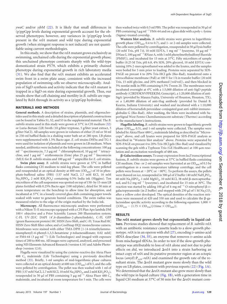
RESULTSThe relA mutant grows slowly but exponentially in liquid cul-ture. Previous studies showed that replacement of B. subtilis relAwith an antibiotic resistance cassette leads to a slow-growth phe-notype. relA is in an operon with dtd (27), encoding D-amino acidtRNA deacylase (34, 35), an enzyme that removes D-amino acidsfrom mischarged tRNAs. In order to test if the slow-growth phe-notype was attributable to loss of relA alone and not due to polareffects on dtd, we introduced �relA into a strain harboring anintact copy of relA and its putative promoter region at an ectopiclocus (amyE::PrelA-relA) and examined the growth rate of the re-sultant strain. The �relA mutant grew more slowly than the wildtype on LB plates, consistent with previous reports (22) (Fig. 1A).We determined that the �relA mutant also grew more slowly thanthe wild type in liquid culture (Fig. 1B), with a generation time inliquid CH medium at 37°C of 50 min for the �relA mutant com-
relA-Dependent Regulation of SigD
January 2015 Volume 197 Number 1 jb.asm.org 129Journal of Bacteriology
on Septem
ber 6, 2018 by guesthttp://jb.asm
.org/D
ownloaded from
pared to 30 min for the wild type (Fig. 1A). Introduction of amyE::PrelA-relA into the �relA strain complemented the slow-growthphenotype both on plates and in liquid medium (Fig. 1A and B),indicating that the slow-growth phenotype was solely attributableto the loss of relA and not related to polar affects on dtd. Despitethe longer generation time of the �relA mutant, the linear phase ofexponential growth mirrored that of the wild type, showing a de-crease in the rate around an OD600 of 0.8 to 1.0. This result sug-gests that the cues triggering the transition out of exponentialgrowth are intact in the �relA mutant background.
The slow-growing �relA mutant was previously shown to beprone to the accumulation of growth rate suppressors in ywaCand yjbM, two other (p)ppGpp synthetase genes (22, 28, 29).When constructing and growing the �relA strain, we also ob-served that spontaneous growth rate suppressors arose frequentlyon plates; sequencing of these suppressors revealed that the faster-growing strains always possessed mutations in ywaC and/or yjbM(data not shown), consistent with prior reports (22, 28, 29). Sincethe �relA mutant frequently accumulates growth rate suppressorsin ywaC and yjbM, the restoration of wild-type growth we ob-served in the presence of amyE::PrelA-relA could be attributable tothe accumulation of one or more suppressor mutations in thesegenes as opposed to true complementation. To rule out this pos-sibility, we sequenced the ywaC and yjbM regions of the comple-mented �relA mutant and found that the regions contained thewild-type alleles (data not shown).
A �relA mutant grows exclusively as unchained cells. While
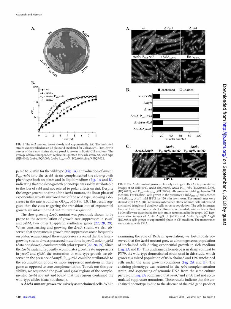
examining the role of RelA in sporulation, we fortuitously ob-served that the �relA mutant grew as a homogeneous populationof unchained cells during exponential growth in rich medium(Fig. 2A and B). This unchained phenotype is in sharp contrast toPY79, the wild-type domesticated strain used in this study, whichgrows as a mixed population of 85% chained and 15% unchainedcells under the same growth conditions (Fig. 2A and B). Thechaining phenotype was restored in the relA complementationstrain, and sequencing of genomic DNA from the same culturepictured in Fig. 2A confirmed that ywaC and yjbM had not accu-mulated suppressor mutations. These results indicate that the un-chained phenotype is due to the absence of the relA gene product
FIG 1 The relA mutant grows slowly and exponentially. (A) The indicatedstrains were streaked on an LB plate and incubated for 24 h at 37°C. (B) Growthcurves of the same strains shown panel A grown in liquid CH medium. Theaverage of three independent replicates is plotted for each strain. wt, wild type(BJH001); �relA, BQA009; �relA PrelA-relA, BQA068; �sigD, BQA022.
FIG 2 The �relA mutant grows exclusively as single cells. (A) Representativeimages of wt (BJH001), �relA (BQA009), �relA PrelA-relA (BQA068), �sigD(BQA022), and Pspac-relAD264G (EUB004) cells grown to mid-log phase in CHmedium. For EUB004, cells grown in the presence (�RelAD264G) and absence(�RelAD264G) of 1 mM IPTG for 120 min are shown. The membranes werestained with TMA. (B) Frequencies of chained (three or more cells linked) andunchained (single and doublet) cells across a population. The cells in imagesfrom at least three independent cultures were counted, and no fewer than1,500 cells were quantitated for each strain represented in the graph. (C) Rep-resentative images of �relA �sigD (BQA059) and �relA Phy-sigD �sigD(BQA083) cells grown to exponential phase in CH medium. The membraneswere stained with TMA.
Ababneh and Herman
130 jb.asm.org January 2015 Volume 197 Number 1Journal of Bacteriology
on Septem
ber 6, 2018 by guesthttp://jb.asm
.org/D
ownloaded from
and not due to polar effects on dtd. Moreover, an inducible alleleof relA that encodes the (p)ppGpp hydrolase activity but lackssynthetase activity (a D264G substitution in RelA) (22), restorednot only the wild-type growth rate, as previously observed (22),but also the cell chaining in a �relA background (Fig. 2A). Incontrast, depletion of RelAD264G in the �relA background resultedin growth as single cells (Fig. 2A). This result is consistent with theunchained phenotype being attributable to the loss of RelA’s(p)ppGpp hydrolase function. The presence of chains in all therelA mutant cultures examined throughout this study correlatedwith growth rate suppressor mutations in the other (p)ppGppsynthetases (data not shown). Therefore, we found that the dis-tinctive, nonchaining phenotype of the relA mutant served as aconvenient marker to monitor the integrity of relA mutant strainsused in our experiments. To determine if the unchained pheno-type could be attributed to the slow growth of the �relA mutant asopposed to a RelA-dependent activity, we examined wild-typecells grown at room temperature in identical media. Wild-typecells grown at room temperature had a generation time of 50min, comparable to that of the �relA mutant, but grew primarilyas chained cells (data not shown), suggesting that a lower growthrate alone does not account for the unchained phenotype.
Deletion of ywaC or yjbM in a �relA mutant restores cellchaining during exponential growth. The �relA mutant lacks themajor (p)ppGpp hydrolase activity, so synthesis of (p)ppGppfrom YwaC and/or YjbM should lead to a net accumulation of(p)ppGpp in a �relA mutant background (22). We hypothesizedthat the unchained phenotype we observed in the �relA mutantmight be due to increased (p)ppGpp levels. Since the basal levels of(p)ppGpp present in the wild-type and �relA mutant back-grounds fall outside the linear range of detection, we tested thisidea genetically using (p)ppGpp synthetase mutants. We hypoth-esized that if higher levels of (p)ppGpp resulted in loss of chaining,then the �relA �yjbM and �relA �ywaC double mutants shouldbe more chained than the �relA single mutant. We observed thatin comparison to the wild type (85% chained) and the relA mutantstrain (0% chained), the �relA �yjbM and �relA �ywaC mutantswere 78% and 34% chained, respectively (Fig. 2B). These resultsare consistent with the idea that accumulation of (p)ppGpp pro-motes the switch from chained to unchained cells and also suggestthat YjbM is a more significant contributor to (p)ppGpp poolsduring exponential growth than YwaC.
The proportion of chained versus unchained cells in a popula-tion varies with strain and culture conditions and was previouslyshown to be regulated by the level of an activated form of thealternative sigma factor SigD (4). Active SigD stimulates tran-scription of several genes, including the cell wall amidase LytF,which promotes postseptational hydrolysis of the peptidoglycanconnecting daughter cells (36). As previously shown, a sigD mu-tant grows exclusively as chains (Fig. 2A and B). In order to deter-mine if the unchained phenotype observed in the relA mutant wasdependent on SigD activity, we generated a �sigD �relA doublemutant by introducing �relA into the �sigD background usingtransformation. We observed, using control DNA, that the trans-formation efficiency of the �sigD mutant was reduced more than10-fold compared to the wild type. For unknown reasons, theefficiency of introducing the �relA mutation into the �sigD mu-tant was even lower, so that we were able to obtain only one trans-formant after 15 attempts. In contrast, �relA could be readilyintroduced into the �sigD background harboring a copy of PrelA-
relA at an ectopic locus. These results hinted that the combinationof �sigD and �relA could be synthetically lethal. To further testthis idea, we introduced �relA into a SigD depletion strain inwhich the only copy of sigD is under the control of an IPTG (iso-propyl-�-D-thiogalactopyranoside)-regulated promoter. To testfor synthetic lethality, the resulting strain was inoculated on mediawith and without inducer. Surprisingly, the strain grew similarlyto the �relA parent on plates whether sigD expression was inducedwith IPTG or not (data not shown). This result suggests that thereduced transformation efficiency we observed is due not to syn-thetic lethality but rather to a reduced ability to obtain transfor-mants. Consistent with this, whole-genome sequencing of the vi-able �sigD �relA mutant revealed no second-site suppressormutations. Microscopic analysis of the �sigD �relA mutant re-vealed an intermediate chaining phenotype different from that ofeither the �sigD or the �relA single mutant (Fig. 2C). Moreover,the SigD depletion strain harboring the �relA mutation appearedsimilar to the �relA strain when SigD was expressed (Fig. 2C) andsimilar to the �sigD �relA mutant when SigD was depleted (Fig.2C). These results indicate that SigD is required for the homoge-neous single-cell phenotype of the �relA mutant.
The �relA mutant displays flagella on the cell surface. Weobserved that the unchained relA mutants observed by micros-copy (Fig. 2A) appeared highly motile in wet mounts (data notshown), indicating that the unchained cells also possessed activeflagella. As mentioned previously, accumulation of active SigDleads to the upregulation of genes responsible for the separation ofchained cells into single cells (5, 36–38). Active SigD also coordi-nately upregulates the synthesis of late flagellar genes (39), includ-ing hag, the gene encoding flagellin (40). We hypothesized thatsince the relA mutants appeared exclusively unchained and motileby microscopy, the relA mutant would also be associated withmore flagella than the wild type. In order to directly visualizeflagella on intact cells, we engineered strains harboring a HagT209C
change in the flagellin subunit, incubated intact cells with a cys-teine-reactive dye that stains surface-exposed cysteine residues,and imaged the flagella by epifluorescence microscopy usingmethodology described previously (39). The relA mutant was un-chained (as shown in Fig. 2A and B) and presented numerousperitrichous flagella (Fig. 3A). In contrast, our wild-type straingrew as both unchained cells that presented flagella and chainedcells that generally lacked flagella (Fig. 3A). We were unable todetermine the numbers of flagella present on the cell surfaces dueto difficulty resolving individual filaments among the dense masson the cell surface. In addition, we observed the presence of sig-nificant numbers of shed flagella in the samples that made it dif-ficult to resolve whether the flagella were cell associated. We be-lieve that the shed flagella likely break off due to the shear forcesrequired to wash and centrifuge the samples during cysteine label-ing (see below). We conclude that the presence of flagella on thesurfaces of the unchained relA mutant cells suggests that SigD ismore active in this strain background than in the wild type grownunder identical conditions.
Although the use of cysteine-reactive dye to observe B. subtilisflagella was previously published (39) and we found that no fluo-rescent signal was associated with wild-type cells lacking HagT209C
(data not shown), we performed an additional control to visualizethe complement of protein species labeled by the cysteine-reactivedye. Intact cells were treated with dye as they would be prior tomicroscopy and then subjected to SDS-PAGE and laser scanning.
relA-Dependent Regulation of SigD
January 2015 Volume 197 Number 1 jb.asm.org 131Journal of Bacteriology
on Septem
ber 6, 2018 by guesthttp://jb.asm
.org/D
ownloaded from

A predominant band emitting in the green channel and running atthe approximate molecular mass of the flagellin monomer (32kDa) was present in strains harboring HagT209C for the wild type,the relA mutant, and the relA-complemented strain, but the bandwas absent from the flagellin mutant (�hag) and the wild-type
PY79 strain, which lacks the HagT209C variant (Fig. 3B and datanot shown). The amounts of flagellin varied greatly even betweenidentical strains in independent trials, despite the fact that sampleswere normalized by optical density; total-protein staining con-firmed that approximately equal amounts of protein were loadedfor each sample (Fig. 3B). We believe the variability seen in flagel-lin levels is due to shearing of the flagella during the sample han-dling required for the labeling reaction. We conclude that thefluorescence observed by microscopy is due primarily to labeledflagella and that, while analysis of labeled proteins using SDS-PAGE followed by laser scanning is useful for determining theapparent molecular weight of stained proteins, it is not useful as aquantitative measure of total flagellin levels in B. subtilis.
The �relA mutant produces more flagellin than the wildtype. In order to quantitate the amount of flagellin associated withthe relA mutant relative to the wild type, we collected cells andsupernatants from exponentially growing cultures and examinedflagellin levels using Western blot analysis (Fig. 3C). The superna-tants were collected so that we could also assess flagellin that mightaccumulate in the supernatants due to shedding, shearing, or celllysis. A predominant band running at the approximate molecularmass of the flagellin monomer (32 kDa) was present in both su-pernatants and cell lysates of the wild type, the relA mutant, andthe relA complemented strain but was absent from the flagellinmutant (�hag) and the sigD mutant (�sigD). The amount offlagellin associated with the supernatants was negligible comparedto the amount associated with the cell bodies, so it was necessary todilute the cell body lysates 50-fold to visualize all of the samples onthe same Western blot without overexposure (Fig. 3C). Since theamount of flagellin present in the supernatant was minor com-pared to that in the cell bodies, we did not analyze these datafurther. The amounts of flagellin present in the wild-type controland the complemented relA strain were similar in each of fourindependent trials and showed no statistically significant differ-ence in flagellin levels. The relA mutant, in contrast, showed a5-fold increase in flagellin levels compared to the wild type, and itwas necessary to dilute the sample 5-fold to avoid overexposure(Fig. 3C). The SigA antibody served as a loading control, confirm-ing that the diluted sample was loaded in the relA mutant lane.These results are consistent with what we observed microscopi-cally (Fig. 3A) and suggest that the unchained relA mutant popu-lation consists primarily of flagellated cells in a SigD on state.
The �relA mutant strain exhibits increased mobility onswim plates. Since the relA mutant population is composed ex-clusively of unchained, flagellated cells, we expected that it wouldalso produce a more motile population than the wild type. Thelaboratory strain used in this study (PY79) does not swarm (33), atype of surfaced-based group motility usually facilitated by a sur-factant produced by bacteria (33, 41). Therefore, in order to ob-tain a quantitative assessment of the motility behavior of the pop-ulation, we performed a swim plate assay that utilizes a lowerconcentration of agar and does not require surfactant production(42). All cultures were grown to mid-log phase and spotted inequal volumes and densities on swim plates. The visible edge of theculture, which we refer to as the “swim front,” was measured rel-ative to the edge of the original spot over a time course (Fig. 4A).An example of a swim plate at the 3.5-h time point is shown in Fig.4B. The absolute distance between the edge of the original spotand the swim front (the “swim radius”) varied slightly from assayto assay (standard deviations are shown in 4A); however, the max-
FIG 3 The relA mutant is comprised of mostly flagellated cells (A) Repre-sentative images of mid-log-phase cultures of wt (BQA057) and �relA(BQA062) strains. The membranes were stained with FM4-64 (red), andflagellin (Hag) was stained with Alexa Fluor 488 C5 maleimide (green).Both strains harbor a point mutation in the chromosomal copy of hag thatcreates HagT209C. (B) Comparison of flagellin (Hag) protein levels in wt(BQA057), �relA (BQA062), �relA PrelA-relA (BQA080), and �hag(BQA076) strains. Samples were collected from mid-log-phase culturesgrown in CH medium and stained with Alexa Fluor 488 C5 maleimide.Proteins from cell lysates were separated by SDS-PAGE, stained with Coo-massie blue (left), and scanned with a laser scanner to visualize fluores-cently labeled protein (right). The arrowhead indicates flagellin. (C) (Top)Western blot analysis showing flagellin levels associated with the culturesupernatants (left; S) and cell pellets (right; P) of the indicated strainsgrown in CH medium to mid-log phase: wt (BJH001), �relA (BQA009),�relA PrelA-relA (BQA068), �sigD (BQA022), and �hag (BQA076). It wasnecessary to dilute the cell pellet lysates to the indicated dilutions in orderto assess the levels of protein associated with each sample without overex-posure. (Bottom) SigA protein served as a loading control for the cell pelletsamples.
Ababneh and Herman
132 jb.asm.org January 2015 Volume 197 Number 1Journal of Bacteriology
on Septem
ber 6, 2018 by guesthttp://jb.asm
.org/D
ownloaded from

imal rate of migration, calculated from linear regions of the data inFig. 4A, remained essentially constant from experiment to exper-iment.
The relA mutant exhibited two notable phenotypes in the swimplate assay. First, unlike the wild type, which exhibited an 2-h lagbefore reaching its maximal swim front migration rate of 2.8mm/h, the relA mutant had a constant migration rate throughoutthe course of the experiment (30 min to 8 h) of 4.9 mm/h. Wild-type cells from exponential-phase cultures, early transition phase(OD600, 1.2), and mid-transition phase (OD600, 1.9) all exhibitedsimilar lags (data not shown). Lag periods for swarming had alsobeen observed previously and may represent a physiological ad-justment to growth on a more solid surface (41). The second ob-servable phenotype was a maximal migration rate of the relA mu-tant of 4.9 mm/h, significantly higher than that of the wild type at2.8 mm/h. The complemented relA strain behaved similarly to thewild type (Fig. 4A). These results suggest that both the absence ofthe lag period and the overall increase in the maximal swim frontmigration rate can be attributed to relA function. As expected, the
swim fronts of strains deficient in either SigD (�sigD) or flagellin(�hag) production did not migrate a measurable distance fromthe culture origin (Fig. 4B). The SigD and flagellin nonswimmingcontrols exhibited wild-type growth rates in liquid culture (Fig.1A and data not shown), suggesting that the growth rate has littleeffect on the distance traveled by the swim front in the 8-h timecourse. We conclude that, consistent with the unchained and flag-ellated phenotype we observed microscopically, the relA mutantpopulation is poised to swim during the exponential growthphase.
The increased chaining we observed in the �relA �yjbM and�relA �ywaC double mutants (Fig. 2C) is consistent with a shifttoward a SigD off state relative to the relA single mutant. If there isreduced SigD activity in the double mutants, then we would alsoexpect a decrease in their ability to swim compared to the relAsingle mutant. To test this hypothesis, we assayed the swim frontmigration of the �relA �yjbM and �relA �ywaC strains. We ob-served that both the �relA �ywaC and �relA �yjbM strains pos-sessed smaller swim radii than the �relA parent alone but largerradii than the wild type (Fig. 4B; see Fig. S1A in the supplementalmaterial). The �relA �ywaC strain, which possesses an intact copyof exponentially expressed yjbM, consistently exhibited a largerswim radius than the �relA �yjbM strain (Fig. 4B; see Fig. S1A inthe supplemental material), indicating that it more closely resem-bles the �relA strain than the �relA �yjbM strain. These results areconsistent with the idea that the switch from the nonmotile (SigDoff) to the motile (SigD on) state is promoted by increasing(p)ppGpp levels; at the same time, we do not rule out the possi-bility that the switch is stimulated by another mechanism unre-lated to (p)ppGpp accumulation.
Loss of relA leads to an increase in SigD levels and activity.The relA mutant grows exclusively as unchained, motile cells dur-ing exponential growth, consistent with the hypothesis that therelA mutant possesses higher levels of active SigD than the wildtype during exponential growth. In order to test this hypothesis,we first examined the levels of SigD protein by performing West-ern blot analysis on strains grown to mid-log phase. Relative to thewild type, the relA mutant consistently showed a nearly 10-foldincrease in SigD levels (Fig. 5A). Approximately wild-type SigDlevels were restored in the relA complementation strain (Fig. 5A),indicating that the increase in SigD levels in the relA mutant aredue to the absence of a functional RelA protein. We conclude thata RelA-dependent function is required to prevent the accumula-tion of SigD protein during exponential growth.
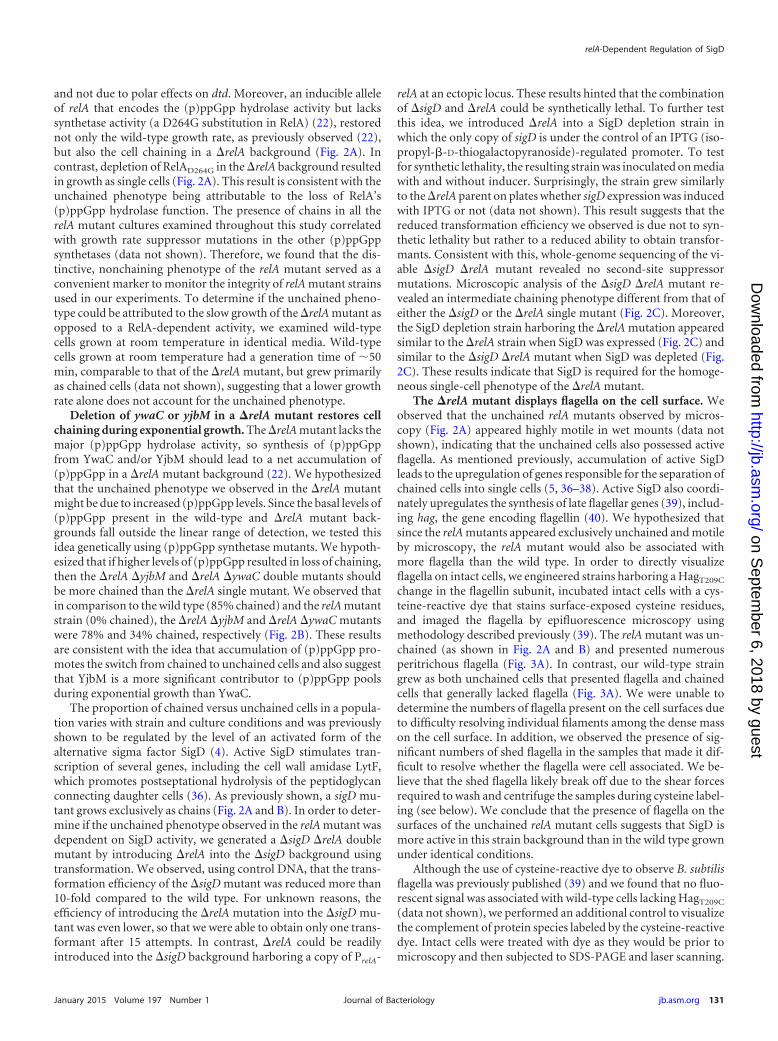
Although SigD protein levels are correlated with SigD activity,the relationship is not linear, and several levels of posttranslationalregulation ultimately determine actual SigD activity (41, 43). Inorder to determine if the increase in SigD levels we observed alsocorresponded to an increase in SigD activity, we analyzed expres-sion from two SigD-dependent promoters using transcriptionalfusions to a lacZ reporter gene. Based on the unchained, flagellatedphenotype, the increase in mobility observed on swimming plates,and the increase in SigD abundance, we expected that the relAmutant would exhibit significantly higher levels of SigD activitythan the wild type. The results of �-galactosidase assays performedon samples collected from mid-log-phase growth are shown inFig. 5B. As expected, both Phag, the flagellin promoter and PlytA,which drives expression of proteins important for flagellar activa-tion through peptidoglycan hydrolysis (37), showed significantlyhigher levels of transcription in the relA mutant than in the wild-
FIG 4 Loss of relA leads to increased mobility on swim plates. (A) Quantita-tion of swim expansion from plate assays performed with wt (BJH001), �relA(BQA009), and �relA PrelA-relA (BQA068) strains on LB fortified with 0.25%agar. Each symbol indicates the average of measurements from five indepen-dent experiments with standard deviations. Measurements were recorded ev-ery 30 min for 8 h. (B) wt (BJH001), �relA (BQA009), �relA PrelA-relA(BQA068), �sigD (BQA022), �hag (BQA076), �relA �yjbM (BQA081), and�relA �ywaC (BQA082) strains were inoculated on LB fortified with 0.25%agar. After 3.5 h of incubation at 37°C, the plates were scanned against a blackbackground. The images were inverted to better show contrast.
relA-Dependent Regulation of SigD
January 2015 Volume 197 Number 1 jb.asm.org 133Journal of Bacteriology
on Septem
ber 6, 2018 by guesthttp://jb.asm
.org/D
ownloaded from
type control. The increased expression in the relA mutant wasdependent on the presence of sigD (Fig. 5B, �relA �sigD). In con-trast, expression was reduced to approximately wild-type levels inthe relA-complemented strain. The �relA �yjbM and �relA�ywaC mutants exhibit intermediate levels of SigD activity (Fig.5B), consistent with the intermediate chaining (Fig. 2B) andswimming motility (Fig. 4B) phenotypes we observed. We con-clude that a RelA-dependent activity regulates the amount of ac-tive SigD that accumulates during the exponential growth phaseand that the �relA mutant is shifted to a SigD on state duringexponential growth.
DISCUSSION
Nucleotide second messengers, like cyclic di-GMP, cyclic AMP(cAMP), and the bacterial alarmone (p)ppGpp, are well-character-ized examples of intracellular signaling molecules that play importantroles in modulating gene expression in response to environmentaland physiological fluxes in bacteria (11, 44, 45). (p)ppGpp is respon-sible for the global downregulation of transcription, translation,
DNA replication, and growth rate that occurs during the stringentresponse to amino acid limitation (10–12). In more recent years,(p)ppGpp has been implicated in a number of nonstringent pro-cesses, including virulence (46–48), persister cell formation (49–51),biofilm production (52), maintaining a balanced growth rate (53),and regulating GTP pools in response to nutrient downshift (54).
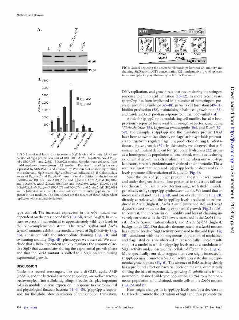
A role for (p)ppGpp in modulating cell motility has also beenpreviously reported for several Gram-negative bacteria, includingVibrio cholerae (55), Legionella pneumophila (56), and E. coli (57–59). For example, (p)ppGpp and the regulatory protein DksAhave been shown to act directly on flagellar biosynthesis promot-ers to negatively regulate flagellum production during E. coli sta-tionary-phase growth (59). In this study, we observed that a B.subtilis relA mutant deficient for (p)ppGpp hydrolysis (22) growsas a homogeneous population of unchained, motile cells duringexponential growth in rich medium, a time when our wild-typelaboratory strain is predominantly chained and nonmotile. Theseresults suggest that increased (p)ppGpp levels or decreased GTPlevels promote differentiation of B. subtilis (Fig. 6).
Since the levels of (p)ppGpp present in the strain backgroundsand under the growth conditions presented in this study fall out-side the current quantitative-detection range, we tested our modelgenetically using (p)ppGpp synthetase mutants. We found that anincrease in cell motility (Fig. 4B) and loss of cell chaining (Fig. 2B)directly correlate with the (p)ppGpp levels predicted to be pro-duced in �relA (highest), �relA �ywaC (intermediate), and �relA�yjbM (lowest) mutants during exponential growth (Fig. 2 and 6).In contrast, the increase in cell motility and loss of chaining in-versely correlate with the GTP levels measured in the �relA (low-est), �relA �ywaC (intermediate), and �relA �yjbM (highest)backgrounds (22). Our data also demonstrate that a �relA mutanthas elevated levels of SigD activity compared to the wild type (Fig.5B), consistent with the homogeneous population of unchainedand flagellated cells we observed microscopically. These resultssupport a model in which (p)ppGpp levels act as a modulator ofSigD activity and, subsequently, cellular differentiation (Fig. 6).More specifically, our data suggest that even slight increases in(p)ppGpp may promote a SigD on activation state during expo-nential growth phase (Fig. 6). The absence of RelA activity clearlyhas a profound effect on bacterial decision making, dramaticallyshifting the bias of exponentially growing B. subtilis cells from anonmotile, chained wild-type population (85%) to a homoge-neous population of unchained, motile cells in the �relA mutant(Fig. 2A and B).
How might changes in (p)ppGpp levels and/or a decrease inGTP levels promote the activation of SigD and thus promote the
FIG 5 Loss of relA leads to an increase in SigD levels and activity. (A) Com-parison of SigD protein levels in wt (BJH001), �relA (BQA009), �relA PrelA-relA (BQA068), and �sigD (BQA022) strains. Samples were collected frommid-log-phase cultures grown in CH medium. Proteins from cell lysates wereseparated by SDS-PAGE and analyzed by Western blot analysis by probingwith either anti-SigD or anti-SigA antibody, as indicated. (B) �-Galactosidaseassays of Phag-lacZ and PlytA-lacZ transcriptional activities conducted on wt(BJH046 and BJH047), �relA (BQA050 and BQA051), �relA �yibM (BQA086and BQA087), �relA �ywaC (BQA088 and BQA089), �sigD (BQA071 andBQA072), �relA PrelA-relA (BQA073 and BQA074), and �relA �sigD (BQA084and BQA085) strains. Samples were collected from mid-log-phase culturesgrown in CH medium. The data shown are the means of three independentreplicates with standard deviations.
FIG 6 Model depicting the observed relationships between cell motility andchaining, SigD activity, GTP concentration (22), and putative (p)ppGpp levelsin various (p)ppGpp synthetase/hydrolase backgrounds.
Ababneh and Herman
134 jb.asm.org January 2015 Volume 197 Number 1Journal of Bacteriology
on Septem
ber 6, 2018 by guesthttp://jb.asm
.org/D
ownloaded from

increased frequency of unchained, swimming cells we observed inrelA mutant backgrounds? SigD activity is regulated at the level ofprotein synthesis (43) and by being held in an inactive statethrough the action of the anti-sigma factor FlgM (60, 61). Weshowed that, compared to the wild type, SigD is both more abun-dant and more active in the relA mutant (Fig. 5A and B). Since theglobal regulator DegU has been shown to repress SigD production(9) and promote FlgM synthesis (62), determining the role ofDegU in the single-cell, swimming phenotype we observed is apotential avenue for future investigations. Interestingly, we ob-served that a �relA �sigD mutant does not grow as long chains likethe �sigD parent (Fig. 2C). This result suggests that the absence ofrelA promotes activation of some SigD-independent autolysins(63). One attractive idea that could explain both the �relA and the�relA �sigD phenotypes is that elevated (p)ppGpp levels in B.subtilis may promote sigma factor switching such as has been re-ported in E. coli (64–66). In E. coli, (p)ppGpp has been shown tobind to RNA polymerase and, in concert with DksA, to directlyregulate gene transcription (15). B. subtilis lacks a known DksAhomolog, and direct regulation of RNA polymerase by (p)ppGpphas not been observed (66). Moreover, B. subtilis lacks conserva-tion in the RNA polymerase (RNAP) residues that make contactwith ppGpp in E. coli (67), suggesting that if (p)ppGpp directlyregulates RNAP in B. subtilis, then the mechanisms will divergesignificantly.
The B. subtilis �relA mutant harbors reduced levels of GTP(22, 28), which may also contribute to the motile, unchained phe-notype we observed. (p)ppGpp synthesis affects GTP levelsthrough at least two independent mechanisms. First, GDP, GTP,and ATP are the substrates for (p)ppGpp-synthetic enzymes, and(p)ppGpp synthesis is correlated with a subsequent drop in GTPlevels (8, 15, 30, 68). Second, a recent study in B. subtilis showedthat (p)ppGpp can also negatively regulate cellular GTP levels bybinding directly to two key enzymes in the GTP synthesis pathway,HptR and Gmk, thereby negatively regulating their ability to syn-thesize GMP and GDP precursors, respectively (54).
It is possible that lower GTP levels present in the relA mutantrelieve transcriptional repression of some promoters in the SigDregulon through CodY derepression. CodY is a GTP-sensing pro-tein that binds DNA and positively or negatively regulates theexpression of over 100 B. subtilis gene products (69). AlthoughCodY has been shown to repress expression from hag (7), a recentglobal analysis found no evidence for binding of CodY to the hagpromoter in vivo (69). Previous studies also showed that tran-scription can be affected by shifts in the GTP and ATP pools ofcells. In B. subtilis undergoing stringent response through aminoacid starvation, (p)ppGpp and ATP levels rise while GTP levels fall(54, 70). Promoters beginning with a �1 G are apparently partic-ularly sensitive to GTP pools, as GTP is rate limiting for initiation(70, 71). Therefore, it is possible that the lower GTP levels of therelA mutant lead to decreased expression from �1 G promoters.Both of the fla-che promoters PD-3 and PA, which regulate the bulkof sigD expression, begin with a �1 A (72), while the flgM pro-moter begins with a �1 G (70).
In summary, our results show that during exponential growth,RelA activity leads to the modulation of gene expression in B.subtilis and support the idea that even minor fluxes in (p)ppGpplevels may greatly influence cellular decision making. More spe-cifically, we show that the B. subtilis �relA mutant is trapped in aSigD on state, biasing cells toward a motile, unchained phenotype
during exponential growth. Future experiments will be aimed atelucidating the molecular mechanisms underlying this develop-mental outcome.
ACKNOWLEDGMENTS
We thank Masaya Fujita for providing �-SigA antibody, Daniel B. Kearnsfor providing anti-Hag antibody and technical advice regarding the mi-croscopy-based cysteine-labeling experiment, and members of the Her-man laboratory and the Center for Phage Technology for providing help-ful feedback.
We acknowledge the Center for Phage Technology and the Depart-ment of Biochemistry and Biophysics at Texas A&M University for pro-viding funding.
REFERENCES1. Dubnau D, Losick R. 2006. Bistability in bacteria. Mol Microbiol 61:564 –
572. http://dx.doi.org/10.1111/j.1365-2958.2006.05249.x.2. Shank EA, Kolter R. 2011. Extracellular signaling and multicellularity in
Bacillus subtilis. Curr Opin Microbiol 14:741–747. http://dx.doi.org/10.1016/j.mib.2011.09.016.
3. Mirouze N, Desai Y, Raj A, Dubnau D. 2012. Spo0AP imposes a temporalgate for the bimodal expression of competence in Bacillus subtilis. PLoS Genet8:e1002586. http://dx.doi.org/10.1371/journal.pgen.1002586.
4. Cozy LM, Phillips AM, Calvo RA, Bate AR, Hsueh YH, Bonneau R,Eichenberger P, Kearns DB. 2012. SlrA/SinR/SlrR inhibits motility geneexpression upstream of a hypersensitive and hysteretic switch at the levelof sigma(D) in Bacillus subtilis. Mol Microbiol 83:1210 –1228. http://dx.doi.org/10.1111/j.1365-2958.2012.08003.x.
5. Kearns DB, Losick R. 2005. Cell population heterogeneity during growthof Bacillus subtilis. Genes Dev 19:3083–3094. http://dx.doi.org/10.1101/gad.1373905.
6. Mirel DB, Estacio WF, Mathieu M, Olmsted E, Ramirez J, Marquez-Magana LM. 2000. Environmental regulation of Bacillus subtilis sig-ma(D)-dependent gene expression. J Bacteriol 182:3055–3062. http://dx.doi.org/10.1128/JB.182.11.3055-3062.2000.
7. Bergara F, Ibarra C, Iwamasa J, Patarroyo JC, Aguilera R, Marquez-Magana LM. 2003. CodY is a nutritional repressor of flagellar gene ex-pression in Bacillus subtilis. J Bacteriol 185:3118 –3126. http://dx.doi.org/10.1128/JB.185.10.3118-3126.2003.
8. Ratnayake-Lecamwasam M, Serror P, Wong KW, Sonenshein AL. 2001. Ba-cillus subtilis CodY represses early-stationary-phase genes by sensing GTP levels.Genes Dev 15:1093–1103. http://dx.doi.org/10.1101/gad.874201.
9. Amati G, Bisicchia P, Galizzi A. 2004. DegU-P represses expression of themotility fla-che operon in Bacillus subtilis. J Bacteriol 186:6003– 6014.http://dx.doi.org/10.1128/JB.186.18.6003-6014.2004.
10. Magnusson LU, Farewell A, Nystrom T. 2005. ppGpp: a global regulatorin Escherichia coli. Trends Microbiol 13:236 –242. http://dx.doi.org/10.1016/j.tim.2005.03.008.
11. Dalebroux ZD, Swanson MS. 2012. ppGpp: magic beyond RNA polymerase.Nat Rev Microbiol 10:203–212. http://dx.doi.org/10.1038/nrmicro2720.
12. Potrykus K, Cashel M. 2008. (p)ppGpp: still magical? Annu Rev Microbiol62:35–51. http://dx.doi.org/10.1146/annurev.micro.62.081307.162903.
13. Sarubbi E, Rudd KE, Cashel M. 1988. Basal ppGpp level adjustmentshown by new spoT mutants affect steady state growth rates and rrnAribosomal promoter regulation in Escherichia coli. Mol Gen Genet 213:214 –222. http://dx.doi.org/10.1007/BF00339584.
14. Ryals J, Little R, Bremer H. 1982. Control of rRNA and tRNA synthesesin Escherichia coli by guanosine tetraphosphate. J Bacteriol 151:1261–1268.
15. Cashel M, Gentry DM, Hernandez VJ, Vinella D. 1996. The stringentresponse, p 1458 –1496. In Neidhardt FC (ed), Escherichia coli and Sal-monella typhimurium: cellular and molecular biology, vol 1. ASM Press,Washington, DC.
16. Nishino T, Gallant J, Shalit P, Palmer L, Wehr T. 1979. Regulatorynucleotides involved in the Rel function of Bacillus subtilis. J Bacteriol140:671– 679.
17. Wang JD, Sanders GM, Grossman AD. 2007. Nutritional control ofelongation of DNA replication by (p)ppGpp. Cell 128:865– 875. http://dx.doi.org/10.1016/j.cell.2006.12.043.
18. Zhang S, Haldenwang WG. 2003. RelA is a component of the nutritional
relA-Dependent Regulation of SigD
January 2015 Volume 197 Number 1 jb.asm.org 135Journal of Bacteriology
on Septem
ber 6, 2018 by guesthttp://jb.asm
.org/D
ownloaded from

stress activation pathway of the Bacillus subtilis transcription factor sigmaB. J Bacteriol 185:5714 –5721. http://dx.doi.org/10.1128/JB.185.19.5714-5721.2003.
19. Primm TP, Andersen SJ, Mizrahi V, Avarbock D, Rubin H, Barry CE,III. 2000. The stringent response of Mycobacterium tuberculosis is re-quired for long-term survival. J Bacteriol 182:4889 – 4898. http://dx.doi.org/10.1128/JB.182.17.4889-4898.2000.
20. Nomura M, Gourse R, Baughman G. 1984. Regulation of the synthesis ofribosomes and ribosomal components. Annu Rev Biochem 53:75–117.http://dx.doi.org/10.1146/annurev.bi.53.070184.000451.
21. Traxler MF, Summers SM, Nguyen HT, Zacharia VM, Hightower GA,Smith JT, Conway T. 2008. The global, ppGpp-mediated stringent re-sponse to amino acid starvation in Escherichia coli. Mol Microbiol 68:1128 –1148. http://dx.doi.org/10.1111/j.1365-2958.2008.06229.x.
22. Nanamiya H, Kasai K, Nozawa A, Yun CS, Narisawa T, Murakami K,Natori Y, Kawamura F, Tozawa Y. 2008. Identification and functionalanalysis of novel (p)ppGpp synthetase genes in Bacillus subtilis. Mol Mi-crobiol 67:291–304. http://dx.doi.org/10.1111/j.1365-2958.2007.06018.x.
23. Smith I, Paress P, Cabane K, Dubnau E. 1980. Genetics and physiologyof the rel system of Bacillus subtilis. Mol Gen Genet 178:271–279. http://dx.doi.org/10.1007/BF00270472.
24. Czarny TL, Perri AL, French S, Brown ED. 2014. Discovery of novel cellwall-active compounds using PywaC, a sensitive reporter of cell wall stressin the model Gram-positive Bacillus subtilis. Antimicrob Agents Che-mother 58:3261–3269. http://dx.doi.org/10.1128/AAC.02352-14.
25. Eiamphungporn W, Helmann JD. 2008. The Bacillus subtilis sigma(M)regulon and its contribution to cell envelope stress responses. Mol Micro-biol 67:830 – 848. http://dx.doi.org/10.1111/j.1365-2958.2007.06090.x.
26. Guariglia-Oropeza V, Helmann JD. 2011. Bacillus subtilis sigma(V) con-fers lysozyme resistance by activation of two cell wall modification path-ways, peptidoglycan O-acetylation and D-alanylation of teichoic acids. JBacteriol 193:6223– 6232. http://dx.doi.org/10.1128/JB.06023-11.
27. Nicolas P, Mader U, Dervyn E, Rochat T, Leduc A, Pigeonneau N,Bidnenko E, Marchadier E, Hoebeke M, Aymerich S, Becher D, Bisic-chia P, Botella E, Delumeau O, Doherty G, Denham EL, Fogg MJ,Fromion V, Goelzer A, Hansen A, Hartig E, Harwood CR, Homuth G,Jarmer H, Jules M, Klipp E, Le Chat L, Lecointe F, Lewis P, Lieber-meister W, March A, Mars RA, Nannapaneni P, Noone D, Pohl S, RinnB, Rugheimer F, Sappa PK, Samson F, Schaffer M, Schwikowski B, SteilL, Stulke J, Wiegert T, Devine KM, Wilkinson AJ, van Dijl JM, HeckerM, Volker U, Bessieres P, Noirot P. 2012. Condition-dependent tran-scriptome reveals high-level regulatory architecture in Bacillus subtilis.Science 335:1103–1106. http://dx.doi.org/10.1126/science.1206848.
28. Natori Y, Tagami K, Murakami K, Yoshida S, Tanigawa O, Moh Y,Masuda K, Wada T, Suzuki S, Nanamiya H, Tozawa Y, Kawamura F.2009. Transcription activity of individual rrn operons in Bacillus subtilismutants deficient in (p)ppGpp synthetase genes, relA, yjbM, and ywaC. JBacteriol 191:4555– 4561. http://dx.doi.org/10.1128/JB.00263-09.
29. Srivatsan A, Han Y, Peng J, Tehranchi AK, Gibbs R, Wang JD, Chen R.2008. High-precision, whole-genome sequencing of laboratory strains fa-cilitates genetic studies. PLoS Genet 4:e1000139. http://dx.doi.org/10.1371/journal.pgen.1000139.
30. Inaoka T, Ochi K. 2002. RelA protein is involved in induction of geneticcompetence in certain Bacillus subtilis strains by moderating the level ofintracellular GTP. J Bacteriol 184:3923–3930. http://dx.doi.org/10.1128/JB.184.14.3923-3930.2002.
31. Chen R, Guttenplan SB, Blair KM, Kearns DB. 2009. Role of the sig-maD-dependent autolysins in Bacillus subtilis population heterogeneity. JBacteriol 191:5775–5784. http://dx.doi.org/10.1128/JB.00521-09.
32. Harwood CR, Cutting SM. 1990. Molecular biological methods for Ba-cillus. Wiley, New York, NY.
33. Patrick JE, Kearns DB. 2009. Laboratory strains of Bacillus subtilis do notexhibit swarming motility. J Bacteriol 191:7129 –7133. http://dx.doi.org/10.1128/JB.00905-09.
34. Soutourina O, Soutourina J, Blanquet S, Plateau P. 2004. Formation ofD-tyrosyl-tRNATyr accounts for the toxicity of D-tyrosine toward Esch-erichia coli. J Biol Chem 279:42560 – 42565. http://dx.doi.org/10.1074/jbc.M402931200.
35. Yang H, Zheng G, Peng X, Qiang B, Yuan J. 2003. D-Amino acids andD-Tyr-tRNA(Tyr) deacylase: stereospecificity of the translation machinerevisited. FEBS Lett 552:95–98. http://dx.doi.org/10.1016/S0014-5793(03)00858-5.
36. Margot P, Pagni M, Karamata D. 1999. Bacillus subtilis 168 gene lytF
encodes a gamma-D-glutamate-meso-diaminopimelate muropeptidaseexpressed by the alternative vegetative sigma factor, sigmaD. Microbiol-ogy 145:57– 65. http://dx.doi.org/10.1099/13500872-145-1-57.
37. Lazarevic V, Margot P, Soldo B, Karamata D. 1992. Sequencing andanalysis of the Bacillus subtilis lytRABC divergon: a regulatory unit en-compassing the structural genes of the N-acetylmuramoyl-L-alanine ami-dase and its modifier. J Gen Microbiol 138:1949 –1961. http://dx.doi.org/10.1099/00221287-138-9-1949.
38. Blackman SA, Smith TJ, Foster SJ. 1998. The role of autolysins duringvegetative growth of Bacillus subtilis 168. Microbiology 144:73– 82. http://dx.doi.org/10.1099/00221287-144-1-73.
39. Blair KM, Turner L, Winkelman JT, Berg HC, Kearns DB. 2008. Amolecular clutch disables flagella in the Bacillus subtilis biofilm. Science320:1636 –1638. http://dx.doi.org/10.1126/science.1157877.
40. Mirel DB, Chamberlin MJ. 1989. The Bacillus subtilis flagellin gene (hag)is transcribed by the sigma 28 form of RNA polymerase. J Bacteriol 171:3095–3101.
41. Patrick JE, Kearns DB. 2012. Swarming motility and the control of masterregulators of flagellar biosynthesis. Mol Microbiol 83:14 –23. http://dx.doi.org/10.1111/j.1365-2958.2011.07917.x.
42. Kearns DB. 2010. A field guide to bacterial swarming motility. Nat RevMicrobiol 8:634 – 644. http://dx.doi.org/10.1038/nrmicro2405.
43. Cozy LM, Kearns DB. 2010. Gene position in a long operon governsmotility development in Bacillus subtilis. Mol Microbiol 76:273–285.http://dx.doi.org/10.1111/j.1365-2958.2010.07112.x.
44. Kalia D, Merey G, Nakayama S, Zheng Y, Zhou J, Luo Y, Guo M, RoembkeBT, Sintim HO. 2013. Nucleotide, c-di-GMP, c-di-AMP, cGMP, cAMP,(p)ppGppsignalinginbacteriaandimplicationsinpathogenesis.Chem.Soc.Rev.42:305–341. http://dx.doi.org/10.1039/c2cs35206k.
45. Camilli A, Bassler BL. 2006. Bacterial small-molecule signaling pathways.Science 311:1113–1116. http://dx.doi.org/10.1126/science.1121357.
46. Vogt SL, Green C, Stevens KM, Day B, Erickson DL, Woods DE, StoreyDG. 2011. The stringent response is essential for Pseudomonas aeruginosavirulence in the rat lung agar bead and Drosophila melanogaster feedingmodels of infection. Infect Immun 79:4094 – 4104. http://dx.doi.org/10.1128/IAI.00193-11.
47. Weiss LA, Stallings CL. 2013. Essential roles for Mycobacterium tuber-culosis Rel beyond the production of (p)ppGpp. J Bacteriol 195:5629 –5638. http://dx.doi.org/10.1128/JB.00759-13.
48. Dalebroux ZD, Svensson SL, Gaynor EC, Swanson MS. 2010. ppGppconjures bacterial virulence. Microbiol Mol Biol Rev 74:171–199. http://dx.doi.org/10.1128/MMBR.00046-09.
49. Lewis K. 2010. Persister cells. Annu Rev Microbiol 64:357–372. http://dx.doi.org/10.1146/annurev.micro.112408.134306.
50. Amato SM, Orman MA, Brynildsen MP. 2013. Metabolic control ofpersister formation in Escherichia coli. Mol Cell 50:475– 487. http://dx.doi.org/10.1016/j.molcel.2013.04.002.
51. Jayaraman R. 2008. Bacterial persistence: some new insights into an oldphenomenon. J Biosci 33:795– 805. http://dx.doi.org/10.1007/s12038-008-0099-3.
52. He H, Cooper JN, Mishra A, Raskin DM. 2012. Stringent responseregulation of biofilm formation in Vibrio cholerae. J Bacteriol 194:2962–2972. http://dx.doi.org/10.1128/JB.00014-12.
53. Potrykus K, Murphy H, Philippe N, Cashel M. 2011. ppGpp is the majorsource of growth rate control in E. coli. Environ Microbiol 13:563–575.http://dx.doi.org/10.1111/j.1462-2920.2010.02357.x.
54. Kriel A, Bittner AN, Kim SH, Liu K, Tehranchi AK, Zou WY, RendonS, Chen R, Tu BP, Wang JD. 2012. Direct regulation of GTP homeostasisby (p)ppGpp: a critical component of viability and stress resistance. MolCell 48:231–241. http://dx.doi.org/10.1016/j.molcel.2012.08.009.
55. Pal RR, Bag S, Dasgupta S, Das B, Bhadra RK. 2012. Functionalcharacterization of the stringent response regulatory gene dksA of Vibriocholerae and its role in modulation of virulence phenotypes. J Bacteriol194:5638 –5648. http://dx.doi.org/10.1128/JB.00518-12.
56. Dalebroux ZD, Yagi BF, Sahr T, Buchrieser C, Swanson MS. 2010.Distinct roles of ppGpp and DksA in Legionella pneumophila differenti-ation. Mol Microbiol 76:200 –219. http://dx.doi.org/10.1111/j.1365-2958.2010.07094.x.
57. Aberg A, Fernandez-Vazquez J, Cabrer-Panes JD, Sanchez A, Balsalo-bre C. 2009. Similar and divergent effects of ppGpp and DksA deficiencieson transcription in Escherichia coli. J Bacteriol 191:3226 –3236. http://dx.doi.org/10.1128/JB.01410-08.
58. Magnusson LU, Gummesson B, Joksimovic P, Farewell A, Nystrom T.
Ababneh and Herman
136 jb.asm.org January 2015 Volume 197 Number 1Journal of Bacteriology
on Septem
ber 6, 2018 by guesthttp://jb.asm
.org/D
ownloaded from
2007. Identical, independent, and opposing roles of ppGpp and DksA inEscherichia coli. J Bacteriol 189:5193–5202. http://dx.doi.org/10.1128/JB.00330-07.
59. Lemke JJ, Durfee T, Gourse RL. 2009. DksA and ppGpp directly regulatetranscription of the Escherichia coli flagellar cascade. Mol Microbiol 74:1368 –1379. http://dx.doi.org/10.1111/j.1365-2958.2009.06939.x.
60. Caramori T, Barilla D, Nessi C, Sacchi L, Galizzi A. 1996. Role of FlgMin sigma D-dependent gene expression in Bacillus subtilis. J Bacteriol 178:3113–3118.
61. Bertero MG, Gonzales B, Tarricone C, Ceciliani F, Galizzi A. 1999.Overproduction and characterization of the Bacillus subtilis anti-sigmafactor FlgM. J Biol Chem 274:12103–12107. http://dx.doi.org/10.1074/jbc.274.17.12103.
62. Hsueh YH, Cozy LM, Sham LT, Calvo RA, Gutu AD, Winkler ME,Kearns DB. 2011. DegU-phosphate activates expression of the anti-sigmafactor FlgM in Bacillus subtilis. Mol Microbiol 81:1092–1108. http://dx.doi.org/10.1111/j.1365-2958.2011.07755.x.
63. Smith TJ, Blackman SA, Foster SJ. 2000. Autolysins of Bacillus subtilis:multiple enzymes with multiple functions. Microbiology 146:249 –262.
64. Osterberg S, del Peso-Santos T, Shingler V. 2011. Regulation of alter-native sigma factor use. Annu Rev Microbiol 65:37–55. http://dx.doi.org/10.1146/annurev.micro.112408.134219.
65. Costanzo A, Nicoloff H, Barchinger SE, Banta AB, Gourse RL, Ades SE.2008. ppGpp and DksA likely regulate the activity of the extracytoplasmicstress factor sigmaE in Escherichia coli by both direct and indirect mech-anisms. Mol Microbiol 67:619 – 632. http://dx.doi.org/10.1111/j.1365-2958.2007.06072.x.
66. Sharma UK, Chatterji D. 2010. Transcriptional switching in Escherichiacoli during stress and starvation by modulation of sigma activity. FEMSMicrobiol Rev 34:646 – 657. http://dx.doi.org/10.1111/j.1574-6976.2010.00223.x.
67. Ross W, Vrentas CE, Sanchez-Vazquez P, Gaal T, Gourse RL. 2013. Themagic spot: a ppGpp binding site on E. coli RNA polymerase responsiblefor regulation of transcription initiation. Mol Cell 50:420 – 429. http://dx.doi.org/10.1016/j.molcel.2013.03.021.
68. Krasny L, Gourse RL. 2004. An alternative strategy for bacterial ribosomesynthesis: Bacillus subtilis rRNA transcription regulation. EMBO J 23:4473– 4483. http://dx.doi.org/10.1038/sj.emboj.7600423.
69. Belitsky BR, Sonenshein AL. 2013. Genome-wide identification of Bacillussubtilis CodY-binding sites at single-nucleotide resolution. Proc Natl Acad SciU S A 110:7026–7031. http://dx.doi.org/10.1073/pnas.1300428110.
70. Krasny L, Tiserova H, Jonak J, Rejman D, Sanderova H. 2008. The identityof the transcription �1 position is crucial for changes in gene expression inresponse to amino acid starvation in Bacillus subtilis. Mol Microbiol 69:42–54. http://dx.doi.org/10.1111/j.1365-2958.2008.06256.x.
71. Tojo S, Hirooka K, Fujita Y. 2013. Expression of kinA and kinB ofBacillus subtilis, necessary for sporulation initiation, is under positivestringent transcription control. J Bacteriol 195:1656 –1665. http://dx.doi.org/10.1128/JB.02131-12.
72. West JT, Estacio W, Marquez-Magana L. 2000. Relative roles of thefla/che P(A), P(D-3), and P(sigD) promoters in regulating motility andsigD expression in Bacillus subtilis. J Bacteriol 182:4841– 4848. http://dx.doi.org/10.1128/JB.182.17.4841-4848.2000.
relA-Dependent Regulation of SigD
January 2015 Volume 197 Number 1 jb.asm.org 137Journal of Bacteriology
on Septem
ber 6, 2018 by guesthttp://jb.asm
.org/D
ownloaded from