relapsing-remitting granulomatous disease in 22q11
TRANSCRIPT
Relapsing-remitting
granulomatous disease in 22q11
deletion syndrome
Dr Paxton Loke
Department of Allergy and Immunology, Royal Children’s Hospital, Melbourne, Victoria.
Background
13 year old girl
• 22q11 deletion (a.k.a. DiGeorge syndrome)
• Diagnosed by FISH at age 3 ½ years in context of developmental delay in Tasmania
• Confirmed on microarray 2014
• Referred to Immunology in 2011 (age 8)
• Long history of recurrent sino-pulmonary infections (Pneumonias/Suppurative Otitis
Media/Chronic Moist cough)
• Immune investigations
• Mild CD8 T cell lymphopenia.
• Markedly reduced naïve T cells.
• Reduced lymphocyte proliferation to PHA and anti-CD3.
• Pan-hypogammaglobulinaemia (IgG 2.33/IgA 0.12/IgM 0.33)
• Reduced memory B cells
• Poor response to vaccines (Pneumovax, tetanus & Hib)
• Diagnosed with antibody deficiency (“CVID-like”)
• Started on IVIG replacement, now on SCIG.
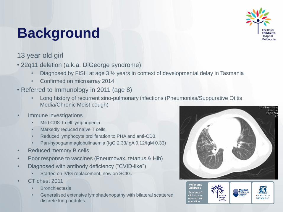
• CT chest 2011
• Bronchiectasis
• Generalised extensive lymphadenopathy with bilateral scattered
discrete lung nodules.

1st episode - Oct 2013
• Presented with cough, lethargy & fever
• BAL – CMV +ve. Treated with IV ganciclovir.
• Anaemic, thrombocytopenic with hepatosplenomegaly
• Workup over the next few months
• Bone Marrow biopsy – no marrow infiltrate
• Lymph node biopsy – granulomatous changes
• CT chest/abdo/pelvis
• Multiple lung nodules. Multiple enlarged mediastinal
lymph nodes
• Bronchiectasis.
• Retroperitoneal lymphadenopathy and splenomegaly
23cm.
• Started on 2mg/kg Prednisolone in Jan 2014 for:
• Granulomas in lung and abdomen
• Cytopaenias (presumed autoimmune).
• Gradual wean over 6 months, ceased 1/7/2014.
• Cytopaenias resolved.
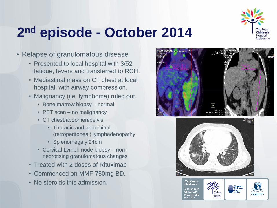
2nd episode - October 2014
• Relapse of granulomatous disease
• Presented to local hospital with 3/52
fatigue, fevers and transferred to RCH.
• Mediastinal mass on CT chest at local
hospital, with airway compression.
• Malignancy (i.e. lymphoma) ruled out.
• Bone marrow biopsy – normal
• PET scan – no malignancy.
• CT chest/abdomen/pelvis
• Thoracic and abdominal
(retroperitoneal) lymphadenopathy
• Splenomegaly 24cm
• Cervical Lymph node biopsy – non-
necrotising granulomatous changes
• Treated with 2 doses of Rituximab
• Commenced on MMF 750mg BD.
• No steroids this admission.

3rd episode – September 2015
• Presented to local hospital with fevers and lethargy.
• Background – 2/12 of issues with her bone-anchored
hearing aid (BAHA) – infected/treated/re-infected
• Cytopaenias recurred and increased splenomegaly at
local hospital.
• IV methypred 1mg/kg prior to transfer to RCH.
• At RCH
• BAHA replaced by ENT.
• Treated for infections (started at local hospital).
• Bone marrow biopsy – no evidence of malignancy.
• B cells have returned – Treated with 2 doses of Rituximab.
• MMF increased to 1g/750mg.
• On weaning regimen of oral Pred.
• CT chest/abdo/pelvis
• Interval reduction in mediastinal/ intra-abdominal lymphadenopathy.
Cervical lymphadenopathy resolved.
• Near-complete resolution of previous numerous small lung nodules.
Reduced splenomegaly persist.
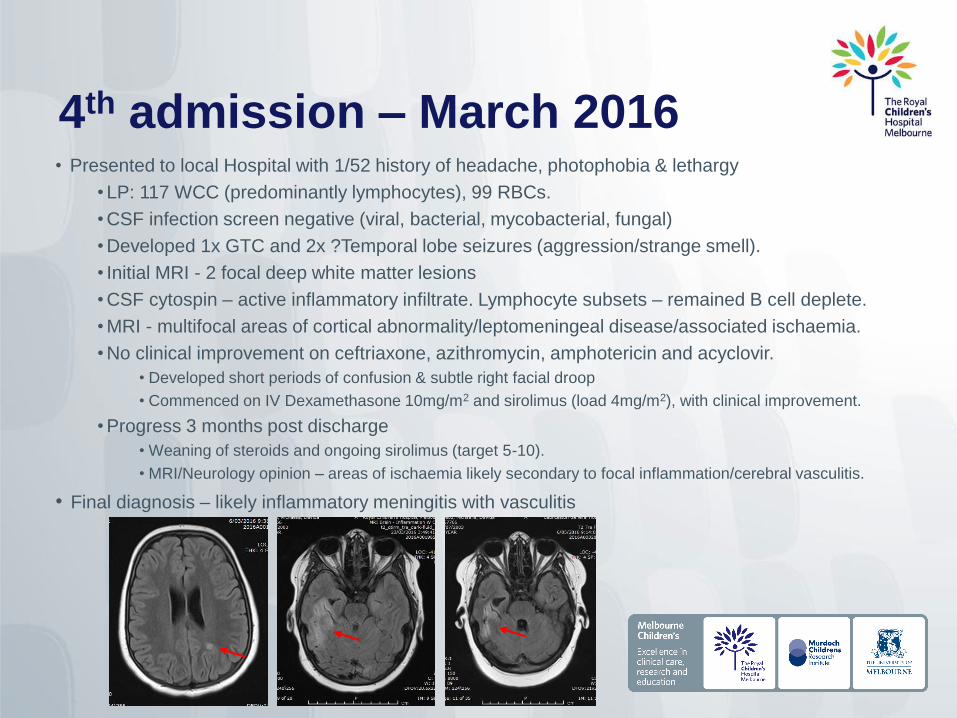
4th admission – March 2016• Presented to local Hospital with 1/52 history of headache, photophobia & lethargy
• LP: 117 WCC (predominantly lymphocytes), 99 RBCs.
• CSF infection screen negative (viral, bacterial, mycobacterial, fungal)
• Developed 1x GTC and 2x ?Temporal lobe seizures (aggression/strange smell).
• Initial MRI - 2 focal deep white matter lesions
• CSF cytospin – active inflammatory infiltrate. Lymphocyte subsets – remained B cell deplete.
• MRI - multifocal areas of cortical abnormality/leptomeningeal disease/associated ischaemia.
• No clinical improvement on ceftriaxone, azithromycin, amphotericin and acyclovir.
• Developed short periods of confusion & subtle right facial droop
• Commenced on IV Dexamethasone 10mg/m2 and sirolimus (load 4mg/m2), with clinical improvement.
• Progress 3 months post discharge
• Weaning of steroids and ongoing sirolimus (target 5-10).
• MRI/Neurology opinion – areas of ischaemia likely secondary to focal inflammation/cerebral vasculitis.
• Final diagnosis – likely inflammatory meningitis with vasculitis
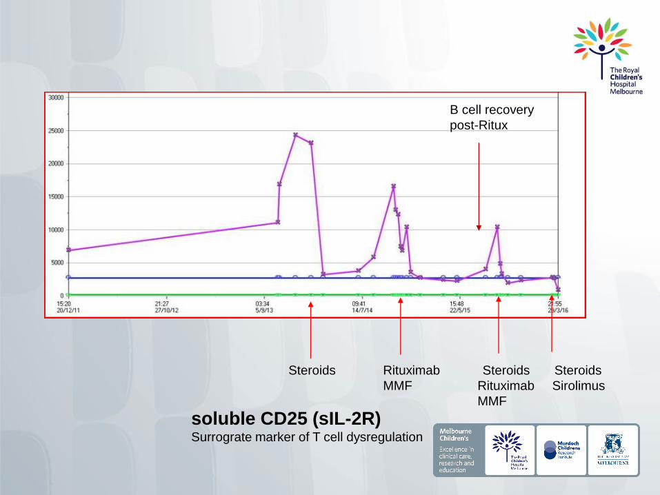
soluble CD25 (sIL-2R)Surrograte marker of T cell dysregulation
Steroids Rituximab Steroids Steroids
MMF Rituximab Sirolimus
MMF
B cell recovery
post-Ritux
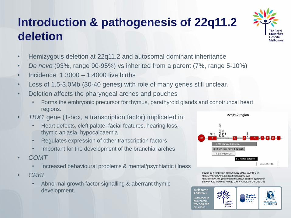
Introduction & pathogenesis of 22q11.2
deletion
• Hemizygous deletion at 22q11.2 and autosomal dominant inheritance
• De novo (93%, range 90-95%) vs inherited from a parent (7%, range 5-10%)
• Incidence: 1:3000 – 1:4000 live births
• Loss of 1.5-3.0Mb (30-40 genes) with role of many genes still unclear.
• Deletion affects the pharyngeal arches and pouches
• Forms the embryonic precursor for thymus, parathyroid glands and conotruncal heart
regions.
Davies G. Frontiers in Immunology 2013; 322(4): 1-9.
http://www.ncbi.nlm.nih.gov/books/NBK1523/
http://ghr.nlm.nih.gov/condition/22q112-deletion-syndrome
Sullivan KE. Immunol Allergy Clin N Am 2008; 28: 353-366
• TBX1 gene (T-box, a transcription factor) implicated in:
• Heart defects, cleft palate, facial features, hearing loss,
thymic aplasia, hypocalcaemia
• Regulates expression of other transcription factors
• Important for the development of the branchial arches
• COMT
• Increased behavioural problems & mental/psychiatric illness
• CRKL
• Abnormal growth factor signalling & aberrant thymic
development.
Immunological features of DGS• Complete DGS (cDGS)
• Complete absence of the thymus (athymia)
• Absolute T Lymphopenia
• SCID-like phenotype (T-B-NK+) with other variable features of DGS
• Absent mitogen (proliferation) responses
• Suffer from opportunistic infections
• Rare and affects < 1% of patients with 22q11 deletion
• Treatment of choice is thymic transplant
• Using donor thymus from infants undergoing cardiac surgery (CMV positive excluded)
• Donor thymus cultured (12-21 days) and implanted in the quadriceps muscle.
• Atypical cDGS
• Presence of some mature T cells via maternal engraftment or oligoclonal expansion of
memory T cells (no thymic processing).
• Erythrodermic rashes, enteropathy and lymphadenopathy (Omenn’s-like)
• Treatment is thymic transplant (pre-conditioning ATG, and cyclosporine)
Davies G. Frontiers in Immunology 2013; 322(4): 1-9.
Gennery A. Curr Opin Pediatr 2013, 25: 730-735

Immunological features of DGS• Partial DGS (pDGS or incomplete DGS)
• Most common clinical scenario with small, often atopic, thymus development
• Variable number of T cells (low to normal) and naïve T cells (reduced or normal)
• Generally normal mitogen responses
• Most pDGS do not suffer opportunistic infections
• Infections are more towards sino-pulmonary (humoral) types
• Humoral deficiency with functional B cell deficit & hypogammaglobinaemia associated with more
severe infections (2.7% on Ig replacement in 2012 study of 1023 DGS cohort).
• Partial DGS with autoimmunity
• Immune dysregulation → autoimmunity
• Between 8.5% to 33% and likelihood increases with lower T-cell numbers.
• Cytopenias, juvenile RA, autoimmune haemolytic disease (ITP, anaemia), hypo/hyperthyroidism.
• Disturbance of central or peripheral tolerance as a consequence of thymic abnormality.
• Reduced numbers of natural T regulatory cells (CD4+ FOXP3+)
• Higher numbers of naïve T cells in early childhood may be protective.
Davies G. Frontiers in Immunology 2013; 322(4): 1-9.
Gennery A. Curr Opin Pediatr 2013, 25: 730-735
Patel et al. J Pediatr, 2012; 161: 950-3
Tison et al. J Allergy Clin Immunol, 2011; 128 (5): 1115-1117
Partial DGS with granulomatous
lymphoproliferation - Case Reports
• 11 yr girl with mutation in TNFRSF13B with granulomatous lymphoproliferation.
• CVID with heterozygous mutation in TACI (encoded by TNFRSF13B - 10% of patients with CVID)
• Developed GLILD and widespread granulomatous-lymphocytic disease
• Treated with steroids, IVIG, hydroxychloroquine and azathioprine
• Deceased following early haemorrhagic post-operative complications following splenectomy.
• 15 yr girl with pulmonary/extranodal marginal zone lymphoma (MALT) & granulomatous
inflammation
• Lung biopsy: inflammation & granulomatous disease & extranodal marginal zone lymphoma of MALT
• Bone marrow: granuloma but no lymphoma
• PET: extensive hypermetabolic pulmonary nodules & lymphadenopathy
• 6 cycles of rituximab/cyclophosphamide/hydroxydaunorubicin/vincristine/prednisolone (R-CHOP) -
achieved remission
• Later new lymphadenopathy - all granuloma and follicular hyperplasia
• 4 doses of weekly rituximab & azathioprine
• 4 other case reports of DGS with LPD has also been reported:
• 23 month girl with widespread EBV+ Diffuse large B-cell lymphoma
• Refused treatment for malignancy, deceased
• 7 month boy with EBV+ Diffuse large B-cell lymphoma
• Treatment not reported, deceased
• 14 yr girl with EBV+ Diffuse large B-cell lymphoma and generalized lymphoadenopathy
• Chemotherapy, thymus transplantation, EBV CTLs
• 25 yr male with EBV+ peripheral T cell lymphoma
• Nil treatment, decreased
Pongruttipan et al. J Pediatr, 2012: 161: 954-8
Mather et al. J Allergy Clin Immunol. 2015 Feb;135(2):559-61

Treatment options
• Treatment of granulomatous disease in CVID• Steroids
• Azathioprine
• Cyclosporine
• Mycophenolate mofetil (MMF)
• Cyclophosphamide
• Infliximab
• Others – thalidomide, hydroxychloroquine, methotrexate
• Treatment of GLILD in CVID
• 10–15 % of patients with CVID develop GLILD (granulomatous lymphocytic interstitial
lung disease)
• Combination therapy - rituximab and azathioprine
• Treatment of autoimmune lymphoproliferative disease• Steroids
• MMF
• Sirolimus
• Rituximab
Chase et. al. J Clin Immunol (2013) 33:30–39
Bonilla et al. JACI in practice (2015) 4(1): 38-59
Maglione P, Curr Allergy Asthma Rep (2016) 16: 19
Verma et al, Lancet Respir Med 2015; 3: 651–60
Franxman et al, J Clin Immunol (2014) 34:820–827
Boursiqout et al J Clin Immunol (2013) 33:84–95
Rao & Oliveira, Blood. 2011;118(22): 5741-5751
Back to the patient
• Aetiology of underlying condition
• 22q11 deletion
• Whole exome sequencing– nothing identified at this stage (i.e. unable
to find 2nd mutation)
• Probable cerebral vasculitis on MRI brain
• No evidence of vasculitis elsewhere at this stage.
• Granulomas, bronchiectasis and cytopaenias appear stable.
• Poor quality of life
• Significantly cushingoid.
• Complications of OSA – snoring, poor sleep.
• Elective admission this week at RCH for further mx
Acknowledgements
• Patient and family
• Medical Team
• Consultants Dr Joanne Smart, Dr Sharon Choo & Dr Theresa Cole
• Past/Present Immunology Fellows and Nurses
• Prof Graham Davies