relevant principal factors affecting the reproducibility ... · results we cultured cells derived...
TRANSCRIPT
Relevant principal factors affecting the reproducibility of insectprimary culture
Norichika Ogata1 & Kikuo Iwabuchi2
Received: 7 September 2016 /Accepted: 2 February 2017 / Editor: Tetsuji Okamoto# The Author(s) 2017. This article is published with open access at Springerlink.com
Abstract The primary culture of insect cells often suffers fromproblems with poor reproducibility in the quality of the final cellpreparations. The cellular composition of the explants (cell num-ber and cell types), surgical methods (surgical duration and sur-gical isolation), and physiological and genetic differences be-tween donors may be critical factors affecting the reproducibilityof culture. However, little is known about where biological var-iation (interindividual differences between donors) ends andtechnical variation (variance in replication of culture conditions)begins. In this study, we cultured larval fat bodies from theJapanese rhinoceros beetle, Allomyrina dichotoma, and evaluat-ed, using linear mixed models, the effect of interindividual var-iation between donors on the reproducibility of the culture. Wealso performed transcriptome analysis of the hemocyte-like cellsmainly seen in the cultures using RNA sequencing and ultra-structural analyses of hemocytes using a transmission electronmicroscope, revealing that the cultured cells have many charac-teristics of insect hemocytes.
Keywords Interindividual variation . Primary explantculture . Coleoptera . Transmission electronmicroscope .
RNA-seq
Introduction
Tissue culture has been defined as the maintenance of isolatedportions of multicellular organisms in artificial containers out-side the individual for considerable periods of time (Murrayand Kopech 1953). It was devised in the twentieth century(Harrison et al. 1907; Carrel 1912) to research the behaviorof animal cells without the effects of homeostasis and exper-imental stress present in in vivo experiments (Freshney 2005).Primary explant culture and dissociated cell culture have beenused to establish insect cell lines (Lynn 2001). However, theprimary culture of insect cells often suffers from problemswith poor reproducibility in the quality of the final cell prep-arations. For example, the freshness of explants markedly af-fects the quality of cultured cells and explants of poor qualityexhibit lower reproducibility (Mothersill et al. 2001; Drobnaet al. 2004; Freshney 2005). These problems are obstructive tostudying developmental biology and molecular biologyin vitro. In this study, we have assessed how interindividualvariation affects the development of primary explant culturesby observing 126 fat body explant cultures dissected from sixAllomyrina dichotoma larvae. That is, we recorded the culturehistory of each explant culture and analyzed correlations be-tween culture development and time course using linearmixed models. The following two models were constructed:(1) a model containing two random effects, donor individualsand culture replicates, and (2) a model containing only culturereplicates; and then the likelihoods of these models werecompared.
Materials and Methods
Japanese rhinoceros beetle A. dichotoma last-instar larvaewere harvested at Fukutsu-shi, Fukuoka, Japan and reared
Electronic supplementary material The online version of this article(doi:10.1007/s11626-017-0140-7) contains supplementary material,which is available to authorized users.
* Norichika [email protected]
1 Nihon BioData Corporation, 3-2-1 Sakado, Takatsu-ku,Kawasaki, Kanagawa 213-0012, Japan
2 Laboratory of Applied Entomology, Faculty of Agriculture, TokyoUniversity of Agriculture and Technology, 3-5-8, Saiwai-cho,Tokyo, Fuchu 183-8501, Japan
In Vitro Cell.Dev.Biol.—AnimalDOI 10.1007/s11626-017-0140-7
in leaf mold. All animals used in this study were in diapause.Adult A. dichotoma deposit their eggs in suitable leaf moldin August and September, which is the end of the Japanesesummer. The eggs absorb water through their surfaces andincrease in size. After hatching, the larvae feed on leaf moldand grow to up to 35 g in weight, twice going throughecdysis by November. When winter arrives, A. dichotomaenter diapause until spring. The larvae were placed in a100-mL beaker with 5 mL of 70% ethanol for 30 min andexposed to the vapor. The larvae regurgitated leaf mold fromtheir guts. The surface of the larvae was washed using waterand dish detergent (Mama Lemon; Lion Corporation, Tokyo,Japan). The larvae were soaked in 0.1% benzalkonium chlo-ride solution (Nihon Pharmaceutical, Tokyo, Japan) for30 min. The larvae were then swabbed with 70% ethanoland flame-sterilized. The surface-sterilized larvae were thenplaced on sterilized filter paper and dissected. The skin onthe lateral side of the larval abdomen was cut open along thelongitudinal axis using ophthalmic scissors. Fat bodies wereremoved with tweezers and placed on polystyrene 35-mmdishes (AS ONE, Osaka, Japan). Explants were washed withShields and Sang M3 insect medium (Sigma-Aldrich, StLouis, MO) containing 10% fetal bovine serum (FBS) usingwide-head (φ = 3 mm) Pasteur pipettes. Washed explantswere transferred into 1 mL of Shields and Sang M3 insectmedium (10% FBS) without antibiotics in polystyrene 35-mm dishes (Corning Incorporated, New York, NY). To se-lect a standard culture medium for the primary culture ofA. dichotoma, we cultured A. dichotoma cells using MMmedium (Mitsuhashi and Maramorsch 1964), MGM-450 in-sect medium (Mitsuhashi and Inoue 1988), and Shields andSang M3 insect medium. In cultures using MM medium, cellmigration and proliferation were not observed. In culturesusing MGM-450 insect medium, cell migration and prolifer-ation were observed, but the cultures did not become con-fluent. In cultures using Shields and Sang M3 insect medi-um, cell migration and proliferation were observed and somecultures became confluent. Cells proliferated in the subcul-tures and were passaged a maximum of three times. Themorphology of the cultured cells was examined using aphase-contrast microscope (Leica DM IRB; Leica CameraAG, Wetzlar, Germany). Cultured cells were categorized bytheir morphological characteristics: size and roundness (size,larger/smaller than 50 μm, cells of intermediate type werescarcely observed). Additionally, the presence of spindle-shaped cells was noted, because these cells are fast-growing.The morphology of the spindle-shaped cells was similar tothat observed in two previously established coleopteran celllines (Iwabuchi 1999; Hoshino et al. 2009). The characteris-tics of the observed cells were recorded during the timecourse. In total, four male larvae and two female larvae wereused, 126 explants were cultured, and 1223 observationswere recorded (Supplementary Material 1). We defined
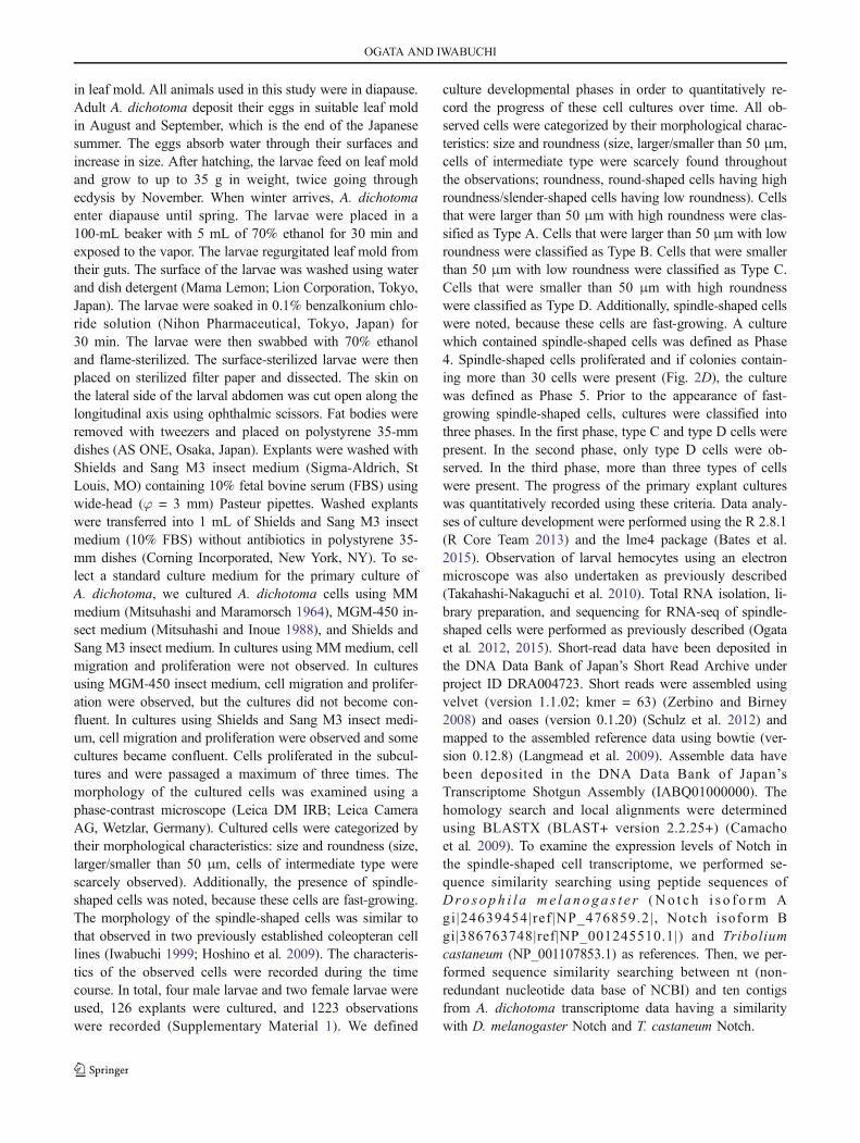
culture developmental phases in order to quantitatively re-cord the progress of these cell cultures over time. All ob-served cells were categorized by their morphological charac-teristics: size and roundness (size, larger/smaller than 50 μm,cells of intermediate type were scarcely found throughoutthe observations; roundness, round-shaped cells having highroundness/slender-shaped cells having low roundness). Cellsthat were larger than 50 μm with high roundness were clas-sified as Type A. Cells that were larger than 50 μm with lowroundness were classified as Type B. Cells that were smallerthan 50 μm with low roundness were classified as Type C.Cells that were smaller than 50 μm with high roundnesswere classified as Type D. Additionally, spindle-shaped cellswere noted, because these cells are fast-growing. A culturewhich contained spindle-shaped cells was defined as Phase4. Spindle-shaped cells proliferated and if colonies contain-ing more than 30 cells were present (Fig. 2D), the culturewas defined as Phase 5. Prior to the appearance of fast-growing spindle-shaped cells, cultures were classified intothree phases. In the first phase, type C and type D cells werepresent. In the second phase, only type D cells were ob-served. In the third phase, more than three types of cellswere present. The progress of the primary explant cultureswas quantitatively recorded using these criteria. Data analy-ses of culture development were performed using the R 2.8.1(R Core Team 2013) and the lme4 package (Bates et al.2015). Observation of larval hemocytes using an electronmicroscope was also undertaken as previously described(Takahashi-Nakaguchi et al. 2010). Total RNA isolation, li-brary preparation, and sequencing for RNA-seq of spindle-shaped cells were performed as previously described (Ogataet al. 2012, 2015). Short-read data have been deposited inthe DNA Data Bank of Japan’s Short Read Archive underproject ID DRA004723. Short reads were assembled usingvelvet (version 1.1.02; kmer = 63) (Zerbino and Birney2008) and oases (version 0.1.20) (Schulz et al. 2012) andmapped to the assembled reference data using bowtie (ver-sion 0.12.8) (Langmead et al. 2009). Assemble data havebeen deposited in the DNA Data Bank of Japan’sTranscriptome Shotgun Assembly (IABQ01000000). Thehomology search and local alignments were determinedusing BLASTX (BLAST+ version 2.2.25+) (Camachoet al. 2009). To examine the expression levels of Notch inthe spindle-shaped cell transcriptome, we performed se-quence similarity searching using peptide sequences ofDro s op h i l a me l a noga s t e r (No t c h i s o f o rm Agi|24639454|ref |NP_476859.2| , Notch isoform Bgi|386763748|ref|NP_001245510.1|) and Triboliumcastaneum (NP_001107853.1) as references. Then, we per-formed sequence similarity searching between nt (non-redundant nucleotide data base of NCBI) and ten contigsfrom A. dichotoma transcriptome data having a similaritywith D. melanogaster Notch and T. castaneum Notch.
OGATA AND IWABUCHI
Results
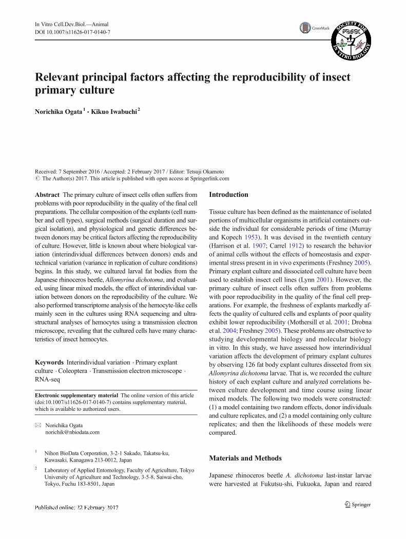
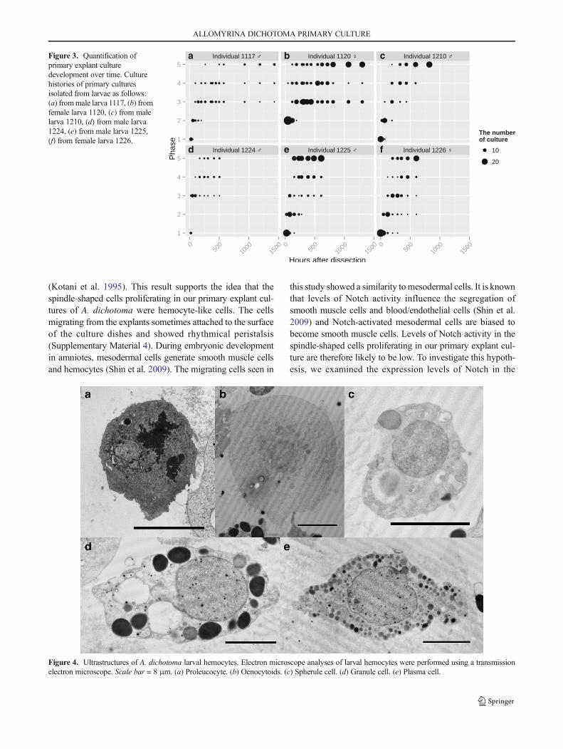
We cultured cells derived from six larvae. Immediately afterdissection, hemocyte-like cells were observed but disappearedwithin a few day. Subsequently, epithelial cell-like cells(Fig. 1), hemocyte-like cells (Fig. 2a), and fibroblast-like cells(Fig. 2b) were observed and then, spindle-shaped cells wereobserved that displayed signs of proliferation (Fig. 2c).Finally, the spindle-shaped cells had increased and some cul-tures became confluent. Progressions of the primary explantcultures were quantitatively recorded (Fig. 3, SupplementaryMaterial 2).
To test whether a random effect was significant in the de-velopment of primary explant cultures, we quantitatively an-alyzed our observations of primary explant culture histories
using a mixed model (Bates et al. 2015). Interindividual andinterdish variations were defined as random effects. We con-structed first-order, second-order, third-order, fourth-order,fifth-order, and sixth-order equations. The phase of each ofthe dishes (phase) was explained by the time elapsed sincedissection (time), interindividual variation (individual), andinterdish variation (dish). The Akaike Information Criterion(AIC) (Akaike 1974) were 3145 (first order eq.), 2978 (secondorder eq.), 2814 (third order eq.), 2777 (fourth order eq.), 2845(fifth order eq.), and 2939 (sixth order eq.). The model that hasthe lowest AIC is optimal; therefore, we selected the fourth-order equation and constructed two models which includeddifferent random effects.
A model that included two random effects, the individualand the dish, was constructed in R language:
model1 < ‐lmer
state∼I time4� �þ I time3
� �þ I time2� �þ timeþ timejindividual
.dish
� �; data ¼ all
� �
A second model, which included a random effect and thedish, was constructed in R language:
model2 < ‐lmer
state∼I time4� �þ I time3
� �þ I time2� �þ timeþ timejdishð Þ; data ¼ all
� �
The AIC of model 1 and model 2 was 2729 and 2873,respectively. Likelihood ratio testing revealed that model 1explained the culture histories better than model 2(p < 0.001). The estimated value of random effects was 0.53(individual, time-independent), 0.61 (dish, time-independent),8.7 × 10−3 (individual, time-dependent), and 3.8 × 10⁻³ (dish,time-dependent). These results show that interindividual var-iation affects the initial state of development of primary ex-plant cultures, which implies a poor reproducibility of primaryexplant culture. Cultured explants of A. dichotoma larvae dis-sected in March and April demonstrated wider interindividualvariation (data not shown).
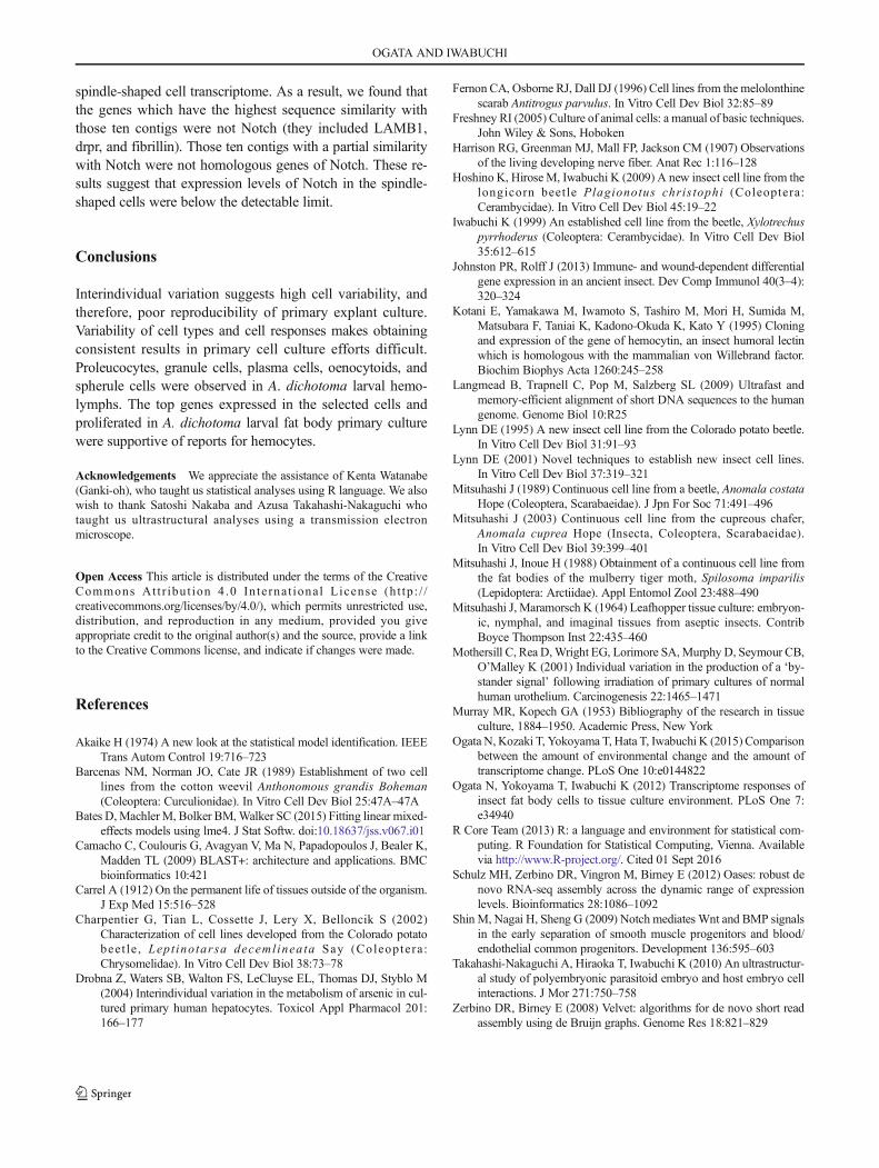
Immediately after dissection, hemocyte-like cells (granulo-cyte-like and plasmatocyte-like cells) were observed. Othertypes of hemocytes that were not adherent might have beenremoved during explant washing. Electron microscope analy-ses of larval hemocytes from the Japanese rhinoceros beetlerevealed the presence of proleucocytes, oenocytoids, spherulecells, granule cells, and plasma cells (Fig. 4).
Our previous study revealed that cultured Bombyx morifat bodies dedifferentiate 80 h after dissection (Ogata et al.
2012). Our statistical analysis showed that interindividualvariation affects the initial state of development of primaryexplant cultures. The interindividual variation detected inthis study could be explained by the interindividual dediffer-entiation ability within cells derived from individual animalswhich would have variations in cell physiology whenharvested.
Discussion
In this study, cell migration from explants and cell prolifera-tion were observed using A. dichotoma larval fat body cellscultured with Shields and Sang M3 insect medium containing10% FBS. Despite the fact that Coleoptera is the largest orderin the animal kingdom, cell lines are vastly underrepresented.Cell culture media which have been successfully used includeMGM-450 insect medium, DCCM media, Schneider’s B me-dia, Shields and Sang M3 insect medium, and EX-CELL 400(Barcenas et al. 1989; Mitsuhashi 1989; Lynn 1995; Fernon
ALLOMYRINA DICHOTOMA PRIMARY CULTURE
et al. 1996; Iwabuchi 1999; Charpentier et al. 2002;Mitsuhashi 2003; Hoshino et al. 2009).
The top ten expressed genes in spindle-shaped cells weresimilar to that of the hemocytin, alpha-l1 nicotinic acetyl cho-line receptor, tubulin alpha-1 chain, apolipophorins, andpolyadenylate-binding protein 4-l ike isoform ×1
(Supplementary Material 3). There was no common gene be-tween these ten genes and previously reported differentialexpressed genes in response to wounding and/or immunechallenge in an insect (Johnston and Rolff 2013). Hemocytinis an adhesive protein and roles in hemostasis or encapsulationof foreign substances for self-defense have been suggested
Figure 1. Morphology of platedcells from A. dichotoma primaryexplants. At 48 h after dissection,epithelial cell-like cells were ob-served. Scale bar = 50 μm. Allpictures were taken using a phase-contrast microscope.
Figure 2. Morphology of cells inA. dichotoma primary culture. Allpictures were taken using a phase-contrast microscope. (a)Hemocyte-like cells. Scalebar = 50 μm. (b) Fibroblast-likecells. Scale bar = 100 μm. (c)Spindle-shaped cells. Scalebar = 50 μm. (d) A colony ofspindle-shaped cells. Scalebar = 100 μm.
OGATA AND IWABUCHI
(Kotani et al. 1995). This result supports the idea that thespindle-shaped cells proliferating in our primary explant cul-tures of A. dichotoma were hemocyte-like cells. The cellsmigrating from the explants sometimes attached to the surfaceof the culture dishes and showed rhythmical peristalsis(Supplementary Material 4). During embryonic developmentin amniotes, mesodermal cells generate smooth muscle cellsand hemocytes (Shin et al. 2009). The migrating cells seen in
this study showed a similarity tomesodermal cells. It is knownthat levels of Notch activity influence the segregation ofsmooth muscle cells and blood/endothelial cells (Shin et al.2009) and Notch-activated mesodermal cells are biased tobecome smooth muscle cells. Levels of Notch activity in thespindle-shaped cells proliferating in our primary explant cul-ture are therefore likely to be low. To investigate this hypoth-esis, we examined the expression levels of Notch in the
Figure 4. Ultrastructures of A. dichotoma larval hemocytes. Electron microscope analyses of larval hemocytes were performed using a transmissionelectron microscope. Scale bar = 8 μm. (a) Proleucocyte. (b) Oenocytoids. (c) Spherule cell. (d) Granule cell. (e) Plasma cell.
a b c
d e f
Figure 3. Quantification ofprimary explant culturedevelopment over time. Culturehistories of primary culturesisolated from larvae as follows:(a) frommale larva 1117, (b) fromfemale larva 1120, (c) from malelarva 1210, (d) from male larva1224, (e) from male larva 1225,(f) from female larva 1226.
ALLOMYRINA DICHOTOMA PRIMARY CULTURE
spindle-shaped cell transcriptome. As a result, we found thatthe genes which have the highest sequence similarity withthose ten contigs were not Notch (they included LAMB1,drpr, and fibrillin). Those ten contigs with a partial similaritywith Notch were not homologous genes of Notch. These re-sults suggest that expression levels of Notch in the spindle-shaped cells were below the detectable limit.
Conclusions
Interindividual variation suggests high cell variability, andtherefore, poor reproducibility of primary explant culture.Variability of cell types and cell responses makes obtainingconsistent results in primary cell culture efforts difficult.Proleucocytes, granule cells, plasma cells, oenocytoids, andspherule cells were observed in A. dichotoma larval hemo-lymphs. The top genes expressed in the selected cells andproliferated in A. dichotoma larval fat body primary culturewere supportive of reports for hemocytes.
Acknowledgements We appreciate the assistance of Kenta Watanabe(Ganki-oh), who taught us statistical analyses using R language. We alsowish to thank Satoshi Nakaba and Azusa Takahashi-Nakaguchi whotaught us ultrastructural analyses using a transmission electronmicroscope.
Open Access This article is distributed under the terms of the CreativeCommons At t r ibut ion 4 .0 In te rna t ional License (h t tp : / /creativecommons.org/licenses/by/4.0/), which permits unrestricted use,distribution, and reproduction in any medium, provided you giveappropriate credit to the original author(s) and the source, provide a linkto the Creative Commons license, and indicate if changes were made.
References
Akaike H (1974) A new look at the statistical model identification. IEEETrans Autom Control 19:716–723
Barcenas NM, Norman JO, Cate JR (1989) Establishment of two celllines from the cotton weevil Anthonomous grandis Boheman(Coleoptera: Curculionidae). In Vitro Cell Dev Biol 25:47A–47A
Bates D,Machler M, Bolker BM,Walker SC (2015) Fitting linear mixed-effects models using lme4. J Stat Softw. doi:10.18637/jss.v067.i01
Camacho C, Coulouris G, Avagyan V, Ma N, Papadopoulos J, Bealer K,Madden TL (2009) BLAST+: architecture and applications. BMCbioinformatics 10:421
Carrel A (1912) On the permanent life of tissues outside of the organism.J Exp Med 15:516–528
Charpentier G, Tian L, Cossette J, Lery X, Belloncik S (2002)Characterization of cell lines developed from the Colorado potatobeet le , Lept inotarsa decemlineata Say (Coleoptera :Chrysomelidae). In Vitro Cell Dev Biol 38:73–78
Drobna Z, Waters SB, Walton FS, LeCluyse EL, Thomas DJ, Styblo M(2004) Interindividual variation in the metabolism of arsenic in cul-tured primary human hepatocytes. Toxicol Appl Pharmacol 201:166–177
Fernon CA, Osborne RJ, Dall DJ (1996) Cell lines from the melolonthinescarab Antitrogus parvulus. In Vitro Cell Dev Biol 32:85–89
Freshney RI (2005) Culture of animal cells: a manual of basic techniques.John Wiley & Sons, Hoboken
Harrison RG, Greenman MJ, Mall FP, Jackson CM (1907) Observationsof the living developing nerve fiber. Anat Rec 1:116–128
Hoshino K, Hirose M, Iwabuchi K (2009) A new insect cell line from thelongicorn beetle Plagionotus chris tophi (Coleoptera:Cerambycidae). In Vitro Cell Dev Biol 45:19–22
Iwabuchi K (1999) An established cell line from the beetle, Xylotrechuspyrrhoderus (Coleoptera: Cerambycidae). In Vitro Cell Dev Biol35:612–615
Johnston PR, Rolff J (2013) Immune- and wound-dependent differentialgene expression in an ancient insect. Dev Comp Immunol 40(3–4):320–324
Kotani E, Yamakawa M, Iwamoto S, Tashiro M, Mori H, Sumida M,Matsubara F, Taniai K, Kadono-Okuda K, Kato Y (1995) Cloningand expression of the gene of hemocytin, an insect humoral lectinwhich is homologous with the mammalian von Willebrand factor.Biochim Biophys Acta 1260:245–258
Langmead B, Trapnell C, Pop M, Salzberg SL (2009) Ultrafast andmemory-efficient alignment of short DNA sequences to the humangenome. Genome Biol 10:R25
Lynn DE (1995) A new insect cell line from the Colorado potato beetle.In Vitro Cell Dev Biol 31:91–93
Lynn DE (2001) Novel techniques to establish new insect cell lines.In Vitro Cell Dev Biol 37:319–321
Mitsuhashi J (1989) Continuous cell line from a beetle, Anomala costataHope (Coleoptera, Scarabaeidae). J Jpn For Soc 71:491–496
Mitsuhashi J (2003) Continuous cell line from the cupreous chafer,Anomala cuprea Hope (Insecta, Coleoptera, Scarabaeidae).In Vitro Cell Dev Biol 39:399–401
Mitsuhashi J, Inoue H (1988) Obtainment of a continuous cell line fromthe fat bodies of the mulberry tiger moth, Spilosoma imparilis(Lepidoptera: Arctiidae). Appl Entomol Zool 23:488–490
Mitsuhashi J, Maramorsch K (1964) Leafhopper tissue culture: embryon-ic, nymphal, and imaginal tissues from aseptic insects. ContribBoyce Thompson Inst 22:435–460
Mothersill C, Rea D,Wright EG, Lorimore SA,Murphy D, Seymour CB,O’Malley K (2001) Individual variation in the production of a ‘by-stander signal’ following irradiation of primary cultures of normalhuman urothelium. Carcinogenesis 22:1465–1471
Murray MR, Kopech GA (1953) Bibliography of the research in tissueculture, 1884–1950. Academic Press, New York
Ogata N, Kozaki T, Yokoyama T, Hata T, Iwabuchi K (2015) Comparisonbetween the amount of environmental change and the amount oftranscriptome change. PLoS One 10:e0144822
Ogata N, Yokoyama T, Iwabuchi K (2012) Transcriptome responses ofinsect fat body cells to tissue culture environment. PLoS One 7:e34940
R Core Team (2013) R: a language and environment for statistical com-puting. R Foundation for Statistical Computing, Vienna. Availablevia http://www.R-project.org/. Cited 01 Sept 2016
Schulz MH, Zerbino DR, Vingron M, Birney E (2012) Oases: robust denovo RNA-seq assembly across the dynamic range of expressionlevels. Bioinformatics 28:1086–1092
Shin M, Nagai H, Sheng G (2009) Notch mediates Wnt and BMP signalsin the early separation of smooth muscle progenitors and blood/endothelial common progenitors. Development 136:595–603
Takahashi-Nakaguchi A, Hiraoka T, Iwabuchi K (2010) An ultrastructur-al study of polyembryonic parasitoid embryo and host embryo cellinteractions. J Mor 271:750–758
Zerbino DR, Birney E (2008) Velvet: algorithms for de novo short readassembly using de Bruijn graphs. Genome Res 18:821–829
OGATA AND IWABUCHI