relief of labor pain by regional analgesia/anesthesia · absence of total spinal anesthesia via the...
TRANSCRIPT

9Relief of Labor Pain
by RegionalAnalgesia/Anesthesia
�Epidural Analgesia . . . . . . . . . . . . . . . . . . . . . 109
Anatomy of the Epidural Space . . . . . . . . . . . . . 109Contents of the Epidural Space . . . . . . . . . . . . . 110Site of Action . . . . . . . . . . . . . . . . . . . . . . . 111Techniques . . . . . . . . . . . . . . . . . . . . . . . . 111Epidural Analgesia Procedure . . . . . . . . . . . . . . 112
Multiorifice Versus Uniorifice Epidural Catheters . . . 113Changes in the Position of Epidural
Catheters Associated with Patient Movement . . . 114Test Dose . . . . . . . . . . . . . . . . . . . . . . . . 115Initial Bolus Administration . . . . . . . . . . . . . . 118Maintenance of Labor Analgesia . . . . . . . . . . . 118Local Anesthetic and Opioid Infusion . . . . . . . . . 120
Possible Block-Related Problems . . . . . . . . . . . . . 121Inadequate Perineal Analgesia . . . . . . . . . . . . . . 121Asymmetric Sensory Block . . . . . . . . . . . . . . . . 121Diminishing Analgesia . . . . . . . . . . . . . . . . . . 121Dense Motor Block . . . . . . . . . . . . . . . . . . . . 122Patchy Block . . . . . . . . . . . . . . . . . . . . . . . 122Miscellaneous . . . . . . . . . . . . . . . . . . . . . . 123Spinal Anesthesia . . . . . . . . . . . . . . . . . . . . 123
Continuous Spinal Anesthesia . . . . . . . . . . . . . . . 123Combined Spinal/Epidural (CSE) . . . . . . . . . . . . . 124Monitoring Following Administration
of Regional Analgesia . . . . . . . . . . . . . . . . . . 126Contraindications . . . . . . . . . . . . . . . . . . . . . . 126Complications of Regional Analgesia . . . . . . . . . . . 127
Paresthesia . . . . . . . . . . . . . . . . . . . . . . . . 127Accidental Dural Puncture . . . . . . . . . . . . . . . . 127Treatment of Headache Following Accidental
Dural Puncture . . . . . . . . . . . . . . . . . . . . . 128
S. Datta et al., Obstetric Anesthesia Handbook,DOI 10.1007/978-0-387-88602-2_9,C© Springer Science+Business Media, LLC 2006, 2010

108 Regional Analgesia/Anesthesia
Conservative Management . . . . . . . . . . . . . . 128Epidural Saline . . . . . . . . . . . . . . . . . . . . . 128Prophylactic Blood Patch . . . . . . . . . . . . . . . 129Therapeutic Blood Patch . . . . . . . . . . . . . . . 129Experimental and Alternative Techniques . . . . . . . 131
Subdural Injection . . . . . . . . . . . . . . . . . . . . 131Massive Epidural Analgesia . . . . . . . . . . . . . . . 132Accidental Intravascular Injection . . . . . . . . . . . . 132Methemoglobinemia . . . . . . . . . . . . . . . . . . . 133Broken Epidural Catheter . . . . . . . . . . . . . . . . 133Shivering . . . . . . . . . . . . . . . . . . . . . . . . . 133Horner’s Syndrome . . . . . . . . . . . . . . . . . . . . 133Backache . . . . . . . . . . . . . . . . . . . . . . . . . 134Major Neurologic Injury . . . . . . . . . . . . . . . . . 134
Obstetric Causes . . . . . . . . . . . . . . . . . . . . 135Anesthesia-Related Causes . . . . . . . . . . . . . . 138
Other Methods of Regional Anesthesia . . . . . . . . . . 141Caudal Anesthesia . . . . . . . . . . . . . . . . . . . . 142Paracervical Block . . . . . . . . . . . . . . . . . . . . 142Lumbar Sympathetic Block . . . . . . . . . . . . . . . 142Pudendal Block . . . . . . . . . . . . . . . . . . . . . . 142
Summary . . . . . . . . . . . . . . . . . . . . . . . . . . 143
By far the most popular form of labor pain relief is regionalanalgesia. A number of techniques are possible for the differentphases of labor and are listed in Table 9-1. This chapter will
Table 9–1. Techniques Used for Relief of Labor Pain
First Stage Second Stage
(1) Epidural analgesia (1) Epidural analgesia(2) Continuous spinal
analgesia and anesthesia (2) Spinal anesthesia
(3) Combined spinal-epiduraltechnique (CSE)
(3) Combined spinal-epiduraltechnique (CSE)
(4) Caudal analgesia (4) Caudal analgesia(5) Paracervical block (5) Pudendal nerve block(6) Bilateral sympathetic block

Regional Analgesia/Anesthesia 109
focus on central neuraxial techniques, including epidural andcombined spinal-epidural analgesia.
Epidural Analgesia
This is the most commonly employed procedure for both thefirst and second stages of labor. It offers greater benefits whencompared with any other anesthetic methods for labor anddelivery.
Anatomy of the Epidural Space
Recent cryomicrotome studies of fresh cadaveric specimenshave advanced the knowledge of epidural spaces for the anes-thesiologist. The epidural space is partially a potential space, inthat it is “empty” but the dura and ligamentum flavum are notadherent, so that it may be expanded to accept catheters anddrugs. Contents of the epidural space as described by Hoganare contained in a series of “metamerically and circumfer-entially discontinuous compartments” (parts at each vertebralsegment filled with fat and other contents) separated by zoneswhere the dura contacts the canal wall. The dura tapers offinferior to the L4–L5 disc in the sacral canal, and the spaceis usually filled by the epidural fat.1 The posterior epiduralspace is occupied at each segment by a fat pad in the triangularspace between the ligamenta flava and dura. Hogan observedno midline fibrous septum. However, the presence of midlinefatty tissue can potentially cause uneven spread of local anes-thetic and result in a patchy or unilateral block. When unilateralblock occurs, it is usually on the right side, though the mech-anism of this finding is unclear.2 The distance from the skin tothe epidural space has been observed with graduated needlesand by using ultrasound and magnetic resonance examination.The depth varies considerably from 3 cm to 9 cm; the averagedepth is 4.5–5.5 cm.3
The posterior epidural space is triangular in shape, with theapex facing posteriorly (Fig. 9-1), and lies between the duramater and the ligamentum flavum. The epidural space extends

110 Regional Analgesia/Anesthesia
Figure 9–1. Contents of the epidural space. (FromAbouleish.79 Used with permission.)
from the base of the skull to the sacral hiatus and is boundedas follows:1. Superiorly by the dura adherent to the skull at the foramen
magnum. The clinical implication of this is related to theabsence of total spinal anesthesia via the epidural route.
2. Inferiorly by the sacrococcygeal ligament at the level of theS2–3 interspace.
3. Anteriorly by the posterior longitudinal ligament (lyinganterior to the dural sac).
4. Posteriorly by the ligamentum flavum.5. Laterally by the dural cuffs, pedicles, and lamina.
Contents of the Epidural Space
The epidural space is sometimes characterized as a “poten-tial” space in that it can be expanded by infused local anes-thetic. However, the space is not empty prior to the block, andindeed contains several important structures:1. Anterior and posterior nerve roots with their coverings.2. Blood vessels that supply the spinal cord.
a. The posterior spinal artery, which originates from theinferior cerebellar artery and supplies the posteriorcolumns and posterior horns.
b. The anterior spinal artery, which originates from the twovertebral arteries at the foramen magnum and suppliesthe anterior portion of the spinal cord.
c. The artery of Adamkiewicz, which is the major feeder ofthe anterior spinal artery and arises from one intercostal

Regional Analgesia/Anesthesia 111
or lumbar artery in the T8–L3 region. It supplies thelower two-thirds of the spinal cord.
d. The vertebral veins, which drain blood from the verte-bral column and the nervous tissue and ultimately formthe vertebral venous plexus. They run via the anterolat-eral part of the epidural space and ultimately drain intothe azygos vein. This venous connection is associatedwith important clinical implications: during pregnancydue to obstruction of the inferior vena cava, epiduraland azygos vein blood flow is markedly increased. Asmall dose of local anesthetic injected accidentally intothe epidural vein, especially during labor, can reach theheart in a higher concentration4 and thus increase thechances of myocardial depression.
3. Fatty areolar tissue, deposited between the nervous andvascular structures.
Site of Action
The site of action of the local anesthetic during epidu-ral analgesia is not exactly known; however, several siteshave been suggested: (1) spinal roots, the most important site;(2) mixed spinal nerve; (3) dorsal root ganglion; and (4) thespinal cord, which might be the ultimate site of action and playsan important role in regression of a prolonged epidural block.
Techniques
For theoretical purposes, segmental block and completeblock techniques have been described; in practice, however,they overlap and form contiguous processes. Segmental blockmay be used in the first stage to limit the extent of sensoryanalgesia to the T10–L1 segments. As labor progresses to thesecond stage, analgesia can be extended to block the sacralinnervations. A top-up dose may be required for this purposewhile the woman is in the sitting position for about 5 min. If aforceps delivery is planned or if cesarean section becomes nec-essary, a higher concentration of local anesthetic may be usedto achieve motor block and perineal relaxation. In a completeblock (T10–S5) sensory analgesia from T10 to S5 is provided

112 Regional Analgesia/Anesthesia
from the very first dose, but theoretically the incidence ofhypotension may be higher than when a segmental approachis used.
Epidural Analgesia Procedure
The following materials are needed for epidural placement:1. An epidural tray (with catheter)2. Local anesthetic agent: Typically, bupivacaine,
0.0625–0.5%, or ropivicaine 0.1–0.2% (more dilutesolutions are popular in present practice); and an epiduralinfusion, either premixed or prepared from sterile salinesolution in a 50- to 100-mL sterile plastic bag. Popular infu-sions include bupivacaine with fentanyl (0.0625–0.125%bupivacaine with 2 mcg/ml fentanyl).
3. A volumetric infusion pump or commercially availableepidural pumps. The latter include important safety featuresnot found on generic infusion pumps.
4. Fentanyl or sufentanil, if no premixed bags of epiduralsolutions are available, to achieve the final desired concen-tration (e.g., 2 mcg/ml fentanyl or 0.5 mcg/ml sufentanil)
5. Resuscitative equipment and drugs, including oxygen anddelivery apparatus (facemasks, resuscitation bag), oral andnasal airways and endotracheal tubes, laryngoscope, car-diac monitor, induction drugs, and succinylcholineAt the Brigham and Women’s Hospital, every effort is
made to consult the parturient before induction of epidu-ral anesthesia and informed consent is signed by thepatient. Brigham and Women’s also provides informationvia printed booklet and website (www.painfreebirthing.com,or http://www.brighamandwomens.org/painfreebirthing). Dep-ending on the preference of the anesthesiologist, the techniqueis performed with the patient either in sitting position or inlateral position. One study showed that epidural catheter inser-tions in the lateral position are associated with decreasedincidence of intravascular catheters.5 The other advantagesclaimed in favor of lateral position are: for the mother, thelateral position is less physiologically demanding and reducesthe need to abandon the technique due to vagal reflexes;the lateral position also enables the provision of neuraxialanalgesia (and anesthesia) in the event of complex presenta-tion; for the fetus, an improvement in blood flow resulting

Regional Analgesia/Anesthesia 113
in better gas exchange.6 The left lateral position is preferredto the right lateral position as the former is associated withbetter maintenance of uterine blood flow. The arguments infavor of sitting position include the technical ease of inser-tion, superior patient comfort, possible improved analgesiafor combined spinal epidural (CSE), and decreased aorto-caval compression.7 However, in obese women, physiciansat Brigham and Women’s, like many others, prefer a sittingposition.
Observation of the fetal heart rate is of paramount impor-tance before the introduction of epidural anesthesia. A vol-ume of 500–1,000 ml of Ringer’s lactate solution is used foracute volume replacement unless contraindicated, althoughevidence supporting this practice in preventing hypotension islacking.8 Sodium citrate, 30 mL, is given p.o. routinely beforethe induction of epidural analgesia. At Brigham and Women’sHospital, the Weiss modification of the Tuohy needle is rou-tinely used. About 50% of anesthesiologists in the authors’institution use the technique of loss of resistance by air. Othersprefer loss of resistance to saline technique. The superiority ofone technique over the other is debatable (Arendt and Segal,Rev Obstet Gynecol 2008;1:49–55). However, using minimalquantities of air or saline to detect epidural space is equallyeffective. The L2–3 or L3–4 interspace is usually used for intro-duction of the epidural needle. Tuffier’s line, a line drawnfrom the top of the iliac crest, coincides with either the L4–5 interspace or the L4 spinous process, though the accuracyof identification of interspaces is quite low. Once the space isidentified by using either the loss-of-resistance to air or saline,3–5 cm of the epidural catheter is inserted into the epiduralspace. The length of the catheter inserted into the epiduralspace depends on the type of catheter as discussed in the fol-lowing section. Some practitioners prefer not inserting morethan 3 cm of the catheter into the epidural space unless thepatient is obese. This technique may decrease the incidence ofa unilateral block.
Multiorifice Versus Uniorifice Epidural Catheters
Multiorifice catheters have recently become increas-ingly popular as they decrease the incidence of unilateral

114 Regional Analgesia/Anesthesia
blocks.9–11 Beilin et al. studied 100 women in a prospective,randomized, and double-blind study. Patients were randomlyassigned to have a multiorifice epidural catheter threaded 3 cm,5 cm, or 7 cm into the epidural space. After placement of thecatheter and administration of a test dose with 3 mL of 0.25%bupivacaine, an additional 10 mL of 0.25% bupivacaine wasadministered in two divided doses. Fifteen minutes later, theadequacy of the analgesia was assessed by a blinded observer.The authors found that catheter insertion to a depth of 7 cmwas associated with the highest rate of insertion complicationswhile insertion to a depth of 5 cm was associated with the high-est incidence of satisfactory analgesia.12 In another study13
800 healthy parturients requesting epidural analgesia were ran-domized to have open-tip (i.e., single-orifice) epidural cathetersinserted 2 cm, 4 cm, 6 cm, or 8 cm within the epidural space.Epidural catheters inserted 8 cm within the epidural spacewere associated with more intravenous cannulation. Epiduralcatheters inserted 2 cm within the epidural space resulted indecreased incidence in unilateral sensory analgesia but weremore vulnerable for dislodgement during movements of thelaboring women. Twenty-three percent of epidural cathetersinserted > 2 cm within the epidural space required manipu-lation. Therefore, if uniorifice catheters are used, the optimumcatheter insertion is a balance between a good bilateral blockand dislodgement. Based on these studies, some clinicianschoose to insert a uniorifice epidural catheter no more than3–4 cm or a multiorifice catheter no more than 5 cm into theepidural space. Recently, the practice at Brigham and Women’sHospital has changed to using single-orifice soft-tip catheters(open ended Arrow Flex-Tip R©) as these catheters decreasethe chances of intravascular insertion and the incidence ofparasthesia without significantly increasing the incidence ofinadequate blocks (when inserted 3–4 cm).14
Changes in the Position of Epidural CathetersAssociated with Patient Movement
Epidural catheter movement has been noted with changein patient position and can result in inadequate anesthesia.This was investigated by Hamilton et al. in 255 parturients

Regional Analgesia/Anesthesia 115
requesting epidural anesthesia for labor or cesarean section,where a multiorificed lumbar epidural catheter was insertedwith the patient in the sitting flexed position.15 The distanceto the epidural space, length of catheter inserted, and amountof catheter position change as the patient moved from the sit-ting flexed to sitting upright and then to the lateral decubitusposition were measured before the catheter was secured to theskin. Data were grouped according to body mass index (BMI): <25 kg/m2, 25–30 kg/m2, and > 30 kg/m2. Catheters frequentlyappeared to be drawn inward with position change from thesitting flexed to lateral decubitus position, with the greatestchange seen in patients with BMI > 30. Maximum epiduralcatheter position change was 4.28 cm in a patient in the >30 BMI group weighing more than 180 kg. Based on theseresults, the authors recommend that multiorificed catheters beinserted at least 4 cm into the epidural space and that patientsassume the sitting upright or lateral position before securing thecatheter to the skin.15
Test Dose
An ideal test dose should be able to detect both accidentalintravascular and subarachnoid injections of local anesthetics.Moore and Batra originally suggested that the use of 15 μgof epinephrine (1:200,000) with local anesthetic will detectaccidental intravascular injections in nonpregnant patients byshowing tachycardia.16 The heart rates of the 175 patientsincreased from a mean of 79 ± 14 beats per minute to 111 ±15 beats per minute. The heart rate increased within 23 ± 6 sfollowing the injection and returned to baseline within 32 ±33 s. However, the investigators used this test dose only in non-pregnant cases that were undergoing elective surgery and wereunder the influence of heavy premedication. When using 3 mLof 0.5% plain bupivacaine via the epidural route in 100 par-turients in active labor, Cartwright and colleagues17 observedheart rate increases of more than 20 beats per minute in 24women and more than 30 beats per minute in 12 women inthe following 60 s even though the catheters were not intravas-cular. Leighton and colleagues, using 15 μg of epinephrineintravenously in term parturients, observed heart rate increases

116 Regional Analgesia/Anesthesia
of greater than 25 beats per minute that lasted longer than 15 sin only 50% of cases.18
From these early studies it appeared that in pregnant women15 μg of epinephrine might not be sensitive or specific enoughto rule out accidental intravascular injection.19 On the otherhand, Abraham and colleagues, in search of an ideal testdose for both accidental intravascular and subarachnoid injec-tions, used 3 mL of 1.5% hyperbaric lidocaine mixed withepinephrine (1:200,000) via an epidural catheter20 (Fig. 9-2).The maternal heart rate increased from 76 ± 2 beats per minuteto 109 ± 6 beats per minute if the solution was injected intra-venously, and the sensory anesthesia reached the S2 level in1.45 ± 0.12 min if the solution was accidentally injected inthe subarachnoid space. Hence, the use of epinephrine, 15 μg,for the diagnosis of accidental intravascular injections remains
Figure 9–2. Time to onset of objective sensory loss (to pinprick) following epidural and spinal administration of a hyper-baric 1.5% lidocaine solution. (From Abraham.20 Used withpermission.)

Regional Analgesia/Anesthesia 117
controversial. If epinephrine is used in the laboring womenas a test dose, one must first observe the maternal heart ratechange from base to peak of a uterine contraction. Then onecan interpret the effect of intravenous epinephrine.
Other approaches have been advocated. Leighton and col-leagues examined air as a useful clinical indicator of intra-venous placement of the epidural catheter. Using 1 mL of airthrough the epidural catheter and monitoring heart tones witha Doppler ultrasound probe, these authors observed only a2% false-positive rate.21 None of the 303 parturients in theirstudy developed any complications due to the injection of1 mL of air; the authors concluded that air, with precordialDoppler detection, is a safe and effective test for identifyingintravenously located epidural catheters. This technique wasnot further evaluated and has not gained popularity in clini-cal practice. In the meantime, while the search continues foran ideal test dose,22 one should exercise caution in preventingand diagnosing an incorrect placement of the epidural catheter:1. For elective cases, unless contraindicated, one can use an
epinephrine (15 μg)-containing solution as a test dose.Continuous electrocardiographs (ECG) monitor or pulseoximetry is essential to detect tachycardia. In this respect,one must remember that parturients who are being treatedwith β-blocking drugs may not show tachycardia even whenthe epinephrine is intravascular.23
2. Negative aspiration findings may not exclude intravascularcatheter placement because the catheter may be againstthe vein wall and because aspiration can collapse thevein lumen. Aspiration immediately following the injectionof local anesthetic may be more effective in recognizingintravascular catheter placement because the local anes-thetic will push the vein wall away and may also dilatethe blood vessels; placing the catheter 45–50◦ below thepatient’s body level at this stage can help the blood to flowthrough the catheter if it is intravascular.
3. Local anesthetics should be injected only 3–5 mL at atime, and the signs and symptoms of intravascular injec-tion should be closely monitored. Fractionation of bolusesof local anesthetic should prevent toxicity even if there isfalse-negative test dose.

118 Regional Analgesia/Anesthesia
4. Many anesthesiologists have argued for a no test dose tech-nique in which “every dose is a test dose” and signs andsymptoms of intravascular injection are sought every timea bolus of local anesthetic is administered. Brigham andWomen’s Hospital currently prefers this approach.
Initial Bolus Administration
Initial labor analgesia is generally provided with a varietyof local anesthetic agents such as bupivacaine, levobupiva-caine, or ropivacaine. At Brigham and Women’s Hospital, wetypically administer 20 ml of 0.125% bupivacaine for the ini-tial establishment of the pain relief. This is administered infractionated quantities of 5 ml boluses each. This initial vol-ume can be decreased to 15–17 ml if 3 ml of lidocaine with15 μg of epinephrine has been used for the epidural test dose.Other institutions favor more dilute initial boluses, such asbupivacaine or ropivicaine 0.1% or less.
Maintenance of Labor Analgesia
After obtaining initial pain relief with fractionated boluses ofloading dose of the local anesthetic agent, further analgesia canbe provided using intermittent injections or continuous infusiontechniques.
Intermittent technique. Reinforcement is needed every1.5–2 h or if the patient is uncomfortable, with the usual aimto maintain sensory analgesia from T10 to S5.
Continuous analgesia. Can be provided with infusion sys-tems and is a more convenient method for providing satisfac-tory labor analgesia. A 50–100-mL sterile plastic bag is filledwith a 0.0625–0.125% solution of bupivacaine and attachedto the high-pressure infusion tubing (Luerlock). The tubing isflushed to remove any air and then connected directly to theepidural catheter. All connections must be secured, and theplastic bag must be labeled. The epidural (or generic volumet-ric) infusion pump is adjusted to deliver the desired dosageper hour. In the past higher concentrations of local anestheticdrugs, such as bupivacaine 0.25%, were used. The presenttrend is to use lower concentrations of local anesthetics in

Regional Analgesia/Anesthesia 119
attempt to minimize the motor effects of labor analgesia. Drugscommonly used are bupivacaine or levobupivacaine (0.0625–0.125%) or ropivacaine (0.1–0.2%) at 8–10 mL/h. The marginaldifferences in effectiveness between these agents are debat-able but they all provide adequate analgesia with no significantinfluence on mode of delivery, duration of labor, or neonataloutcome.24,25 In some studies bupivacaine has been attributedto more motor block than ropivacaine. However, this is notconclusively proved in other studies and may simply reflecta potency difference between the drugs.26 In addition to thelocal anesthetic agents that are in use currently, other drugssuch as 1% 2-chloroprocaine and 1% lidocaine have alsobeen used in the past. They can be used under exceptionalcircumstances.
Continuous infusion for epidural analgesia in obstetrics wasfirst described in 1963.27 However, the technique did notbecome popular, mainly because of the lack of availabilityof proper instruments as well as local anesthetics. With theadvent of better mechanical infusion pumps as well as bet-ter local anesthetics, continuous infusion has indeed becomethe technique of choice for vaginal delivery. Different authorshave compared the intermittent injection technique with con-tinuous epidural infusion (CEI), and the potential advantages ofCEI include28:1. a more stable depth of analgesia, which obviously becomes
an important part of patient satisfaction;2. the possibility of lower blood concentrations of local anes-
thetic, both by absorption from the epidural space and if thecatheter is accidentally placed in the vein;
3. a reduced risk of total spinal block in the presence of aninadvertent injection of local anesthetic in the subarachnoidspace;
4. a lower incidence of hypotension due to the possibility ofdecreased sympathetic blockadePatient-controlled epidural analgesia (PCEA). PCEA has
become popular at the present time29,30 and is standard prac-tice at Brigham and Women’s Hospital. At BWH it is usedwith a background infusion (bupivacaine 0.125% with fentanyl2 μg/ml, 6 ml/h, 6 ml patient demand bolus, 15 min lockoutbetween demands, no 4 h limit). This technique offers more

120 Regional Analgesia/Anesthesia
patient control over the level of the block and requires fewerphysician interventions compared to the continuous infusiontechnique.
General considerations during epidural maintenance.During the maintenance phase of labor analgesia with anyof the techniques described above, parturients should bepositioned head-up with left uterine displacement. Head-upposition facilitates adequate perineal anesthesia as the patientprogresses from first stage to second stage of labor. Routinemonitoring of maternal vital signs, the fetal heart rate, anduterine contractions are essential during this phase. Notationsabout the block and vital signs should be made every 1–2 hon the hospital record. One must remember that even withthe continuous infusion technique one should frequently checkthe block to verify uniformity and rule out subarachnoid orintravascular migration of the catheter.
Local Anesthetic and Opioid Infusion
A combination of lower concentrations of local anesthet-ics and lipophilic opioids has popularized CEI and PCEAtechniques even further. This combination offers a superioranalgesia than local anesthetics alone.31 Different authors haveinvestigated the efficacy of low concentrations of local anes-thetics combined with opioids and have claimed moderateto good success.32–34 The most popular cocktail at presentis the combination of 0.0625% or 0.125% bupivacaine and2 μg/ml of fentanyl infused at the rate of 8–10 mL/h. Alfentanil(5 μg/mL) has been tried in our institution in combinationwith bupivacaine (0.125% at 8–10 mL/h) with excellent suc-cess. However, placental transfer of alfentanil is significantand hence this drug has never become popular. Sufentanil0.5 μg/ml is used in some institutions, although no clearadvantage has been demonstrated, and it is more expensive.Morphine and hydromorphone, which are more hydrophilicopioids, have not proven satisfactory in labor analgesia (thoughthey may be quite effective after cesarean delivery in largerdoses).
Bupivacaine in even lower concentrations of 0.04–0.0625%with opioid has also been tried with varying success; obviously,

Regional Analgesia/Anesthesia 121
the lower concentration will be associated with minimal motorblockade, which might benefit the parturients if the sensoryanalgesia is adequate.33 Other local anesthetics such as ropiva-caine (0.2%) and levobupivacaine (0.125%) with fentanyl havealso been used with success.24
Possible Block-Related Problems
Inadequate Perineal Analgesia
At the time of delivery one must make sure of the pres-ence of adequate perineal analgesia. If additional analgesia isdeemed necessary, bupivacaine 0.125% (6–8 ml), or 0.25%(3–4 ml) with fentanyl 50–100 μg offers good pain relieffor pushing the baby without augmenting the motor block.Placing the parturient in the semi-Fowler’s position for “push-ing” also helps achieve a more complete perineal block.Additionally, lidocaine 1.5–2%, bupivacaine 0.25–0.5%, or2–3% 2-chloroprocaine may be required to provide sufficientanalgesia or anesthesia if forceps delivery or episiotomy iscontemplated.
Asymmetric Sensory Block
If a unilateral block is encountered after initial dosing, theepidural catheter can be withdrawn by 1 cm and in mostcases the block will become bilateral after supplemental 3–5 ml of bupivacaine 0.25% or 6–10 ml of 0.125%.2 If aparturient lies continuously on one side, the level of sensoryblock may become asymmetric. The situation should be cor-rected by repositioning the patient, and administering 4–6 mLof 0.25% or 8–12 mL of 0.125% bupivacaine mixed with fen-tanyl 2 μg/ml solution (bolus dose) after appropriate aspiration,and then the infusion should be restarted. The patient shouldbe encouraged to turn from side to side to maintain uterinedisplacement.
Diminishing Analgesia
Progressive diminution of the sensory block and loss of theblock may be due to a number of factors:

122 Regional Analgesia/Anesthesia
1. The pump on/off switch may be off.2. The tubing may be disconnected.3. The reservoir bag may be empty.4. The catheter may no longer be in the epidural space, and
intravascular migration must be ruled out.The differential diagnosis should consist of rechecking the
infusion pump setup and testing to determine the correct posi-tion of the catheter. After aspiration, a 3-mL test dose of 1.5–2%lidocaine or 0.25% bupivacaine with or without 1:200,000epinephrine is injected. The patient is observed for signs ofintravascular placement of the catheter, and if there is nega-tive response, an attempt is made to re-establish the block with3–5-mL incremental doses of 0.125–0.25% bupivacaine. If theblock cannot be re-established or if aspiration and testing indi-cate intravascular migration of the tip, then the catheter mustbe removed. Depending on the clinical setting, either a newcatheter may be inserted via a second placement, or alterna-tive analgesia may be initiated. At the above infusion rates,bupivacaine probably will not produce symptoms of intravas-cular injection. The only clue may be diminishing or absentanalgesia.
Dense Motor Block
Patients given a continuous infusion of 0.0625–0.125%bupivacaine usually exhibit mild motor blockade of the lowerextremities. If a progressively dense motor blockade resem-bling a subarachnoid block ensues, the catheter must bedisconnected immediately and careful aspiration performedto rule out subarachnoid migration. A suspicion of subarach-noid migration after testing mandates either withdrawal of thecatheter and reinsertion at another site or management as acontinuous spinal catheter (see below).
Patchy Block
If a spotty or patchy block occurs, one should attempt tosolidify the block by aspirating to determine catheter place-ment and injecting 4–6 mL of 0.125–0.25% bupivacaine. Then

Regional Analgesia/Anesthesia 123
the pump is reconnected. Initially patchy blocks may be dueto misplacement of the epidural catheter (subdural, paraverte-bral, catheter threaded over a nerve root) and consideration ofimmediate replacement should be given.
Miscellaneous
If a woman requires an acute change in the character ofthe block for operative delivery, simply increasing the infusionrate will be inadequate. The parturient must be disconnectedfrom the pump, the catheter placement must be checked, andthe epidural catheter should then be “topped up” with 0.5%bupivacaine, 2% lidocaine with or without epinephrine, or3% 2-chloroprocaine to obtain the desired level of sensoryanesthesia (to at least a bilateral T4 level for cesarean delivery).
Spinal Anesthesia
Single-shot spinal anesthesia has a very limited role in laborand vaginal delivery. Spinal anesthesia will relax the pelvicfloor muscle and will thus disturb the integrity of the birth pas-sage; the expulsive powers can also be diminished by blockadeof the abdominal segments. Because spinal anesthesia pro-duces an intense motor block of the pelvic floor muscle, itis a desirable technique for forceps delivery. At Brigham andWomen’s Hospital, we aim for a T10–S5 block in all forcepsdeliveries except in the case of a trial of forceps, where wemay aim for a higher block (T4) in case cesarean delivery isrequired. Consultation with the obstetrician regarding the like-lihood of success with forceps can help guide the level ofanesthesia required. Drugs that can be used are (1) lidocaine,(2) mepivacaine, and (3) bupivacaine.
Continuous Spinal Anesthesia
Continuous spinal anesthesia can be used if there is an acci-dental or intentional dural puncture. The epidural catheter isinserted 3 cm in the subarachnoid space. Advantages of the

124 Regional Analgesia/Anesthesia
continuous spinal catheter technique include the following:(1) small doses of local anesthetics are needed, (2) rapid onsetof action, (3) quick recovery because of the small dose, (4) theabsence of accidental intravenous injections of large doses oflocal anesthetic, and (5) the possibility of the use of smalldoses of intraspinal opioids for the relief of labor pain in afew special situations. At Brigham and Women’s, we administer2.5–3 mg bupivacaine with 25–30 μg of fentanyl via the spinalcatheter as an initial bolus, followed by a continuous infusionof bupivacaine 0.125% with fentanyl 2 μg/ml at 1 ml/h. Ifthe patient becomes uncomfortable during the course of thelabor, an additional bolus of 1 ml of the mix or 15–25 μgfentanyl provides comfort. Occasionally, the infusion has tobe gradually increased to 2 ml/h in a graded fashion. Sincethe local anesthetic mixture is hypobaric at body temperature,there is a tendency of inadequate block on the dependent sidein lateral positions. This is usually remedied by changing thepatient one side to the other before reinforcement with localanesthetic mix.
Combined Spinal/Epidural (CSE)
The CSE technique has become popular since the introduc-tion of neuraxial opioids. The epidural needle is first insertedin the epidural space with loss-of-resistance technique. Then along pencil-point spinal needle (25- or 27-gauge) is insertedvia the epidural needle. This spinal needle usually extends12 mm beyond the tip of the epidural needle. With theappearance of free-flowing CSF, a mixture of lipid-soluble opi-oid (fentanyl or sufentanil) mixed with plain bupivacaine isinjected via the spinal needle. At this point the spinal needleis withdrawn, the epidural catheter is inserted, and the epidu-ral needle is removed. Different lipid-soluble opioids havebeen tried; 10 μg sufentanil or 25 μg of fentanyl are mostpopular. The advantages of CSE may include (1) faster onsetof analgesia, (2) decreased or nonexistent motor blockade,(3) less cardiovascular instability, (4) lower amount of localanesthetic in the systemic circulation, (5) shorter first stage

Regional Analgesia/Anesthesia 125
of labor in nulliparous women compared to CEI technique,and (6) improved analgesia when administered in advancedlabor.35,36
Continuous epidural anesthesia, or PCEA, can be startedonce the expectant mother is comfortable. No loading bolusesare needed. However, if the patient is not comfortable, addi-tional epidural boluses should be administered cautiouslybecause of the possible synergistic effect of the local anestheticand opioid.
Side effects of CSE technique include:(1) Pruritus, usually mild and short-lasting. If severe, intra-
venous nalbuphine (Nubaine) in 5–10 mg doses can begiven. Small doses of nalaxone (40–80 μg) or propofol(10 mg) also have been used with success. Eight milligramsof ondansetron has also been used successfully for spinalopioid-induced pruritus.37
(2) Nausea and/or vomiting. Less lipophilic morphine maybe associated with higher incidences of this sideeffect.
(3) Respiratory depression is extremely rare with lipophilicopioid.
(4) Fetal bradycardia may be associated with CSE technique.38
The mechanism is not understood at present. Postulatedmechanisms include:1. Decrease in maternal epinephrine concentrations;
unopposed norepinephrine effect may be associatedwith increased uterine tone or uterine vessel (macroand micro) vasoconstriction and hence increased uter-ine tone.39,40
2. Maternal hypotension also may decrease uteroplacen-tal perfusion.
3. Finally, direct vagotonic effect of the sufentanil on thefetus has been suggested; however, this effect may bejust theoretically important.
Treatment should include intravenous ephedrine to increasematernal cardiac output; if the FHR does not improve tocolyticagents (intravenous terbutaline, nifedipine, or nitroglycerin)should be considered. Occasionally urgent delivery may berequired in persistent cases.

126 Regional Analgesia/Anesthesia
Monitoring Following Administrationof Regional Analgesia
The blood pressure and pulse rate are routinely noted by thenursing staff before and immediately after the induction ofanesthesia. Blood pressure is measured every minute for thefirst 5 min and every 3–5 min thereafter up to 30 min. If theblood pressure remains stable after 30 min, the nursing staffmonitors the blood pressure routinely every 15 min through-out labor and delivery. Routine pulse rate measurements aremade during the time of blood pressure readings. Continuouspulse oximetry may also be an important tool in selected cases(maternal cardiovascular or respiratory disease, maternal som-nolence). In most high-risk pregnant women (e.g., diabetes,preeclampsia) or if there is any sign of fetal distress, a oxygenmask should be used with high-flow oxygen where indicated.Continuous fetal heart rate monitoring becomes absolutelyessential after the administration of epidural analgesia. If theparturient complains of tinnitus, circumoral numbness, a metal-lic taste, dizziness, high sensory anesthesia, or excessive motorblockade, the nursing staff should immediately inform the anes-thesiology team for possible migration of the epidural catheterinto either the vascular or subarachnoid space.
Contraindications
Besides the usual contraindications for regional anesthesia(e.g., local infection, coagulation problems), continuous infu-sion or PCEA should be used carefully in a patient with anaccidental dural puncture. Frequent checks for excessive sen-sory and motor block should be performed. If high block isrecognized, the infusion rates should be decreased appropri-ately. In the past, epidural placements were contraindicatedif platelet count is below 100,000/mm3. However, recenttechniques of studying coagulation using thromboelastographyhave shown that the coagulation status is not altered even withlower platelet counts.41,42 It is presently accepted as reason-able practice to administer epidural anesthesia if platelet countsare above 70,000/mm3, provided there is no other coagulationabnormality.

Regional Analgesia/Anesthesia 127
Complications of Regional Analgesia
Major anesthesia-related neurological problems are extremelyrare; the rate may vary from 1:40,000 to 1:100,000.43
Obstetric-related major complications are much more com-mon, varying between 1:2600 and 1:6400.44
Paresthesia
The incidence of transient paresthesia varies from 5% to25%. If paresthesia persists, the catheter should be removedand reinserted in another space. Modern soft-tipped epidu-ral catheters (e.g., the Arrow Flex-Tip R©) are associated witha lower incidence of paresthesia. The incidence of persistentparesthesia lasting for 4–6 weeks varies between 5 and 42.3per 10,000 (see Table 9-2).45 The causal relationship betweentransient paresthesia during catheter insertion and serious neu-rologic injury is controversial but it appears to be a significantassociation.46
Table 9–2. Incidence of Paresthesia After EpiduralAnesthesia∗
Source Number of Cases Incidence per 10,000
Crawford 2,035 14.7Eisen et al. 9,532 16.8Abouleish 1,417 42.3Lund 10,000 5.0Bonica et al. 3,637 24.7
From Ong et al.45 Used with permission.
Accidental Dural Puncture
The incidence of dural puncture varies among institutions,which may relate to practitioner experience or supervision,characteristics of the patient population (e.g., prevalence ofobesity), and equipment and techniques used. The incidenceat the Brigham and Women’s Hospital is between 1% and2%. Dural puncture by the epidural catheter is very rare; how-ever, the clinical implication of this is important because of

128 Regional Analgesia/Anesthesia
the possibility of total spinal anesthesia. Post-dural punctureheadache (PDPH) is the most frequent complication, with arate varying from 76% to 85% and which appears to have beenconsistent over several decades of experience.47,48
Treatment of Headache Following AccidentalDural Puncture
Following an accidental dural puncture, the anesthesiolo-gist usually places the epidural catheter in a different space.One should exercise caution while injecting local anestheticvia this newly placed catheter because of direct connectionwith the dural hole as well as seepage of a small amount oflocal anesthetic via the dural hole. Alternatively, an epidu-ral catheter may be threaded into the intrathecal space andcontinuous spinal analgesia may be employed for labor. Thismaneuver may reduce the incidence of headache, althoughthere is considerable controversy. Other measures to prevent ortreat PDPH include epidural saline, prophylactic blood patch,and therapeutic blood patch.
Conservative Management
Women are informed about the occurrence of an acciden-tal dural puncture. Although patients often feel better while notupright, absolute bed rest is not mandatory (Fig. 9-3). Womenare advised to drink liberal quantities of fluids, although there isno convincing human data to prove that this reduces headachesymptoms. At BWH, most clinicians prescribe analgesictablets (e.g., acetaminophen/caffeine/butalbital [FioricetTM], oracetaminophen with hydrocodone) if there is a headache.Some have advocated abdominal binders, though there is lim-ited data on effectiveness, and this treatment is impractical ifthere is an abdominal incision.
Epidural Saline
At the termination of the procedure, several investigatorshave suggested the use of preservative-free normal saline viathe epidural catheter (1) in a single dose (60 mL),49 (2) every

Regional Analgesia/Anesthesia 129
Figure 9–3. Difference in incidence of spinal headachebetween patients who are ambulatory and on bed rest. (FromCarbalt and VanCrevel.80 Used with permission from Elsevier.)
6 h (30–60 mL),50 and (3) as a continuous infusion of 1,000 mLover a 24-h period.51 The success rate has varied considerably.
Prophylactic Blood Patch
A prophylactic blood patch has been used with somesuccess.52 However, the ultimate validity of this techniqueis controversial, and a randomized trial failed to show anyreduction in PDPH.53 In our experience, efficacy has beenpoor, and the majority of the anesthesiologists have abandonedthe technique.
Therapeutic Blood Patch
A therapeutic blood patch is indicated (1) if the headache ispostural and does not improve within 48 h; (2) if there is severeor debilitating headache limiting activities of daily living orinfant care (for example, if accompanied by nausea and vom-iting); (3) if there is blurred vision or diplopia that is related tostretching of the sixth cranial nerve (abducens), which suppliesthe lateral rectus muscle of the eye; and (4) if there is hearing

130 Regional Analgesia/Anesthesia
loss following dural puncture, which occurs rarely. Blood patchis contraindicated in the presence of any untreated coagulopa-thy, fever of unknown origin or untreated systemic infection, orlocalized infection at the injection site.
Procedures for the use of therapeutic blood patches are asfollows:1. An intravenous catheter is inserted and hydration begun
if the patient has not been consuming liberal oral fluids.Overhydration should be avoided, because it will cause aneed to urinate and require ambulation.
2. The woman’s back is aseptically prepared.3. If there is only one puncture, then the same interspace
is selected; however, if there are multiple punctures, oneshould use the lowermost interspace because it is easierfor epidurally injected blood to spread cephalad than cau-dad.54 Some advocate loss of resistance by saline to preventfurther intensification of the headache by introducing airthrough the dural hole. Others emphasize using the tech-nique most familiar to the operator, but care should be takennot to inject significant air into the epidural space.
4. Once the epidural needle is positioned properly, bloodshould be drawn from a large vein (20–30 ml) after properaseptic care.
5. Blood should be injected slowly until patient feels consis-tent pressure in the back.
6. The woman should lie supine with pillows under the kneesfor 1–2 h.
7. The woman should be monitored carefully for a few days.Strenuous activity and extreme bending of the back shouldbe avoided for 24–48 h. Modest back pain is commonfor the first day and may be treated with nonsteroidalanti-inflammatory drugs.The incidence of success from the first blood patch has been
observed to be as high as 70–75%; in a few cases, a second orthird blood patch may be necessary. The most common causesof failure of the blood patch are wrong diagnosis and improperplacement of the patch. If the blood patch fails, one has to re-evaluate the diagnosis, and if necessary, a neurologist should beconsulted. Cortical vein thrombosis, which mimics symptomsafter a dural puncture, has to be excluded because a blood

Regional Analgesia/Anesthesia 131
patch can make the clinical condition worse. CT scans mayconfirm the diagnosis.55
Experimental and Alternative Techniques
Recently, alternative techniques for the treatment of post-dural puncture headache have been reported. In one study,56
a prospective, randomized, double-blind trial was conductedto study the effect of epidural morphine in prevention of post-dural puncture headache in 25 parturients after inadvertentdural puncture. Women were randomly allocated to receivetwo epidural injections, 24 h apart, of either 3 mg morphine in10 ml saline (morphine group) or 10 ml saline (saline group).The incidence of headache and need for therapeutic epiduralblood patch were reported. There was a significant difference inthe incidence of headache between the two groups: 3/25 (12%)in the morphine group and 12/25 (48%) in the saline group(p = 0.014). Therapeutic epidural blood patches were requiredin six patients in the saline group and none of the patients in themorphine group (p= 0.022). Another group reported the use ofintravenous hydrocortisone 200 mg, followed by 100 mg threetimes daily for 48 h decreased the intensity of headache follow-ing post-dural puncture as compared to the control group (bedrest, acetaminophen, meperidine, and fluids).57 Anecdotal suc-cess has also been reported with adrenocorticotrophin (ACTH)80 i.u. administered intramuscularly in the treatment of duralpuncture headache.58
Subdural Injection
This involves an injection of local anesthetic between thedura mater and arachnoid; because of decreased compli-ance of this space, a higher spread is possible in comparisonwith epidural anesthesia. The following are characteristics ofsubdural injection:1. Incidence, 0.1–0.82%2. Incidence increased during rotation of the epidural needle
after a loss of resistance3. Incidence increased in patients with prior back surgery

132 Regional Analgesia/Anesthesia
4. Widespread sensory anesthesia with the use of a smallamount of local anesthetic
5. Block usually weak and patchy and spread mainly in acephalad direction
6. Delayed onset of 10–30 min7. Hypotension possibly the initial symptom8. Faster resolution, in comparison with epidural or subarach-
noid blockadeTreatment is supportive as the block resolves, and in most
cases the catheter must be replaced.
Massive Epidural Analgesia
This represents an excessive segmental spread from a rel-ative overdosage of local anesthetic. This problem is moreoften seen in morbidly obese individuals as well as in parturi-ents with severe arteriosclerosis and diabetes. The onset of thisproblem is more gradual, and very rarely does it spread highenough to produce unconsciousness. Treatment is supportivewhile the block recedes (airway, ventilation, blood pressure).With careful dosing and confirmation of an epidural position,the catheter may continue to be used.
Accidental Intravascular Injection
An accidental intravascular injection of local anesthetic canhappen either at the time of induction of epidural analgesia oranesthesia or as a result of subsequent migration of the epiduralcatheter in the intravascular space. Injection of local anestheticdirectly into an epidural vein can give rise to a systemic reac-tion causing convulsions as well as possible cardiovascularcollapse. Initiation of immediate management is important:1. Assure left uterine displacement.2. Airway patency must be maintained, if necessary, by an
endotracheal tube and ventilation with 100% oxygen.3. Convulsions are usually short-lived, but if they continue,
benzodiazepines (diazepam 5–10 mg, midazolam 1–2 mg,or lorazepam 0.5–1 mg) or a small amount of thiopental(50–100 mg) are indicated.

Regional Analgesia/Anesthesia 133
4. Fetal heart rate monitoring will ultimately govern the nextstep: if the fetal heart rate is normal, labor can continue fora vaginal delivery, but in the presence of a non-reassuringtracing, an immediate cesarean section with general anes-thesia should be planned, and active resuscitation of thefetus may be necessary.Cardiovascular toxicity of local anesthetics may also occur,
particularly with lipophilic drugs (e.g., bupivacaine).
Methemoglobinemia
This rare condition is associated with prilocaine especiallywhen the dose exceeds 600 mg. It can also be associated withtopical benzocaine. These drugs are rarely used in modernobstetric anesthesia practice. Treatment includes 1 mg/kg ofmethylene blue, a reducing agent which restores hemoglobinto the normal ferrous state.
Broken Epidural Catheter
The exact incidence is not known. Most authors advisethat a broken catheter be left in place if it is in the lumbarepidural space.4 Studies in cats showed that implanted epidu-ral catheters were ultimately covered with fibrous tissue afterabout 3 weeks.
Shivering
The cause of this common event (20–35%) is unknown;however, this side effect can be treated with epidural sufentanilor intravenous or epidural meperidine.
Horner’s Syndrome
Although rarely of clinical significance, at least some signsoccur in 25–75% of parturients who receive high blocks, forexample, for cesarean delivery. The incidence is likely lowerin labor analgesia, though obstetric patients may experience iteven after volumes of local anesthetic typical of such blocks.59
Nerves involved are the upper four thoracic nerves, which form

134 Regional Analgesia/Anesthesia
the cervical sympathetic ganglion. Signs include ptosis, miosis,anhydrosis, enophthalmos, and ecchymosis.
Backache
This is a frequent problem following delivery in obstet-ric patients, occurring in approximately 40% of postpartumwomen. Multiple attempts with needles may cause tempo-rary localized back pain at the injection site by causing directtrauma and hemorrhage into the intervertebral ligament andvertebral periosteum. More diffuse and longer-lived pain ismore common, even after uneventful regional anesthesia. Pre-existing conditions like arthritis or osteoporosis may exacerbatethe problem. Importantly, however, epidural analgesia does notincrease this risk of ongoing back pain. Breen et al. observedthe incidence of back pain 1–2 months postpartum was simi-lar in parturients with or without epidural analgesia (44% vs.45%).60 A similar study followed women for up to 1 year. Theauthors observed no increased risk of back pain in women whohad used epidural analgesia compared with those who did not(10% vs. 14%).61
Major Neurologic Injury
Neurological complications in the obstetric population mustbe divided into obstetric causes and anesthesia-related causes.Anesthesiologists are often requested to consult postpartumwomen with neurological problems even if these problemsare unrelated to regional anesthesia. A clear clinical picture isabsolutely necessary to make a proper diagnosis. The followingsteps will help with the differential diagnosis: history, physicalexamination, X-ray films, coagulation studies, electromyog-raphy to possibly define the timing of the lesion (Fig. 9-4),computed tomographic (CT) scans, and magnetic resonanceimaging (MRI). The authors suggest a neurological consulta-tion for any woman with a complicated neurological deficitthat does not resolve within a reasonable period.

Regional Analgesia/Anesthesia 135
Figure 9–4. Electromyography in the differential diagnosisof postoperative neurological complications. (Adapted fromBromage, p. 703.4 Used with permission.)
Obstetric CausesThe incidence of neurological complications related to
obstetric causes varies from 1:2,600 to 1:6,400.62,63 Theseneurological complications are often associated with pro-longed labor and forceps delivery. Changes in the obstetricpractice of difficult deliveries might have decreased the inci-dence of major obstetric-related neurological complications.45
Peripheral nerves that might be involved include (Table 9-3):

136 Regional Analgesia/Anesthesia
Table 9–3. Neurological Complications Unrelated toRegional Anesthesia: Obstetric Cause
Clinical Findings
Nerve Sensory deficit Motor deficit
Lumbosacraltrunk (L4, L5)
Hypoesthesia of thelateral aspect ofthe calf and foot
Weakness ofthe hipabductor
Foot dropUnilateral
weaknessof thequadriceps
Femoral nerve (L2,L3, L4)
Hypoesthesia of theanterior aspect ofthe thigh andmedial aspect ofthe calf
Quadricepsparalysis
Lateral femoralcutaneous nerve(L2, L3)
Numbness of theanterolateralaspect of the thigh
Sciatic nerve (L4,L5, S1, S2, S3)
Pain in posteriorgluteal region withradiation to thefoot
Inability offlexion ofthe leg
Obturator nerve(L2, L3, L4)
Decreased sensationover the medialaspect of the thigh
Inability toadduct theleg
Common peronealnerve (L4, L5,S1, S2)
Sensory deficit overthe anterolateralaspect of the calfand dorsum of thefoot and toes
Plantar flexionwith aninversiondeformity
Saphenous nerve(L2, L3, L4)
Loss of sensationover the medialaspect of the footand anteromedialaspect of the lowerportion of the leg
Lumbosacralplexus (L1, L2,L3, L4, L5, S1,S2, S3, S4)
Variable Variable

Regional Analgesia/Anesthesia 137
1. A prolapsed intervertebral disk may occur because of theexertional efforts of labor. This may cause spinal root com-pression, the incidence of which has been documented tobe 1 in 6,000 deliveries.
2. The lumbosacral trunk (L4, L5) may be compressed betweenthe descending fetal head and the ala of the sacrum. Itmight be associated with the use of mid-to-high forceps.Clinical findings may include foot drop, hypoesthesia of thelateral aspect of the foot and calf, a slight weakness of hipadductors, and quadriceps weakness.
3. The femoral nerve (L2, L3, L4) can be injured in the litho-tomy position because of hyperacute hip flexion as wellas the use of retractors during cesarean section. There willbe impaired knee extension due to quadriceps paralysis,an absence of the patellar reflex, and hypoesthesia of theanterior portion of the thigh and medial aspect of the calf.
4. The lateral femoral cutaneous nerve (L2, L3) can be injuredby retractors during cesarean section or during incorrectlithotomy positioning. In addition, palsy of this nerve,known as meralgia paresthetica, occurs spontaneously inmany women in the third trimester, from musculoskeletalchanges of pregnancy. There will be transient numbness ofthe thigh at the anterolateral aspect.
5. The sciatic nerve (L4, L5, S1, S2, S3) can be injured byincorrect lithotomy positioning along with knee extensionand external hip rotation. There will be pain in the glutealregion that radiates to the foot and an inability to flex theleg.
6. The obturator nerve (L2, L3, L4) may be injured due tolithotomy positioning. Acute flexion in the thigh to thegroin area, particularly in an obese woman, may lead tocompression and cause weakness or paralysis of the thighadductors.
7. The common peroneal nerve (L4, L5, S1, S2) may beinvolved in a pressure injury during lithotomy positioningas a result of prolonged compression of the lateral aspect ofthe knee. The woman may have difficulty standing from aseated position. There will be associated foot drop.
8. The saphenous nerve (L2, L3, L4) can be affected duringlithotomy positioning. There will be a loss of sensation over

138 Regional Analgesia/Anesthesia
the medial aspect of the foot and anteromedial aspect of thelower portion of the leg.
9. The lumbosacral plexus (L1, L2, L3, L4 [portion], L5, S1,S2, S3, S4 [portion]) can be injured by the descendingfetal head compressing it against the sacrum, particularlyin women with unfavorable pelvic anatomy or a large baby.In addition, forceps and retractors during cesarean deliv-ery can compress the plexus. These injuries present withgreatly varying sensory and motor deficits, reflecting the siteof injury between the nerve roots and the peripheral nerves.When an injury appears not to follow a specific nerve dis-tribution nor be confined to a single root, a plexus injuryshould be suspected.
Anesthesia-Related Causes
Regional anesthesia used for the relief of labor pain orcesarean section is associated with certain neurological prob-lems. The incidence of motor deficits after the epidural tech-nique varies from 0 to 15/10,000, with the largest studiesdemonstrating rates of approximately 1/10,000 (Table 9-4).
Prolonged neural blockade. Delayed recovery followingepidural analgesia for labor has been described. This was usu-ally associated with the use of tetracaine or bupivacaine inhigh doses or concentrations. Patchy sensory anesthesia andmotor deficit occasionally may last as long as 10–48 h but willultimately resolve.64 The etiology of this problem is unknownand may be related to individual patient variation, total doseof local anesthetic used, or physiologic changes of pregnancyaltering the pharmacokinetics of the drugs.
Bladder dysfunction. Overstretching of the bladder dueto prolonged continuous epidural blockade can produce thisproblem. Longer-acting local anesthetics are more often asso-ciated with this complication, as is intrathecal or epiduralmorphine. Many labor units routinely place urinary cathetersonce epidural analgesia has begun.
Trauma to nerve roots. Direct trauma by the needle andcatheter to the nerve root is extremely rare, but if it occurs,
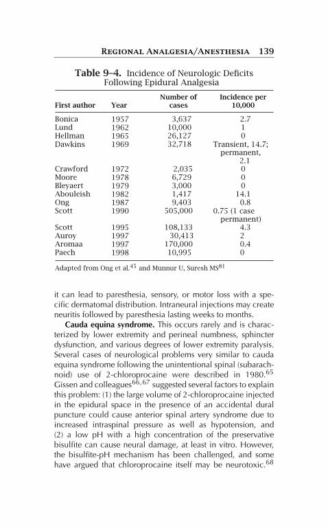
Regional Analgesia/Anesthesia 139
Table 9–4. Incidence of Neurologic DeficitsFollowing Epidural Analgesia
First author YearNumber of
casesIncidence per
10,000
Bonica 1957 3,637 2.7Lund 1962 10,000 1Hellman 1965 26,127 0Dawkins 1969 32,718 Transient, 14.7;
permanent,2.1
Crawford 1972 2,035 0Moore 1978 6,729 0Bleyaert 1979 3,000 0Abouleish 1982 1,417 14.1Ong 1987 9,403 0.8Scott 1990 505,000 0.75 (1 case
permanent)Scott 1995 108,133 4.3Auroy 1997 30,413 2Aromaa 1997 170,000 0.4Paech 1998 10,995 0
Adapted from Ong et al.45 and Munnur U, Suresh MS81
it can lead to paresthesia, sensory, or motor loss with a spe-cific dermatomal distribution. Intraneural injections may createneuritis followed by paresthesia lasting weeks to months.
Cauda equina syndrome. This occurs rarely and is charac-terized by lower extremity and perineal numbness, sphincterdysfunction, and various degrees of lower extremity paralysis.Several cases of neurological problems very similar to caudaequina syndrome following the unintentional spinal (subarach-noid) use of 2-chloroprocaine were described in 1980.65
Gissen and colleagues66,67 suggested several factors to explainthis problem: (1) the large volume of 2-chloroprocaine injectedin the epidural space in the presence of an accidental duralpuncture could cause anterior spinal artery syndrome due toincreased intraspinal pressure as well as hypotension, and(2) a low pH with a high concentration of the preservativebisulfite can cause neural damage, at least in vitro. However,the bisulfite-pH mechanism has been challenged, and somehave argued that chloroprocaine itself may be neurotoxic.68

140 Regional Analgesia/Anesthesia
Conversely, there are recent reports of safe use of preservative-free chloroprocaine in human spinal anesthesia.69
Another cause of cauda equina syndrome is 5% hyperbariclidocaine given via spinal microcatheter. The mechanism mightbe related to high doses of lidocaine that cause nerve dam-age because of improper mixing with cerebrospinal fluid.70
Microcatheters were removed from the market in the US in1991 but are still used in other countries. A large random-ized trial recently demonstrated the safety of such deviceswhen appropriate drugs were used (excluding concentratedlidocaine).71
Transient neurologic symptoms. A more recently appre-ciated anesthesia-related neurologic problem that has stirredcontroversy is transient neurological symptoms (TNS; for-merly transient radicular irritation, or TRI). This problem, firstdescribed by a group from Switzerland, is characterized by(1) aching pain in the buttocks radiating to both dorsolateralsides of the thigh and calves; (2) association with subarachnoidlidocaine; (3) short duration of symptoms of less than 4–6 days;and (4) surgery in the lithotomy position.72 Subsequent stud-ies observed that TNS was observed in 30% of cases with 5%hyperbaric lidocaine, 3% with use of 2% hyperbaric prilocaine,and 0% with 0.5% hyperbaric bupivacaine.73 Interestingly, theincidence of TNS increases significantly when the lithotomyposition is used compared to the supine position (13% vs. 5%)with lidocaine, but does not differ between concentrations oflidocaine or addition of dextrose.74 More recent studies con-firmed no difference in the concentrations of lidocaine in themanifestation of TNS in patients undergoing surgery in thelithotomy position.75 The lithotomy position may stretch L5–S1 nerve roots, which remain in the most dorsal position inthe spinal canal. Under this condition, blood perfusion of thenerves or a subset of nerve fibers may be hampered and thusincrease the vulnerability to injury. However, careful neuro-logic studies in volunteers demonstrate no evidence of directnerve injury in patients developing TNS.76 Hence TNS is con-troversial; judicious care is required with the use of drugs thatmay increase the incidences of TNS.

Regional Analgesia/Anesthesia 141
Epidural hematoma. This is very rare following regionalanesthesia, but it may happen following trauma to epiduralblood vessels, especially if the clotting parameters are abnor-mal due to the use of anticlotting medications or because ofassociated medical problems such as severe preeclampsia orHELLP (hemolysis, elevated liver enzymes, and low plateletcount) syndrome. An immediate diagnosis is necessary, andsurgical decompression should be performed within 6 h.
Epidural abscess and meningitis. Although extremely rare,infection in the spinal canal is usually secondary to infec-tion elsewhere in the body. Four important clinical featuresinclude (1) severe back pain, (2) local overlying tenderness,(3) fever, and (4) leukocytosis. Some case reports of epiduralabscess have been reported and found to be caused by skinflora, and rarely meningitis has been traced to oral commensalsin the anesthesiologist. Meticulous attention to aseptic tech-nique is mandatory.77 One should avoid regional anesthesia,if possible, in the presence of untreated bacteremia or sep-ticemia. Antibiotic pretreatment appears to be protective inanimal models.78
Adhesive arachnoiditis. This can take place as a result ofclinical irritation of the structures in the subarachnoid spacedue to contamination of spinal needles or solutions. Observedsymptoms include headache, nausea and vomiting, nuchalrigidity, fever, and Kernig’s sign.
Anterior spinal artery syndrome. This is an extremely rarecomplication in which the anterior part of the spinal cord expe-riences ischemic degeneration associated with motor deficit.This portion of the cord is more vulnerable because of its singlearterial supply as well as lack of collateral supply. Hypotensionand unfavorable anatomy are risk factors.
Other Methods of Regional Anesthesia
These techniques are largely of historical interest, though somemay occasionally still be used in lieu of modern epidural andspinal techniques.

142 Regional Analgesia/Anesthesia
Caudal Anesthesia
The caudal space is the lowermost part of the epidural spaceand lies in the sacral canal. This technique involves the intro-duction of a 17-gauge epidural or 19-gauge 3.8–7.6-cm needle.A catheter can be introduced for continuous use, or a one-shottechnique can be used just before the delivery for perineal anal-gesia. This technique has become unpopular because of therequirement of higher doses of local anesthetics.
Paracervical Block
This technique involves blocking nerve impulses from theuterine body and cervix by injecting local anesthetics in theparacervical tissues. Usually it is performed during the firststage of labor. A paracervical block does not relieve the per-ineal pain. A continuous technique has also been attempted.This technique is rarely used at the present time mainly becauseof its depressant effects on the fetus. Fetal bradycardia followinga paracervical block is mainly due to two factors: (1) constric-tion of uteroplacental blood vessels by local anesthetic and(2) vascular absorption of a large amount of local anestheticthat will directly depress the fetal myocardium.
Lumbar Sympathetic Block
This technique is technically cumbersome, requires specialskill, must be performed bilaterally to be effective, and there-fore is seldom used at present; it is useful only for the first stageof labor.
Pudendal Block
A pudendal block is performed by the obstetrician justbefore the delivery by blocking the pudendal nerves while pass-ing over the ischial spine. This technique will provide analgesiaof the perineum and is useful only for second-stage labor. Inselected cases of unblocked sacral nerves, it may find a placein modern practice even when an epidural block is employed.

Regional Analgesia/Anesthesia 143
Uptake of local anesthetic from this technique is very similar tothat in an epidural block.
Summary
Epidural and combined spinal-epidural analgesia are the mostcommonly employed techniques for labor analgesia. Althoughvery safe and usually quite effective, adequate safety prepara-tions, careful technique and knowledge of potential problemsand how to address them, and awareness of possible com-plications are necessary for the provision of these popularprocedures.
References
1. Hogan H. Epidural anatomy: new observations. Can J Anaesth.1998;45(5):R40–R44.
2. Beilin Y, Zahn J, Bernstein HH, Zucker-Pinchoff B, Zenzen WJ,Andres LA. Treatment of incomplete analgesia after placementof an epidural catheter and administration of local anesthetic forwomen in labor. Anesthesiology. 1998;88(6):1502–1506.
3. Segal S, Beach M, Eappen S. A multivariate model to predictthe distance from the skin to the epidural space in an obstetricpopulation. Reg Anesth. 1996;21(5):451–455.
4. Bromage P, ed. Epidural Analgesia. Philadelphia: WB Sauders Co;1978.
5. Bahar M, Chanimov M, Cohen ML, et al. Lateral recumbent head-down posture for epidural catheter insertion reduces intravascularinjection. Can J Anaesth. 2001;48(1):48–53.
6. Tsen LC. Neuraxial techniques for labor analgesia should beplaced in the lateral position. Int J Obstet Anesth. 2008;17(2):146–149.
7. Polley LS. Neuraxial techniques for labor analgesia should beplaced in the lateral position. Int J Obstet Anesth. 2008;17(2):149–152.
8. Rout CC, Rocke DA, Levin J, Gouws E, Reddy D. A reevaluation ofthe role of crystalloid preload in the prevention of hypotension

144 Regional Analgesia/Anesthesia
associated with spinal anesthesia for elective cesarean section.Anesthesiology. 1993;79(2):262–269.
9. D’Angelo R, Foss ML, Livesay CH. A comparison of multiportand uniport epidural catheters in laboring patients. Anesth Analg.1997;84(6):1276–1279.
10. Dickson MA, Moores C, McClure JH. Comparison of single, end-holed and multi-orifice extradural catheters when used for con-tinuous infusion of local anaesthetic during labour. Br J Anaesth.1997;79(3):297–300.
11. Segal S, Eappen S, Datta S. Superiority of multi-orifice oversingle-orifice epidural catheters for labor analgesia and cesareandelivery. J Clin Anesth. 1997;9(2):109–112.
12. Beilin Y, Bernstein HH, Zucker-Pinchoff B. The optimal distancethat a multiorifice epidural catheter should be threaded into theepidural space. Anesth Analg. 1995;81(2):301–304.
13. D‘Angelo R, Berkebile BL, Gerancher JC. Prospective examinationof epidural catheter insertion. Anesthesiology. 1996;84(1):88–93.
14. Jaime F, Mandell GL, Vallejo MC, Ramanathan S. Uniport soft-tip,open-ended catheters versus multiport firm-tipped close-endedcatheters for epidural labor analgesia: a quality assurance study.J Clin Anesth. 2000;12(2):89–93.
15. Hamilton CL, Riley ET, Cohen SE. Changes in the posi-tion of epidural catheters associated with patient movement.Anesthesiology. 1997;86(4):778–784, discussion 729A.
16. Moore DC, Batra MS. The components of an effective testdose prior to epidural block. Anesthesiology. 1981;55(6):693–696.
17. Cartwright PD, McCarroll SM, Antzaka C. Maternal heartrate changes with a plain epidural test dose. Anesthesiology.1986;65(2):226–228.
18. Leighton BL, Norris MC, Sosis M. Limitations of an epinephrineepidural anesthesia test dose in laboring patients (abstract).Anesthesiology. 1986;65:403.
19. Leighton BL, Norris MC. Epidural test dose and intravascu-lar injection in obstetrics: sensitivity, specificity. Anesth Analg.1993;76(5):1174–1175.
20. Abraham RA, Harris AP, Maxwell LG, Kaplow S. The efficacy of1.5% lidocaine with 7.5% dextrose and epinephrine as an epidu-ral test dose for obstetrics. Anesthesiology. 1986;64(1):116–119.
21. Leighton BL, Norris MC, DeSimone CA, Rosko T, Gross JB. The airtest as a clinically useful indicator of intravenously placed epiduralcatheters. Anesthesiology. 1990;73(4):610–613.
22. Guay J. The epidural test dose: a review. Anesth Analg.2006;102(3):921–929.
Regional Analgesia/Anesthesia 145
23. Popitz-Bergez F, Datta S, Ostheimer GW. Intravascularepinephrine may not increase heart rate in patients receivingmetoprolol. Anesthesiology. 1988;68(5):815–816.
24. Atienzar MC, Palanca JM, Torres F, Borras R, Gil S, Esteve I.A randomized comparison of levobupivacaine, bupivacaine andropivacaine with fentanyl, for labor analgesia. Int J Obstet Anesth.2008;17(2):106–111.
25. Beilin Y, Guinn NR, Bernstein HH, Zahn J, Hossain S, Bodian CA.Local anesthetics and mode of delivery: bupivacaine versus ropi-vacaine versus levobupivacaine. Anesth Analg. 2007;105(3):756–763.
26. Polley LS, Columb MO, Naughton NN, Wagner DS, van de Ven CJ.Relative analgesic potencies of ropivacaine and bupivacaine forepidural analgesia in labor: implications for therapeutic indexes.Anesthesiology. 1999;90(4):944–950.
27. Scott DB, Walker LR. Administration of continuous epidural anal-gesia. Anaesthesia. 1963;18:82–83.
28. Smedstad KG, Morison DH. A comparative study of continuousand intermittent epidural analgesia for labour and delivery. Can JAnaesth. 1988;35(3 Pt 1):234–241.
29. Chen SH, Liou SC, Hung CT, et al. Comparison of patient-controlled epidural analgesia and continuous epidural infusion forlabor analgesia. Chang Gung Med J. 2006;29(6):576–582.
30. Okutomi T, Saito M, Mochizuki J, Amano K, Hoka S. A double-blind randomized controlled trial of patient-controlled epiduralanalgesia with or without a background infusion following initialspinal analgesia for labor pain. Int J Obstet Anesth. 2008.
31. Celleno D, Capogna G. Epidural fentanyl plus bupivacaine0.125 per cent for labour: analgesic effects. Can J Anaesth.1988;35(4):375–378.
32. Skerman J, Thompson B, Goldstein M. Combined continuousepidural fentanyl and bupivacaine in labor: a randomized study.Anesthesiology. 1985;63:450.
33. Chestnut DH, Owen CL, Bates JN, Ostman LG, Choi WW,Geiger MW. Continuous infusion epidural analgesia duringlabor: a randomized, double-blind comparison of 0.0625%bupivacaine/0.0002% fentanyl versus 0.125% bupivacaine.Anesthesiology. 1988;68(5):754–759.
34. Phillips G. Continuous infusion epidural analgesia in labor: theeffect of adding sufentanil to 0.125% bupivacaine. Anesth Analg.1988;67(5):462–465.
35. Tsen LC, Thue B, Datta S, Segal S. Is combined spinal-epiduralanalgesia associated with more rapid cervical dilation in nul-

146 Regional Analgesia/Anesthesia
liparous patients when compared with conventional epiduralanalgesia? Anesthesiology. 1999;91(4):920–925.
36. Wong CA, Scavone BM, Peaceman AM, et al. The risk of cesareandelivery with neuraxial analgesia given early versus late in labor.N Engl J Med. 2005;352(7):655–665.
37. Borgeat A, Stirnemann HR. Ondansetron is effective to treatspinal or epidural morphine-induced pruritus. Anesthesiology.1999;90(2):432–436.
38. Friedlander JD, Fox HE, Cain CF, Dominguez CL, SmileyRM. Fetal bradycardia and uterine hyperactivity following sub-arachnoid administration of fentanyl during labor. Reg Anesth.1997;22(4):378–381.
39. Segal S, Csavoy AN, Datta S. The tocolytic effect ofcatecholamines in the gravid rat uterus. Anesth Analg.1998;87(4):864–869.
40. Segal S, Wang SY. The effect of maternal catecholamineson the caliber of gravid uterine microvessels. Anesth Analg.2008;106(3):888–892, table of contents.
41. Sharma SK, Philip J, Whitten CW, Padakandla UB, LandersDF. Assessment of changes in coagulation in parturientswith preeclampsia using thromboelastography. Anesthesiology.1999;90(2):385–390.
42. Orlikowski CE, Rocke DA, Murray WB, et al.Thromboelastography changes in pre-eclampsia and eclampsia.Br J Anaesth. 1996;77(2):157–161.
43. Scott DB, Hibbard BM. Serious non-fatal complications associ-ated with extradural block in obstetric practice. Br J Anaesth.1990;64(5):537–541.
44. Donaldson J. Neurology of Pregnancy. 2nd ed. London: W.B.Saunders; 1989.
45. Ong BY, Cohen MM, Esmail A, Cumming M, Kozody R, PalahniukRJ. Paresthesias and motor dysfunction after labor and delivery.Anesth Analg. 1987;66(1):18–22.
46. Auroy Y, Narchi P, Messiah A, Litt L, Rouvier B, Samii K. Seriouscomplications related to regional anesthesia: results of a prospec-tive survey in France. Anesthesiology. 1997;87(3):479–486.
47. Brownridge P. The management of headache following acciden-tal dural puncture in obstetric patients. Anaesth Intensive Care.1983;11(1):4–15.
48. Sprigge JS, Harper SJ. Accidental dural puncture and post duralpuncture headache in obstetric anaesthesia: presentation andmanagement: a 23-year survey in a district general hospital.Anaesthesia. 2008;63(1):36–43.

Regional Analgesia/Anesthesia 147
49. Craft JB, Epstein BS, Coakley CS. Prophylaxis of dural-punctureheadache with epidural saline. Anesth Analg. 1973;52(2):228–231.
50. Smith BE. Prophylaxis of epidural wet tap headache (abstract).American Society of Anesthesiologists, San Fancisco. 1979:119.
51. Crawford JS. The prevention of headache consequent upon duralpuncture. Br J Anaesth. 1972;44(6):598–600.
52. Colonna-Romano P, Shapiro BE. Unintentional dural puncture andprophylactic epidural blood patch in obstetrics. Anesth Analg.1989;69(4):522–523.
53. Scavone BM, Wong CA, Sullivan JT, Yaghmour E, Sherwani SS,McCarthy RJ. Efficacy of a prophylactic epidural blood patch inpreventing post dural puncture headache in parturients after inad-vertent dural puncture. Anesthesiology. 2004;101(6):1422–1427.
54. Szeinfeld M, Ihmeidan IH, Moser MM, Machado R, Klose KJ,Serafini AN. Epidural blood patch: evaluation of the volume andspread of blood injected into the epidural space. Anesthesiology.1986;64(6):820–822.
55. Gewirtz EC, Costin M, Marx G. Cortical vein thrombosis maymimic postdural puncture headache. Reg Anaesth. 1987;12:188.
56. Al-metwalli RR. Epidural morphine injections for prevention ofpost dural puncture headache. Anaesthesia. 2008;63(8):847–850.
57. Noyan Ashraf MA, Sadeghi A, Azarbakht Z, Salehi S,Hamediseresht E. Evaluation of intravenous hydrocortisone inreducing headache after spinal anesthesia: a double blind con-trolled clinical study [corrected]. Middle East J Anesthesiol.2007;19(2):415–422.
58. Ghai A, Wadhera R. Adrenocorticotrophic hormone – is asingle dose sufficient for post-dural puncture headache? ActaAnaesthesiol Scand. 2007;51(2):266.
59. Skaredoff MN, Datta S. Horner’s syndrome during epiduralanaesthesia for elective caesarean section. Can Anaesth Soc J.1981;28(1):82–85.
60. Breen TW, Ransil BJ, Groves PA, Oriol NE. Factors associated withback pain after childbirth. Anesthesiology. 1994;81(1):29–34.
61. Macarthur AJ, Macarthur C, Weeks SK. Is epidural anesthesia inlabor associated with chronic low back pain? A prospective cohortstudy. Anesth Analg. 1997;85(5):1066–1070.
62. Hill EC. Maternal obstetric paralysis. Am J Obstet Gynecol.1962;83:1452–1460.
63. Cole JT. Maternal obstetric paralysis. Am J Obstet Gynecol.1946;52:372.
64. Bromage PR. An evaluation of bupivacaine in epidural analgesiafor obstetrics. Can Anaesth Soc J. 1969;16(1):46–56.

148 Regional Analgesia/Anesthesia
65. Ravindran RS, Bond VK, Tasch MD, Gupta CD, Luerssen TG.Prolonged neural blockade following regional analgesia with2-chloroprocaine. Anesth Analg. 1980;59(6):447–451.
66. Gissen AJ, Datta S, Lampbert D. The chloroprocaine contro-versy. I. A hypothesis to explain the neural complications ofchloroprocaine epidural. . Reg Anaesth. 1984;9:124.
67. Gissen AJ, Datta S, Lampbert D. The chloroprocaine controversy.II. Is chloroprocaine neurotoxic? Reg Anaesth. 1984;9:135.
68. Taniguchi M, Bollen AW, Drasner K. Sodium bisulfite:scapegoat for chloroprocaine neurotoxicity? Anesthesiology.2004;100(1):85–91.
69. Sell A, Tein T, Pitkanen M. Spinal 2-chloroprocaine: effective dosefor ambulatory surgery. Acta Anaesthesiol Scand. 2008;52(5):695–699.
70. Rigler ML, Drasner K, Krejcie TC, et al. Cauda equina syndromeafter continuous spinal anesthesia. Anesth Analg. 1991;72(3):275–281.
71. Arkoosh VA, Palmer CM, Yun EM, et al. A randomized,double-masked, multicenter comparison of the safety of con-tinuous intrathecal labor analgesia using a 28-gauge catheterversus continuous epidural labor analgesia. Anesthesiology.2008;108(2):286–298.
72. Schneider M, Ettlin T, Kaufmann M, et al. Transient neurologic tox-icity after hyperbaric subarachnoid anesthesia with 5% lidocaine.Anesth Analg. 1993;76(5):1154–1157.
73. Hampl KF, Heinzmann-Wiedmer S, Luginbuehl I, et al. Transientneurologic symptoms after spinal anesthesia: a lower inci-dence with prilocaine and bupivacaine than with lidocaine.Anesthesiology. 1998;88(3):629–633.
74. Pollock JE, Neal JM, Stephenson CA, Wiley CE. Prospective studyof the incidence of transient radicular irritation in patients under-going spinal anesthesia. Anesthesiology. 1996;84(6):1361–1367.
75. Pollock JE, Liu SS, Neal JM, Stephenson CA. Dilution of spinallidocaine does not alter the incidence of transient neurologicsymptoms. Anesthesiology. 1999;90(2):445–450.
76. Pollock JE, Burkhead D, Neal JM, et al. Spinal nerve func-tion in five volunteers experiencing transient neurologic symp-toms after lidocaine subarachnoid anesthesia. Anesth Analg.2000;90(3):658–665.
77. Hebl JR. The importance and implications of aseptic tech-niques during regional anesthesia. Reg Anesth Pain Med.2006;31(4):311–323.
78. Carp H, Bailey S. The association between meningitis and duralpuncture in bacteremic rats. Anesthesiology. 1992;76(5):739–742.

Regional Analgesia/Anesthesia 149
79. Abouleish E. Pain Control in Obstetrics. Philadelphia: JBLippincott; 1977:261.
80. Carbaat PA, Van Crevel H. Lumbar puncture headache. Controlledstudy on the preventive effect of 24 hours bed rest. Lancet1981;2:1133–1135.
81. Munnur U, Suresh MS. Backache, headache, and neurologicdeficit after regional anesthesia. Anesthesiol Clin North America.2003;21(1):71–86.