rem sleep twitches rouse nascent cerebellar circuits ... sleep twitches rouse nascent cerebellar...
TRANSCRIPT

REM Sleep Twitches Rouse Nascent CerebellarCircuits: Implications for Sensorimotor Development
Greta Sokoloff,1,2 Brandt D. Uitermarkt,1 Mark S. Blumberg1,2,3
1 Department of Psychology, University of Iowa, Iowa City, Iowa
2 Delta Center, The University of Iowa, Iowa City, Iowa
3 Department of Biology, University of Iowa, Iowa City, Iowa
Received 23 December 2013; revised 8 March 2014; accepted 25 March 2014
ABSTRACT: The cerebellum is critical for sensori-motor integration and undergoes extensive postnataldevelopment. During the first postnatal week in rats,climbing fibers polyinnervate Purkinje cells and, beforegranule cell migration, mossy fibers make transient, directconnections with Purkinje cells. Activity-dependent proc-esses are assumed to play a critical role in the develop-ment and refinement of these and other aspects ofcerebellar circuitry. However, the sources and patterningof activity have not been described. We hypothesize thatsensory feedback (i.e., reafference) from myoclonictwitches in sleeping newborn rats is a prominent driver ofactivity for the developing cerebellum. Here, in 6-day-oldrats, we show that Purkinje cells exhibit substantial state-dependent changes in complex and simple spike activity—
primarily during active sleep. In addition, this activityincreases significantly during bouts of twitching. More-over, the surprising observation of twitch-dependentincreases in simple spike activity at this age suggests afunctional engagement of mossy fibers before the parallelfiber system has developed. Based on these and otherresults, we propose that twitching comprises a uniqueclass of self-produced movement that drives criticalaspects of activity-dependent development in the cerebel-lum and other sensorimotor systems. VC 2014 Wiley
Periodicals, Inc. Develop Neurobiol 00: 000–000, 2014
Keywords: cerebellum; myoclonic twitching; sleep; reaf-ference; corollary discharge
INTRODUCTION
During rapid eye movement (REM, or active) sleep,skeletal muscles throughout the body twitch, causingjerky movements of the limbs (Blumberg et al.,2013a), eyes (Seelke et al., 2005), and even whiskers(Tiriac et al., 2012). Across many species of mam-mals, twitching is one of the most conspicuous of all
infant behavior (Gramsbergen et al., 1970; Jouvet-Mounier et al., 1970). The numbers tell the story:each day, across all skeletal muscles, infant rats pro-duce hundreds of thousands, if not millions, oftwitches (Blumberg et al., 2013b).
Similar to other forms of spontaneous activity inthe developing nervous system (Katz and Shatz,1996; O’Donovan, 1999), sensory feedback (or reaf-ference) from twitching limbs has been implicated inthe self-organization of spinal circuits and somato-topic maps (Petersson et al., 2003; Khazipov et al.,2004; Blumberg et al., 2013b). This implication restsin part on the quantity and patterning of neural activ-ity associated with twitch-related reafference. First,using novel methods developed for in vivo recordingin rat pups, Khazipov et al. (2004) discovered that
Correspondence to: M.S. Blumberg ([email protected]).
Contract grant sponsor: NICHD; contract grant number:HD63071.! 2014 Wiley Periodicals, Inc.Published online 00 Month 2014 in Wiley Online Library(wileyonlinelibrary.com).DOI 10.1002/dneu.22177
1

twitches and other movements trigger oscillatoryevents, called spindle bursts, in primary somatosen-sory cortex. Second, subsequent work has demon-strated that, at least under some testing conditions,twitch-related reafference potently increases neuralactivity, whereas wake-related reafference does not(Mohns and Blumberg, 2010; Tiriac et al., 2012).These and other observations are building a case forconsidering twitching as a unique form of spontane-ous activity that contributes to the development ofsensorimotor systems (Blumberg et al., 2013b).
Any discussion of sensorimotor integration mustinclude the cerebellum, a hindbrain structure thatreceives sensory and motor signals from spinal cord,brainstem, and cerebral cortex (Apps and Garwicz,2005; Odeh et al., 2005; Huang et al., 2013). Inputsto the cerebellum arrive primarily through one of twopathways (Eccles et al., 1967): climbing fibers thatarise exclusively from the inferior olive and mossyfibers that arise from several brainstem nuclei (e.g.,pontine gray, external cuneate). Climbing fibers andmossy fibers directly or indirectly modulate the activ-ity and plasticity of Purkinje cells (Ito, 1989), thesole output of the cerebellar cortex.
Cerebellar circuitry develops substantially over thefirst three postnatal weeks in rats (Altman, 1972; Shi-mono et al., 1976; Wang and Zoghbi, 2001).Although molecular cues play important roles inshaping cerebellar development (Wang and Zoghbi,2001; Sillitoe and Joyner, 2007), activity-dependentmechanisms may be equally important. For example,neural activity contributes to synapse elimination andclimbing fiber translocation at Purkinje cells (Kaki-zawa et al., 2000; Andjus et al., 2003; Kano andHashimoto, 2011; Watanabe and Kano, 2011; Mikuniet al., 2013), as well as spinocerebellar afferenttopography (Tolbert et al., 1994). It is also likely thatactivity-dependent processes guide the convergenceof somatotopically related climbing fiber and mossyfiber inputs onto Purkinje cells (Odeh et al., 2005;Pijpers et al., 2006; Huang et al., 2013).
Little is currently known about the sources of neu-ral activity that drive activity-dependent developmentof the infant cerebellum (Hashimoto and Kano,2013). Spontaneous neural activity may contribute.Also, sensory experiences arising from interactionswith the mother and littermates could drive neuralactivity in the cerebellum. Indeed, despite theirimmaturity in newborns, climbing fibers and mossyfibers transmit sensory information from the periph-ery to Purkinje cells (Puro and Woodward, 1977a,b).
Here we test the novel hypothesis that sensoryfeedback from myoclonic twitching drives activity innascent cerebellar circuits. Recording from cerebellar
cortex in 6-day-old rats, we report that Purkinje cellactivity increases substantially and predominantlyduring periods of twitching. These results reveal forthe first time a prodigious source of spatiotemporallystructured activity to this critical sensorimotor struc-ture during a period of extreme immaturity and rapiddevelopmental change.
MATERIALS AND METHODS
Subjects
Six-day-old male and female Sprague-Dawley Norway rats(Rattus norvegicus) from nine litters were used. Litterswere culled to eight pups within 3 days of birth. Mothersand their litters were housed and raised in standard labora-tory cages (48 3 20 3 26 cm). Food and water were avail-able ad libitum. All animals were maintained on a 12-hlight–dark schedule with lights on at 0700 h. All experi-ments were carried out in accordance with the NationalInstitutes of Health Guide for the Care and Use of Labora-tory Animals (NIH Publication No. 80-23) and wereapproved by the Institutional Animal Care and Use Com-mittee of the University of Iowa.
Surgery
A pup with a visible milk band was removed from the homecage. Under 2–5% isoflurane anesthesia, 2–4 bipolar stainlesssteel hook electrodes (50 lm diameter; California Fine Wire,Grover Beach, CA) were implanted bilaterally into thenuchal muscle and in one or both of the hindlimbs andsecured with collodion. A ground wire on the back waslooped transdermally and secured with collodion. Asdescribed previously (Karlsson et al., 2005), a custom-builthead-fix apparatus, secured to the skull with cyanoacrylateadhesive, allowed for attachment to the earbars of a stereo-taxic apparatus (David Kopf Instruments, Tujunga, CA). Atthe end of the surgery, which lasted approximately 10 min,the pup’s trunk was lightly wrapped in gauze and the pupwas placed in a humidified incubator maintained at thermo-neutrality (35!C) to recover for at least 1 h. Before placingthe subject into the testing apparatus, the subject was reanes-thetized with 2–5% isoflurane anesthesia and three smallholes were drilled in the skull for insertion of the electrode,ground wire, and thermocouple (the last for measuring braintemperature). After 1–2 h of acclimation in the testing appa-ratus, pups typically cycle regularly between sleep and wake.Despite prolonged isolation from their mother, pups exhibitregular sleep–wake cycles in these temperature-controlledconditions for many hours (Seelke and Blumberg, 2005).
Cerebellar Neurophysiology
A total of nine 6-day-old rats were used to produce 30 unitsfor analysis. To record from Purkinje cells, 16-channel
2 Sokoloff et al.
Developmental Neurobiology

silicon depth electrodes (NeuroNexus, Ann Arbor, MI; Mod-els A1x16, A1x16-Poly2), with impedances ranging from 1to 4 MX, were connected to a data acquisition system(Tucker-Davis Technologies, Alachua, FL) that amplified(10,0003) and filtered (500–5000 Hz band-pass) the neuralsignals. Prior to insertion, the electrode surface was coatedwith fluorescent DiI solution (Life Technologies, GrandIsland, NY) for subsequent histological verification of elec-trode placement. A Ag/AgCl ground electrode (Medwire,Mt. Vernon, NY, 0.25 mm diameter) was placed into the cer-ebral cortex. Electromyographic (EMG) electrodes wereconnected to a differential amplifier (A-M Systems, Carls-borg, WA) that amplified (10,0003) and filtered (300–5000Hz band-pass; 60 Hz notch filter) the EMG signal. Neuraland EMG signals were sampled at 12.5 and 1 kHz, respec-tively, using a digital interface and Spike2 software (Cam-bridge Electronic Design, Cambridge, UK). Braintemperature, maintained at 36–37!C, was measured using afine-wire thermocouple (Omega Engineering, Stamford, CT)placed in the cerebral cortex contralateral to the ground wire.
Using a stereotaxic apparatus, the electrode was posi-tioned above cerebellar cortex (coordinates: 1.0–1.5 mmcaudal to lambda, 1.5–2.0 mm lateral to midline, 10–12!
angle) and was slowly lowered until action potentials weredetected on one or more recording channels. Purkinje cellactivity was identified by the occurrence of large-amplitudeaction potentials (signal-to-noise" 2:1) and the presence ofcomplex spike waveforms as described by others (Crepel,1971; Puro and Woodward, 1977a). Using procedures simi-lar to those described previously (Karlsson et al., 2005;Mohns and Blumberg, 2010; Tiriac et al., 2012), data acqui-sition began after Purkinje cell unit activity was identifiedand had stabilized for at least 10 min. Each recording ses-sion lasted for 15 min and comprised continuous collectionof neural and EMG data as the pup cycled freely betweensleep and wake. During acquisition, an experimenter moni-tored the subject’s behavior and digitally marked the occur-rence of wake and active sleep (AS) movements insynchrony with the physiological data. The experimenterwas always blind to the physiological data while scoringbehavior. Following the first recording session, the elec-trode was lowered further and additional recordings wereacquired if new Purkinje cell unit activity was identified.
Histology
After the final recording session, the pup was overdosedwith sodium pentobarbital (1.5 mg/g) and perfused trans-cardially with phosphate buffered saline followed by 4%paraformaldehyde. Recording sites were verified by visual-izing the DiI tract with fluorescence illumination at 5–103magnification using a Leica microscope (Leica Microsys-tems, Buffalo Grove, IL). Using a calibrated reticle, thelocation of the unit recordings was determined.
Data Analysis
Spike Sorting and Burst Analysis. Spike sorting was per-formed using template matching in Spike2 (Cambridge
Electronic Design). The distributions of waveforms com-prising each template were analyzed using the principalcomponent analysis tool; waveforms greater than 3.5 stand-ard deviations outside of each distribution were excluded.After spikes were sorted into units and converted intoevents, a second analysis was performed to separate com-plex spikes from simple spikes using a burst script forSpike2. Bursts were identified as multispike events withinterspike intervals #15 ms. All unit activity comprising aburst was subsequently extracted from the raw record ofunit activity, resulting in separate simple spike andcomplex spike records for each unit.
Identification of Behavioral State. Sleep and wake peri-ods were defined by calculating mean EMG activity, basedon methods described previously (Karlsson et al., 2005;Mohns and Blumberg, 2010; Tiriac et al., 2012). Briefly,each nuchal or hindlimb EMG record was rectified andsmoothed (s 5 0.001 s). The mean amplitude of high mus-cle tone and atonia was calculated from five representative1-s EMG segments, and the midpoint between the two wasused to establish a threshold for determining state. TheEMG signal was then dichotomized into periods of hightone (indicative of wake) and atonia (indicative of sleep).Sleep bouts were further divided into periods of quiet sleepand AS: quiet sleep was characterized by hypotonia/atoniaand behavioral quiescence, and AS was characterized bythe appearance of myoclonic twitches against a backgroundof muscle atonia (Seelke and Blumberg, 2008; Blumbergand Seelke, 2010). For three pups with more than one use-able EMG record, nuchal and hindlimb EMG events weremerged into one event channel.
Analysis of State-Dependency. Mean unit firing rateswere determined across all periods of wake, quiet sleep,and AS for each pup. For any single period, firing rates thatexceeded three times the standard deviation were excludedas outliers. To assess whether units were significantly moreactive during different behavioral states, independent pair-wise comparisons of the mean firing rates during wake,quiet sleep, and AS were assessed using the Wilcoxonmatched-pairs signed-ranks test (SPSS, IBM, Armonk,NY). After characterizing units as AS-On, Sleep-On,Wake-On, AS/Wake-On, or State-Independent, mean firingrates for each unit were used in a summary analysis. Meanfiring rates were also assessed for all units during eachstate. For all tests, including those described below, alphawas set at 0.05 and a Bonferroni correction was used whenappropriate.
Event Correlations. Using methods similar to thosedescribed previously (Mohns and Blumberg, 2010; Tiriacet al., 2012), twitches were identified as discrete EMGevents with amplitudes exceeding three times the meanEMG baseline during atonia. For pooled data across all sub-jects, a twitch-triggered cross-correlogram of complexspike activity was constructed for a 1-s window (10-msbins) around twitch onset. We tested statistical significance
Infant Twitching and Cerebellar Activity 3
Developmental Neurobiology
for the cross-correlogram by jittering twitch events 1000times within a 400-ms window using PatternJitter (Harrisonand Geman, 2009; Amarasingham et al., 2012). Finally,using a custom-written Matlab program, a 95% probabilitythreshold (p< 0.05) was determined for the bins spanning250 ms on either side of a twitch. For presentation pur-poses, cross-correlograms were smoothed using a 3-binmoving window. An identical method was used to test therelationship between simple spikes and twitching.
Window Analysis. As a population, complex spikesexhibited rhythmic activity after a twitch. For each unit, a“separation procedure” (Bloedel and Ebner, 1984; Nichol-son and Freeman, 2003) was used to quantify the numberof complex spikes within each of three time windows aftera twitch: Window 1 at 0–50 ms, Window 2 at 100–150 ms,and Window 3 at 200–250 ms. If 45% or more of the com-plex spikes for that unit were associated with only one win-dow, it was designated as either a W1 unit (n 5 16), W2unit (n 5 4), or W3 unit (n 5 3). Seven units exhibited noconsistent window preference (NP). For each type of unit,cross-correlational analyses were performed to determinetheir relationship with twitching. Simple spike activity wasassessed based on that unit’s complex spike window assign-ment and cross-correlational analyses were performed.Again, significance was determined using the jitter proce-dure described above.
Complex Spike Autocorrelations. Autocorrelations ofcomplex spikes were analyzed in 5 ms bins using the timeseries command in JMP (SAS, Cary, NC). For each cere-bellar unit, autocorrelograms and spectral density plotswere created. Using a white noise test, we tested against thenull hypothesis that the production of complex spikes hadno periodic component. Both Fisher’s Kappa and Bartlett’sKolmogorov–Smirnov statistic were relied on to establishsignificant periodicity.
RESULTS
Identifying Complex and Simple Spikes
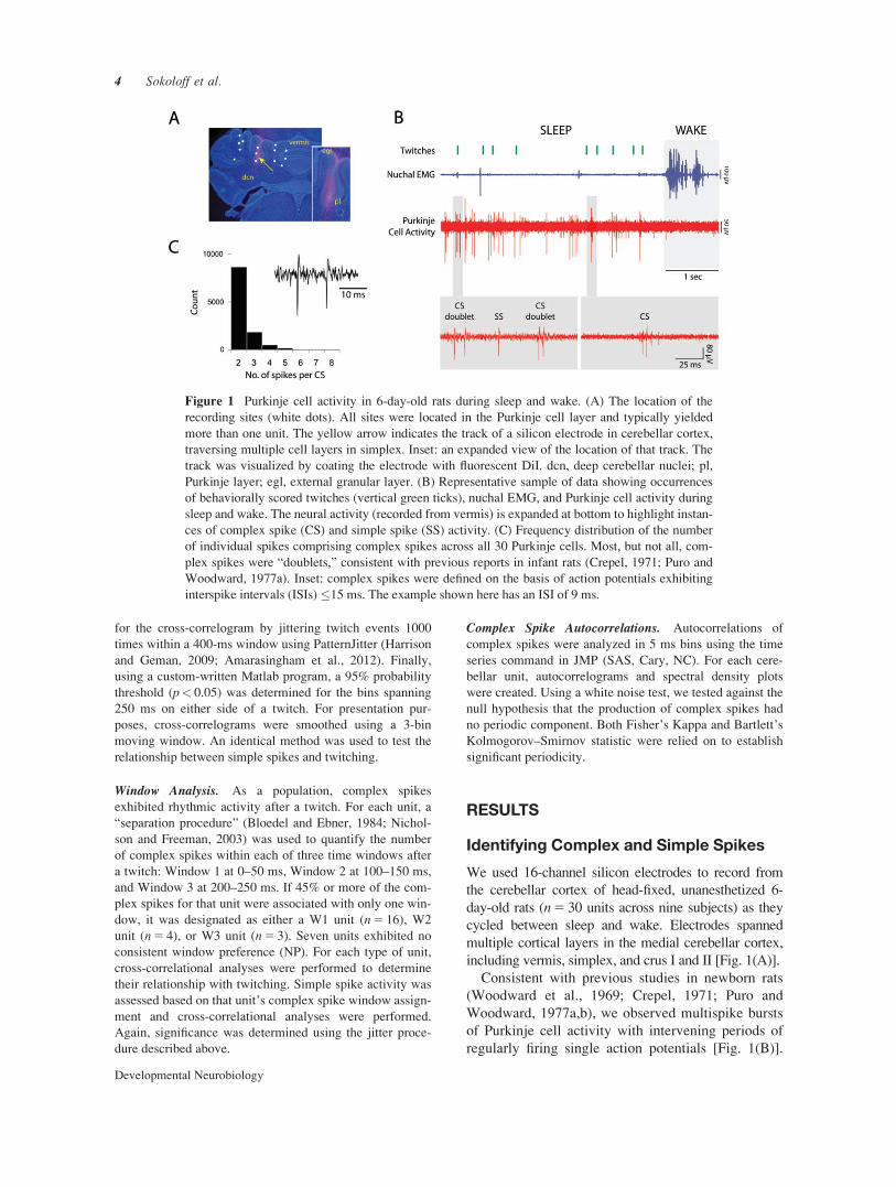
We used 16-channel silicon electrodes to record fromthe cerebellar cortex of head-fixed, unanesthetized 6-day-old rats (n 5 30 units across nine subjects) as theycycled between sleep and wake. Electrodes spannedmultiple cortical layers in the medial cerebellar cortex,including vermis, simplex, and crus I and II [Fig. 1(A)].
Consistent with previous studies in newborn rats(Woodward et al., 1969; Crepel, 1971; Puro andWoodward, 1977a,b), we observed multispike burstsof Purkinje cell activity with intervening periods ofregularly firing single action potentials [Fig. 1(B)].
Figure 1 Purkinje cell activity in 6-day-old rats during sleep and wake. (A) The location of therecording sites (white dots). All sites were located in the Purkinje cell layer and typically yieldedmore than one unit. The yellow arrow indicates the track of a silicon electrode in cerebellar cortex,traversing multiple cell layers in simplex. Inset: an expanded view of the location of that track. Thetrack was visualized by coating the electrode with fluorescent DiI. dcn, deep cerebellar nuclei; pl,Purkinje layer; egl, external granular layer. (B) Representative sample of data showing occurrencesof behaviorally scored twitches (vertical green ticks), nuchal EMG, and Purkinje cell activity duringsleep and wake. The neural activity (recorded from vermis) is expanded at bottom to highlight instan-ces of complex spike (CS) and simple spike (SS) activity. (C) Frequency distribution of the numberof individual spikes comprising complex spikes across all 30 Purkinje cells. Most, but not all, com-plex spikes were “doublets,” consistent with previous reports in infant rats (Crepel, 1971; Puro andWoodward, 1977a). Inset: complex spikes were defined on the basis of action potentials exhibitinginterspike intervals (ISIs) #15 ms. The example shown here has an ISI of 9 ms.
4 Sokoloff et al.
Developmental Neurobiology
Importantly, stimulation of the inferior olive early indevelopment elicits complex spikes, predominantlyin the form of “doublets” comprising two individualspikes (Crepel, 1971; Puro and Woodward, 1977a).Doublets were also the predominant form of complexspikes observed here [Fig. 1(C)].
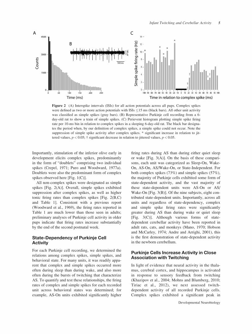
All non-complex spikes were designated as simplespikes [Fig. 2(A)]. Overall, simple spikes exhibitedsuppression after complex spikes, as well as highertonic firing rates than complex spikes [Fig. 2(B,C)and Table 1]. Consistent with a previous report(Woodward et al., 1969), the firing rates reported inTable 1 are much lower than those seen in adults;preliminary analyses of Purkinje cell activity in olderpups indicate that firing rates increase substantiallyby the end of the second postnatal week.
State-Dependency of Purkinje CellActivity
For each Purkinje cell recording, we determined therelations among complex spikes, simple spikes, andbehavioral state. For many units, it was readily appa-rent that complex and simple spikes occurred moreoften during sleep than during wake, and also moreoften during the bursts of twitching that characterizeAS. To quantify and test these relationships, the firingrates of complex and simple spikes for each recordedunit across behavioral states was determined; forexample, AS-On units exhibited significantly higher
firing rates during AS than during either quiet sleepor wake [Fig. 3(A)]. On the basis of these compari-sons, each unit was categorized as Sleep-On, Wake-On, AS-On, AS/Wake-On, or State-Independent. Forboth complex spikes (73%) and simple spikes (57%),the majority of Purkinje cells exhibited some form ofstate-dependent activity, and the vast majority ofthese state-dependent units were AS-On or AS/Wake-On [Fig. 3(B)]. Of the nine subjects, eight con-tributed state-dependent units. Importantly, across allunits and regardless of state-dependency, complexand simple spike firing rates were significantlygreater during AS than during wake or quiet sleep[Fig. 3(C)]. Although various forms of state-dependent cerebellar activity have been reported inadult rats, cats, and monkeys (Mano, 1970; Hobsonand McCarley, 1974; Andre and Arrighi, 2001), thisis the first demonstration of state-dependent activityin the newborn cerebellum.
Purkinje Cells Increase Activity in CloseAssociation with Twitching
In light of evidence that neural activity in the thala-mus, cerebral cortex, and hippocampus is activatedin response to sensory feedback from twitching(Khazipov et al., 2004; Mohns and Blumberg, 2010;Tiriac et al., 2012), we next assessed twitch-dependent activity of all recorded Purkinje cells.Complex spikes exhibited a significant peak in
Figure 2 (A) Interspike intervals (ISIs) for all action potentials across all pups. Complex spikeswere defined as two or more action potentials with ISIs #15 ms (black bars). All other unit activitywas classified as simple spikes (gray bars). (B) Representative Purkinje cell recording from a 6-day-old rat to show a train of simple spikes. (C) Perievent histogram plotting simple spike firingrate per 10-ms bin in relation to complex spikes in a sleeping 6-day-old rat. The black bar designa-tes the period when, by our definition of complex spikes, a simple spike could not occur. Note thesuppression of simple spike activity after complex spikes. * significant increase in relation to jit-tered values, p< 0.05; † significant decrease in relation to jittered values, p< 0.05.
Infant Twitching and Cerebellar Activity 5
Developmental Neurobiology

activity immediately after twitching and a secondpeak approximately 100 ms after the first, alongwith a third, weak peak approximately 100 ms afterthe second [Fig. 4(A)]. Simple spikes also exhibiteda significant peak in activity immediately aftertwitching; this peak was broader than that of com-plex spikes [Fig. 4(B)].
Next, using a “separation procedure” used previ-ously in adult cats and weanling rats (Bloedel andEbner, 1984; Nicholson and Freeman, 2003), wetested the possibility that the multiple peaks in com-
plex spike activity were a population-level feature ofensembles of Purkinje cells, arising when Purkinjecells fire during the first peak or the second peak orthe third, but rarely in more than one. To do this, wenoted the occurrence of complex spikes within eachof three 50-ms windows defined in relation totwitch-dependent activity: 0–50 ms [Fig. 4(A), red],100–150 ms (green), and 200–250 ms (blue). Unitsthat fired preferentially during Windows 1, 2, or 3were designated as W1, W2, or W3 units, respec-tively. Cross-correlational analyses showed that
Table 1 Descriptive Statistics of Complex Spike and Simple Spike Activity Across All 30 Units in 6-Day-Old Rats
Unit Activity Mean Interspike Interval (ms) Mean Firing Rate (Hz) Mean Burst Duration (ms)
Complex spikes 4.03 (0.10)a 0.36 (0.11) 9.15 (0.26)Simple spikes 240.98 (14.1) 2.62 (0.49) NA
Mean (SEM).aInterspike interval within a complex spike.
Figure 3 Complex and simple spike activity is state-dependent. (A) Firing rate (Hz) of complexspikes (top) and simple spikes (bottom) during wake, quiet sleep (QS), and active sleep (AS) in Pur-kinje cells characterized as AS-On and AS/Wake-On (data for Sleep-On, Wake-On, and State-Independent categories not shown). * significantly different from the other two groups (p< 0.017).(B) Percentage of complex and simple spikes (n 5 30 in each group) characterized as AS-On, AS/Wake-On, Sleep-On, Wake-On, and State-Independent. (C) Firing rate (Hz) of complex and simplespikes during wake, quiet sleep, and active sleep in Purkinje cells regardless of state. * significantlydifferent from the other two groups (p< 0.017). [Color figure can be viewed in the online issue,which is available at wileyonlinelibrary.com.]
6 Sokoloff et al.
Developmental Neurobiology

W1, W2, and W3 units increased their complexspike activity exclusively within their associatedwindow [Fig. 4(C)]. Units exhibiting NP did notexhibit any twitch-related increase in complex spikeactivity. A similar result was obtained for simplespikes, but with only the W1 units exhibiting a sig-nificant increase in activity after a twitch [Fig.4(D)]. Thus, twitching triggers short-latency com-plex and simple spike responses (W1), as well as aseparate longer-latency complex spike response(W2) that may result from resetting of the intrinsicolivary rhythm (Bloedel and Ebner, 1984; Nichol-son and Freeman, 2003).
Based on the complex spike data presented in Fig-ure 4(A), we repeated the twitch-triggered cross-cor-relational analyses separately for the AS-On units.Complex spikes again exhibited pronounced and sig-
nificant W1 and W2 peaks after twitches [Fig. 5(A)],and simple spikes again exhibited a significant W1peak [Fig. 5(B)]. In contrast, neither AS/Wake-Onnor State-Independent units exhibited pronouncedpeaks, thus indicating that AS-On units contributedpredominantly to the overall twitch-related activitypresented in Figure 4. For one representative AS-Onunit recorded from crus II [Fig. 5(C)], the tight cou-pling between Purkinje cell activity and AS is appa-rent. Importantly, Purkinje cell activity decreasedmarkedly during periods of active wake when thepup was moving its limbs.
Complex Spike Autorhythmicity
Autorhythmicity in climbing fiber activity, at a frequencyof about 10 Hz, has been reported in adult rats during
Figure 4 Complex and simple spike activity increases immediately after twitches. (A) Perieventhistogram plotting the number of complex spikes per 10-ms bin in relation to myoclonic twitchingin sleeping 6-day-old rats. Data were pooled across all 30 units. The vertical black bar is the lastbin before a twitch and the horizontal red bars indicate statistical significance (p< 0.05). (B) Sameas in (A) except for simple spikes (p< 0.05). (C) Perievent line histograms for complex spikes bro-ken down for the W1 (red), W2 (green), and W3 (blue) units, as well as the units that showed nowindow preference (NP, black). For a small subset (10%) of all twitches analyzed, a unit that firedin one window fired again in one or both of the other windows. There was no clear relationshipbetween a unit’s window assignment and its state-dependency. The data were smoothed using a 3-bin moving window. * significant peak within window (p< 0.0125). (D) Same as in (C) except forsimple spikes. * significant peak within window (p< 0.0125).
Infant Twitching and Cerebellar Activity 7
Developmental Neurobiology

wakefulness, but its occurrence is variable both withinand across species (Llinas and Sasaki, 1989; Kitazawaand Wolpert, 2005). To assess whether such autorhyth-micity occurred in our subjects, we performed autocorre-lation analyses. Of the 30 units examined, six exhibitedstatistically significant autorhythmicity. Of these, threeexhibited strong rhythms at frequencies of 8–12 Hz (Fig.6). Importantly, this analysis shows that the inferior oliveis capable of rhythmic activity as early as 6 days ofage. However, as the window analysis suggests, complexspike autorhythmicity is not responsible for the twitch-related multiple peaks in complex spike activity.
DISCUSSION
We found that the Purkinje cells of 6-day-old ratsexhibit substantial state-dependent increases in com-
plex and simple spike activity in close temporal asso-ciation with twitching. These results are strikinggiven the lack of several key cerebellar features con-sidered integral for its mature function, including thegranule cell–parallel fiber network and innervation ofPurkinje cells by inhibitory interneurons and basketcells (Shimono et al., 1976). In addition, before thegranule cell–parallel fiber network develops, mossyfibers form direct, transient connections onto Purkinjecell somas (Mason and Gregory, 1984; Takeda andMaekawa, 1989; Kalinovsky et al., 2011). Conse-quently, the observed twitch-dependent increases insimple spike activity at the age tested here indicate,for the first time in unanesthetized animals, func-tional engagement of these direct mossy fiber connec-tions. All together, the current findings introduce anew and unanticipated mechanism for drivingactivity-dependent development of mossy and
Figure 5 Twitch-related complex and simple spike activity for AS-On units. Perievent line histo-grams plotting complex (A) and simple spike (B) counts per 10-ms bin in relation to myoclonictwitching in sleeping 6-day-old rats. Each plot is broken down for the W1 (red), W2 (green), andunits that showed no window preference (NP, black). All data were smoothed using a 3-bin movingwindow. * significant peak within window (p< 0.017). (C) Representative data showing occur-rences of behaviorally scored twitches from all visible limbs and tail (vertical green ticks), complexspikes (vertical purple ticks), nuchal EMG, and Purkinje cell activity (recorded from crus I). Notethe increased Purkinje cell and complex spike activity during periods of active sleep.
8 Sokoloff et al.
Developmental Neurobiology

climbing fiber connectivity (Takeda and Maekawa,1989; Kalinovsky et al., 2011; Kano and Hashimoto,2011).
Twitching, Reafference, and CorollaryDischarge
Although the function of the cerebellum continues toelude simple description, most current theories tend tofocus on its role in motor timing or motor learning(Kitazawa and Wolpert, 2005). Of critical importance
to these theories are the spatial and temporal detailsregarding the transmission of sensory and motor signalsthrough mossy and climbing fibers. Toward that end,investigators seek to understand how the cerebellumprocesses the two established classes of sensory input:reafference, arising from self-produced movements, andexafference, arising from other-produced movements(Sperry, 1950; Von Holst and Mittelstaedt, 1950; Pouletand Hedwig, 2007; Crapse and Sommer, 2008). In addi-tion to reafference, motor commands trigger corollarydischarge (or efference copy), which serves the function
Figure 6 Complex spike autorhythmicity in sleeping 6-day-old rats. For three individual units,autocorrelograms are shown at left and spectral density plots are shown at right. (A) A unit exhibitingstrong 10-Hz rhythmicity. This unit was Sleep-On and did not exhibit a window preference. (B) Aunit exhibiting strong 8-Hz rhythmicity. This unit was State-Independent and exhibited a preferencefor Window 2 (W2). (C) A unit exhibiting weaker rhythmicity at 11.5 Hz. This unit was AS-On andexhibited a preference for Window 1 (W1 unit). These three units were recorded from two pups.
Infant Twitching and Cerebellar Activity 9
Developmental Neurobiology

of helping animals distinguish between expected (i.e.,reafferent) and unexpected (i.e., exafferent) signals(Crapse and Sommer, 2008). Importantly, both reaffer-ence and corollary discharge signals are conveyed tothe adult cerebellum via climbing and mossy fibers (vanKan et al., 1993; Wolpert et al., 1998; Huang et al.,2013). Further investigation of the infant cerebellummay provide insight into the development of both sig-nals in this essential sensorimotor structure.
The top-left and bottom-right cells in Figure 7denote the two conventional classes of sensory input:reafference from self-produced movements that ismodified by corollary discharge and exafference thatis not. There is, however, a potential third class ofinput that is biologically plausible but rarely dis-cussed: self-produced movements that are not accom-panied by corollary discharge (bottom-left cell in Fig.7). Here we propose the working hypothesis thattwitches embody this novel class of self-producedmovement.
We were led to this hypothesis for several reasons.Foremost among them is the observation that, similarto previous findings in hippocampus (Mohns andBlumberg, 2008, 2010) and sensory thalamus (Tiriacet al., 2012), Purkinje cell activity was prominent dur-ing AS in close temporal proximity to twitching,whereas wake-related activity was weak or absent. Itshould be stressed that we cannot be certain that thetwitch-related Purkinje cell activity observed herereflects reafference alone. Nonetheless, the totality ofevidence from cerebellum, hippocampus, and thala-mus strongly suggests that—at least in rat pups underthese testing conditions—reafferent signals associatedwith twitches are processed differently from thoseassociated with wake movements. One possible expla-nation for this differential processing is that corollarydischarge is absent during twitching. This notion ofstate-dependent modulation of corollary discharge hasnot, to our knowledge, been considered previously.
There is intriguing but indirect support for thehypothesis that twitches are not accompanied by corol-lary discharge. First, human adults awaking fromREM-related dreams display deficits in their ability todistinguish self- from other-produced stimulation, sug-gesting suspended corollary discharge (Blagrove et al.,2006). Second, REM-dream narratives exhibit qual-ities suggestive of suspended corollary discharge, sim-ilar to the hallucinations of schizophrenics (Feinbergand Guazzelli, 1999; Ford et al., 2008). Accordingly, abetter understanding of differential processing of cor-ollary discharge during sleep and wakefulness couldgreatly aid our understanding of how we learn to dis-tinguish self from other, and how such processes goawry.
Limitations of This Study
The present results are clear in showing that Purkinjecell activity is strongly sleep- and twitch-dependentin rats at 6 days of age. We are confident that werecorded from Purkinje cells because of the locationof the electrode sites within the Purkinje layer andthe detection of doublets in the neurophysiologicalrecord. Doublets are considered an immature form ofcomplex spike based on evidence that they are pro-duced by climbing fibers arising from the inferiorolive (Crepel, 1971; Puro and Woodward, 1977a).However, a fully accurate characterization of theactivity triggered by climbing and mossy fibers earlyin development will require selective inactivation ofthe inferior olive and other structures that project tothe cerebellum.
As mentioned in the previous section, another con-cern relates to the precise timing of the Purkinje cellactivity in relation to twitching. For the analyses oftwitch-related neural activity reported here and else-where, we rely on the fact that, because of highcross-correlations in the twitch activity of skeletalmuscle groups, any given muscle can act as a reason-able proxy for another one (Mohns and Blumberg,2010; Tiriac et al., 2012). This reliance on statisticalassociation has not presented interpretative problemswhen recording from forebrain sites where the laten-cies between twitches and reafferent-related activityare approximately 200 ms. In the cerebellum, how-ever, the first peak in twitch-related activity was 50ms or less, which may or may not be too short for areafferent signal at this age (Puro and Woodward,
Figure 7 Hypothesized framework relating self-producedand other-produced movements with the accompaniment ofcorollary discharge. Conventionally, self-produced move-ments generate reafference that is accompanied by corol-lary discharge, whereas other-produced movementsgenerate exafference that is not accompanied by corollarydischarge. For obvious reasons, other-produced movementscannot be accompanied by corollary discharge (n/a: notapplicable). Here we propose a third possible categorywhereby sleep-related twitches are self-produced move-ments that are not accompanied by corollary discharge.
10 Sokoloff et al.
Developmental Neurobiology

1977a; Takeda and Maekawa, 1989). To resolve thisissue, it will be important to record from locations inthe cerebellum that can be definitively related totwitching in specific muscle groups. Once that isaccomplished, it may also then be possible to assesshow twitch-related activity guides the developmentof convergent and somatotopically organized mossyand climbing fiber inputs onto Purkinje cells (Odehet al., 2005; Pijpers et al., 2006; Huang et al., 2013).
Finally, we recorded here from 6-day-olds becausethis is an age when Purkinje cells are functional butgranule cells have not yet migrated to the internalgranular layer. It is also an age when the Purkinjecells themselves are undergoing rapid developmentalchange (Watanabe and Kano, 2011). Of great interestwill be documenting changes in state- and twitch-dependent activity in the cerebellum at younger andolder ages and relating that activity to known featuresof cerebellar circuitry (Shimono et al., 1976). Such adevelopmental study is currently underway.
A New Era in DevelopmentalNeurophysiology
As a contribution to this special issue on “NeuralMechanisms of Behavioral Maturation,” it seemsworthwhile to place the present work in a broaderhistorical context. Before 2004, there were only ahandful of papers devoted to state-dependent neuralactivity in infant animals, including recordings at thecortical surface and multiunit recordings in the brain-stem (Gramsbergen, 1976; Tam!asy and Kor!anyi,1980; Tam!asy et al., 1980; Mirmiran and Corner,1982). For a variety of technical and methodologicalreasons, these studies were unable to document clearand convincing relationships between neural activityand sleep in early infancy. Moreover, with regard tothe cerebral cortex, the conclusion drawn from theseearlier studies was that this structure does not exhibitstate-dependent activity before the emergence ofslow waves at P11 (for review, see Blumberg andSeelke, 2010).
Then, beginning only a decade ago, new in vivorecording methods were introduced and refined forrecording hippocampal (Leinekugel et al., 2002;Karlsson and Blumberg, 2003) and cortical (Khazi-pov et al., 2004) activity in freely moving and head-fixed infant rats. These methods have made it possi-ble to investigate state-dependent neural activity inthe infant brainstem (Karlsson and Blumberg, 2005;Karlsson et al., 2005) and forebrain (Mohns et al.,2006), discover previously undetected state-dependent cortical activity patterns and trace theirearly development (Yang et al., 2009; Seelke and
Blumberg, 2010), assess the contributions to infantcortical activity of subplate neurons (Hanganu et al.,2008; Tolner et al., 2012) and the corpus callosum(Marcano-Reik and Blumberg, 2008), and investigatethe consequences of twitch-related reafference forneural activity throughout the neuraxis (Khazipovet al., 2004; Mohns and Blumberg, 2008, 2010; Tiriacet al., 2012). Imaging techniques, including the useof voltage-sensitive dyes, can now capture broaderfeatures of twitch-related cortical activity (McVeaet al., 2012; Tiriac et al., 2012).
Although similar in vivo recording and imagingmethods are providing new insights into sensoryprocessing in the visual system (e.g., Hanganu et al.,2006, 2007; Ackman et al., 2012), these methods canand should be expanded to the study of other behav-ioral contexts and the interactions among behavioralsystems. For example, even in head-fixed pups, itshould now be possible to investigate neural mecha-nisms associated with ingestion (Hall and Williams,1983), thermoregulation (Blumberg, 2001), andlearning and memory (Johanson and Hall, 1979;Campolattaro and Freeman, 2008), among otherbehaviors and processes. Such advances, when fullyrealized, hold the promise to reshape our understand-ing of brain–behavior relations and how they emergeacross early development.
CONCLUSIONS
Spontaneous activity is a ubiquitous feature ofdeveloping nervous systems (O’Donovan, 1999).For example, spontaneous activity in the developingretina (Galli and Maffei, 1988; Wong, 1999) andcochlea (Tritsch et al., 2007) is thought to contributeto the development of the visual and auditory sys-tems, respectively. Twitching is different from spon-taneous retinal and cochlear activity in that it entailsmotor as well as sensory components. As such, it isideally suited to provide information to the develop-ing sensorimotor system about the functional prop-erties of limbs and their neural control (Blumberget al., 2013b). Previous findings have highlightedhow reafference from twitching profoundly andselectively activates forebrain structures, includingthalamus, cerebral cortex, and hippocampus (Khazi-pov et al., 2004; Mohns and Blumberg, 2010; Tiriacet al., 2012). Here we have shown for the first timethat twitch-related processes modulate the activityof the cerebellum, a hindbrain structure that is criti-cal for sensorimotor integration and motor learning.This finding introduces a potent source of structuredactivity that can plausibly account for activity-
Infant Twitching and Cerebellar Activity 11
Developmental Neurobiology

dependent development in the cerebellar system. Indoing so, these findings lend additional support tothe counterintuitive notion that, contrary to the still-popular view of twitching as a functionless by-product of dreams (Blumberg, 2010), twitching is avital source of neural activity for the developinginfant.
The authors thank Dan Nicholson, John Freeman, Car-los Del Rio Bermudez, Alan Plumeau, and Alex Fanningfor helpful comments.
REFERENCES
Ackman JB, Burbridge TJ, Crair MC. 2012. Retinal wavescoordinate patterned activity throughout the developingvisual system. Nature 490:219–225.
Altman J. 1972. Postnatal development of the cerebellarcortex in the rat. I. The external germinal layer and thetransitional molecular layer. J Comp Neurol 145:353–397.
Amarasingham A, Harrison MT, Hatsopoulos NG, GemanS. 2012. Conditional modeling and the jitter method ofspike resampling. J Neurophysiol 107:517–531.
Andjus PR, Zhu L, Cesa R, Carulli D, Strata P. 2003. Achange in the pattern of activity affects the developmentalregression of the purkinje cell polyinnervation by climbingfibers in the rat cerebellum. Neuroscience 121:563–572.
Andre P, Arrighi P. 2001. Modulation of Purkinje cellresponse to glutamate during the sleep–waking cycle.Neuroscience 105:731–746.
Apps R, Garwicz M. 2005. Anatomical and physiologicalfoundations of cerebellar information processing. NatRev Neurosci 6:297–311.
Blagrove M, Blakemore S, Thayer B. 2006. The ability toself-tickle following rapid eye movement sleep dreaming.Conscious Cogn 15:285–294.
Bloedel JR, Ebner TJ. 1984. Rhythmic discharge of climb-ing fibre afferents in response to natural peripheral stim-uli in the cat. J Physiol (Lond) 352:129–146.
Blumberg MS. 2001. The developmental context of thermalhomeostasis. In: Blass EM, editor. Handbook of Develop-mental Neurobiology, Vol. 13: Developmental Psychobi-ology. New York: Plenum Press, pp 199–228.
Blumberg MS. 2010. Beyond dreams: Do sleep-relatedmovements contribute to brain development? Front Neu-rol 1:140.
Blumberg MS, Coleman CM, Gerth AI, McMurray B.2013a. Spatiotemporal structure of REM sleep twitchingreveals developmental origins of motor synergies. CurrBiol 23:2100–2109.
Blumberg MS, Marques HG, Iida F. 2013b. Twitching insensorimotor development from sleeping rats to robots.Curr Biol 23:R532–R537.
Blumberg MS, Seelke AMH. 2010. The form and functionof infant sleep: From muscle to neocortex. In: BlumbergMS, Freeman JH, Robinson SR, editors. The Oxford
Handbook of Developmental Behavioral Neuroscience.New York: Oxford University Press, pp 391–423.
Campolattaro M, Freeman JH. 2008. Eyeblink conditioningin 12-day-old rats using pontine stimulation as the condi-tioned stimulus. Proc Natl Acad Sci USA 105:8120–8123.
Crapse TB, Sommer MA. 2008. Corollary discharge acrossthe animal kingdom. Nat Rev Neurosci 9:587–600.
Crepel F. 1971. Maturation of climbing fiber responses inthe rat. Brain Res 35:272–276.
Eccles JC, Ito M, Szentagothai J. 1967. The Cerebellum asa Cognitive Machine. New York: Springer-Verlag.
Feinberg I, Guazzelli M. 1999. Schizophrenia—A disorderof the corollary discharge systems that integrate themotor systems of thought with the sensory systems ofconsciousness. Br J Psychiatry 174:196–204.
Ford JM, Roach BJ, Faustman WO, Mathalon DH. 2008.Out-of-synch and out-of-sorts: Dysfunction of motor-sensory communication in schizophrenia. Biol Psychiatry63:736–743.
Galli L, Maffei L. 1988. Spontaneous impulse activity ofrat retinal ganglion cells in prenatal life. Science 242:90–91.
Gramsbergen A. 1976. The development of the EEG in therat. Dev Psychobiol 9:501–515.
Gramsbergen A, Schwartze P, Prechtl HFR. 1970. Thepostnatal development of behavioral states in the rat. DevPsychobiol 3:267–280.
Hall WG, Williams CL. 1983. Suckling isn’t feeding, or isit? A search for developmental continuities. Adv StudyBehav 13:219–254.
Hanganu IL, Ben-Ari Y, Khazipov R. 2006. Retinal wavestrigger spindle bursts in the neonatal rat visual cortex. JNeurosci 26:6728–6736.
Hanganu IL, Okabe A, Lessmann V, Luhmann HJ. 2008.Cellular mechanisms of subplate-driven and cholinergicinput-dependent network activity in the neonatal ratsomatosensory cortex. Cereb Cortex 19:89–105.
Hanganu IL, Staiger J, Ben-Ari Y, Khazipov R. 2007. Cho-linergic modulation of spindle bursts in the neonatal ratvisual cortex in vivo. J Neurosci 27:5694–5705.
Harrison MT, Geman S. 2009. A rate and history-preserving resampling algorithm for neural spike trains.Neural Comput 21:1244–1258.
Hashimoto K, Kano M. 2013. Synapse elimination in thedeveloping cerebellum. Cell Mol Life Sci 70:4667–4680.
Hobson JA, McCarley RW. 1974. Multiple firings by catcerebellar Purkinje cells in sleep and waking. Exp Neurol44:41–48.
Huang CC, Sugino K, Shima Y, Guo C, Bai S. 2013. Con-vergence of pontine and proprioceptive streams onto mul-timodal cerebellar granule cells. eLife 2:e00400.
Ito M. 1989. Long-term depression. Annu Rev Neurosci12:85–102.
Johanson I, Hall W. 1979. Appetitive learning in 1-day-oldrat pups. Science 205:419–421.
12 Sokoloff et al.
Developmental Neurobiology

Jouvet-Mounier D, Astic L, Lacote D. 1970. Ontogenesisof the states of sleep in rat, cat, and guinea pig during thefirst postnatal month. Dev Psychobiol 2:216–239.
Kakizawa S, Yamasaki M, Watanabe M, Kano M. 2000.Critical period for activity-dependent synapse eliminationin developing cerebellum. J Neurosci 20:4954–4961.
Kalinovsky A, Boukhtouche F, Blazeski R, Bornmann C,Suzuki N, Mason CA, Scheiffele P. 2011. Developmentof axon-target specificity of ponto-cerebellar afferents.PLoS Biol 9:e1001013.
Kano M, Hashimoto K. 2011. Activity-dependent matura-tion of climbing fiber to Purkinje cell synapses duringpostnatal cerebellar development. Cerebellum 11:449–450.
Karlsson KÆ, Blumberg MS. 2003. Hippocampal theta inthe newborn rat is revealed under conditions that promoteREM sleep. J Neurosci 23:1114–1118.
Karlsson KÆ, Blumberg MS. 2005. Active medullary con-trol of atonia in week-old rats. Neuroscience 130:275–283.
Karlsson KÆ, Gall AJ, Mohns EJ, Seelke AMH, BlumbergMS. 2005. The neural substrates of infant sleep in rats.PLoS Biol 3:e143.
Katz L, Shatz C. 1996. Synaptic activity and the construc-tion of cortical circuits. Science 274:1133–1138.
Khazipov R, Sirota A, Leinekugel X, Holmes GL, Ben-AriY, Buzs!aki G. 2004. Early motor activity drives spindlebursts in the developing somatosensory cortex. Nature432:758–761.
Kitazawa S, Wolpert DM. 2005. Rhythmicity, randomnessand synchrony in climbing fiber signals. Trends Neurosci28:611–619.
Leinekugel X, Khazipov R, Cannon R, Hirase H, Ben-AriY, Buzs!aki G. 2002. Correlated bursts of activity in neo-natal hippocampus in vivo. Science 296:2049–2052.
Llinas R, Sasaki K. 1989. The functional organization ofthe olivo-cerebellar system as examined by multiple Pur-kinje cell recordings. Eur J Neurosci 1:587–602.
Mano NN. 1970. Changes of simple and complex spikeactivity of cerebellar Purkinje cells with sleep and wak-ing. Science 170:1325–1327.
Marcano-Reik AJ, Blumberg MS. 2008. The corpus cal-losum modulates spindle-burst activity within homotopicregions of somatosensory cortex in newborn rats. Eur JNeurosci 28:1457–1466.
Mason CA, Gregory E. 1984. Postnatal maturation of cere-bellar mossy and climbing fibers: Transient expression ofdual features on single axons. J Neurosci 4:1715–1735.
McVea DA, Mohajerani MH, Murphy TH. 2012. Voltage-sensitive dye imaging reveals dynamic spatiotemporalproperties of cortical activity after spontaneous muscletwitches in the newborn rat. J Neurosci 32:10982–10994.
Mikuni T, Uesaka N, Okuno H, Hirai H, Deisseroth K, BitoH, Kano M. 2013. Arc/Arg3.1 Is a postsynaptic mediatorof activity-dependent synapse elimination in the develop-ing cerebellum. Neuron 78:1024–1035.
Mirmiran M, Corner M. 1982. Neuronal discharge patternsin the occipital cortex of developing rats during activeand quiet sleep. Brain Res 255:37–48.
Mohns EJ, Blumberg MS. 2008. Synchronous bursts ofneuronal activity in the developing hippocampus: Modu-lation by active sleep and association with emerginggamma and theta rhythms. J Neurosci 28:10134–10144.
Mohns EJ, Blumberg MS. 2010. Neocortical activation ofthe hippocampus during sleep in newborn rats. J Neurosci30:3438–3449.
Mohns EJ, Karlsson KÆ, Blumberg MS. 2006. The pre-optic hypothalamus and basal forebrain play opposingroles in the descending modulation of sleep and wakeful-ness in infant rats. Eur J Neurosci 23:1301–1310.
Nicholson DA, Freeman JH. 2003. Developmental changesin evoked Purkinje cell complex spike responses. J Neu-rophysiol 90:2349–2357.
O’Donovan MJ. 1999. The origin of spontaneous activityin developing networks of the vertebrate nervous system.Curr Opin Neurobiol 9:94–104.
Odeh F, Ackerley R, Bjaalie JG, Apps R. 2005. Pontinemaps linking somatosensory and cerebellar cortices are inregister with climbing fiber somatotopy. J Neurosci 25:5680–5690.
Petersson P, Waldenstr€om A, Fahraeus C, Schouenborg J.2003. Spontaneous muscle twitches during sleep guidespinal self-organization. Nature 424:72–75.
Pijpers A, Apps R, Pardoe J, Voogd J, Ruigrok TJH. 2006.Precise spatial relationships between mossy fibers andclimbing fibers in rat cerebellar cortical zones. J Neurosci26:12067–12080.
Poulet JFA, Hedwig B. 2007. New insights into corollarydischarges mediated by identified neural pathways.Trends Neurosci 30:14–21.
Puro DG, Woodward DJ. 1977a. Maturation of evokedclimbing fiber input to rat cerebellar Purkinje cells (I.).Exp Brain Res 28:85–100.
Puro DG, Woodward DJ. 1977b. Maturation of evokedmossy fiber input to rat cerebellar Purkinje cells (II.). ExpBrain Res 28:427–441.
Seelke AMH, Blumberg MS. 2005. Thermal and nutritionalmodulation of sleep in infant rats. Behav Neurosci 19:603–611.
Seelke AMH, Blumberg MS. 2008. The microstructure ofactive and quiet sleep as cortical delta activity emerges ininfant rats. Sleep 31:691–699.
Seelke AMH, Blumberg MS. 2010. Developmental appear-ance and disappearance of cortical events and oscillationsin infant rats. Brain Res 1324:34–42.
Seelke AMH, Karlsson KÆ, Gall AJ, Blumberg MS. 2005.Extraocular muscle activity, rapid eye movements andthe development of active and quiet sleep. Eur J Neurosci22:911–920.
Shimono TT, Nosaka SS, Sasaki KK. 1976. Electrophysio-logical study on the postnatal development of neuronalmechanisms in the rat cerebellar cortex. Brain Res 108:279–294.
Infant Twitching and Cerebellar Activity 13
Developmental Neurobiology

Sillitoe RV, Joyner AL. 2007. Morphology, molecular codes,and circuitry produce the three-dimensional complexity ofthe cerebellum. Annu Rev Cell Dev Biol 23:549–577.
Sperry R. 1950. Neural basis of the spontaneous optokineticresponse produced by visual inversion. J Comp PhysiolPsychol 43:482–489.
Takeda TT, Maekawa KK. 1989. Transient direct connec-tion of vestibular mossy fibers to the vestibulocerebellarPurkinje cells in early postnatal development of kittens.Neuroscience 32:99–111.
Tam!asy V, Kor!anyi LL. 1980. Multiunits in the mesence-phalic reticular formation: Ontogenetic development ofwakefulness and sleep cycle in the rat. Neurosci Lett 17:143–147.
Tam!asy V, Kor!anyi L, Liss!ak K. 1980. Early postnataldevelopment of wakefulness-sleep cycle and neuronalresponsiveness: A multiunit activity study on freely mov-ing newborn rat. Electroencephalogr Clin Neurophysiol49:102–111.
Tiriac A, Uitermarkt BD, Fanning AS, Sokoloff G,Blumberg MS. 2012. Rapid whisker movements in sleep-ing newborn rats. Curr Biol 22:2075–2080.
Tolbert DLD, Pittman TT, Alisky JMJ, Clark BRB. 1994.Chronic NMDA receptor blockade or muscimol inhibi-tion of cerebellar cortical neuronal activity alters thedevelopment of spinocerebellar afferent topography. DevBrain Res 80:268–274.
Tolner EA, Sheikh A, Yukin AY, Kaila K, Kanold PO.2012. Subplate neurons promote spindle bursts and thala-
mocortical patterning in the neonatal rat somatosensorycortex. J Neurosci 32:692–702.
Tritsch N, Yi E, Gale J, Glowatzki E, Bergles D. 2007. Theorigin of spontaneous activity in the developing auditorysystem. Nature 450:50–55.
van Kan P, Gibson A, Houk J. 1993. Movement-relatedinputs to intermediate cerebellum of the monkey. J Neu-rophysiol 69:74–94.
Von Holst E, Mittelstaedt H. 1950. The principle of reaffer-ence: Interactions between the central nervous systemand the peripheral organs. Naturwissenschaften 37:464–476.
Wang VYV, Zoghbi HYH. 2001. Genetic regulation of cer-ebellar development. Nat Rev Neurosci 2:484–491.
Watanabe M, Kano M. 2011. Climbing fiber synapse elimi-nation in cerebellar Purkinje cells. Eur J Neurosci 34:1697–1710.
Wolpert DM, Miall CR, Kawato M. 1998. Internal modelsin the cerebellum. Trends Cogn Sci 2:338–347.
Wong R. 1999. Retinal waves and visual system develop-ment. Annu Rev Neurosci 22:29–47.
Woodward DJ, Hoffer BJ, Lapham LW. 1969. Postnataldevelopment of electrical and enzyme histochemicalactivity in Purkinje cells. Exp Neurol 23:120–139.
Yang J-W, Hanganu-Opatz IL, Sun J-J, Luhmann HJ. 2009.Three patterns of oscillatory activity differentially syn-chronize developing neocortical networks in vivo. J Neu-rosci 29:9011–9025.
14 Sokoloff et al.
Developmental Neurobiology