renal cysts and homoeopathy - ningapi.ning.com/.../renalcystsandhomoeopathy.pdf · arpkd is due to...
TRANSCRIPT
Renal Cysts and Homoeopathy © Dr. Rajneesh Kumar Sharma MD (Homoeopathy)
Homoeo Cure Research Institute NH 74- Moradabad Road Kashipur (UTTARANCHAL) - INDIA Ph- 09897618594 E. mail- [email protected] www.homeopathictreatment.org.in www.homeopathyworldcommunity
Definition
A renal cyst is a fluid-filled sac (Pseudopsora) that grows on the surface of or within the
kidney. Its origin is unclear, but is thought to come from a defect in the collecting tubules of
the kidney (Syphilis). It has recently been reported that up to 40% of people over age 60 have
renal cysts.
Classification of Renal cysts
Broad categories of cystic disease include the following:
Developmental -Multicystic dysplastic kidney (MCDK) (Pseudopsora)
Genetic -Autosomal recessive polycystic kidney disease (ARPKD), autosomal
dominant polycystic kidney disease (ADPKD), juvenile nephronophthisis (JNPHP),
medullary cystic kidney disease (MCKD), glomerulocystic kidney disease (GCKD)
(Syphilis)
Cysts associated with systemic disease -Von Hippel-Lindau syndrome (VHLS),
tuberous sclerosis (TS) (Psora/ Syphilis)
Acquired - Simple cysts, acquired cystic renal disease, medullary sponge kidney
(MSK) (Psora/ Sycosis)
Malignancy -Cystic renal cell carcinoma (RCC) (Cancerous)
Note -The most common larger cysts include acquired cysts, simple cysts, and cysts
associated with ADPKD. Smaller cysts characterize ARPKD, JNPHP, MCKD, and MSK. In
adults, renal angiomyolipomas and RCC may also have cystic components.
Pathophysiology
Cysts develop from renal tubule segments and most detach from the parent tubule after they
grow to a few millimeters in size (Psora/ Syphilis). Cyst development is generally attributed
to increased proliferation (Psora/Sycosis) of tubular epithelium, abnormalities in tubular cilia,
and excessive fluid secretion (Psora).
Developmental cystic renal disease
MCDK represents abnormal development or formation of the kidney and may involve part, or
all of, one or both kidneys. (Pseudopsora))
Inherited cystic renal disease
ADPKD is due to mutations in the genes PKD1 and PKD2, which encode polycystin proteins
(Psora).
ARPKD is due to mutations in PKHD1, a large gene that encodes fibrocystin/polyductin,
which plays critical roles in collecting-tubule and biliary development (Psora).
GCKD is often confused with ADPKD, as it is common in individuals with a family history
of ADPKD (Psora).
JNPHP and medullary cystic disease are two diseases that some consider a disease complex.
They share similar pathologic features but are due to different genetic mutations and have
different inheritance patterns. (Psora/ Pseudopsora)
Systemic disease with associated renal cysts
Tuberous Sclerosis is caused by mutations in the suppressor genes TSC1 and TSC2 (psora).
Renal cysts and angiomyolipomas are part of a syndrome that includes seizures and
dermatologic findings (Pseudopsora).
VHLS is due to mutations in the VHL gene (Psora), which increases the risk for malignancy,
including RCC. Affected individuals develop cysts in multiple organs, including the kidney,
pancreas, liver, and epididymis.(Psora/ Syphilis/ Sycosis)
Acquired cystic renal disease
The exact cause of this disease is not known. It occurs exclusively in patients on dialysis
(Causa occasionalis).
History
Developmental cystic renal disease
Multicystic dysplastic kidney (MCDK) is identified during prenatal sonographic examination.
The involved kidney partially or completely improves with age in 40-90% of patients.
Bilateral renal involvement is not compatible with life. MCDK can exist independently or as
part of syndromes such as the vertebral defects, anal atresia, tracheoesophageal fistula with
esophageal atresia, and radial and renal anomalies association i.e. Zellweger syndrome; or
BOR syndrome.
Inherited cystic renal disease
Autosomal dominant polycystic kidney disease
Patients present in the fourth decade of life with flank pain or intermittent haematuria. Their
may also be cyst hemorrhage, renal infection, or nephrolithiasis. Hypertension and chronic
renal failure are noted in the fifth decade of life, and patients progress to end-stage renal
disease (ESRD) in the sixth decade of life. Hypertension is seen in 50% of patients with
ADPKD aged 20-34 years.
The disease course varies considerably among affected individuals. Only 50% of carriers
actually progress to renal failure.
Kidney size increases exponentially over time and appears symmetric in a given individual,
with an equal growth rate in both kidneys.

Hepatic cysts are the most common extrarenal manifestation of autosomal dominant
polycystic kidney disease (ADPKD). Other clinical associations include cardiac valve disease
(particularly mitral valve prolapse 25%), diverticulosis, cerebral aneurysms (5-10%),
pancreatic cysts, and seminal vesicle cysts.
Autosomal recessive polycystic kidney disease
Autosomal recessive polycystic kidney disease (ARPKD) affects renal and hepatic
development (dysgenesis of the portal triad), but the degree of organ involvement varies in
relation to the age of onset.
In the neonatal period, pulmonary disease, resulting from nephromegaly and
oligohydramnios, dominates the presentation. Typically, the neonate has profound respiratory
compromise, often exacerbated by pneumothorax. This presentation may result in neonatal
death.
Symptoms in an infant include hypertension (80%), diminished urine concentrating ability,
and renal insufficiency. In older children (4-8 y), the kidneys often are less severely affected,
while hepatic disease may predominate. Hepatic involvement usually presents with
symptoms secondary to portal hypertension, particularly varices and splenomegaly.
Glomerulocystic kidney disease (GCKD)
This occurs in early (neonatal) and late (adult) forms. Neonates present with hypertension,
abdominal masses, and variable degrees of renal failure. Adults typically present with flank
pain, hematuria, and hypertension. Hepatic cysts may also develop.
Juvenile nephronophthisis (JNPHP)
This has several different phenotypic expressions depending on the gene involved. Infantile
(NPHP2), juvenile (NPHP1, NPHP4) and adolescent (NPHP3) forms of the disease exist, but
most symptoms appear during the first decade of life. These include growth retardation, urine
concentrating defects, skeletal dysplasia, and progressive renal failure. Some degree of
hepatic fibrosis and biliary duct enlargement is usually present.
Medullary cystic kidney disease
This is clinically milder than JNPHP, occurs later in life (third to fourth decades), and has
limited extrarenal manifestations.
Systemic disease with associated renal cysts
Tuberous sclerosis (TS): Clinical features of TS include facial nevi, cardiac rhabdomyomas,
epilepsy, angiofibromas, and mental retardation.
Von Hippel-Lindau syndrome (VHLS): Clinical features of VHLS include retinal and
cerebellar hemangioblastomas, pheochromocytomas, and cystic disease of the kidneys,
pancreas, and epididymis.

Acquired cystic renal disease
Acquired cystic disease may be found in patients with all etiologies of ESRD, particularly in
patients who are dialysis-dependent. Hemorrhagic cysts occur in 50% of patients.
Medullary sponge kidney (MSK)
It is usually detected on radiographic evaluation of adults with nephrolithiasis. Fifteen to 20%
of patients with calcium oxalate and calcium phosphate renal calculi have MSK. Patients may
also have a history of hematuria or urinary tract infection (UTI).
Simple cysts
These are usually are clinically silent, although they occasionally hemorrhage and cause
acute pain.
Physical
Developmental cystic renal disease
MCDK may be palpable as a flank mass in an otherwise healthy infant and is the most
common cause of a renal mass and the second most common cause of a palpable abdominal
mass in neonates.
Inherited cystic renal disease
ARPKD: Bilateral flank masses are palpable in 30% of neonates and infants with this
disease. Older children may demonstrate signs of portal hypertension.
ADPKD: The enlarged kidneys and liver may be palpable.
Acquired cystic renal disease
Simple cysts rarely become large enough to be palpable.
Causes
Developmental cystic renal disease: MCDK is thought to arise from abnormal
development of the metanephros. (Syphilis)
Inherited cystic renal disease: The vast majority of mutations affect the primary
cilia of the tubular epithelium, indicating that disruption of this structure relates to
disease development. Additionally, dedifferentiation and increased proliferation of
tubular epithelium, along with abnormal fluid secretion, appear to be common
elements in cystic disease. (Syphilis)
o ADPKD: Inheritance is autosomal dominant, with close to 100% penetrance.
PKD1 (chromosome 16) encodes for the transmembrane protein polycystin-1
(PC1), which is responsible for cell-to-cell and cell–to–extracellular matrix
binding. Mutations in this gene are responsible for 85-90% of cases. Mutations
in polycystin-2 (PKD2, chromosome 4), a calcium channel important for PC1
localization and function, account for the remaining 10-15%. Interestingly,
while this is a genetic disease that affects every cell in the kidney, cysts
involve only 1-2% of the nephrons or collecting ducts, supporting the
hypothesis that a "second hit," or mutation of the abnormal allele, must occur.
Five to 8% of cases do not involve a family history and are the result of
spontaneous mutations. (Psora/ Syphilis)
o ARPKD: Inheritance is autosomal recessive. All cases are caused by
mutations in PKHD1, a large gene that encodes fibrocystin/polyductin, which
appears to be related to the polycystin complex and controls epithelial
proliferation, secretion, and structure and development of the renal tubules and
biliary ducts. The genetic defect is located on chromosome 6p21.1-p12.
o In both ADPKD and ARPKD, epidermal growth factor (EGF) has been
identified as an important stimulus for proliferation of cystic epithelium.
o GCKD is a rare disease that is transmitted in an autosomal dominant manner.
The involved gene has not been identified, and both familial and sporadic
forms exist.
o JNPHP is inherited in an autosomal recessive manner and is due to mutations
in the NPHP genes (NPHP1-NPHP5) which are located on multiple different
chromosomes and encode nephrocystins and inversin. All of the gene products
are found in the primary cilium. Ten to 20% of cases are associated with
retinal disease and are termed Senior-Loken syndrome.
NPHP1 is located on chromosome 2q12-13 and encodes nephrocystin.
NPHP2 is found on chromosome 9q22-31 and encodes inversin.
NPHP3 is found on chromosome 3q21-22 and encodes nephrocystin-3.
NPHP4 is located at chromosome 1q36 and encodes nephrocystin-4.
NPHP5 (chromosome 3q13.33-21.2) encodes nephrocystin-5 and is
found only in cases associated with Senior-Loken syndrome.
o Medullary cystic kidney disease (MCKD) is due to mutations in the
MCKD1 (chromosome 1q21) and MCKD2 (chromosome 16p12) genes and is
inherited in an autosomal dominant manner.
Systemic disease with associated renal cysts
o TS: Inheritance is autosomal dominant, with variable penetrance. Sixty to
70% of cases are due to sporadic mutations. Genetic markers have been
identified at chromosome band 9q34 (TSC1, which encodes hamartin) and
chromosome band 16p13 (TSC2, which encodes tuberin). TSC2 accounts for
two thirds of TS cases. While the functions of these genes are not understood,
TSC2 is adjacent to the PKD1 gene, which is involved in the most common
form of ADPKD. In some cases, a contiguous gene syndrome has been
described, involving large deletions that affect both TSC2 and PKD1.
o VHLS: Inheritance is autosomal dominant, with variable penetrance. The
genetic defect has been localized to chromosome band 3p25.
Biochemical analyses have identified a protein (mTOR) that may be part of a
common pathway in several of the genetic forms of cystic disease. mTOR activity is
related to cell growth, proliferation, apoptosis, and differentiation. Levels of mTOR
have been demonstrated to be increased in cyst epithelium. Under normal conditions,
PC1 (mutated in ADPKD) and TSC2 (mutated in TS) suppress or inactivate mTOR.
When these genes, as well as others that relate to the primary cilia, mutate, mTOR
activity becomes dysregulated, possibly allowing cyst formation.
Acquired cystic renal disease: The exact cause of cyst formation has not been
identified. One theory suggests that the development of cysts in acquired renal cystic
disease (ARCD) is secondary to obstruction of the tubules by fibrosis or oxalate

crystals. Another hypothesis invokes the accumulation of growth factors and
stimulatory chemicals (uremia), including EGF, which leads to the development of
cysts. The disease occurs in patients on all types of dialysis and appears to regress
after transplantation.
Differentials
Neuroblastoma
Renal Corticomedullary Abscess
Wilms Tumor
Laboratory Studies
Developmental cystic disease (MCDK): In MCDK, because of the associated
ureteral obstruction, the patient may have pyelonephritis in spite of an unremarkable
urine specimen. However, blood cultures and clinical examination should readily
suggest this diagnosis.
Inherited cystic renal disease
o Autosomal dominant polycystic kidney disease (ADPKD): Diagnosis is
primarily clinical, but, in presymptomatic patients with a family history, gene
linkage analysis can be used in combination with sonography for screening.
The combination of these 2 modalities can achieve a detection sensitivity of
88.5% in patients younger than 30 years and 100% in patients older than 30
years. Some authors suggest that until effective treatments become available,
the adverse effects from presymptomatic diagnosis in children (psychological,
educational, career, and insurability issues) outweigh the benefits.
o Autosomal recessive polycystic kidney disease (ARPKD): Genetic testing
for mutations at PKHD1 is currently available, with 80-85% detection rates. A
neonate may have hyponatremia during the first few weeks of life. The infant
subsequently may demonstrate diminished urine osmolality (ie, < 500
mOsm/kg) secondary to reduced concentrating ability and metabolic acidosis
secondary to decreased urinary acidification capacity. The patient may also
have recurrent pyuria. Bilirubin and hepatic enzyme values may also be
elevated.
o Juvenile nephronophthisis (JNPHP) and medullary cystic kidney disease
(MCKD): The urine has elevated sodium levels and low specific gravity with
minimal proteinuria and normal sediment. Renal tubular acidosis may result in
alkalotic urine and systemic acidosis. Genetic linkage analysis may be used to
establish the diagnosis.
Systemic disease with associated renal cysts: Prenatal screening is available for
tuberous sclerosis (TS) if the diseased allele can be identified in an affected family
member. In the absence of this, no reliable genetic marker for TS is known. Genetic
screening techniques can be used to identify likely disease-causing mutations in 58-
68% of cases.
Imaging Studies
Developmental cystic renal disease (MCDK)
o Prenatal sonography is the diagnostic tool of choice and can be used to
identify MCDK as early as 15 menstrual weeks. It demonstrates multiple

variably sized, noncommunicating cysts outlined by hyperechoic intervening
renal parenchyma. The corresponding ureter and renal pelvis are typically not
visualized.
o A prenatal sonogram of a fetus with a multicystic dysplastic kidney. The right
kidney is appreciated as a large multicystic paraspinal mass. The left kidney
and bladder are normal, and a normal amount of amniotic fluid is present.
o After birth, serial (one within days of life and another one month later) high-
quality sonography should be performed to confirm the diagnosis and to
evaluate the contralateral kidney and the rest of the urinary tract.
o Intravenous pyelography (IVP) may show a nonfunctioning kidney or a
deformed mass with faint specks of contrast corresponding to small areas of
functioning renal tissue. No collecting system or ureter is identified. Shell-like
calcifications outlining some of the cysts may be noted.
o Ureteral obstruction with collecting system dilatation may be difficult to
differentiate from MCDK. In these cases, nuclear medicine functional studies
can be helpful and demonstrate a rim of functional tissue in the obstructive
cases.
o An association with contralateral ureteropelvic junction obstruction, as well as
with renal ectopia, exists. Previously, voiding cystourethrography (VCUG)
was routinely performed to rule out reflux into the contralateral kidney. Recent
data suggest, however, that VCUG is of little value if serial high-quality
ultrasonography findings are consistent with MCDK and demonstrate a
normal bladder and contralateral kidney.
Inherited cystic renal disease
o Autosomal dominant polycystic kidney disease
Typically, cysts first are observed radiographically in the second to
third decades of life. With progression, the kidneys become enlarged
with multiple spherical fluid-filled cysts (1-3 cm) that are appreciated
readily with CT scanning, ultrasonography, or MRI. Sonographic
criteria for ADPKD depend on patient age. Sonographic diagnosis in
individuals at 50% risk for the disease involves 2 unilateral or bilateral
cysts in patients younger than 30 years, 2 cysts in each kidney in
individuals aged 30-59 years, and 4 cysts in each kidney in individuals
60 years or older.

CT examination of the abdomen of a 70-year-old woman with
autosomal dominant polycystic kidney disease (ADPKD) is shown.
The kidneys are bilaterally enlarged with multiple cysts.
CT scan of the same patient (70-year-old woman with autosomal
dominant polycystic kidney disease [ADPKD]) demonstrating multiple
hepatic cysts.
Debris may produce heterogeneous cyst attenuation, and cysts may
have fluid-fluid levels from hemorrhage. Hemorrhagic cysts
demonstrate unenhanced CT attenuation values of 40-100 Hounsfield
units (HU). Symptomatic episodes of gross hematuria underestimate
the true incidence of hemorrhage, as up to 90% of patients with
ADPKD have cysts that are hyperdense on CT. Calcification may be
observed in the cyst walls or in the parenchyma between cysts, and
nephrocalcinosis or nephrolithiasis is observed in as many as 50% of
patients. Calcification likelihood increases with age and is fairly
common in patients older than 50 years. Contrast enhancement of the
renal parenchyma provides an indication of the amount of functioning
renal parenchyma that remains. The likelihood of hepatic cysts
increases with age; 40% of patients demonstrate liver cysts by the
fourth decade of life, and nearly 90% of patients have them by the
sixth decade of life.
When ADPKD presents in childhood, ultrasonography may reveal
hyperechoic enlarged cystic kidneys, a pattern that may be difficult to
differentiate from ARPKD. In this situation, family history and
possible ultrasonography of the parents' or grandparents' kidneys is
recommended.
When malignancy or infected cysts are a concern, a contrast-enhanced
CT scan can be performed.
Patients should be screened for intracranial aneurysms. This can be
readily accomplished noninvasively with magnetic resonance
angiography (MRA).
o Autosomal recessive polycystic kidney disease

Severe cases of this disease can be are detected with sonography in
utero, with most cases detected in the third trimester of gestation.
Features include enlarged kidneys that maintain their reniform shape
and have increased echogenicity. With severe renal disease, urine may
be absent in the bladder, and oligohydramnios, pulmonary hypoplasia,
and a small thorax may be observed. At birth, neonates require assisted
ventilation, and pneumothorax is common.
In children, kidney size is typically at least 2 standard deviations
greater than normal and diffusely hyperechogenic. Loss of
corticomedullary differentiation may be observed, and small cysts
oriented in a radial pattern in the distribution of the collecting ducts
may be evident. The cysts tend to enlarge over time.
Precontrast CT scan images show enlarged smooth kidneys with low
attenuation (likely representing the large volume of fluid in the
collecting tubules). Renal calcifications are frequently noted. With
contrast, poor opacification of the kidneys may be observed (with
severe renal failure), and the physician may appreciate radial streaks of
contrast extending from the cortical surface to the inner medulla. The
classic radial streak pattern is best appreciated with IVP.
Liver disease: Ultrasonography demonstrates hepatomegaly with
echogenic parenchyma (secondary to fibrosis), hepatic cysts, and
dilatation of the peripheral hepatic ducts with fibrous bridging.[4]
Magnetic resonance cholangiography is more sensitive in detecting
dilated biliary ducts.
o Glomerulocystic kidney disease
The kidneys appear either hypoplastic or normal in size on sonography
and maintain their reniform shape. Cysts are small (< 1 cm) and are
observed in an echogenic cortex; the medulla is spared.
Corticomedullary differentiation is lost.
On CT and MRI, glomerulocystic kidney disease (GCKD) appears as
numerous small cortical cysts. These do not enhance with gadolinium
during MRI.
o JNPHP and MCKD: Sonography and CT scan reveal bilaterally shrunken
kidneys. On sonography, cysts are observed at the corticomedullary junction
in a background of diffusely echogenic renal parenchyma.
Acquired cystic renal disease
o Acquired renal cystic disease (ARCD): Diagnosis can be made if
involvement is bilateral, with at least 4 cysts per kidney. Once cysts are
observed sonographically, further evaluation with contrast-enhanced CT scan
is indicated to rule out carcinoma. Contrast-enhanced helical CT scanning has
96% sensitivity and 95% specificity in detecting carcinoma. In patients who
cannot tolerate ionic contrast, MRI may be useful to evaluate for neoplasms.
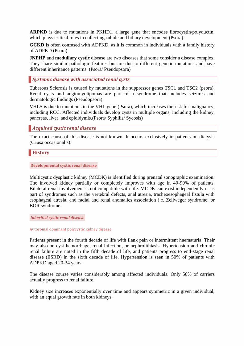
This CT scan demonstrates acquired renal cystic disease (ARCD) in a 70-year-
old man who is dialysis-dependent. The CT scan demonstrates bilateral
atrophic kidneys with multiple renal cysts.
o Medullary sponge kidney (MSK): Findings on plain radiographs may be
normal, or they may exhibit medullary nephrocalcinosis (represented by
multiple discrete calculi clustered in the renal pyramids). At least one renal
calculus (typically < 5 mm) is often observed. IVP demonstrates a "bouquet of
flowers" or "paintbrush" pattern. Ectatic tubules are observed as dense streaks
of contrast material radiating from the calyces, while papillary cysts are
observed as round opacifications in the papillae. The "brush" pattern of the
ectatic tubules must be differentiated from a dense papillary blush, which may
be observed in healthy patients; with low-osmolar contrast, papillary blush is
observed in as many as 13% of routine IVPs. A greater than 0.3-mm cylinder
or streak diameter has been recommended to help differentiate between
pathologic tubular ectasia and normal variant physiology. CT scan may show
calcifications at the corticomedullary junction.
o Simple cyst: The most clinically significant aspect of a simple cyst is
differentiating it from carcinoma. Simple-cyst walls occasionally calcify and,
thus, radiographically mimic malignancy. Sonographic features that support
the diagnosis of simple cyst include an anechoic round mass with a smooth
and sharply demarcated wall and through-transmission with strong posterior
wall echo. If the ultrasonography findings are suspicious or equivocal, a CT
scan is warranted. CT scan criteria for a benign cyst include (1) sharp
demarcation cyst with a smooth thin wall, (2) homogenous fluid within the
cyst (typically with density < 20 HU, although higher measurements may be
found with a benign proteinaceous cyst or if hemorrhage is present in a benign
cyst), and (3) no contrast enhancement. Enlargement of the cyst can raise the
concern of malignancy, although the natural history of benign renal cysts does
show progressive slow enlargement.
o Bosniak classification: Bosniak has described a classification scheme for renal
cysts based on CT scan findings.
Category I (simple cyst) - Thin wall without septa, calcifications, or
solid components; measures water density (< 20 HU) and does not
enhance (< 2% chance of malignancy)
Category II (minimally complex cyst) - Thin wall (< 1 mm) and no
enhancement; may contain 1 or 2 hairline-thin septa, fine calcification,
or short segment of slightly thickened calcification; includes high-
attenuation lesions that are smaller than 3 cm (Malignancy rates in

series range from 0-14%. Series with higher malignancy rates include
IIF lesions.)
Category IIF (indeterminate) - Minimal enhancement and/or
thickening of a hairline-thin smooth septum or wall; mildly thickened
or nodular calcification; no enhancing soft-tissue components; includes
nonenhancing high-attenuation lesions that are 3 cm or larger
(approximately 20% likelihood of malignancy)
Category III (suspicious indeterminate) - Multilocular lesion with
multiple enhancing septae, uniform wall thickening, nodularity, or
thick or irregular calcification (30-60% likelihood of malignancy)
Category IV (malignant) - Contains enhancing (>10 HU) large nodules
or clearly solid components (>90% likelihood of malignancy)
o Multiphasic CT in combination with Bosniak class can improve diagnostic
accuracy. To predict a renal cell carcinoma, a corticomedullary phase minus
precontrast phase value of >42 HU resulted in 97.1% sensitivity and 85.7%
specificity. This technique can further help guide treatment options.
o Another option for patients with renal impairment or allergy to iodinated
contrast is contrast-enhanced ultrasound (CE-US). CE-US is a technique that
has been shown to be equivalent to CT, and in one experience CE-US was
found to be better than CT in the diagnosis of malignancy in Bosniak IIF and
III renal cysts.[22]
o MRI may be used to help evaluate renal lesions in patients with either renal
impairment or allergy to iodinated contrast material. Contrast-enhanced MRI
and CT scan reveal similar findings in most cystic renal lesions. However,
MRI suggests a higher classification for some lesions by identifying more
septae, areas of wall thickening, or enhancement. Additionally, calcification
may not be appreciated with MRI.
Procedures
Aspiration: In the evaluation of an intermediate renal cyst, fine-needle aspiration has a
limited role. Some centers report a sensitivity of more than 70% for core biopsy and cyst
aspiration with cytology. The enzyme CA9 is being studied as a new marker for clear cell
renal cell carcinoma. One report showed that CA9 can be detected in the fluid of malignant
cystic, but not benign, renal tumors. This marker may be useful to help guide decisions of
treatment versus observation in select populations.
Developmental cystic renal disease
Multicystic dysplastic kidney: Cystic dysplasia is a subset of renal dysplasia. In this
form, typical renal configuration is lost. The disease is usually a unilateral process,
but it ranges from involving a portion of one kidney to completely involving both
kidneys. Grossly, the kidney appears to be an enlarged mass of cysts among immature
primitive tissue, often with surrounding fibrosis and an atretic collecting system.[10]
The ureter is often stenotic or hypoplastic, and the renal artery is often small or
absent.[4] Microscopy reveals small areas of otherwise normal-appearing glomeruli
and tubules interspersed with cysts lined with cuboidal epithelium and surrounded by
collars of spindle cells. The cysts are filled with proteinaceous or sanguinous fluid. In
addition, immature-appearing cartilage is often present in the tissue.

Cut surface of a nephrectomy specimen from a patient with a multicystic dysplastic
kidney (MCDK).
Inherited cystic renal disease
Autosomal dominant polycystic kidney disease
o The kidneys are enlarged and distorted by multiple renal cysts. Cystic kidneys
can exceed 40 cm in length and weigh as much as 5 kg. Cysts range in size
from a few millimeters to several centimeters and are distributed relatively
uniformly through the medulla and cortex. Cyst fluid ranges from clear to
hemorrhagic.
o Microscopic evaluation shows cystic dilatations in all segments of the
nephron, with loss of connection to the tubule. While all segments are
involved, the cysts derived from the collecting duct are the largest and most
numerous. The cysts are lined by a single layer of flattened-to-cuboidal
epithelium. The intervening parenchyma demonstrates interstitial fibrosis,
tubular atrophy, chronic inflammation, and vascular sclerosis.
External surface of a nephrectomy specimen from a patient with autosomal
dominant polycystic kidney disease (ADPKD).
Cut surface of the same nephrectomy specimen from a patient with autosomal
dominant polycystic kidney disease (ADPKD).

Autosomal recessive polycystic kidney disease
o The kidneys are enlarged bilaterally, but a reniform shape is preserved. With
neonatal presentation, the kidneys may be 10-20 times normal size. Radial
cysts are typically smaller than 3 mm in diameter and extend perpendicularly
from the papillary tips to the surface of the cortex. Microscopically, the cysts
are lined by flattened (undifferentiated) epithelium and represent fusiform
dilation of collecting tubules that retain their connection to the afferent and
efferent tubules. The parenchyma adjacent to the cysts progressively develops
interstitial fibrosis and glomerulosclerosis.
o The liver is grossly enlarged, and microscopic evaluation demonstrates bile
duct dilatation and periportal fibrosis. This histologic pattern is known as
congenital hepatic fibrosis (CHF) and is always present in ARPKD. However,
CHF is not specific to this disease.
GCKD is characterized by dilatation of Bowman space without involvement of the
related tubule. The dilated Bowman spaces are lined by a flattened epithelium and
contain rudimentary glomerular tufts.
Juvenile nephronophthisis and medullary cystic kidney disease: These diseases
are characterized by thickening and wrinkling of the tubular basement membrane,
tubular atrophy, and interstitial fibrosis, leading to bilaterally small kidneys with a
pitted surface.[3] The renal cortex is uniformly thinned, and cysts are located at the
corticomedullary junction and are derived from the collecting ducts and distal
tubules.[12] The number of cysts varies (5-50), and cysts measure from several
millimeters to 1 cm. However, 25% of cases do not involve grossly visible cysts.
Microscopic evaluation demonstrates that the cysts are lined by single layers of
cuboidal epithelium.
Systemic disease with associated renal cysts
TS: Renal cysts are uncommon and usually not extensive, but diffuse cystic renal
disease that involves both the cortex and the medulla is occasionally noted,
particularly in children. Cysts vary in size from several millimeters to 3 cm. Diffuse
renal cystic disease grossly resembles kidneys affected by ADPKD. Microscopically,
the cysts are lined by large eosinophilic cells with enlarged hyperchromatic nuclei.[1]
Von Hippel-Lindau syndrome (VHLS): Multiple renal cysts develop bilaterally.
Renal cysts are lined with glycogen-rich, clear-appearing cells (similar to those
observed with grade I clear-cell renal cell carcinoma [RCC]). Atypia and epithelial
hyperplasia are common in the cysts.
Acquired cystic renal disease
Acquired renal cystic disease
o Gross evaluation of early disease reveals cortical cysts filled with clear fluid.
Cysts are usually smaller than 0.5 cm in diameter but may be as large as 3 cm
in diameter. With more advanced disease, medullary cysts are observed. The
disease may progress to numerous diffusely distributed cysts and resemble a
small kidney affected by ADPKD.
o Microscopy reveals a flattened, hyperplastic tubular epithelial lining. Foci of
epithelial hyperplasia or renal adenomas are common. The remaining renal
tissue exhibits sclerotic glomeruli, atrophic tubules, and interstitial fibrosis.
Oxalate crystals are common in the walls of cysts.

MSK: Gross evaluation reveals normal-sized kidneys, which may be unremarkable
with the exception of at least one enlarged and pale renal pyramid. The disease is
bilateral in 70% of cases. Microscopic evaluation reveals dilated collecting ducts lined
by cuboidal or flattened epithelium. The cystlike cavities range in size from 1-7.5 mm
(usually 1-3 mm) and are present in the papillary portions of the pyramids. Roughly
half of the dilated channels contain calcifications. Inflammatory infiltrate is found
adjacent to the dilated tubules.
Simple cysts: Cysts measure 1-5 cm in diameter and are filled with clear fluid. The
cysts are usually lined by a flattened layer of epithelium, although they may lack an
epithelial lining.
Nephrectomy specimen from a patient with a large benign simple cyst.
Cut section of nephrectomy specimen demonstrating renal cell carcinoma (RCC),
with an adjacent simple cyst.
Close-up photograph of the cut surface of the same nephrectomy specimen
demonstrating a simple cyst adjacent to a renal cell carcinoma (RCC).

Medical Care
Effective means of prevention or modulation of disease have not yet been identified. Current
treatment is aimed at symptom control. In general, therapy is reserved for pain, hypertension,
infection, renal salt wasting, and nephrolithiasis.
Inherited cystic renal disease
o Autosomal dominant polycystic kidney disease
Patients have decreased ability to concentrate urine and should be
encouraged to drink 1-2 L of water daily.
Generally, 130/80 is considered the treatment goal for hypertension in
this population. Moderate hypertension may be treated with sodium
restriction (ie, < 100 mEq/d), exercise, and weight control.
Angiotensin-converting enzyme (ACE) inhibitors and angiotensin
receptor blockers (ARBs) are effective in controlling hypertension in
autosomal dominant polycystic kidney disease (ADPKD). However,
ACE inhibitors have been associated with reversible renal failure in
polycystic kidney disease. Calcium channel blockers also are effective
in managing hypertension in ADPKD.
Hypertension appears to correlate with the size of the cyst, and
aspiration of renal cysts results in a reduction of blood pressure.[25]
Prevention of infection with appropriate precautions is important,
particularly in women. Avoid urinary tract instrumentation whenever
possible.
Treatment of infection involving cystic kidneys requires a prolonged
course of antibiotics. Most cyst walls are permeable to polar
antibiotics, including cephalosporins, penicillin derivatives, and
aminoglycosides. Occasionally, cysts are relatively impermeable to
these agents and require parenteral lipophilic antibiotics, such as
ciprofloxacin, erythromycin, chloramphenicol, or a tetracycline.
Clinical evaluation findings, including sterile urine, lack of fever, and
no renal pain on deep palpation, should guide the route and duration of
antibiotic therapy.
o Autosomal recessive polycystic kidney disease (ARPKD): The newborn is
provided supportive therapy while the degree of pulmonary insufficiency and
the etiology is reviewed. Dialysis may be required for renal failure. With less
severe childhood disease, edema often is a problem and is managed with
sodium restriction and loop diuretics. Hypertension is controlled with salt
restriction and antihypertensives, with particular emphasis on the use of ACE
inhibitors and ARBs.
o Juvenile nephronophthisis (JNPHP) and medullary cystic kidney disease:
With severe salt wasting, salt supplementation may improve renal function
and slow renal demise. End-stage renal insufficiency necessitates dialysis or
renal transplantation.
Acquired cystic renal disease
o Acquired renal cystic disease (ARCD): Mild bleeding episodes may be
managed with bed rest and analgesics.
o Medullary sponge kidney (MSK): Encourage patients with nephrolithiasis to
produce 2 L of urine daily. Patients with hypercalcuria may benefit from oral

thiazide diuretics. Patients may develop UTI and should be taught preventative
measures.
o Simple cyst: An infected simple cyst usually requires a combination of
antimicrobial and surgical management. Pathogens encountered most
frequently in infected simple cysts include Enterobacteriaceae, staphylococci,
and Proteus species.
Recent research is beginning to identify biochemical targets that may allow disease-
modifying therapy.
Inhibitors of the EGF receptor tyrosine kinase have been shown to slow cyst
development and extend the life span in polycystic mice. Clinical trials with these
agents are underway.
Vasopressin receptor activation results in increased levels of cAMP. cAMP has been
shown to be cystogenic and provides the rationale for vasopressin receptor blockade.
Tolvaptan is a vasopressin receptor antagonist with high affinity in humans and is
currently in Phase III clinical trials in patients with ADPCK.
The identification of mTOR as a possible common pathway to cyst development
makes this protein an attractive target for therapy. Rapamycin inhibits mTOR and has
been shown to stop kidney growth and even allow regression in kidney size in a
mouse model. Additionally, a retrospective comparison of patients treated with
rapamycin to those not treated demonstrated a 25% decrease in kidney volume in the
treatment group. These animal studies have failed to correlate with human clinical
trials. In adults with ADPKD and early chronic kidney disease, 18 months of
treatment with sirolimus, an mTOR inhibitor , did not halt polycystic kidney growth.[
Surgical Care
Multicystic dysplastic kidney (MCDK): Previously, the involved kidney was
routinely removed to prevent the subsequent development of symptoms. Today,
however, surgical excision is indicated only if the dysplastic kidney interferes with
respiratory or digestive function or if significant hypertension has developed.
Additionally, cyst rupture, which can occur spontaneously or secondary to trauma,
may require emergent surgical intervention.
Inherited cystic renal disease
o ADPKD: Significant chronic pain may result from expansion of renal cysts.
Needle aspiration is usually the first-line approach to symptomatic cysts.
Initial resolution and then return of symptoms with reaccumulation of cyst
fluid increases the chance that a laparoscopic cyst decortication will eliminate
the patient's pain. However, for the management of severe pain, hypertension,
hematuria, or infection, surgical excision may be preferred. Complex cysts can
be explored laparoscopically and treated appropriately based on intraoperative
frozen sections. Laparoscopic techniques have been used with good results.
New studies have reported good outcomes of laparoscopic cyst decortication
using a retroperitoneal approach especially for posterior or lower pole lesions.
o Percutaneous endocystolysis is another technique described for treatment of
symptomatic cysts. The technique involves obtaining percutaneous access,
dilating the tract, and then introducing a resectoscope with rollerball electrode
to cauterize the internal surface of the cyst. A 13-year experience with this
technique reported clinical improvement in 100% of the patients with minimal
complications.
o Nephrectomy may be performed simultaneously with renal transplantation to
create space for the transplanted kidney and to relieve symptoms associated
with the native polycystic kidney. The timing of performing the nephrectomy
in the transplant patient has been debated. Data suggest that open ipsilateral
nephrectomy at the time of transplantation with staged contralateral native
nephrectomy has fewer perioperative complications than performing a
laparoscopic bilateral nephrectomy. In extreme cases of liver enlargement,
severe pain and wasting may result. Partial hepatectomy may alleviate these
symptoms.
o ARPKD: In patients with severe portal hypertension, sclerotherapy or
portosystemic shunt placement may be necessary to control bleeding.
Splenectomy may be indicated for splenomegaly with significant
complications.
o JNPHP and medullary cystic kidney disease (MCKD): If transplantation is
considered, selecting an older or unrelated donor is advisable to minimize the
risk of the transplanted kidney also being affected with these diseases.
Acquired cystic renal disease
o ARCD: Persistent or severe hemorrhage may necessitate nephrectomy or
renal embolization. If a 3-cm renal mass suggestive of renal cell carcinoma
(RCC) is noted, a partial or radical nephrectomy is indicated.
o Simple, intermediate, and suspicious cysts: Simple renal cysts rarely require
surgical management to relieve pain or obstruction. Treatment options include
aspiration, sclerosis, open resection, endoscopic marsupialization and
fulguration, percutaneous resection, and laparoscopic resection.
o Bosniak category III and IV renal cysts require surgical exploration.
Approximately 50% of Bosniak category III cystic renal lesions are malignant.
Management depends on the appearance of the lesion and varies from
exploration and biopsy to nephrectomy. The current standard approach is open
exploration with anticipated partial nephrectomy. However, as the experience
with laparoscopic exploration and nephrectomy grows, this technique may
prove equally reasonable.
Cystic clear cell renal cell carcinoma: Whether the patient has known
pathologically diagnosed malignancy from biopsy or suspected malignancy based on
Bosniak classification, a urologist can anticipate good surgical outcomes after
resection. A study evaluating laparoscopic nephrectomy for cystic clear cell renal cell
carcinoma revealed that all patients treated were alive after 5 years and that no patient
had extrarenal disease at the time of surgery. These data suggest that patients with
cystic RCC should expect to be cured after surgical resection, and furthermore should
undergo nephron-sparing surgery when possible.
Medication Summary
No specific medical therapies are available for the renal cysts themselves. Complications of
cystic renal diseases, such as hypertension, infection, and pain, are treated with standard
medical therapy.
Homoeopathic Treatment
Anilinum
SchroyeF. Synthesis Repertory- 9th version- KIDNEYS - TUMORS – Ureters
Colchicum
JULIAN O. A., Materia Medica of New Homeopathic Remedies- Urinary and genital organs Renal lithiasis-
Pyelitis and nephritis in a lithiasic or prostatic case.
Numerous cysts on the kidney.
Kalium bichromicum
Liga Medicorum Homoeopathica Internationalis- Kalium bichromicum- Essential hypertension- In the
cardiac zone, Kali Bichromicum plays a dominant role in the control of essential Hypertension due to Polly-cystic
Kidney with striated masses of cysts and parenchyma, causing fibrosis and sclerosis of the renal artery. There is
much of shooting pain at the renal angles due to vaso-constriction. Renal Ischaemia follows with essential
Hypertension due to excessive liberation of Rennin and reaction of Hypertensionogen.
Kresol
SchroyeF. Synthesis Repertory- 9th version- Kidney- Cysts
Medorrhinun
Kroschewski Konig Friedel- Kidneys Cysts.
Plumbum met
HEMPEL C. J., A New and Comprehensive System of Matera medica- Both kidneys have dwindled to one-
half their size; the surface of the cortical substance shows large degenerations or granulations of the size of a
millet-seed, and slightly raised above the surface, of a dingy, yellowish white, very firm, tenacious; a deposit of
a blackish-blue pigmentum was deposited here and there through the degenerated portion of the kidneys, with
a few scattered cysts of the size of a pea, containing a brownish serum. The bladder was contracted, contained
a few drops of turbid urine; in the abdominal cavity six pounds of gray serum were discovered (in the case of an
engraver, thirty years old, who had handled preparations of lead since his childhood, and had had ten attacks
of the lead-colic, the last attack ten years ago).
As consequences of a chronic poisoning by lead were discovered : shrinking and considerable contraction of the
inner cavities, particularly of the stomach and bowels, hardness of the parenchymatous organs, which had
become much smaller in consequence of a morbid contractions,; great thinness and almost inorganic hardness
of the muscles; complete shrivelling of the mucous and adipose tissues. A special examination of the
neurilemma and the nervous substance is entirely wanting.
Although the toxicological effects of lead are exceedingly striking and varied, yet its importance as a
therapeutic agent is not proportionate to the position it occupies in the works of toxicologists. I will endeavour
to condense from the work of Tanquerel des Planches, from Frank's Magazine, and from toxicological treatises,
such toxicological symptoms as may be of any possible use to us in a therapeutic point of view.
Merc cor
HUGHES R. and DAKE J. P., A Cyclopaedia of Drug Pathogenesis- Kidneys of normal consistence, the
cortex showing many little red points, especially between the infundibula, apparently from incipient
inflammation.

On the back of the right kidney was a cyst the size of a marble, filled with clear fluid.
Bladder very much contracted, containing about 1/2 oz. of turbid urine.
Bibliography
Chapter 38. Urology > Simple Renal Cysts CURRENT Diagnosis & Treatment: Surgery, 13e
Chapter 9. Laparoscopic Surgery > Renal Cyst Decortication Smith and Tanagho's General Urology, 18e
Chapter 38. Urology > Simple Renal Cyst CURRENT Diagnosis & Treatment: Surgery, 13e
Chapter 8. Percutaneous Endourology & Ureterorenoscopy > Renal Cysts Smith and Tanagho's General
Urology, 18e
Chapter 46. Cystic Diseases of the Kidney > Simple Renal Cysts CURRENT Diagnosis & Treatment:
Nephrology & Hypertension
Chapter 32. Disorders of the Kidneys > Simple (Solitary) Cyst Smith and Tanagho's General Urology, 18e
Chapter 56. Urinary Tract Infections > Infected Cysts Principles of Critical Care, 3e
Chapter 16. Pathology of the Kidney and Bladder > Simple Cysts Pathology: The Big Picture
Chapter 46. Cystic Diseases of the Kidney > General Considerations CURRENT Diagnosis & Treatment:
Nephrology & Hypertension
Encyclopedia Homoeopathica
New England Journal of Homoeopathy (_New_Engl_J_Hom) Baryta sulphurica ...ear infections on the right side. Also infection of the kidneys, appendicitis, ovarian cysts and cysts in her right breast.
SCHOLTEN J., Homoeopathy and minerals (stj1) " ...ear infections on the right side. Also infection of the kidneys, appendicitis, ovarian cysts and cysts in her right breast. Seasickness (2).
JULIAN O. A., Materia Medica of New Homeopathic Reme... Colchicum autumnale ...Numerous cysts on the kidney.

Liga Medicorum Homoeopathica Internationalis (~LMHI) Kalium bichromicum ...role in the control of essential Hypertension due to Polly-cystic Kidney with striated masses of cysts and parenchyma, causing fibrosis and sclerosis of the renal artery
HUGHES R. and DAKE J. P., A Cyclopaedia of Drug Path... Mercurius corrosivus ...On the back of the right kidney was a cyst the size of a marble, filled with clear fluid.
HEMPEL C. J., A New and Comprehensive System of Mat... Plumbum metallicum ...deposited here and there through the degenerated portion of the kidneys, with a few scattered cysts of the size of a pea, containing a brownish serum
American Institute of Homoeopathy (Am_Inst_Hom) cyst may have more physical signs in common with a cyst of the kidney than with any pathologically allied growth, ovarian or uterine. " ...or corpuscles have been found in the fluid obtained from cysts of the kidney.
GUPTA R. L., Directory of Diseases and Cures in Homoeo... ...kidney). Sometimes kidney failure is due to the disease polycystic kidneys (the formation of numerous cysts in both the kidneys) and symptoms of polycystic kidneys, like haematuria; infection of the
Homeopathic Medical Society Pennsylvania, The (_Hom_... ...In the more advanced stages of the disease, the granular kidney almost always contains cysts. They are found both in the cortex and in the " ...causes the tube to remain fixedly open, even after death. Cysts also arise, whether the kidneys be granular or waxy, from the constricting action of the
International Hahnemannian Association (Int_Hahn_Ass) ...dilated to the size of a little finger. The right kidney, although not enlarged, was cystic, with points of suppuration. " ...and there are others). One said it was a dermoid cyst, another a floating kidney. " ...was passed on to me after the removal of one kidney for cystic degeneration, the remaining one being so diseased that it was
RAUE C. G., Diseases of Children (res1) ...unless they attract attention by attaining considerable size. Infants with cystic kidneys die shortly after birth as a rule. The condition is
RISQUEZ F., Psychiatry and Homeopathy (rq1) ...smear IIIb. Laboratory tests within normal range. Abdominal echo: Small cyst in the right kidney, with no pathological relevance. Chest X rays: Normal. There is..
Radar 10