renal drug metabolism - pharmacological...
TRANSCRIPT
Renal Drug MetabolismJAMES W. LOHRa, GAIL R. WILLSKY, AND MARGARET A. ACARA
Departments of Pharmacology and Toxicology, Biochemistry, and Medicine, School of Medicine and Biomedical Sciences, State Universityof New York, Buffalo and V.A. Medical Center, New York
I. Introduction. . . . . . . . . . . . . . . . . . . . . . . . . . . . . . . . . . . . . . . . . . . . . . . . . . . . . . . . . . . . . . . . . . . . . . . . . . . . 107A. History . . . . . . . . . . . . . . . . . . . . . . . . . . . . . . . . . . . . . . . . . . . . . . . . . . . . . . . . . . . . . . . . . . . . . . . . . . . . . 108B. Methodology . . . . . . . . . . . . . . . . . . . . . . . . . . . . . . . . . . . . . . . . . . . . . . . . . . . . . . . . . . . . . . . . . . . . . . . . 108
1. Clearance . . . . . . . . . . . . . . . . . . . . . . . . . . . . . . . . . . . . . . . . . . . . . . . . . . . . . . . . . . . . . . . . . . . . . . . . 1082. Sperber technique in chickens . . . . . . . . . . . . . . . . . . . . . . . . . . . . . . . . . . . . . . . . . . . . . . . . . . . . . 1093. Isolated perfused kidney . . . . . . . . . . . . . . . . . . . . . . . . . . . . . . . . . . . . . . . . . . . . . . . . . . . . . . . . . . 1104. Tissue preparations. . . . . . . . . . . . . . . . . . . . . . . . . . . . . . . . . . . . . . . . . . . . . . . . . . . . . . . . . . . . . . . 1105. Molecular biology . . . . . . . . . . . . . . . . . . . . . . . . . . . . . . . . . . . . . . . . . . . . . . . . . . . . . . . . . . . . . . . . . 111
II. Renal pathways for the biotransformation of drugs. . . . . . . . . . . . . . . . . . . . . . . . . . . . . . . . . . . . . . . . . 111A. Cytochrome P450 dependent mixed function oxidase system . . . . . . . . . . . . . . . . . . . . . . . . . . . . . 111
1. Cytochrome P450 in the kidney . . . . . . . . . . . . . . . . . . . . . . . . . . . . . . . . . . . . . . . . . . . . . . . . . . . . 1122. Drugs that induce cytochrome P450 proteins . . . . . . . . . . . . . . . . . . . . . . . . . . . . . . . . . . . . . . . . 1133. Specific renal cytochrome P450 enzymes . . . . . . . . . . . . . . . . . . . . . . . . . . . . . . . . . . . . . . . . . . . . 1144. Nondrug factors that affect cytochrome P450 enzymes in the kidney that may modulate
kidney drug metabolism. . . . . . . . . . . . . . . . . . . . . . . . . . . . . . . . . . . . . . . . . . . . . . . . . . . . . . . . . . . 115B. N-oxidation (flavin-containing monooxygenases) . . . . . . . . . . . . . . . . . . . . . . . . . . . . . . . . . . . . . . . . 116C. Alcohol oxidation . . . . . . . . . . . . . . . . . . . . . . . . . . . . . . . . . . . . . . . . . . . . . . . . . . . . . . . . . . . . . . . . . . . . 116D. Aldehyde oxidation . . . . . . . . . . . . . . . . . . . . . . . . . . . . . . . . . . . . . . . . . . . . . . . . . . . . . . . . . . . . . . . . . . 117E. Oxidative deamination (monoamine oxidase) . . . . . . . . . . . . . . . . . . . . . . . . . . . . . . . . . . . . . . . . . . . 118F. Aldehyde and ketone reduction . . . . . . . . . . . . . . . . . . . . . . . . . . . . . . . . . . . . . . . . . . . . . . . . . . . . . . . 118
1. Aldehyde reductase . . . . . . . . . . . . . . . . . . . . . . . . . . . . . . . . . . . . . . . . . . . . . . . . . . . . . . . . . . . . . . . 1192. Ketone reductase . . . . . . . . . . . . . . . . . . . . . . . . . . . . . . . . . . . . . . . . . . . . . . . . . . . . . . . . . . . . . . . . . 1193. Other . . . . . . . . . . . . . . . . . . . . . . . . . . . . . . . . . . . . . . . . . . . . . . . . . . . . . . . . . . . . . . . . . . . . . . . . . . . 119
G. Hydrolysis mechansims . . . . . . . . . . . . . . . . . . . . . . . . . . . . . . . . . . . . . . . . . . . . . . . . . . . . . . . . . . . . . . 1191. Ester and amide hydrolysis/carboxylesterase and amidase . . . . . . . . . . . . . . . . . . . . . . . . . . . . 1192. Epoxide hydrolysis. . . . . . . . . . . . . . . . . . . . . . . . . . . . . . . . . . . . . . . . . . . . . . . . . . . . . . . . . . . . . . . . 120
III. Phase II: synthetic conjugation pathways . . . . . . . . . . . . . . . . . . . . . . . . . . . . . . . . . . . . . . . . . . . . . . . . . 121A. Glucuronidation . . . . . . . . . . . . . . . . . . . . . . . . . . . . . . . . . . . . . . . . . . . . . . . . . . . . . . . . . . . . . . . . . . . . . 121
1. Isoforms . . . . . . . . . . . . . . . . . . . . . . . . . . . . . . . . . . . . . . . . . . . . . . . . . . . . . . . . . . . . . . . . . . . . . . . . . 1222. Substrates and kinetics . . . . . . . . . . . . . . . . . . . . . . . . . . . . . . . . . . . . . . . . . . . . . . . . . . . . . . . . . . . 1233. Induction of renal glucuronyl transferase . . . . . . . . . . . . . . . . . . . . . . . . . . . . . . . . . . . . . . . . . . . 1234. Localization . . . . . . . . . . . . . . . . . . . . . . . . . . . . . . . . . . . . . . . . . . . . . . . . . . . . . . . . . . . . . . . . . . . . . . 1245. Relative contribution of renal glucuronidation . . . . . . . . . . . . . . . . . . . . . . . . . . . . . . . . . . . . . . . 124
B. Sulfation . . . . . . . . . . . . . . . . . . . . . . . . . . . . . . . . . . . . . . . . . . . . . . . . . . . . . . . . . . . . . . . . . . . . . . . . . . . 124C. Methylation. . . . . . . . . . . . . . . . . . . . . . . . . . . . . . . . . . . . . . . . . . . . . . . . . . . . . . . . . . . . . . . . . . . . . . . . . 125
1. N-methylation. . . . . . . . . . . . . . . . . . . . . . . . . . . . . . . . . . . . . . . . . . . . . . . . . . . . . . . . . . . . . . . . . . . . 1252. O-methylation. . . . . . . . . . . . . . . . . . . . . . . . . . . . . . . . . . . . . . . . . . . . . . . . . . . . . . . . . . . . . . . . . . . . 1263. S-methylation . . . . . . . . . . . . . . . . . . . . . . . . . . . . . . . . . . . . . . . . . . . . . . . . . . . . . . . . . . . . . . . . . . . . 126
D. Acetylation . . . . . . . . . . . . . . . . . . . . . . . . . . . . . . . . . . . . . . . . . . . . . . . . . . . . . . . . . . . . . . . . . . . . . . . . . 127E. Glutathione conjugation. . . . . . . . . . . . . . . . . . . . . . . . . . . . . . . . . . . . . . . . . . . . . . . . . . . . . . . . . . . . . . 128F. Mercapturic acid synthesis . . . . . . . . . . . . . . . . . . . . . . . . . . . . . . . . . . . . . . . . . . . . . . . . . . . . . . . . . . . 129G. Amino acid conjugation . . . . . . . . . . . . . . . . . . . . . . . . . . . . . . . . . . . . . . . . . . . . . . . . . . . . . . . . . . . . . . 130
1. Glycine conjugation . . . . . . . . . . . . . . . . . . . . . . . . . . . . . . . . . . . . . . . . . . . . . . . . . . . . . . . . . . . . . . . 1312. Glutamine conjugation . . . . . . . . . . . . . . . . . . . . . . . . . . . . . . . . . . . . . . . . . . . . . . . . . . . . . . . . . . . . 132
H. Cysteine conjugate b-lyase . . . . . . . . . . . . . . . . . . . . . . . . . . . . . . . . . . . . . . . . . . . . . . . . . . . . . . . . . . . 132IV. Localization of drug metabolizing enzymes in the kidney . . . . . . . . . . . . . . . . . . . . . . . . . . . . . . . . . . . 132V. Effects of renal metabolism . . . . . . . . . . . . . . . . . . . . . . . . . . . . . . . . . . . . . . . . . . . . . . . . . . . . . . . . . . . . . . 133
0031-6997/98/5001-0107$03.00/0PHARMACOLOGICAL REVIEWS Vol. 50, No. 1Copyright © 1998 by The American Society for Pharmacology and Experimental Therapeutics Printed in U.S.A.
107
by guest on May 24, 2018
Dow
nloaded from

VI. Acknowledgments. . . . . . . . . . . . . . . . . . . . . . . . . . . . . . . . . . . . . . . . . . . . . . . . . . . . . . . . . . . . . . . . . . . . . . . 134VII. References . . . . . . . . . . . . . . . . . . . . . . . . . . . . . . . . . . . . . . . . . . . . . . . . . . . . . . . . . . . . . . . . . . . . . . . . . . . . . 135
I. Introduction
A. History
The kidneys have important physiological functionsincluding maintenance of water and electrolyte balance,synthesis, metabolism and secretion of hormones, andexcretion of the waste products from metabolism. Inaddition, the kidneys play a major role in the excretionof drugs, hormones, and xenobiotics. Mechanisms in-volved in the transport of drugs in the proximal tubulein the secretory direction have been amply reviewed(Bessighir and Roch-Ramel, 1988; Pritchard and Miller,1993). Reabsorptive transport for organic compounds,particularly amino acids (Zelikovic and Chesney, 1989;Silbernagl, 1992) and choline (Acara and Rennick, 1973;Acara et al., 1979) also have been studied. The conceptsassociated with pH dependence in the nonionic passiveback diffusion of drugs are well-described (Roch-Ramelet al., 1992). However, the role of the kidney in themetabolism of both endogenous and exogenous com-pounds has not received appropriate attention.
Most of the current knowledge about drug metabolismis based on studies in which the liver was the experi-mental organ. It is now clear that the kidney activelymetabolizes many drugs, hormones, and xenobiotics(Anders, 1980; Bock et al., 1990). In some cases, certainbiotransformations occur at a faster rate in the kidneythan in the liver; e.g., glycination of benzoic acid (Poonand Pang, 1995). Bowsher et al. (1983) found histamineN-methyltransferase activity to be higher in concentra-tion in rat renal tissue than in any other organ. Gammaglutamyl transferase activity in mammalian tissues is atits highest in the kidney (Goldbarg et al., 1960; seeSection III.F.).
The heterogeneity of the kidney makes it important todefine the regional distribution of enzyme systems on acellular and subcellular level. The human kidney hastwo distinct regions: an outer cortical region and theinner medullary region. The medulla is divided intoseveral pyramids, the base of which is at the corticomed-ullary junction and the apex of which approaches therenal pelvis, forming a papilla. This heterogeneity iscaused by three successive excretory systems that de-velop during embryonic development; and the latter two,the mesonephros and metanephros, contribute to theformation of the kidney. The ureteric bud, a specializedstructure of the mesonephric duct, gives rise to the col-lecting ducts, calyces, pelvis, and ureter. The metane-
phros gives rise to the glomerulus, proximal, and distaltubules. Whereas most studies have been performedeither in whole kidney or in cortical tissue, biotransfor-mations have also been identified in the medullary re-gion (Toback et al., 1977a; Lohr and Acara, 1990).
Information accumulated over the past 20 years dem-onstrates a large capacity for metabolism in the kidney,leading to activation or inactivation of numerous com-pounds and providing a major route for drug disposition.In addition, the metabolic products produced by thekidney may exert significant toxic effects. The pattern ofblood flow through the kidney, the acidity of the urine,and the urinary concentrating mechanism provide anenvironment that facilitates the concentration of partic-ular compounds in the medullary/papillary zone of thekidney, and sometimes even, their precipitation (e.g.,uric acid) with resultant damage. Such reactions will bepresented in a general way because the action of toxinson the kidney is beyond the scope of this review.
In this review, various methods will be described thathave been used to study renal metabolism of drugs,xenobiotics, hormones, and endogenous compounds. Thevarious types of metabolic reactions that occur in thekidney will be presented along with the compounds thatoccupy those particular routes. The contribution of theparticular metabolic pathways to the direction of move-ment of metabolite, into blood or into urine, provides aninterrelationship between transport and metabolism.
B. Methodology
Several different methods have been used to study therole of the kidney in the metabolism of drugs and xeno-biotics. These vary from in vivo techniques, such asclearance and the Sperber chicken preparation, to invitro studies of metabolism using organelles such asmitochondria and microsomes and molecular biology inwhich genes encoding specific enzymes of metabolismhave been identified. Each technique has contributeddifferent information regarding the way in which com-pounds are handled by the kidney.
1. Clearance. Historically, the contribution of the kid-ney to the elimination of a particular drug was mea-sured as renal clearance (Moller et al., 1928). The term“clearance” must be defined because it is being used todescribe an ever increasing number of functional equa-tions. Renal clearance as described by Homer Smith(1956) is “the volume of plasma required to supply thequantity X excreted in urine each minute” or the volumeof plasma completely cleared of that substance in 1minute time. However, these two definitions are not thesame because what is “cleared” may not necessarily ap-pear in the urine.
a Address correspondence to: Margaret Acara, Department ofPharmacology and Toxicology, School of Medicine and BiomedicalSciences, State University of New York at Buffalo, Buffalo, NY14214.
108 LOHR ET AL.

Clearance when it is applied to the kidney, is gener-ally defined by the equation: CX 5 UX
. V/PX where UX isthe concentration of compound (x) in the urine, PX is theconcentration in the plasma, V is the urine flow rate andthe result (Cx) expressed in volume per unit time, e.g.,ml/min.
An important point is that this equation provides in-formation on the amount of substance appearing in theurine but does not account for the portion of that sub-stance that undergoes metabolism or synthesis in thekidney. When, as is frequently done, the ClX is related toglomerular filtration rate, a value ,1 is taken to indi-cate a reabsorptive component or removal from tubularfluid. The disappearance of compound can be attributedto metabolism as well as reabsorption. Thus, the term“renal clearance” may be thought of as a general expres-sion of the removal of compound by all the routes of thekidney as in (A-V/A)Q (the concentration of compound inthe renal artery (A) minus the concentration of com-pound in the renal vein (V) divided by the concentrationin the renal artery times renal blood flow (Q)). “Urinaryclearance” is that clearance associated with compoundappearing in the urine. The difference between thesetwo clearance terms is attributable to storage and me-tabolism (Acara, 1992).
Clearance methods, developed to quantify renal func-tion, were the first methods used to study in vivo renalmetabolism (Toretti and Weiner, 1976; Tucker, 1981).Metabolism may be detected during infusion of a radio-labeled compound when the label in the urine is identi-fied as something other than the original compound(Quebbemann and Rennick, 1969; Acara and Rennick,1972; Toback et al., 1977a,b).
Monitoring the fate of radiolabeled compounds afterinfusion into the renal artery has provided some insightinto the kidney’s capacity to metabolize. Diamond andQuebbemann (1981) demonstrated clearance of radiola-beled metabolite during steady-state infusion of radiola-beled metabolite (true clearance) and measurement ofthe clearance of unlabeled metabolite during steady-state infusion of radiolabeled metabolite and unlabeledprecursor (apparent clearance). The difference betweenthe apparent clearance and true clearance is the renalcontribution to formation of urinary metabolite.
Tremaine, Diamond and Quebbemann (1984, 1985)developed another radioisotope technique, termed thespecific activity difference ratio (SADR
b
) technique. Thisinvolves the infusion of radiolabeled precursor as well asthe infusion of unlabeled metabolites if not present en-dogenously. The technique permits the quantification ofrenal formation and excretion of several metabolites ifthey are excreted in measurable amounts. Specific ac-tivity ratio technique is a standard isotope dilutionmethod. This is performed by a constant infusion ofradiolabeled metabolite along with a constant infusionof unlabeled precursor. The specific radioactivities ofmetabolites in plasma and urine are subsequently mea-sured. The ratio of the specific activity of metabolite inurine to that in plasma when subtracted from one indi-cates the fraction of urinary metabolite formed in thekidney and excreted; thus, determining the in vivo renalcontribution to formation of a compound.
2. Sperber technique in chickens. Birds have a renalportal circulation, accessible through a leg vein, whichpermits the administration of substances to the ipsilat-eral kidney. Sperber (1946) demonstrated that whensubstrates transported by organic excretory transportcarriers were infused into the leg vein, they were ex-creted in excess in the urine from the ipsilateral kidney.Substances entering the general circulation were ex-creted by both kidneys equally. Because the chicken hasno bladder, ureters from either side may be isolated forurine collection (Campbell, 1960). Thus, when contralat-eral excretion is subtracted from ipsilateral excretionand blood flow through this system is considered, avalue is obtained that describes the efficiency by whichthe compound is removed from the blood by the kidney.In measuring the excretion of the metabolites of aninfused substance, those excreted in excess by the in-fused kidney represent the results of intrarenal metab-olism.
An advantage of the Sperber technique include an invivo system in which pico- to nanomole amounts of ra-dioactive substrate may be studied. Because systemiccontributions may be accounted for, it acts as an in vivoperfused kidney. The technique can be used to measurethe metabolic capacity of the renal parenchyma and theeffects of drugs and chemicals on the function of certainrenal enzymes (Rennick, 1981).
b Abbreviations: ADH, alcohol dehydrogenase; ALDH, aldehydedehydrogenase; AP, aminopyrine; BBM, brush border membrane;b-NF, b-napthoflavone; BP, benzpyrene; CCbL, cysteine conjugateb-lyase; cDNA, complementary deoxyribonucleic acid; COMT, cate-cholamine-O-methyltransferase; CYP, cytochrome P450; DNA, de-oxyribonucleic acid; mEH, microsomal epoxide hydroxylase; FAD,flavin adenine dinucleotide; FMO, flavin-containing monoxygenase;GFR, glomerular filtration rate; GSH, glutathione; GST, glutathioneS-transferase; HMT, histamine N-methyltransferase; 5-HT, 5-hy-droxytryptamine; LPS, lipopolysaccharide; MAO, monoamineoxi-dase; MB-COMT, membrane bound COMT; 3-MC, 3-methylcholan-threne; mEH, microsomal epoxide hydrolase; MFO, mixed functionoxidase; mRNA, messenger ribonucleic acid; NAD, nicotinamide ad-enine dinucleotide; NADH, nicotinamide adenine dinucleotide, re-duced; NADPH, nicotinamide adenine dinucleotide phosphate; NAT,N-acetyltransferases; PABA, para-aminobenzoic acid; PAH, r-amin-ohippuric acid; PAPS, 39-phosphoadenosine 59 phosphosulfate; PB,phenobarbital; PCB, polychlorinated biphenyl; PEA, phenylethyl-amine; PNMT, phenylethanolamine N-methyl-transferase; SADR,specific activity difference ratio; SAM, S-adenosylmethionine; SAR,specific activity ratio; sEH, cystolic (soluble) epoxide hydrolase; ST,sulfotransferase; STZ, streptozotocin; TEMT, cytosolic thioether-S-methyltransferase; TMT, thiol-methyltransferase; TPMT, solublethiopurine-methyltransferase; TTCD, tetrachlorodibenzo-p-dioxin;UDP, uridine diphosphate; UDPGA, UDP-glucuronic acid; UDPGT,uridinediphosphoglucuronyl transferase; UGT, UDP-glucuronyl-transferase.
RENAL DRUG METABOLISM 109

Whereas urinary metabolites clearly reflect active bio-chemical pathways qualitatively, conversion rates aredifficult to determine. The technique does not accountfor the reabsorptive route of the metabolites but only forthat route that ends with the appearance of the metab-olite in the urine.
3. Isolated perfused kidney. The isolated perfused kid-ney preparation permits the measurement of excretion,reabsorption, and renal metabolism. (Nishiitsutsuji-Uwo, 1967; Bowman, 1978; Nizet, 1978). Because thekidney is removed from the animal, the influence ofother organs and tissues is not present. Renal clearanceand urinary clearance may be determined for a givencompound. As previously indicated, a large renal clear-ance associated with a low urinary clearance suggests ametabolic component. Kidneys may be perfused for up to2 h and samples of perfusate and urine collected forappropriate analyses.
The kidney itself may be analyzed at the end of theexperiment. Thus, the total disposition of substrate inurine, perfusate, and kidney, the direction of transport,and the associated metabolic routes can be measured, ascan the effects of other agents on these compartments(Acara, 1979). The conversion of enalapril, an inhibitorof angiotensin converting enzyme, to its metabolite, ena-laprilat, and the transport of drug and metabolite acrossthe basolateral and luminal membranes using constantflow single pass and recirculating isolated perfused ratkidney preparations provided an intrinsic clearance forrenal drug metabolism as well as identifying membranetransport steps (de Lannoy et al., 1990).
Glutathione content of isolated perfused kidney is con-sistently lower than that observed in vivo (Ross et al.,1980) and the maximum rates of drug metabolism maynot be observed. Functional shortcomings include lowglomerular filtration rate relative to perfusate flow andexcessive delivery of filtrate to the distal tubule withdecreased concentrating ability (Maack, 1980, 1986).
4. Tissue Preparations.a. KIDNEY SLICES. Kidney slices have been used for the
study of renal uptake and metabolism for decades (For-ster, 1948). Kidneys are removed quickly after onset ofanesthesia and kept chilled during slicing and until thestart of incubation. Slices, thin enough to permit oxygento reach all of the tissue, may be incubated for up to 2 h.At the termination of the experiment, slices are blottedon filter paper and weighed. Analyses of media andtissues for substrate and metabolites can reveal accu-mulation of substrate against a concentration gradient;as well as metabolic routes and intracellular and extra-cellular amounts of substrate and metabolites.
Phospholipid metabolism from (14C)-choline was stud-ied in mouse kidney slices during renal compensatorygrowth by Toback et al. (1974). Inner cortical slices werefound to have enhanced synthesis of choline containinglipids during kidney regeneration (Toback et al., 1977b).Relative specific activities of enzymes have been studied
in dissected cortex, outer medulla, and inner medulla.Choline dehydrogenase activity was shown to increasein cortex and not change in the inner medulla duringhypernatremia (Grossman and Hebert, 1989). Additionof dimethylaminoethanol, an analogue of choline and aninhibitor of choline oxidase, to isolated perfused kidneysas well as dissected tissue regions decreased betaineproduction from 14C-choline (Lohr and Acara, 1990).Toback et al. (1977b) studied the phosphorylation ofcholine into choline phosphoglycerides in different kid-ney regions.
However, information regarding the direction ofmovement, i.e., reabsorptive versus excretory, is not ob-tained using kidney slices. Some controversy exists re-garding whether or not tubules are collapsed. Unifor-mity of slices is important because substrate and oxygenshould reach all cells. Sometimes the innermost cells ofthe slice are not exposed to the same concentrations asthe surface cells (Foulkes, 1996). The reproducibilityand viability of tissue slices were greatly improved bythe more recent introduction of automated procedures toproduce “precision cut” kidney slices (Smith et al., 1985).
b. ISOLATED TUBULES. Tubule segments may be ob-tained by several methods. Microdissection of largenumbers of tubule segments may be performed afterperfusion of the kidney with a collagenase containingbuffer and incubation with collagenase. The tubule seg-ments can then be used for metabolic studies in whichthe tubules are typically incubated with a radiolabeledcompound. After incubation, the tubules can be sepa-rated from the medium either by centrifugation or rapidfiltration. Specific nephron segments can be obtained bythis method but the tissue yield is low. Development ofmicroassays has permitted the study of enzyme activi-ties in these segments (Endou, 1983a; Schlondorff,1986).
Relatively homogeneous proximal tubule suspensionscan be obtained in greater quantity by density gradientcentrifugation, and, with oxygenation, remain viable forapproximately 2 h. Obtaining suspensions of tubule seg-ments other than proximal is more difficult because theyconstitute lower percentages of the kidney mass.
c. CELL CULTURE. Cultured cells have become a morepopular tool for metabolic studies. In addition to pri-mary cultures, there are now many continuous kidneycell lines (e.g., MDCK, LLC-PK1, OK, A6, JTC-12,BSC1) available for study (Handler and Burg, 1992).
The LLC-PK1, OK, and JTC-12 cell lines are of prox-imal tubular origin. Of these the LLC-PK1 cells, derivedfrom the Hampshire pig, have been best characterized.They have characteristics such as Na-dependent glucoseuptake, Na-dependent phosphate uptake, and Na-de-pendent amino acid uptake, and high activities of sev-eral brush border enzymes. The MDCK cell line is mostcharacteristic of distal tubule, and the A6 cell line re-sembles the collecting duct.
110 LOHR ET AL.

Primary cultures of proximal renal tubules may alsobe grown for use in studies of renal drug metabolism.This may be accomplished with tubules obtained bymicrodissection or by macro separation techniques usingrabbit, rat, dog or human kidney.
Metabolic studies of cells in culture are generally per-formed on cells which have reached confluence. The cellsare rinsed with a buffer free of the metabolic substrateto be studied and then incubated for an appropriate timewith substrate generally radiolabeled. The experimentis ended by aspirating the medium, rinsing the cells, anddisrupting the cells with NaOH, scraping, or sonication.The cell fractions can then be further analyzed.
Fry and colleagues (1978) found that liver and kidneycells demonstrate similar conjugated metabolite pat-terns. Jones et al. (1979) found that the specific activi-ties for formation of glucuronide and sulfate derivativesin the kidney were approximately 5% of those for livercells, although formation of sulfhydryl derivatives wasproportionately higher in kidney cells with paracetamolas substrate.
d. SUBCELLULAR FRACTIONS. Small (100 nm diameter)closed vesicles which form when cells are disrupted byhomogenization and sediment at 100,000 3 g are iden-tified as microsomes. They consist primarily of mem-branes of endoplasmic reticulum and have proven to bea valuable tool in studying synthetic and metabolic func-tions of the cell. Microsomes from kidney cortex havebeen used to study drug metabolism. After homogeniza-tion and centrifugation at low speed, the resulting su-pernatant is centrifuged at high speed for 60 min. Themicrosomal fraction is the pellet that is resuspended foruse in drug metabolism studies. Substrates are added tothe microsomal incubation medium to study rates ofconversion to metabolites. Animals may be pretreatedwith various drugs and xenobiotics, and the effects ofthese compounds on microsomal enzyme activity deter-mined in vitro. In addition to studying metabolism byuse of microsomes, the cytosolic fraction (i.e., the super-natant from the microsomal fractionation) of cells can beisolated and used alone or in conjunction with the mi-crosomal fraction.
5. Molecular biology. The increased use of molecularbiology techniques in the past 15 years has heavily im-pacted on how we classify and identify proteins. Whenavailable, IUBMB enzyme numbers are given for theenzymes discussed in this review. However, many en-zyme activities have only been studied in membranefractions or using nucleic acid probes, and IUBMB num-bers are not available. A parallel system based on ge-netic information has arisen. The advances in molecularbiology have allowed isolated proteins to be cloned andsequenced. In many cases, especially caused by the easeof analysis and amplification of small amounts of nucleicacid materials, it is easier to study the genetic materialrather than the protein. Genes are isolated in differentorgans and species on the basis of homology to known
genes whose enzymatic activities have been studied atthe protein level. The transcriptional regulation ofthese genes can be studied and hypothetical proteinsequences deduced. The explosion of molecular biologydata has led to the same gene being isolated duringstudies of different phenotypes. To put some order intothe system, there are organizations devoted to specificorganisms: HUGO Gene Nomenclature Committee forhuman genes (accessed through http://gdb.org/gdb) andthe Mouse Gene Nomenclature Committee for mousegenes (accessed through the Jackson Laboratory website: http://www.informatics.jax.org). In addition, spe-cific gene families have their own nomenclature organi-zations, such as the P450 Nomenclature Committeeidentified in the P450 section.
Molecular biology studies can never completely super-sede the biochemical studies of isolated enzymes. Thestudy of a gene isolated in the kidney as a homologue ofa well-studied liver enzyme is extremely useful. How-ever, it is not guaranteed that the enzyme encoded bythat gene, even if it shows the same transcriptionalregulation, has the same function in the kidney. Theresearcher is advised to check the gene banks for homol-ogous genes, but to realize also that the strictly molec-ular biology studies do not indicate that the proteinencoded by that gene has the implied function underphysiological conditions in the organ of interest. Withthese caveats in mind, the authors of this review haveattempted to use the most recent molecular biology des-ignations of specific genes isolated in the kidney. Enzy-matic activities of genes isolated in the kidney as homo-logues of liver genes are mentioned in the review, butthey may be described more generally if their specificactivity in the kidney has not been demonstrated.
II. Renal Pathways for the Biotransformation ofDrugs
The authors have organized the description of thespecific pathways by first presenting an overview of thegeneral reaction, then a discussion of the specific en-zyme involved, and finally the role of this reaction inkidney drug metabolism.
A. Cytochrome P450 Dependent Mixed FunctionOxidase System
The most well-studied drug metabolism reaction inthe kidney (as well as in the liver) is the cytochromeP450 (CYP) mixed function oxidase (MFO) reaction,which catalyzes the hydroxylation of a diverse group ofdrugs as shown below:
H1 1 NADPH 1 R 1 O2
cyto P450OOOO¡ NADP1 1 H2O 1 RO [1]
In the above equation, R is an oxidizable substrateand RO is the metabolite formed by the addition ofoxygen.
RENAL DRUG METABOLISM 111
The localization of P450 MFO in the kidney has beenknown since the early 1960s, and the early work in thisarea has been reviewed (Anders et al., 1980). Except forfatty acid hydroxylation (Oliw, 1994), which is found tohave greater activity in the kidney than in the liver, it isclear that the renal metabolic contribution of the MFOsystem is much less than that of the liver.
There are multiple components of the MFO, and dif-ferent proteins are described below. Cytochrome P450 isa heme containing enzyme that serves as the terminaloxidase component of the electron transfer systempresent in the endoplasmic reticulum. The usual secondcomponent of the system is the flavoprotein nicoti-namide adenine dinucleotide phosphate (NADPH) de-pendent cytochrome P450 reductase that transfers re-ducing equivalents from NADPH to cytochrome P450. Inaddition, phospholipid is required for MFO activity. Thelipid phosphatidylcholine appears to be required for thecoupling of the cytochrome P450 to NADPH-dependentcytochrome P450 reductase. In addition, cytochrome b5and cytochrome b5 reductase can also donate an electronfrom nicotinamide adenine dinucleotide, reduced (NADH)to cytochrome P450 (Guengerich, 1993).
In contrast to the wide range of cytochrome P450proteins present in the cell, there appears to be a limitednumber of NADPH cytochrome P450 reductases. Thisenzyme contains 1 mole of flavin adenine dinucleotide(FAD) and 1 mole of flavin mononucleotide per mole offlavoprotein and is found in close association with cyto-chrome P450 in the endoplasmic reticulum. NADPHcytochrome P450 oxidoreductase (NADPH:ferricytochro-moxidoreductase, E.C. 1.6.2.4) has a Mr of 78.275 kDaand is found in close association with cytochrome P450in the endoplasmic reticulum (O’Leary et al., 1996).
The enzyme activity of NADPH-cytochrome c reduc-tase has been determined to be 34 and 77 nmol/mgprotein/min in rabbit and mouse kidney, respectively(Litterst et al., 1975). Human kidney was found to have10.9 nmol reduced product/mg protein/min (Jakobssonet al., 1978). These values range from 15 to 70% of thatconcentration found in the liver of the respective species.
Microsomal NADPH cytochrome c reductase activitywas found in decreasing amounts from cortex to innermedulla (Zenser et al., 1978; Endou, 1983a,b). Whenisolated tubules were examined, the activity was great-est in the proximal tubule, although detectable in theglomerulus, distal tubule, and collecting tubule (Endou,1983a,b). Induction by xylene and its isomers was ob-served in rat kidney by Toftgard and Nilsen (1982).
1. Cytochrome P450 in the kidney. The various cyto-chrome P450 proteins not only display different sub-strate activities, but they also display different regionaland stereo selectivities so that the fate of a chemical ina tissue will be determined not only by the total cyto-chrome P450 concentration but also by the form(s)present in that tissue. There are a variety of oxidativereactions catalyzed by the cytochrome P450 system.
These include aliphatic hydroxylations, aromatic oxida-tion, alkene epoxidation, nitrogen dealkylation, oxida-tive deamination, oxygen dealkylation, nitrogen oxida-tion, oxidative desulfurization, oxidative dehalogenationand oxidative denitrification (Wislocki et al., 1980). Notall isoforms of cytochrome P450 have been identified inthe kidney.
The study of the cytochrome P450 system has beenaided by the agreement among the workers in this areaon a common nomenclature for genes and gene products.The reader is referred to the P450 home page on theInternet for the latest information: http://www.icgeb.trieste.it/p450/. In 1989, Gonzalez summarized the cur-rent molecular biology data, while in 1990, Ioannidesand Parke summarized the current protein work. Themost current form of the nomenclature system, found inNelson et al. (1996), will be given whenever possible.The reader is referred to this review for information onenzyme functions and species location of the P450 fam-ilies and subfamilies. The italicized root symbol (CYP forhuman) will be followed by an Arabic number denotingthe family, a letter representing the subfamily, and anArabic number representing the gene within the sub-family. The gene products of the CYP genes, messengerribonucleic acid (mRNA), and proteins will be referred tousing all capital letters. Information from Nelson et al.(1993) was used to provide the new name replacing theold common names for P450 enzymes. However, much ofthe earlier work was done using enzyme assays or im-munological techniques. Because it is not possible toverify that the P450 enzymes studied biochemically ac-tually represent the enzymes encoded by the isolatedgenes common names will appear in this review in ad-dition to the current nomenclature of Nelson et al.(1996).
The following protein isoforms of the CYP genes havebeen found in the kidney from studies of enzymes.CYP1A1 and CYP1A2 enzyme activities were found(Ioannides and Parke, 1990). CYP4A1 (P450 LAv) waspresent in normal kidneys (Hardwick et al, 1987.) Un-treated kidney was shown to have low CYP1A enzymeactivity, which this was induced by polycyclic aromatichydrocarbons, b-napthoflavone, and 2-acetylaminoflou-rine (Ioannides and Parke, 1990).
Molecular biology techniques have helped to find cy-tochrome P450 genes in the kidney. Members of theCYP2 and CYP4 gene families were among the earliestgenes found in the kidney (Gonzalez, 1989). CYP4A11deoxyribonucleic acid (DNA) was cloned from a humankidney complementary deoxyribonucleic acid (cDNA) li-brary independently by Palmer et al. (1993) and Imaokaet al. (1993). Messenger ribonucleic acid (mRNA) relatedto CYP4A11 was found in liver and kidney (with thehighest abundance in the kidney) using Northern blotanalysis and ribonuclease protection assays (Palmer,1993). The CYP4A5, A6, and A7 genes have been foundin rabbit kidney by Johnson et al. (1990), whereas the
112 LOHR ET AL.

rat CYP4A2 was isolated by Kimura et al. (1989a,b).CYP4A3 is also present in the kidney, and CYP3A4 isexpressed in 80% of human kidneys (Parkinson, 1996).
a. LOCALIZATION. The localization of microsomal cyto-chrome P450 in various regions of the kidney has beenexamined (table 1). P450 is found in highest concentra-tion in the cortex, with smaller amounts in the outermedulla and more in the inner medulla in rabbits(Zenser et al., 1978; Armbrecht et al., 1979; Mohandas etal., 1981b). Using an isolated tubule preparation, Endou(1983b) investigated the distribution of P450 in rat andrabbit kidney. P450 was found to be localized to theproximal tubule, with the highest concentration in theS2 and S3 segments. At the same time using proximaland distal tubule suspensions and spectrophotometricmeans cytochrome P450 was found to be present inlargest quantities in the proximal tubules (Cojocel et al.,1983). Other investigators subsequently confirmed thelocalization of cytochrome P450 to the proximal tubule(Foster et al., 1986). Immunochemical analysis hasshown that P450IIE1 is located primarily in proximaltubules with some staining in distal tubules (Hu et al.,1993).
The study of the induction of various forms of P450 bybenzpyrene (BP) and 3-methylcholanthrene (3-MC)have helped to establish that these enzymes exist in thekidney. BP induced a two-fold increase in rabbit kidneyP450 in the S2 segment of the proximal tubule (Endou,1983a).
In rabbits, a five-fold increase in cortical renal cyto-chrome P450 content was seen after exposure to 3-MC,whereas the outer medullary activity was only able to beidentified after 3-MC treatment (Zenser et al., 1978;Armbrecht et al., 1979). Likewise, 3-MC caused induc-
tion of renal microsomal P450 in rats (Endou, 1983b;Funae et al., 1985; Wilson et al., 1990). A 3-MC-induc-ible form of cytochrome P448 (CYP4A7), which catalyzedthe hydroxylation of BP, has been isolated from thecortex of rabbit kidneys (Kusunose et al., 1989) andcytochrome P1450 (CYP1A1), but not Ps450, was in-duced by 3-MC (Tuteja et al., 1985). In vivo hybridiza-tion of CYP4A8 to rat kidney sections showed this DNAto be present in the outer stripe of the outer medulla orproximal tubule (Stromstedt et al., 1990).
2. Drugs that induce cytochrome P450 proteins. Thefollowing sections describe systems in which P450 pro-tein have been reported to be induced by various drugs.The researcher is directed to these references for morespecific information.
a. ANALGESICS. Oxidation of acetaminophen by P450in the kidney was reported by Mohandas et al. (1981a,b).Immunohistochemical studies have been done that colo-calized acetaminophen and CYP2E1 protein in damagedkidney tissue after administration of acetaminophen tomice (Hart et al., 1995). Acetaminophen, a commonlyused analgesic, induced nephrotoxicity in males or fe-males treated with testosterone, which correspondedwith the induction of renal CYP2E1 protein in mice asshown by Western blot analysis and enzymatic activity(Hoivik et al., 1995). These results are consistent withmetabolism of acetaminophen by CYP2E1 protein caus-ing toxicity. Note that nephrotoxicity has also been hy-pothesized to be related to prostaglandin synthesis ac-tivity in the kidney (cf section in lipid metabolism).
b. ANESTHETICS. In rats, ether anesthesia caused low-ering of total CYP protein, CYP1A, and CYP2B activi-ties, whereas increases in CYP2E1 activity were found.These changes were more pronounced in fasted rats (Liuet al., 1993).
c. BARBITURATES. Isozymes of renal P450 have beenfound to be inducible by polycyclic aromatic compoundsand barbiturates. Phenobarbital (PB) induced CYP en-zymes in cortical microsomes from rabbit but not fromrat (Kuo et al., 1982). Specifically CYP1A1 and A2 en-zymes were induced by PB (Ioannides and Parke, 1990).Multiple forms of CYP2B and CYP4B1 were shown to beinduced in the kidney by PB using molecular biologystudies (Ryan et al., 1993).
d. CHEMOTHERAPEUTIC AGENTS. It has been known fora while that cis-diaminedichloroplatinum caused an in-crease in CYP enzyme activity as measured by spectralmethods (Jollie and Maines, 1985). Cis-platinum wasfound to specifically induce renal CYP2C23 (Ohishi etal., 1994), and this immunological result was not corre-lated with any increase in lauric acid v-hydroxylationactivity.
e. ALCOHOL. The CYP2E1 gene is an ethanol inducibleform of P450 that metabolizes substrates such as etha-nol, acetone, diethyl ether, p-nitrophenol, halothane,benzene, and N-nitrosodimethylamine, and is expressedin rabbit kidney (Khani et al., 1988). The protein en-
TABLE 1Cytochrome P450 content
Species Location Concentration Reference(s)(nmol/mg protein)
Rat Proximal tubule 0.014 Cojocel et al., 1983Rat Microsomes 0.14 Toftgard and Nilsen,
1982Rat Microsomes 0.15 Funae et al., 1985Rat Microsomes 0.09 Hasamura et al., 1983Rat Microsomes 0.07 McMartin et al., 1981Rat Microsomes 0.07 Jollie and Manes,
1985Rat Cortical
microsomes0.13 Orrhenius et al., 1973
Rat Corticalmicrosomes
0.08 Kuo et al., 1982
Rabbit Microsomes 0.18 Liem et al., 1980Rabbit Microsomes 0.10 Ding et al., 1986Rabbit Cortical
microsomes0.11 Kuo et al., 1982
Rabbit Corticalmicrosomes
0.10 Mohandas et al.,1981b
Rabbit Outer medullamicrosomes
0.01 Mohandas et al.,1981b
Hamster Corticalmicrosomes
0.8 Liehr et al., 1987
Hamster Microsomes 0.25 Smith et al., 1986Guinea pig Microsomes 0.13 Smith et al., 1986
RENAL DRUG METABOLISM 113

coded by CYP2E1 is sometimes referred to a P450 3a,P4502E1, or P450alc.
Alcohol was shown to induce P450 isozyme 3a(CYP2E1) in the kidney using antibodies to the liverprotein. This increased expression accompanied a seven-fold increase in the isozyme 3a dependent rate of anilineand butanol metabolism in kidney microsomes (Ding etal., 1986). Ethanol has been found to induce specificallyisozyme CYP2E1 in the kidney by 50 to 80% (Ueng et al.,1987) and 500% (Ding et al., 1986). Cyp2e-1 (the mouseortholog of CYP2E1) is present in kidney and induced byalcohol (Thomas et al., 1987). In addition to alcoholCYP2E1 protein was induced by bacterial lipopolysac-charide (LPS) irritants. Bacterial LPS also inducedCYP4A2 and 4A3 along with CYP2E1 (Sewer et al.,1997).
Inhalation of alcohol vapor was found to be the bestway to induce the protein encoded by CYP2E1, as de-tected by immunoblotting, in the proximal convolutedtubule of the rat kidney. This result correlated withinduction of chlorzoxazone hydroxylation in rat kidneymicrosomes (Zerilli et al., 1995). The half-life of immu-noreactive CYP2E1 protein in kidney was found to beapproximately 6 h (Roberts et al., 1994). The CYP2E1protein and gene can also be induced by pyridine (Kim etal., 1992).
f. IMMUNOSUPPRESSIVE AGENTS. Total kidney cyto-chrome P450 levels were increased after exposure tocyclosporin, whereas hepatic P450 decreased (Mayer etal., 1989). Using Western blot analysis, Yoshimura et al.(1989) showed that cyclosporin A induced a protein inrat kidney that cross reacted to rabbit CYP2B protein.In rat renal microsomes obtained after treatment of theanimal with cyclosporin A, immunoblotting with anti-body to rabbit CYP2B4 protein did not show any in-crease of this protein. In addition, levels of cross reactingmaterial to CYP4A5 (P450 kd) was also found aftertreatment (Yoshimura et al., 1993b). Western blots ofthe membrane protein from rat kidney obtained usingantibodies to enzyme P450LM2, (CYP2B4) showed noinduction of this protein by rapamycin. However, therewas an increase in aminopyrine N-demethylase activityafter rapamycin treatment (Yoshimura et al., 1993a).Cyclosporin A has been shown to induce CYP4A2 in ratkidney (Nakamura et al., 1994).
3. Specific renal cytochrome P450 enzymes. The follow-ing sections discuss representative types of reactionsattributed to kidney P450 enzymes (CYP) by purificationand enzyme assay. These studies have found differencesin activities in P450 enzymes isolated from kidneys ofdifferent species. If a specific P450 protein designationhas been reported in the cited article the correct CYPnomenclature is given.
a. AROMATIC OXIDATION. This common reaction fordrugs and xenobiotics that contain a benzene ring hasbeen shown to occur in kidney microsomes. The hydroxy-lation of BP has been examined in rat (Mayer et al.,
1989), rabbit (Zenser et al., 1978) hamster, and guineapig renal microsomes (Smith et al., 1986). Likewise, thehydroxylation of aniline and biphenyl in various specieswas found to occur at low levels of activity (Litterst etal., 1975).
b. N-DEALKYLATION. N-dealkylation occurs in the me-tabolism of drugs containing an alkyl group attached toan amine. In all instances, its activity in the kidney isfound to be equal to or less than that of the liver (Or-rhenius et al., 1973; Litterst et al., 1975). The mostcommonly used substrate, aminopyrine (AP), undergoesN-dealkylation in mouse, rat, rabbit, hamster, guineapig, and human kidney. Using AP as substrate, catalyticactivity of human renal cytochrome P450 was demon-strated in a reconstituted system using rat NADPH-cytochrome P450 reductase and rat cytochrome b5(Imaoka et al., 1990a).
Zenser et al. (1978) examined the demethylation of APin microsomes from cortex, outer medulla, and innermedulla of rabbit kidney and found activity only in thecortex, which was approximately one-third that of liver.Pretreatment with 3-MC induced cortical activity andcaused appearance of activity in the outer medulla(Zenser et al., 1978).
N-demethylation of benzphetamine was demonstratedin rabbit renal microsomes, but not in rat. The activitywas induced three-fold by PB treatment (Kuo et al.,1982). Likewise, benzphetamine demethylation was in-duced by PB in renal microsomes from hamsters. Activ-ity was 5 to 10% that seen in liver (Smith et al., 1986).
O-DEALKYLATION. Oxygen dealkylation occurs in themetabolism of drugs containing an ether group. Renalcytochrome P450 2C23 (k2), A8(k-4), and 4A2(k-5) havebeen shown to catalyze the O-deethylation of 7 ethoxy-coumarin in a reconstituted system (Imaoka et al.,1990a). Enzyme activity was demonstrated in micro-somes from rat and rabbit kidney. PB induced activityonly in rabbits (Kuo et al., 1982) and a two-fold inductionwas seen with ethanol (Ueng et al., 1987). O-deethyla-tion of 7-ethoxycoumarin and exthoxyresorufin in renalmicrosomes was approximately 15% that of liver in ham-ster, but much less in guinea pig (Smith et al., 1986). Noinduction of ethoxycoumarin deethylase by PB, polybro-minated byphenyl or b-napthoflavone (b-NF) occurred.Ethoxyresorufin deethylase activity in renal microsomeswas less than 10% that of liver in both hamster andguinea pig and activity was induced by b-NF (Smith etal., 1986). Rat kidney activity was induced by xylene(Toftgard and Nilsen, 1982). Cytochrome b5 independentO-dealkylation using 7-ethoxycoumarin was found in ratand human kidney (Imaoka et al., 1990a). Cojocel et al.(1983) reported O-deethylation of 7-ethoxycoumarin tobe highest in the proximal tubule fraction of the kidney.O-dealkylation of ethoxyresorufin was shown in rat kid-ney microsomes exposed to the polychlorinated biphenyl(PCB), Aroclor, by Beebe et al. (1995) and by exposure tothe pesticide Fenarimol (Paolini et al., 1996).
114 LOHR ET AL.

c. OXIDATIVE DEHALOGENATION. Halogenated organiccompounds, such as general anesthetics, undergo oxida-tive dehalogenation to the corresponding alcohol or acid.Renal microsomes metabolize dichloromethane at a rate5% that of liver microsomes (Kubic and Anders, 1975).
4. Nondrug factors that affect cytochrome P450 en-zymes in the kidney that may modulate kidney drugmetabolism. In addition to being induced by drugs, var-ious cytochrome P450s are induced by endogenous sub-strates, hormones, toxins, and various metabolic states.The presence of these additional effectors of P450 me-tabolism could affect the metabolism of drugs in thekidney. Below are representatives of these nondrugmodulators on P450 function and expression in the rab-bit kidney.
a. ENDOGENOUS LIPID METABOLISM. MFO and thereforeP450 enzymes have been well-documented as playing arole in fatty acid and steroid metabolism in the liver.The kidney has been recently shown to be a site of thesemetabolic reactions. Expression of CYP3A, which cata-lyzes the 6-hydroxylation of endogenous steroids, hasbeen found in amphibian (A6) renal cells and rat kidneyand human kidney microsomes (Schuetz et al., 1992). Inimmunochemistry, studies using antibodies raised to anadrenal cortex enzyme (steroid 21-hydroxylase), immu-noreactive protein was localized in the distal, cortical,and medullary collecting tubules of the kidney.CYP21A1 (P450C21) enzymatic activity had previouslybeen reported in the bovine kidney (Sasano et al., 1988).
The kidney has many P450s that are involved in fattyacid metabolism. The general role of cytochrome P450 inthe arachidonate cascade has been reviewed by Capdev-ila and colleagues (1992). Zenser recognized in 1979 thatthere were at least two separate pathways for arachi-donic acid metabolism in the kidney. One pathway wasthe prostaglandin cyclooxygenase pathway, whereas asecond pathway involved the NADPH-dependent mixedfunction oxidase P450 system. The action of the CYP4Afamily (arachidonic acid v/v-1 oxygenase activity) me-tabolism in the kidney has been related to hypertension(Laniado-Schwartzman et al., 1996; Makita et al., 1996)and the action of CYP2A, the arachidonic acid epoxyge-nase, in the kidney may also be related to disease states(Makita et al., 1996). Thus, these target enzymes couldbecome the targets of drugs of the future.
The renal cytochrome P450 arachidonic acid systemhas been reviewed by Laniado-Schwartzman and Abra-hams (1992). A gene encoding a putative rat kidneyarachidonic acid epoxygenase was isolated from a kid-ney cDNA library. Overexpression of this gene in COS-1cells produced a protein that catalyzed the NADPH-dependent metabolism of arachidonic acid with similarspecificity to P450 2C23 (Karara et al., 1993).
A P450 isozyme, responsible for the oxygenation ofpolyunsaturated fatty acids, has been well-documentedin the kidney (Oliw, 1994). Cytochrome P450K(CYP2C6) protein, catalyzing the hydroxylation of vari-
ous saturated fatty acids to the corresponding v- and v-1hydroxy derivatives has been studied in rat kidney cor-tex by spectral and enzymatic methods (Ellin et al.,1972).
The CYP4 gene family, expressed in rat kidney tissue,includes isoforms that catalyze the v/(v-1) hydroxyla-tion of prostaglandin A (CYP4A7) (Kusunose et al.,1989) and fatty acids (Imaoka et al., 1990b). Members ofthis family of CYP4A proteins have been shown to beinduced in kidney by peroxisome proliferators such asdi-(2-ethylhexyl)phthalate (Okita et al., 1993). In the ratkidney CYP4A1 mRNA was induced by methylclofe-napate (Bell et al., 1992) and CYP4A2 mRNA was in-duced by dehydroepiandrosterone, an adrenal steroidthat is a peroxisome proliferator at high dosages (Webbet al., 1996).
b. TOXIN BIOTRANSFORMATION. The biotransformationof many toxins by P450 enzymes has been shown tooccur in the kidney. Renal microsomal cytochrome P450was induced by xylene in the rat kidney and studied asthe O-deethylation of 7-ethoxyresorufin (Toftgard andNilsen, 1982). Enzyme assays and immunoblots wereused to demonstrate that dietary exposure to Aroclor1254, a PCB, induced P450 1A2 (Beebe et al., 1995)whereas exposure to the pesticide, Fenarimol, inducedCYP 1A1 (Paolini et al., 1996).
Not all toxic agents stimulate kidney P450 enzymes.The mutagenic compound, 3-chloro-4-(dichloromethyl)-5hydroxy-2(5H)-furone (MX) has been found in chlori-nated drinking water and shown to inhibit kidneyEROD (7-ethoxyresorufin-O-deethylase) activity. How-ever, note that MX induced UDP glucuronosyltrans-ferase activity in kidney (see glucuronidation section).
Studies have shown tissue specific expression of di-oxin induced P450 enzymes. Form 6 (CYP1A1 protein inrabbit) was found to be induced by dioxin (TCDD) in ratkidney, whereas form 4 (CYP1A2 in humans) was foundto be localized in the rabbit liver as measured by enzy-matic assay (Liem et al., 1980). Using more sensitiveimmunohistochemistry, it was found that P-450 iso-forms 4 and 6 stained proximal tubules and endotheliumintensely after TCDD treatment.
The aromatic hydrocarbon receptor is a small cyto-plasmic protein required for the induction of CYP1A1 atthe transcriptional level. This protein has the highestaffinity for TCDD and is sometimes called the Dioxininhibitor. (Parkinson, 1996). The general action of thearomatic hydrocarbon receptor and it’s localization inthe kidney has been reported by Ioannides and Parke(1990).
In rabbit kidney of untreated animals it was foundthat forms 2 and 3 were localized to proximal tubules(Dees et al., 1982). In addition, the P450 gene family,P450 1A1 has been found in kidney tissue after induc-tion with TCDD (Gonzalez, 1989).
c. SEX HORMONES. As in other organs, sex related ex-pression and metabolism of various xenobiotics and en-
RENAL DRUG METABOLISM 115

dogenous substrates by P450 enzymes have been shownin the kidney (Hawke and Welch, 1985; Hu et al., 1993).Renal P450 enzymes have been induced both in males ortestosterone treated females (Williams et al., 1986) andby estrogen (Liehr et al., 1987). Inducers such as 3-MC,PB, and purazole changed induction profiles of CYP2Aby significantly increasing 7 a-hydroxylase activity inmale kidney microsomes, whereas only PB increasedactivity in females (Hoivik et al., 1995; Pelkonen et al.,1994).
Male specific induction of the gene encoding P450 4A2as tested by Northern blot analysis after treatment withthe peroxisome proliferator, clofibrate, has been re-ported in the rat kidney (Sundseth and Waxman, 1992).Administration of androgens to female mice changed themouse kidney pattern of expression of P450 enzymesfrom female to male in 8 days. Male mice missing theandrogen receptor were not responsive to testosteroneand maintained the female P450 kidney distribution.(Henderson and Wolf, 1991). CYP4A2, a P450 isozymefound in greater abundance in female rat kidney, wasalso induced by growth hormone under certain condi-tions (Imaoka et al., 1992).
d. FASTING, OBESITY, EXERCISE, AND DIABETES. TheP450 content/g of kidney was increased by 50% over thatof controls in the obese overfed rat (Corcoran andSalazar, 1988). Starvation has been shown by enzymeassay, spectrophotometric methods, and Western blots(Imaoka et al., 1990b) to increase activity of CYP4A2(P450 K-5) extending early work showing starvationincreased amounts of P450 and laurate-v-oxidation ac-tivity in rat kidney microsomes (Hasumura et al., 1983;McMartin et al., 1981). In addition, exercise has beendemonstrated to increase renal microsomal P450 by upto 60% in male rats and age to reduce it (Piatkowski etal., 1993).
In rats with streptozotocin (STZ)-induced diabetes,the P450 content was reduced by 50% (Del Villar et al.,1995). P450 2EI, 4A2, and 4A8 (K-4) in renal micro-somes were induced by diabetes along with v and v-1hydroxylation activity (Shimojo et al., 1993). It is possi-ble that although total P450 content may decline, spe-cific P450’s may increase in STZ-induced diabetes.
B. N-Oxidation (Flavin-Containing Monooxygenases)
Flavin containing monooxygenases (FMOs) are foundin liver, kidney, and lung and can oxidize the nucleo-philic nitrogen, sulfur, and phosphorus heteroatom of avariety of xenobiotics. They require NADPH and O2 andcatalyze some of the same reactions as cytochrome P450.These are mostly detoxication reactions, and metabo-lites produced generally result from the chemical reac-tion between a peracid or peroxide. FMO plays a role inthe N- and S-oxygenation of numerous xenobiotics.
FAD is reduced by NADPH and oxidized NADP1 re-mains bound to the enzyme, which then binds oxygenproducing a relatively stable peroxide. During oxygen-
ation, the 4a-hydroperoxyflavin is converted to 4a-hy-droxyflavin and the flavin peroxide oxygen is trans-ferred to the substrate.
cDNAs for five different FMOs (FMO1, FMO2, FMO3,FMO4, FMO5) have been cloned and sequenced. Each ofthese genes has been mapped to the long arm of chro-mosome 1. The open reading frames deduced from theDNA sequence of FMO1, 2, 3, and 5 contain between 532and 535 amino acid residues and the calculated molec-ular mass is approximately 60 kDa. FMO 4 is believed toencode 25 more amino acid residues. Each of these genesis expressed in a species and tissue specific manner inhumans and other mammals. The kidney of mouse, rat,and human contains relatively high levels of FMO1, andFMO3 is high in the mouse and rat but not in the humankidney (Dolphin et al., 1991; Parkinson, 1996). Theforms of FMO are distinct gene products with differentphysical properties and substrate specificities. HumanFMOs 1 and 3 have been expressed in bacterial andinsect systems, and the proteins found to be functionallyactive in catalyzing the N-oxidation of N-ethyl-N-methy-laniline and pargyline (Phillips et al., 1995).
Many basic drugs, such as benadryl, imipramine,chlorpromazine, nicotine, morphine, methaqualone,methadone, and meperidine, form N-oxides. The chickenkidney was found to produce 7.2 mmol/hr/g kidney oftrimethylamine oxide from trimethyl amine in vivo(Acara et al., 1977). The same metabolism in chickenliver homogenates occurred at a rate of 9.3 mmol/hr/gliver (Baker et al., 1963). After meperidine perfusion ofthe isolated rat kidney, meperidine N-oxide was identi-fied by GC/MS as the major renal metabolite (Acara etal., 1981).
Vickers et al. (1996) found that N-oxidation was themajor renal biotransformation pathway for the 5HT3antagonist, tropisetron. Although the overall contribu-tion to tropisetron metabolism was very small, 2 to 12pmol/hr/mg slice for human rat and dog kidney werecomparable to human and rat liver (but not dog). In thekidney, the only metabolite formed of imipramine wasits N-oxide (Lemoine et al., 1990). The kinetic analysisindicated an affinity of 7 mM for human liver micro-somes versus 0.3 mM in kidney.
C. Alcohol Oxidation
The principle route of elimination of alcohol is byoxidation to the aldehyde and subsequently to the car-boxylic acid. Alcohols can also be directly conjugatedwith glucuronic acid and metabolized by a microsomalP450 enzyme. Although 90% of ethanol metabolism oc-curs in the liver, the enzyme is ubiquitous and renalmetabolism also plays a role in elimination.
Alcohol dehydrogenase (ADH) (E.C. 1.1.1.1) is a cyto-plasmic NAD1 dependent zinc metalloenzyme that cat-alyzes the reaction oxidizing an alcohol to an aldehydeand reduces NAD1 to NADH. At this time, the humanADH family consists of seven genes, which have evolved
116 LOHR ET AL.

from a common ancestral gene. The genes encode pro-teins belonging to one of five classes of ADH isoenzymesbased on structural and kinetic features. Class I (ADH1,ADH2, ADH3) has a low KM for ethanol and is sensitiveto inhibition by pyrazoles. Classes II (ADHP) and III(ADH5) have a higher KM for ethanol, greater affinity forlong chain alcohols, and are insensitive to pyrazole in-hibition. Class IV (ADH7), isolated from rat stomach,has enzyme characteristics of class II but substantialstructural differences (Pares et al., 1992). Class V(ADH6) has been described in liver and stomach (Yasu-nami et al., 1991).
Kidney ADH has been studied in several species(Moser et al., 1968). Five major isozymes were isolatedfrom various tissues in baboons, with the kidney extractshowing the highest activity of Class I isozymes termedADH 1 and ADH 2 (Holmes et al., 1986). The specificactivity of ADH from kidney extract with 5 mM ethanolas substrate was 476 nmol/min/g tissue, roughly one-third that seen in liver extract.
ADH mRNA was found to be present in the innercortex and medulla of kidneys from female Wistar rats(Qulali et al., 1991). Treatment with estradiol inducedADH mRNA resulting in a three-fold increase in ADHactivity. ADH activity of liver was 5 times that of kidneybut showed no induction with estradiol. Subsequentstudies localized estradiol-induced ADH mRNA only tokidney tubule cells and further elucidated the role ofhormones in the control of rat kidney ADH (Qulali et al.,1993). Fasting, hyperthyroidism, and treatment of malerats with estradiol increased ADH activity. Androgenswere found to induce ADH mRNA in mouse kidney(Felder et al., 1988; Tussey and Felder, 1989; Watsonand Paigen, 1990) and although androgen treatmentcaused a difference in the transcription rate of mRNA inthe kidney, liver ADH level was controlled posttran-scriptionally.
D. Aldehyde Oxidation
Aldehydes are produced as intermediates in manybiological reactions. The most common source of alde-hyde in humans is acetaldehyde formed from the metab-olism of ethanol. In addition, aldehyde formation mayresult from biogenic amine metabolism, amino acid me-tabolism, and lipid peroxidation of polyunsaturatedfatty acids (Ambruziak and Pietruszko, 1993).
Aldehydes may be oxidized to their corresponding car-boxylic acid by enzymes such as aldehyde dehydroge-nase (ALDH) (E.C. 1.2.1.3), aldehyde oxidase, and xan-thine oxidase. ALDH activity in the kidney was firstdescribed by Deitrich (1966). The overall activity ofALDH in the rat kidney varies from 20 to 80% of that inrat liver (Deitrich, 1966; Vasilou and Marselos, 1989;Dipple and Crabb, 1993). Because proximal tubule cellscontain cytochrome P450 and ADH, the cells have the
potential to oxidize a variety of compounds to aldehydesthat are potential cytotoxins. The presence of ALDH inthese cells is thus beneficial.
Three major classes of ALDH (E.C.1.2.1.3) containingseveral isozymes have been described. Class 1 are cyto-solic ALDHs that have been localized primarily in theliver and exhibit broad substrate specificity and a low KM
with acetaldehyde as a substrate. Class 2 ALDHs aremitochondrial enzymes that are present at significantlevels in human (Agarwal et al., 1989), opossum (Holmeset al., 1991), rat (Dipple and Crabb, 1993), and mouse(Ront et al., 1987) kidney, as well as the liver. Class 2ALDHs also show a low KM for acetaldehyde and areactive in aliphatic and biogenic amine metabolism. Class3 ALDHs include the major corneal/stomach ALDH witha high KM for acetaldehyde and have not been describedin the kidney.
The genomic structure of the gene encoding humanclass 1 ALDH protein (Hsu et al., 1989) and class 2ALDH protein (Hsu et al., 1988) have been described.Each has approximately 50 kb and consists of 13 codingexons separated by 12 introns. Human class 3 ALDHhas also been cloned and sequenced. Recently severaladditional human ALDH genes have been identified buthave not yet been assigned to gene classes. In particular,ALDH7 cDNA was cloned from human kidney tissue(Hsu et al., 1994). Information on ALDH genes maybe obtained from the Internet at the “Vasilou Labora-tory Home Page” (http://www.uchsc.edu/sp/sp/alcdbase/alcdbase.html).
An isozyme termed ALDH5 was found to be present inboth cytosolic and mitochondrial fractions of many opos-sum organs including the kidney (Holmes et al., 1991).The isozyme ALDH4, a mitochondrial enzyme with ahigh KM was found to have significant activity in kidney(Agarwal et al., 1989; Holmes et al., 1991). On purifica-tion, it was found to be identical with glutamate-semi-ALDH (E.C. 1.5.1.2) (Forte-McRobbie and Pietruszko,1986). Isozymes have been shown to vary in time ofappearance during development (Ront et al., 1987). Arat kidney ALDH isozyme that catalyzes the oxidation ofretinol to retinoic acid has been isolated (Labrecque etal., 1995) and the cDNA encoding this protein has beencloned (Bhat et al., 1995).
Subcellular fractionation of proximal tubule frag-ments from rabbit kidney using propionaldehyde as asubstrate showed ALDH activity to be bimodally distrib-uted in the mitochondrial and cytosolic fractions. Kineticcharacteristics suggested the presence of two isoen-zymes (Hjelle et al., 1983).
3-MC induced ALDH activity in rat kidney using ben-zaldehyde/NADP as substrate but no change was seenafter PB treatment (Vasilou and Marselos, 1989). Itappears that the pattern of ALDH induction is depen-dent on the inducer.
RENAL DRUG METABOLISM 117

E. Oxidative Deamination (Monoamine Oxidase)
Monoamine oxidase (MAO) amine:oxygen oxidoreduc-tase (deaminating flavin-containing E.C.1.4.3.4.) cata-lyzes the oxidative metabolism of amines. MAO istightly associated with the outer mitochondrial mem-brane. The reaction catalyzed by MAO consists of reduc-tive and oxidative half reactions. In the reductive halfreaction, the amine substrate is oxidized and the co-valently attached FAD reduced to the hydroxyquinolone.In the second half-reaction, the FAD is reoxidized byoxygen with formation of H2O2 and an aldehyde (Weyleret al., 1990). The sum of the MAO partial reactions is:
RCH2NH2 1 O2 1 H2O → RCHO 1 NH3 1 H2O2 [2]
The kinetics of oxidation by MAO appear to be sub-strate dependent. In most instances, the substrates areoxidized in a ping-pong mechanism, whereas in othersthere is a ternary mechanism.
There are two isoforms of MAO, designated A and B,which are widely distributed in mammalian tissues andfunction to breakdown not only biogenic amines but awide variety of amines including aliphatic, aromatic,primary, secondary, and tertiary amines. The two formswere originally demonstrated through selective inhibi-tion by clorgyline (MAO-A) (Johnston, 1968) and depry-nel (MAO-B) (Knoll and Magyar, 1972). Subsequently,they have been distinguished by differences in substratepreference (Lyles and Shaffer, 1979), tissue and cellulardistribution (Weyler et al., 1990), immunological prop-erties (Denny et al., 1983), and most recently determi-nation of nucleotide sequences of cDNA (Bach et al.,1988). cDNA cloning of human liver monoamine oxidaseA and B has determined that they are derived fromseparate genes. The MAO-A and MAO-B human genesare located next to each other on the human X chromo-some. The A and B forms have subunits with molecularweights of 59.7 and 58.8 kDa, respectively with 70%sequence identity. The obligatory cofactor FAD is co-
valently bound to cysteine in the same pentapeptidesequence in both isoforms. The genes for monoamineoxidase A and B have identical exon-intron organization,suggesting a common ancestral gene (Grimsby et al.,1991).
The kidney contains high concentrations of bothMAO-A and -B. Most of the activity has been shown to bein tubular cells and the enzyme is of particular impor-tance in the metabolism of amines filtered by the kidney(Fernandes and Soares-da-Silva, 1990).
Table 2 shows the kinetic parameters of MAO fromhomogenates of human and rat kidney. In human kid-ney, the similar activities toward 5-hydroxytryptamineand phenylethylamine suggest that differences in Vmaxare caused by differences in concentration of the enzyme(Fernandes and Soares-da-Silva, 1992). The activity ofMAO-A is similar in cortex and medulla, whereas that ofMAO-B is higher in the cortex. Studies in rabbit renalproximal tubule using Western blot analysis and en-zyme assays show MAO-B to be the predominant iso-form. This isoform holds the I2 imidazoline binding site,a regulator of MAO (Gargalidis-Mondanase et al., 1997).In rat tissue, MAO-A is the predominant form. The MAOactivity of human kidney is approximately one-thirdthat of liver (Vogel et al., 1983).
It has been demonstrated that in rat renal slices asubstantial amount of newly formed dopamine is deami-nated to 34-dihydroxyphenylacetic acid in renal tubularcells (Fernandes and Soares-da-Silva, 1990). The effectof aging on MAO activity in the kidney has been studied,with little change found in rabbits or mice (Feldman andRoche, 1978) but a small decrease in MAO-A andMAO-B activity was seen in aging rats (Lai et al., 1982).
F. Aldehyde and Ketone Reduction
Aldehydes and ketones are widely distributed andhave several biological functions. In addition to ADH,there are several enzymes in the aldo-keto reductase
TABLE 2Kidney monoamine oxidase
Species Tissue Enzyme Substrate Vmaxa KM
b Reference(s)
Human Cortex MAO-A 5HT 142 302 Fernandes and Soares-da-Silva, 1992Medulla MAO-A 5HT 153 309 Fernandes and Soares-da-Silva, 1992Cortex MAO-B 5HT 166 58 Fernandes and Soares-da-Silva, 1992Medulla MAO-B PEA 93 20 Fernandes and Soares-da-Silva, 1992Whole kidney MAO-A 5HT 53 392 Vogel et al., 1983Whole kidney MAO-B PEA 117 374 Vogel et al., 1983Whole kidney MAO-A 5HT 353 218 Sullivan et al., 1986Whole kidney MAO-B PEA 112 5 Sullivan et al., 1986Whole kidney MAO-A Tyramine 456 183 Sullivan et al., 1986Whole kidney MAO-B Tyramine 882 360 Sullivan et al., 1986Whole kidney MAO-A Tryptamine 435 28 Sullivan et al., 1986Whole kidney MAO-B Tryptamine 108 41 Sullivan et al., 1986Whole kidney MAO-A 5HT 145 Haenick, 1981Whole kidney MAO-B PEA 32 Haenick, 1981
Rat Cortex MAO-A 5HT 62 220 Fernandes and Soares-da-Silva, 1992Medulla MAO-A 5HT 59 214 Fernandes and Soares-da-Silva, 1992Cortex MAO-B PEA 31 39 Fernandes and Soares-da-Silva, 1992Medulla MAO-B PEA 15 67 Fernandes and Soares-da-Silva, 1992
a Vmax 5 nmoles/mg protein/hr; b KM 5 1026 M.
118 LOHR ET AL.

family that may participate in the metabolism of alde-hydes and ketones in the kidney. Aldose reductase (E.C.1.1.1.21), aldehyde reductase (E.C. 1.1.1.2), and car-bonyl reductase (E.C. 1.1.1.184), also referred to as ke-tone reductase, are members of this enzyme family thatare found in relatively high amounts in the kidney.These enzymes reduce aldehydes and ketones to theircorresponding alcohols. Aldose reductase has been foundto metabolize only endogenous compounds in the kidney.Therefore it will not be discussed further in this review.
1. Aldehyde reductase. The aldehyde reductases aremembers of a superfamily of NADPH-dependent reduc-tases that contribute to the metabolism of certain car-bonyl compounds (Bohren et al., 1989). The enzymes areof monomeric structure and have broad substrate sensi-tivity in reducing xenobiotics and naturally occurringcarbonyl compounds. The enzymes are widely distrib-uted with kidney having the highest activity in mostspecies (Bosron and Prairie, 1973), and a concentrationof up to 0.5% of the total protein in tissue homogenates.Aldehyde reductase was first purified to homogeneity inpig kidney (Bosron and Prairie, 1972) and has beendemonstrated to be active in the reduction of p-nitroben-zaldehyde, indole-3-acetaldehyde, DL-glyceraldehyde,and D-glucuronate (Flynn, 1986). The cDNA of humanaldehyde reductase has been cloned and sequenced andencodes a protein with a deduced Mr of 37 kDa.
Immunohistochemical studies in rats have shown al-dehyde reductase to be present in the kidney cortex,localized primarily to the proximal convoluted tubules(Terubayashi et al., 1989). Aldehyde reductases arecharacterized by inhibition by PB (Flynn, 1982).
2. Ketone reductase. Ketone reductase (also known ascarbonyl reductase) is another member of the aldo-ketoreductase superfamily that may reduce certain unoxi-dized carbonyl substances. Aromatic, acyclic, and unsat-urated ketones may be reduced to free or conjugatedalcohols. The best substrates are quinones (Wermuth etal., 1986). The various ketone reductases have commonfeatures such as ubiquitous tissue distribution, primar-ily cytosolic localization, and preference for NADPH as acoenzyme.
The enzymes are monomers with molecular weights of28 to 40 kDa, and occur in multiple forms. This hetero-geneity may be caused by autocatalytic reductive alky-lation by 2-oxocarboxylic acid (Wermuth et al., 1993).The structure and gene sequence of human carbonylreductase has now been identified and found to be amember of the short-chain dehydrogenase family (Wer-muth et al., 1988).
In the human kidney, carbonyl reductase was found tobe localized to the proximal convoluted and straighttubules (Wirth and Wermuth, 1992). The enzyme wasfound to be identical with a prostaglandin 9-ketoreduc-tase from human and pig kidney (Shieber et al., 1992).
The enzyme purified from rabbit kidney reduces sev-eral drugs with a ketone group, such as acetohexamide,
befunolol, metapyrone, levobunolol, daunorubicin, halo-peridol, moperone, trifluperidol, and loxoprofen, alongwith several other aldehydes and ketones (Imamura etal., 1993; Higuchi et al., 1993). Dihydromorphinone re-ductase has been isolated from chicken and rabbit kid-ney (Roerig et al., 1976), warfarin reductase from hu-man, rabbit, and rat kidney (Moreland and Hewick,1975), and oxisuran, daunorubicin, and metapyrone re-ductase from rabbit and human kidney (Ahmed et al.,1979). The specific activities of renal reductase towardthese substrates was generally high, second only to liverin terms of tissue distribution (Ahmed et al., 1979).Although generally found to be cytosolic enzymes, themicrosomal fraction in mouse, rat, and guinea pig kid-ney had significant metapyrone reductase activity (Op-perman et al., 1991). The enzymes have been found to beinhibited by quercetin (Felsted and Bachur, 1980) andby nonsteroidal drugs such as indomethacin (Higuchi etal., 1994).
3. Other. Other enzymes that have been shown to beinvolved in the reduction of xenobiotic carbonyl com-pounds in the kidney are dihydrodiol dehydrogenasesand 11-b-hydroxysteroid dehydrogenase. Dihydrodioldehydrogenase (E.C. 1.3.1.20) catalyzes the reduction ofdicarbonyl compounds and some aldehydes in the pres-ence of NADPH (Hara et al., 1989) and plays a role inthe detoxification of endogenous ketoaldehydes such asmethylglyoxal and 3-deoxyglucosone. Dimeric dihydro-diol dehydrogenase has been isolated from monkey kid-ney (Nakagawa et al., 1989). 11-b-hydroxysteroid dehy-drogenase has been assayed in mouse kidney anddisplays xenobiotic carbonyl reductase activity towardthe drug metapyrone as well as endogenous glucocorti-coid 11-B-oxidoreduction (Maser et al., 1994).
G. Hydrolysis Mechanisms
1. Ester and amide hydrolysis/carboxylesterase andamidase. Carboxylesterases/amidases (E.C. 3.1.1.1) cat-alyze hydrolysis of carboxylesters, carboxyamides, andcarboxythioesters, as seen below in equations 3 to 5,respectively (Heymann, 1980). The specificity of the car-boxylesterase/amidase action depends on the nature ofR, R9, R0.
carboxylester:
R(CO)OR9 1 H2O → R(CO)OH 1 HOR9 [3]
carboxylamide:
R(CO)NR9R0 1 H2O → R(CO)OH 1 HNR9R0 [4]
carboxythioester:
R(CO)SR9 1 H2O → R(CO)OH 1 HSR9 [5]
Several chemicals have been shown to be detoxified byliver carboxylesterase, including insecticides (organo-phosphates, and carbamates), herbicides (esters of phe-
RENAL DRUG METABOLISM 119

noxy acid and picinolic acid) and drugs. Common classesof drugs hydrolyzed by carboxylesterases include anes-thetics (procaine, lidocaine), analgesics (acetyl salicylicacid, phenacetin), and antibiotics (chloramphenicol).Metabolic studies of these drugs have generally beenperformed using liver microsomes. Few have been stud-ied in kidney tissue.
The carboxylesterases represent a multigene family.Using cDNA clones of various carboxylesterases, it hasbeen shown that the sequences required for hydrolyticactivity at the active sites of all the esterases testedincluding acetylcholinesterase are highly conserved (Sa-toh and Hosokawa, 1995).
Because proteins classified as carboxylesterases havealso been shown to be potential amidases, and viceversa, we will refer to this class of enzymes simply ascarboxylesterases. As with several other enzymes, thereare differences in classification schemes. In genetic no-menclature, each esterase is termed “Es” followed by anumber. However, this is not available for most of theenzymes discussed in this review. A classificationscheme introduced by Aldridge (1953) is based on theinhibition of certain carboxylesterases by paraoxon. TheA-esterases (arylesterases) act preferentially on aro-matic esters, use paraoxon as a substrate, and are in-hibited by 4-chloromercuribenzoate or Hg21. The B-es-terases (carboxylesterases or serine hydrolases) havealiphatic esters as the preferred substrate, are inhibitedby paraoxon, and Hg21 is without effect. The C-esterases(acetylesterases) act on acetyl esters, are not inhibitedby paraoxon, and are activated by 4-chloromercuriben-zoate or Hg21 (Bergmann et al., 1957). Although thesethree classes (along with cholinesterase) have been de-scribed in renal tissue, the best studied in the kidney arethe B-esterases (Ecobichon and Kalow, 1964), wherethere are relatively high amounts localized predomi-nantly to the proximal tubule (Wienker et al., 1973;Bocking et al., 1976; Yan et al., 1994).
Ecobichon and Kalow (1964) first described aliester-ase and acetylesterase activities present in human kid-ney. In further studying the species distribution of renalcarboxylesterase activity, Ecobichon (1972) found thathorse, dog, ox, guinea pig, and female mouse kidneytissue homogenates had activity comparable with thatfound in human kidney, approximately 10% of that inliver. Cat, rat, and male mouse kidneys had activitiescomparable with that seen in liver with a-napthylac-etate as substrate.
Carboxylesterases have been purified from kidneys ofdifferent species. A trimeric B-esterase with a Mr ofapproximately 160 kDa was purified from pig kidney(Heymann and Iglesia, 1974). A testosterone-dependentcarboxylesterase termed esterase-9A, with a Mr of 45 to51 kDa was purified from mouse kidney (Goeppinger etal., 1978), which was later shown to be a component ofthe ES-20 trimer (DeLooze et al., 1985).
A carboxylesterase was purified from rat kidney,which was found to have a Mr of approximately 60 kDaand primarily localized to the microsomal fraction, al-though activity was also found in the cytosol. Maximumactivity toward methylbutyrate occurred at a concentra-tion of 200 mM at a pH of 8. This carboxylesterasecatalyzed the hydrolysis of mono-acylglycerols andshort-chain triacylglycerols but not long-chain triacylg-lycerols (Tsujita et al., 1988).
A rat kidney decarboxylase, termed hydrolase B, hasbeen cloned and sequenced and found to be indistin-guishable from rat liver hydrolase B (Yan et al., 1994).Immunocytochemical studies have shown the enzyme tobe localized to the proximal tubule. It catalyzes thehydrolysis of p-nitrophenylacetate with low affinity andis insensitive to inhibition by phenylmethylsulfonyl flu-oride. The active site had the catalytic triad composed ofSer203, His448, and Asp97, or Glu228 (Morgan et al.,1994). Palmitoyl-coenzyme A hydrolase was subse-quently found to have the same N-terminal amino acidsequence as hydrolase B (Tsujita and Okuda, 1993).
Carboxylesterase activity is found in the microsomalfraction of kidney homogenates (Ashour et al., 1987;Morgan et al., 1994; Yan et al., 1994). Electron micros-copy of proximal tubule kidney cells showed that most ofthe B-esterase activity is localized in the endoplasmicreticulum, although some is seen on the periphery of themitochondria. However, another study showed B-ester-ase localized to the mitochondria in mouse kidney (Bock-ing et al., 1976). Low levels of A-esterase activity havebeen demonstrated in the microsomal fraction of ratkidney (Pond et al., 1995).
The substrate specificity of kidney carboxylesterasehas principally been studied using simple aliphatic es-ters, such as methyl-butyrate (Tsujita et al., 1988) andphenol esters such as p-nitrophenyl acetate (Ecobichon,1972). Acetylsalicylic acid esterase activity has beendemonstrated in rat kidney (Ali and Kaur, 1983).
The physiological regulation of kidney carboxylester-ase appears to be similar to that of the liver. There areseveral testosterone-dependent esterases in male mousekidney. Using p-nitrophenylacetate as a substrate, car-boxylesterase activity was induced in rat kidney by thepolycyclic hydrocarbons BP and 3-MC (Nousainen et al.,1984).
2. Epoxide hydrolysis. The epoxide hydrolases are afamily of enzymes that hydrolyze various epoxides totheir corresponding diols. There are four known princi-pal epoxide hydrolases: microsomal epoxide hydrolase(mEH), cytosolic (soluble) epoxide hydrolase (sEH), cho-lesterol epoxide hydrolase, and leukotriene epoxide hy-drolase.
Aromatic and olefinic compounds may be metabolizedby MFOs to reactive electrophiles such as epoxides.These epoxides can be formed as metabolites of drugssuch as cyproheptadine, carbamazepine, and protripty-line. The epoxides can then (a) rearrange to alcohols; (b)
120 LOHR ET AL.

be metabolized by glutathione transferase; or (c) be me-tabolized to transdihydrodiols by mEH (E.C.4.2.1.63).The latter product is usually nontoxic but may serve asa precursor to more reactive dihydrodiol epoxides.
mEH plays a more important role in the metabolism ofxenobiotics. The mEH is in the same compartment asthe epoxide producing monooxygenase system. It cata-lyzes the conversion of highly reactive arene and alkeneoxides to the less reactive dihydrodiols by cleavage of theoxirane ring. The enzyme appears to be predominantlylocalized to the endoplasmic reticulum. The best sub-strates are oxiranes with 1 to 2 hydrophobic constitu-ents.
The liver microsomal enzyme system has been se-quenced (Heinemann and Ozols, 1984; Porter et al.,1986) and well characterized as to substrate specificityas well as enzyme properties and kinetics (Wixtrom andHammock, 1985). Constitutive expression of mEH RNAwas observed in rabbit kidney (Hassett et al., 1989). Inaddition to the mRNA characterized for one form ofmEH in rat (mEHb), two other mEHb mRNAs that dif-fered at the 59 end were isolated in rat liver that werethe result of alternative mRNA splicing (Honscha et al.,1991), but only one mRNA was expressed in kidney. It isbelieved that all of the mRNAs encode the same protein.mEH has been found in kidney from rat, mouse, andhamster (Oesch et al., 1977; Oesch and Schmassman,1978) and localized to proximal tubules and collectingducts (Tsuda et al., 1987; Sheehan et al., 1991). In hu-man kidney immunohistochemical studies demon-strated mEH to be localized primarily to proximal anddistal tubule, but there was also staining in the collect-ing ducts and vascular endothelial cells (McKay et al.,1995). mEH activity in human kidney was found to bepresent at 5% that in liver (Pacifici et al., 1988a).
mEH is commonly increased by treatment with xeno-biotics. In rat kidney, where a control level of 1.2 mg/mgprotein of mEH was found, there was a two-fold increaseafter treatment with clofibrate (Moody et al., 1987).Treatment of rats with thiazole caused a six- to eight-fold increase in kidney mEH mRNA levels with a two-fold increase in mEH protein (Kim et al., 1994). Induc-tion of rat proximal tubule epoxide hydrolase byadministration of lead acetate has been shown in theabsence of induction of the hepatic enzyme (Sheehan etal., 1991).
Whole kidney EH mRNA in rat was found to risepostpartum but remained stable after 35 days of age(Simmons and Kasper, 1989). As opposed to liver wherethere were age and gender related changes in mEHexpression, microsomes from rat kidney showed no age-dependent change in either sex (Kim and Kim, 1992).
sEH has differing substrate specificity than the mi-crosomal enzyme. Many compounds that are poor sub-strates for mEH are good substrates for sEH. Thesesubstrates include epoxides derived from polycyclic aro-matic compounds, unsaturated fatty acids, and trans-
substituted styrene derivatives. Poorly metabolized ep-oxides are the best inhibitors, and induction studiessuggest that sEH is peroxisomal in origin.
The cDNA encoding rat liver sEH has been character-ized and expressed in Escherichia coli (Knehr et al.,1993). Human liver sEH cDNA encoding the sEH pro-tein has also been isolated (Beetham et al., 1993). Thehuman gene EPHX2, encoding the sEH protein has beenlocalized to chromosome 8 and its structural organiza-tion determined. The gene consists of 19 exons and isapproximately 40 kb (Sandberg and Meijer, 1996).
sEH has been detected in rat and mouse kidney tis-sues. In the rat, sEH activity using transstilbene oxideand transethylstyrene oxide as substrates was greaterin kidney than liver (Schladt et al., 1986). The hypolipi-demic agent, clofibrate, increased sEH two-fold.
sEH has been detected in mouse kidney (Kaur andGill, 1985) at a level two-fold less than that of the liverenzyme. sEH activity was greater in liver than in kid-neys of male and female mice and shown to increasewith age (Pinot et al., 1995). There was greater activityof sEH in both the liver and kidneys of adult males thanfemales and this sexual dimorphism was more pro-nounced in the kidneys (283% higher) than in the liver(55% higher). The same study showed that testosteroneand clofibrate independently regulate sEH activity invivo and that kidneys and liver respond differently tothese agents.
Cholesterol 56-oxide hydrolase is a microsomal en-zyme which catalyzes the hydroxylation of several ste-roid 56-oxides by trans-addition of water (Guenther,1990). It has been identified in rat kidney.
Leukotriene A4 hydrolase catalyzes the hydrolyticcleavage of the LTA4 oxirane ring. It is a cytosolic en-zyme that has been found to have significant activity inguinea pig kidney (Izumi et al., 1986).
III. Phase II: Synthetic Conjugation Pathways
In conjugation reactions, a xenobiotic is linked to anendogenous moiety through a functional group that maybe present on the original drug or which results from a(phase I) reaction of oxidation, reduction or hydrolysis.In many conjugation reactions a proton, present in ahydroxyl, amino or carboxyl group, is replaced by theconjugating agent. In general, conjugated metabolitesare highly water soluble and have no pharmacologicalactivity. Glucuronidation, sulfation, acetylation and con-jugation with glutathione or amino acids form the majorphase II reactions.
A. Glucuronidation
One of the major routes of inactivation and elimina-tion of xenobiotics, as well as certain endogenouscompounds, is conjugation with uridine diphosphate(UDP) glucuronic acid (UDPGA). This reaction, cata-lyzed by UDP-glucuronyltransferases (UGT) (E.C.2.4.1.17), results in compounds that generally are less
RENAL DRUG METABOLISM 121

biologically active and more polar. The latter character-istic facilitates their excretion in bile and urine (Siest etal., 1987). Glucuronidation is an Sn2 reaction in which anucleophile acceptor group on the substrate attacks anelectrophilic C-1 atom of the glucuronic acid group.Thus, many electrophilic groups such as hydroxyl, car-boxyl, sulfhydryl, or phenol can serve as an acceptor.N-glucuronides may be formed by certain nitrogen con-taining groups such as tertiary or aromatic amines.
Esterification of the hemiacetyl hydroxyl group of glu-curonic acid to organic acids forms acyl or ester glucu-ronides. The acyl glucuronides, unlike glucuronidesformed with phenols and alcohols, have a great suscep-tibility to nucleophilic substitution and intramolecularrearrangement (Faed, 1984). Although it has beenthought for some time that phase II metabolites ofdrugs, such as the O- or N-glucuronides, are rapidlyexcreted, this is not true for the reactive acyl glucu-ronides (Spahn-Langguth and Benet, 1992). Nonsteroi-dal antiinflammatory drugs of the aryl-alkyl class with ahigh incidence of adverse drug reactions including ne-phritis and acute renal failure were removed from themarket (e.g., benoxaprofen, indoprofen, alclofenac, tic-rynafen and ibufenac). It has been proposed that theformed acyl glucuronides acting as electrophiles andreacting with sulfhydryl and hydroxyl groups of cellmacromolecules might be responsible for this toxicity(Spahn-Langguth and Benet, 1992).
Glucuronidation requires a sufficient supply of UD-PGA and its concentration in the cytosol may determinethe transferase activity. This may be more critical inextrahepatic tissues than in the liver. The concentrationof UDPGA in the kidney has been found to be one-fifteenth that of liver in human tissues (Cappiello et al.,1991) and also substantially lower than in livers ofguinea pig, rat, and mouse (Wong, 1977).
Once glucuronidation has occurred, whether in theliver, kidney, or other organs, the glucuronide conju-gates are excreted either in bile or urine. The renalclearance of glucuronides has been studied in severalanimal species. Active tubular secretion has been dem-onstrated for certain glucuronide conjugates, such asthose of catecholglucuronides in the chicken(Quebbemann and Rennick, 1969) and 1-naphtholglucu-ronide in the isolated perfused kidney of the rat (Re-degeld and Noordhoek, 1986).
As with other drug metabolizing enzymes in the kid-ney, UGTs can be activated and induced by factors al-tering the metabolic state of the animal. For example,kidney microsomes of STZ diabetic rats have beenshown to have a lower UGT substrate efficiency that wasnot reversed by insulin treatment (Del Villar et al.,1995).
Extrahepatic glucuronidation can easily be demon-strated in whole animal experiments (Cassidy andHouston 1980; Mazoit et al., 1990; Gray et al., 1992; andVree et al., 1992). Much of this extrahepatic metabolism
is assumed to occur in the kidney. This review will focuson examples of UGTs that can definitely be localized tothe kidney.
1. Isoforms. Molecular probes have recently been de-veloped that have helped to characterize the large num-ber of isoforms in the UGT gene family. Over 25 UGTcDNAs have been cloned and sequenced (Burchell,1994). The human hepatic UGT cDNAs cloned have beenclassified into two families, UGT1 And UGT2. The prod-uct of the phenol/bilirubin UGT1 gene has many iso-forms and is active in the glucuronidation of a variety ofdrugs. The product of the steroid/bile acid UGT2 genecatalyzes the glucuronidation of steroids, fatty acids,
TABLE 3Glucuronidation
Therapeutic agentsAcetaminophen Hjelle et al., 1986Azidothymidine (AZT) Howe et al., 1992Chloramphenicol Hjelle et al., 1986Frusemide Vree et al., 1992Harmol Mulder et al., 1984Harmol Dawson et al., 19854-Methylumbelliferone Aitio, 19744-Methylumbelliferone Chowdhury et al., 19854-Methylumbelliferone Mulder et al., 19854-Methylumbelliferone Rush and Hook, 1984Morphine Watrous and Fujimoto, 1971Morphine Paul et al., 1989Paracetamol Jones et al., 1979Paracetamol Ross et al., 1980Paracetamol Emslie et al., 1981Paracetamol Bock et al., 1993Propofol LeGuellec et al., 1995Ritodrine Pacifici et al., 1993Sulfamethazine Nouws, 1988
Endogenous compoundsAndrosterone Chowdhury et al., 1985Bilirubin Chowdhury et al., 1985Bilirubin Coughtrie et al., 1987Bilirubin Koster et al., 1986Bilirubin Mottino et al., 1991Catechol Rennick and Quebbemann, 1970Diethylstilbesterol Hjelle et al., 1983Estradiol Coughtrie et al., 1987Estrone Chowdhury et al., 1985Estrone Hjelle et al., 1983Testosterone Chowdhury et al., 1985
Enzyme-specific substrates1-Aminophenol Litterst et al., 19753,6-Quinone Koster et al., 19863,6-Quinol Koster et al., 19861-Naphthol Chowdhury et al., 19851-Naphthol Diamond et al., 19821-Naphthol Hjelle et al., 19871-Naphthol Koster et al., 19861-Naphthol Rush and Hook, 19844-Nitrophenol Chowdhury et al., 19854-Nitrophenol Diamond et al., 19824-Nitrophenol Rush and Hook, 19844-Nitrothiophenol Coughtrie et al., 19874-Nitrothiophenol Diamond et al., 1982
122 LOHR ET AL.
and certain carboxylic drugs. Further development inthis area will permit a unified nomenclature structure.
The different isoforms have different substrate speci-ficities (see Section III.A.2.). They act on aglycones, prin-cipally those which have hydroxyl, carboxyl, sulfhydryl,or amino groups. Certain isozymes of UGT1 (previouslycalled GT1) (i.e., UGT1*02) accept bulky substrates suchas 4-hydroxybiphenyl morphine and chloramphenicol,whereas others (previously called GT2) (i.e., UGT1*6)accept flat planar substrates such as 1-naphthol, 3-hy-droxy-BP. Using DNA probes to HP1 and HP4 humankidneys have been found to have mRNA that hybridizedto the specific UGT1 subfamily. In these same experi-ments, UGT activity was demonstrated in kidney micro-somes (Sutherland et al., 1993; Burchell et al., 1994).
Isozymes have also been distinguished on the basis ofphysiological criteria, by induction studies, and morerecently by purification. As mentioned above, the prin-ciple enzyme forms involved in xenobiotic transforma-tions have been denoted “GT1” (induced by 3-MC,TCDD, Aroclor 1254, and b-NF) and “GT2” (induced byPB). Coughtrie and colleagues (1987) purified rat kidneyUGT, which was active toward 1-naphthol, 4-nitrophe-nol, phenol, bilirubin, and estradiol. Activity was in-duced after treatment of rats with b-NF but not aftertreatment with PB. Bilirubin UGT was specifically in-duced after treatment with clofibrate.
Differences in renal UGT isoforms between speciesare clearly apparent. For example, renal glucuronida-tion of morphine is found in microsomes from humanfetal (Pacifici and Rane, 1982) and adult (Yue et al.,1988) kidney but not in rat kidney microsomes (Rushand Hook, 1984; Koster et al., 1986). Thus, human kid-ney contains the functional “GT2”, not found in the rat.Using recombinant UGT1A1, (R)-naproxen was found tobe a stereoselective substrate of rat UGT1A1 but not ofthe orthologous human UGT1A1 (Mouelhi et al., 1993).Induction of UGT activity by TCDD, as measured inkidney microsomes, has been shown to be species spe-cific. In rat kidney, UGT was induced five-fold, whereasin rabbit and guinea pig kidney microsomes, no induc-tion was seen (Hook et al., 1975).
2. Substrates and kinetics. The activity of UGT inkidney microsomes has been found to be substrate de-pendent (Chowdhury et al., 1985). Rates of glucuronida-tion of 1-naphthol and morphine are comparable in hu-man tissue but the lower concentration of UDPGA inkidney provides evidence that the renal contribution tototal glucuronidation may be limited in vivo (Capiello etal., 1991). There are a large number of xenobiotics aswell as endogenous compounds that have been shown toundergo glucuronidation in the kidney (table 3).
Rat renal microsomal UGT toward 1-naphthol wasfully activated by 0.03% deoxycholate whereas the he-patic enzyme required 0.05% deoxycholate. Activity wasfound to be 0.25 nmol/min/mg protein in kidney micro-
somes and 1.21 nmol/min/mg protein in liver (Rush andHook, 1984).
Bilirubin glucuronidation rate in rat renal micro-somes was found to be approximately one-half that ofthe liver in untreated control animals (Chowdhury,1985; Coughtrie et al., 1987). A comparison of formationof codeine 6-glucuronide by Yue et al. (1990) showed thatthe rate for human liver microsomes was 0.77 nmol/mg/min whereas that for kidney microsomes was 14-foldless.
Kinetic studies of UGTs are difficult to compare be-cause phospholipids had differential effects on microso-mal preparations isolated from rat liver and kidney(Yokota et al., 1991). The activity of the hepatic enzymetoward naphthol was increased eight-fold by lysophos-phatidylcholine but the renal activity increased onlythree-fold. Dilauroylphosphatidylcholine increased ac-tivity of the hepatic transferase two-fold but reducedthat of the renal enzyme.
3. Induction of renal glucuronyl transferase. Moststudies demonstrating induction of this enzyme in thekidney have been performed in the rat (table 4). Induc-tion of UGT isozymes of 54 kDa and 56 kDa by 3-MC orA1254 was demonstrated in renal as well as hepaticmicrosomes (Koster et al., 1986). This increase in pro-tein correlated with an increase in glucuronidation ca-pacity for “GT1” substrates. Gel electrophoresis revealedprotein bands of 54 and 55 kDa. In addition, immunoblotanalyses with anti-rat liver uridinediphosphoglucuronyltransferase (UDPGT) revealed polypeptides of 56 and 54
TABLE 4Induction of glucuronidation in the kidney
Inducer Substrate I/Ca Reference(s)
3,4 benzpyrene O-aminophenol 1.0 Hanninen and Aitio, 1968Cinchophen 3.2–4.6 Fry et al., 1978Cinchophen 4-MUF 1.4 Hanninen and Aitio, 19683-MC p-nitrophenol 1.6 Aitio, 19733-MC p-nitrophenol 1.9 Yokota and Yuasa, 19903-MC p-nitrophenol 1.5 Aitio et al., 19723-MC O-aminophenol 1.0 Fry et al., 19783-MC O-aminophenol 1.0 Hanninen and Aitio, 19683-MC 4-MUF 1.6 Yokota et al., 19893-MC 1-naphthol 2.0 Rush and Hook, 1984B-naphthoflavone p-nitrophenol 2.5 Rush and Hook, 1984B-naphthoflavone 4-MUF 2.0 Rush and Hook, 1984B-naphthoflavone 1-naphthol 2.0 Rush and Hook, 1984B-naphthoflavone 4-hydroxybiphenyl 4.0 Yokota et al., 1989B-naphthoflavone Bilirubin 2.0 Coughtrie et al., 1987B-naphthoflavone Estradiol 1.6 Coughtrie et al., 1987B-naphthoflavone 1-napthol 3.0 Coughtrie et al., 1987B-naphthoflavone 4-nitrophenol 3.0 Coughtrie et al., 1987B-naphthoflavone Phenol 3.0 Coughtrie et al., 1987Phenobarbital 4-nitrophenol 1.0 Yokota and Yuasa, 1990Phenobarbital O-aminophenol 1.0 Hanninen and Aitio, 1968Phenobarbital 1-naphthol 1.0 Yokota et al., 1989Phenobarbital O-aminophenol 1.0 Fry et al., 1978Salicyclic acid O-aminophenol 1.8 Fry et al., 1978Salicyclic acid O-aminophenol 1.8 Hanninen and Aitio, 1968TCDD 4-MUF 1.4 Aitio and Parkki, 1978TCDD Paracetamol 1.8 Bock et al., 1993TCDD p-nitrophenol 5.4 Hook et al., 1975Trans-stilbene p-nitrophenol 2.5 Rush and Hook, 1984Oxide 4-MUF 2.1 Rush and Hook, 1984
a I/C 5 ratio of induced UGT activity to control UGT activity.
RENAL DRUG METABOLISM 123
kDa that catalyzed glucuronidation of 1-naphthol andbilirubin. Yokota and colleagues (1989) have isolatedUDPGT from rat kidney (termed “GT-2”), which immu-nochemically corresponds to a form of UDPGT (“GT-1”)purified from rat liver. It is inducible by 3-MC and hasactivity toward phenolic xenobiotics. In addition, theyfound that UDPGT activity toward phenolic xenobioticsand serotonin was enhanced several-fold by treatment ofthe animal with b-NF. They purified this form and foundit to be a 54 kDa protein consisting of a polypeptide withhigh mannose oligosaccharide chains. The NH2 terminalresidues of this “GT-2” were found to differ by two resi-dues in the final seven from the “GT-1” form. Kidneymicrosome UDPGT activity toward pNP was increasedafter induction by 3-MC but not by PB. An increase inkidney microsomal protein of 54 kDa, corresponding toGT-1, was recognized by immunoblotting (Yokota andYuasa, 1990). Salicylic acid induces glucuronidation ofo-aminophenol and catechol in the chicken kidney asseen using whole animal SADR studies (Diamond et al.,1982) and hexabromobiphenyl induces the glucuronida-tion of 4-methylumbelliferone in mouse kidney (Aitio etal., 1972).
4. Localization. With respect to localization within thekidney, UGT activity is primarily limited to the corticalregion (Stevenson and Dutton, 1962; Rush and Hook,1984), and the activity is greatest in cells of the proximaltubule (Hjelle et al., 1986). Renal UGT activity has beendemonstrated in isolated tubule fragments (Fry et al.,1978) and isolated tubule cells (Jones et al., 1979) aswell as the microsomes obtained from homogenization ofrenal cortical tissue. GT activity toward morphine hasbeen demonstrated in human renal medullary tissue butat approximately half the level of that seen in the cortex(Yue et al., 1988). Because the proximal tubule cells areexposed to the initial filtrate through the glomerulus, itwould be logical for them to play the primary role inxenobiotic conjugation. Within the individual cells, theUDP glucuronyltransferases (UGTs) are a family ofmembrane bound enzymes located in the endoplasmicreticulum. The enzyme, including the active site, isthought to be primarily within the lumen of the ER(Mulder, 1992).
5. Relative contribution of renal glucuronidation. Mea-surement of tissue activities showed that after the liver,the highest activity (one-third to one-half) of UDPGT isfound in kidney (Krishna and Klotz, 1994). It is moredifficult to determine the actual renal contribution toglucuronidation in vivo. Using the SADR technique, re-nal conjugation of pNP was studied in the chicken (Di-amond and Quebbemann, 1981). Like humans, thechicken excreted pNP almost entirely in the urine. Aminimum of 38% of pNP conjugation (sulfate and glucu-ronidation) was nephrogenic in origin at an infusion rateof 100 nmol/min/kg. At a 20-fold higher infusion rate, thepercentage of renal contribution to conjugation dropped
markedly, indicating increased participation by otherorgans.
Studies examining in vivo conjugation of 1-naphtholand pNP in the rat revealed that renal metabolism ac-counted for a minimum of 20% of conjugation of eithersubstrate in the urine, with a majority of the conjugationcontributed by glucuronidation (Tremaine et al., 1984,1985). Extrahepatic conjugation of harmol was found tobe greater than that of the liver, but kidney localizationfor this activity was not specifically demonstrated (Mul-der et al., 1984).
B. Sulfation
In addition to being a major conjugation pathway forphenols, sulfate contributes to the biotransformation ofxenobiotic alcohols, amines, and thiols. It is also impor-tant in the metabolism of endogenous compounds suchas neurotransmitters and steroid hormones. The result-ing compounds are generally less active, more polar, andmore readily excreted in the urine.
Sulfate conjugation is a multistep process. Inorganicsulfate is inert and must first be converted to adeno-sine-59 phosphosulfate (APS) and then to an activatedform, 39-phosphoadenosine 59 phosphosulfate (PAPS) bythe following reactions:
ATP 1 SO422
ATP-sulfuraseOOOOOOO¡ APS 1 PPi [6]
APS 1 ATPAPS-phosphokinaseOOOOOOOO¡ PAPS 1 ADP [7]
The enzymes ATP-sulfurase and APS-kinase arepresent in the cytosol. PAPS is synthesized in all mam-malian cells. The concentration is highest in the liver(Hazelton et al., 1985), but kidney cells synthesize sig-nificant amounts, with the concentration higher in thecortex than in the medulla (Hjelle et al., 1986).
The reaction by which sulfotransferases catalyze thetransfer of a sulfuryl group from PAPS to the acceptormolecule (i.e., phenol) is shown in the following reaction:
R-OH 1 PAPSsulfotransferaseOOOOOOO¡ R-OSO3H
1 3-phosphoadenosine 59 phosphate [8]
These reactions are catalyzed by the enzyme, sulfo-transferase (ST). The sulfotransferases are a family ofsoluble enzymes with different substrate specificitiessuch as phenol ST, alcohol ST, amine-N-ST, and aryl-sulfate ST. Sulfotransferases have been isolated from avariety of organs including the kidney (Mulder and Ja-koby, 1990), although the individual forms may not havebeen identified there.
Sulfate conjugation is regulated by (a) the sulfateconcentration, (b) availability of inorganic sulfate, and(c) the synthesis of PAPS. Studies on the sulfate conju-
124 LOHR ET AL.

gation of 7-hydroxycoumarin by isolated kidney cellsshowed that inorganic sulfate was the most effectiveprecursor for PAPS formation, but that cysteine, N-ace-tylcysteine, and glutathione could also be used (Dawsonet al., 1983).
Capiello et al. (1989) showed that the concentration ofPAPS in human kidney was 4.8 nmol/mg tissue, 21%that of the liver, and sulfotransferase activity was 0.034nmol/min/mg. The conjugation of 2-naphthol by the kid-ney proceeded at 2% that of the liver. The reduced avail-ability of PAPS in the kidney may reflect the smallrequirements for this substrate.
Sulfotransferase enzymes are members of a gene su-perfamily that includes phenol ST, hydroxysteroid ST,and flavonol ST. There are currently five human cytoso-lic ST enzymes: three phenol STs, a hydroxy steroid ST,and an estrogen ST. The cDNAs and genes have beencloned for each of these (Weinshilboum et al., 1997) andtheir chromosomal locations determined. At this time,30 eukaryotic ST cDNAs have been reported. An STenzyme classification system has been devised based onthe primary amino acid sequence of the enzymes (Wein-shilboum et al., 1997). Most of the cloning experimentshave been performed using liver tissue. Recently, acDNA of human ST, named SULT1C1, has been clonedand dot blot analysis of mRNA indicated that this cDNAwas expressed in human kidney as well as other tissues(Her et al., 1997).
Overall, sulfate conjugation in the kidney, as well asin the liver, plays a lesser role quantitatively than glu-curonidation in the metabolism of xenobiotics (Hjelle etal., 1986; Elbers et al., 1980; Redegeld et al., 1988).However, for certain compounds, such as harmol, renalsulfate conjugation predominates (Mulder et al., 1984).There has been relatively little study of renal sulfateconjugation compared with glucuronidation.
Sulfate of the steroids dehydroepiandrosterone andestrone, as well as the compound pNP was described byHolcenberg and Rosen (1965). Incubation of 17-b-estra-diol with human renal slices produced estrone sulfateand estradiol-3-sulfate metabolites (Mellor andHobkirk, 1975). Dehydroepiandrosterone sulfotrans-ferase has been identified in the human fetus (Barker etal., 1994). Sulfate of the b-adrenoreceptor agonist rito-drine was found to be substantially higher in humanfetal tissue than in adult kidney (Pacifici et al., 1993).
Pacifici et al. (1988b) demonstrated that sulfotrans-ferase activity using 2-naphthol as substrate waspresent in the cytosolic fraction of human fetal as well asadult kidney. Activity in fetal kidney was twice as highas that of liver whereas adult kidney activity was 15-foldless than observed in the cytosolic fraction of adult liver.Naphthol was sulfated (as well as glucuronidated) byrabbit kidney (Hjelle et al., 1986). Incubation of humankidney cortex with biphenyl formed 4-hydroxyphenyl-sulfate at a rate one-tenth that of liver (Powis et al.,1987). 1-naphthol was metabolized to the sulfate and
glucuronide conjugate in the isolated perfused rat kid-ney as well as guinea pig tubule fragments (Redegeld etal., 1988; Schwenk and Locher, 1985).
Sulfotransferase activity in the kidney has been local-ized in cytosolic as well as membrane fractions of rabbitproximal tubules (Hjelle et al., 1986). Measurement ofactivity in homogenates of cortex and outer medulla, aswell as in isolated proximal tubules of rabbit, showedgreatest activity in the proximal tubule, which was 2.2-fold that of the cortex.
Using the Sperber technique in chickens,Quebbemann and Anders (1973) demonstrated renal tu-bule formation and excretion of sulfate (and glucu-ronide) conjugates of phenol and pNP. When infusedinto the saphenous vein at 1 nmol/min, 80% of the phe-nol and 50% of the pNP that reached the kidney wereexcreted in the urine. All the excreted phenol and pNPwere identified as sulfate or glucuronide conjugates. Us-ing the SADR technique, renal sulfate and glucuronideconjugation of pNP was studied in the chicken (Diamondand Quebbemann, 1981). pNP sulfate was found to bethe major urinary metabolite, and approximately 38% ofthe pNP sulfate was nephrogenic at an infusion rate of100 nmol/min/kg. Renal sulfate conjugation of 1-naph-thol and pNP has been found to account for a minimumof 20% of the endogenously formed conjugates excretedin the urine of the rat.
Sulfate conjugation contributed to paracetamol me-tabolism in the isolated perfused kidney and hydroxybi-phenyl conjugation in human kidney (Pacifici et al.,1991a). The maximal rate of 4-MUF conjugation as sul-fate in kidney tubule fragments was 17% of that found inisolated hepatocytes (Fry et al., 1978). Formation ofacetaminophen sulfate in isolated kidney cells was 5%that of liver, and was unchanged by treatment of ratswith 3-MC (Jones et al., 1979).
C. Methylation
Methylation reactions are primarily involved in themetabolism of small endogenous compounds such asepinephrine, norepinephrine, dopamine, and histaminebut also play a role in the metabolism of macromoleculessuch as nucleic acids and in the biotransformation ofcertain drugs. Unlike most other conjugative reactions,methylation leads to less polar compounds that may beless readily excreted from the body. N-,O-, and S-meth-yltransferases are present in the kidney. The cofactor,S-adenosylmethionine (SAM) is required as a methyldonor in reactions catalyzed by these enzymes. SAM isprimarily formed by the condensation of ATP and L-methionine, and is present at varying levels in differenttissues (Eloranta, 1977). In the rat, kidney tissue levelsare approximately two-thirds that of the liver.
1. N-methylation. There are three N-methyltrans-ferase enzymes that play a role in renal drug metabo-lism.
RENAL DRUG METABOLISM 125

a. Histamine N-methyltransferase (HMT) (E.C.2.1.1.8) is a cytoplasmic enzyme that requires SAM as amethyl donor and histamine as a methyl acceptor. Thesubstrates for HMT are limited to histamine and similaramine compounds in which positions 12 and 3 are un-substituted and there is a positive charge on the sidechain. There are a large number of inhibitors of HMTincluding H1 and H2 receptor antagonists, diuretics,and local anesthetics (Tachibana et al., 1988).
Bowsher et al. (1983) found HMT activity in higherconcentration in rat renal tissue than in any other or-gan. Interestingly, the renal artery contained a highlevel of this enzyme. The level of HMT in the kidneyappears to control the urinary excretion rate of hista-mine in the rat (Snyder and Axelrod, 1965) and the dog(Lindell and Schayer, 1958). Purification of rat kidneyHMT revealed a protein with a molecular weight of 33.4to 35 kDa (Bowsher et al., 1983; Harvima et al., 1985).The cDNA of 1.3 kb encodes a protein of 295 amino acidresidues with a calculated molecular weight of 34 kDa.After introduction of the cDNA, Escherichia coli cellsexpressed HMT activity with characteristics similar tothe native enzyme (Takemura et al., 1992). More re-cently, human HMT cDNA has been cloned and ex-pressed from human kidney (Girard et al., 1994). Theamino acid sequence of this protein was 84% identicalwith the HMT from rat kidney. The partially purifiedHMT from the cytosol of transfected COS cells had prop-erties similar to isolated renal cortical HMT. The struc-tural organization and chromosomal localization of hu-man HMT have subsequently been described (Aksoy etal., 1996).
HMT activity is higher in male rats and may be re-duced by castration (Snyder and Axelrod, 1965). Signif-icant increases in rat renal HMT activity have beenobserved after hydronephrosis (Barth et al., 1975). Phar-macogenetic studies showed minor strain variations inlevels of renal enzyme activity among inbred mousestrains (Scott et al., 1991).
b. Phenylethanolamine N-methyl-transferase (PNMT)(E.C. 2.1.1.28) has been demonstrated in human kidneyby histochemical staining and found to be located pri-marily in glomeruli, endothelial cells, and distal tubulecells (Kennedy et al., 1995). Unlike amine-N-methyl-transferase, this enzyme is specific for its methyl accep-tor substrates, requiring the presence of a phenyleth-anolamine compound. Its endogenous substrates includenorepinephrine and epinephrine. PNMT cDNA was orig-inally cloned from bovine adrenal medulla (Baetge et al.,1986). The cDNA was expressed in hamster kidney cellsproducing PNMT with a molecular mass of 21 kDa,which was enzymatically active. The amino acid se-quence of human PNMT has been determined (Kanedaet al., 1988). The human enzyme consists of 282 aminoacids and has a Mr of 30.9 kDa.
c. Amine-N-methyltransferase (E.C. 2.1.1.49), alsotermed indolethylamine N-methyltransferase, an en-
zyme that catalyzes the transfer of a methyl group fromSAM to the amino group of indoleamines has been foundin both human and rabbit kidney (Axelrod, 1962;Bhikharidis et al., 1975; Kennedy et al., 1995).
2. O-methylation. O-methylation of phenolic groups isimportant in the metabolism of neurotransmitters suchas the catecholamines and structurally related drugs.Isoproterenol is metabolized to 3-O-methyl isoproterenolin the isolated perfused rat kidney (Szefler and Acara,1979). Catecholamine-O-methyltransferase (COMT)(E.C. 2.7.1.6) is present in cytosolic (S-COMT) and mem-brane bound (MB-COMT) forms that appear to havesimilar properties such as using SAM as the methyldonor and requiring the presence of Mg11 for activity.S-COMT is the predominant form, although MB-COMThas a higher affinity for catechol substrates. Theseforms have now been characterized and found to havediffering structures at the N-terminus. COMT cDNAshave been isolated from several tissues and there ap-pears to be only one gene for COMT in rat and human.This gene is regulated by two promoters, P1 and P2, withP1 expressing S-COMT RNA and P2 expressing MB-COMT mRNA (Tenhunen and Ulmanen, 1993).
Both forms of COMT have been described in kidneytissue. Using in situ hybridization histochemistry, Meis-ter et al. (1993) detected S-COMT mRNA in the S3segment of the proximal tubule and the thick ascendinglimb of Henle in adult rat and human kidney. The S-COMT mRNA was detected in prenatal rat kidney asearly as 18 days. Another study using antiserum torecombinant rat COMT showed staining of proximal,distal, and collecting duct cells of rat kidney but nostaining in glomeruli (Karkunen et al., 1994). MB-COMT was also found in kidney tissue (Nissinsen,1984).
COMT catalyzes the transfer of a methyl group fromSAM to a phenolic group of a catechol. Substrates in-clude catecholamines, amino acids such as L-dopa, andcertain alkaloids and steroids. S-adenosylhomocysteineand isosteres of catechol substrates inhibit COMT invitro (Thakker and Creveling, 1990).
The enzyme has been isolated and partially purifiedfrom rat kidney (Darmenton et al., 1976). Rat kidneyCOMT activity was found to be present at one-fourth thelevel found in liver (Weinshilboum et al., 1979) and atcomparable levels in human kidney (Weinshilboum,1978). Pharmacogenetic studies from inbred rat strainshave revealed differences in inherited phenotype activ-ity between strains (Weinshilboum et al., 1979). COMTactivity is particularly high in newborn rat kidney ascompared with other organs (Goldstein et al., 1980) andaging is associated with a decrease in COMT affinity forsubstrate (Vieira-Coelhl and Soares-da-Silva, 1996).
3. S-methylation. Thiol methylation is important inthe metabolism of many sulfhydryl drugs such as capto-pril, D-penicillamine, azathioprine, and 6-mercaptopu-rine (6MP). Thiol methyltransferases are important in
126 LOHR ET AL.

the detoxification of toxic thiols and are generally ex-creted as sulfoxides or sulfones. In addition, anotherimportant pathway for xenobiotic metabolism is termedthe thiomethyl shunt, acting on compounds in whichsulfur has been added from glutathione. It starts withthe addition of glutathione followed by conversion to thecysteine conjugate. This cysteine conjugate is cleaved bycysteine conjugate b-lyase to pyruvate, ammonia, andthiol. The thiol is then methylated. Thus intermediatesformed during mercapturic acid synthesis (described be-low in Section F.) may be diverted to undergo methyl-ation and excretion rather than N-acetylation (Stevensand Bakke, 1990).
There are three enzymes with varied characteristicsinvolved in S-methylation: microsomal thiol-methyl-transferase (TMT), cytosolic thioether-S-methyltrans-ferase (TEMT), and soluble thiopurine-methyltrans-ferase (TPMT). Each of these enzymes have been foundin kidney tissue and require SAM as a cofactor.
TMT is a membrane-bound enzyme that catalyzesD-methylation of aliphatic sulfhydryl compounds such ascaptopril and D-penicillamine. Microsomal TMT wasfound in the kidney by Fujita and Suzuoki (1973). ThecDNA sequence of cytosolic thioether-S-methyltrans-ferase has been determined and the enzyme purified(Warner et al., 1995). It has a molecular mass of 29.46kDa. Both the nucleotide and amino sequences havesignificant homology with PNMT.
The role of thioether methyltransferase to methylateseleno, telluro, and thioethers to more water solubleonium ions suitable for urinary excretion was proposedby Mozier and Hoffman (1990). They also demonstratedthat the renal activity of this enzyme was approximatelyone-third that of the liver for substrates tetrahydrothio-phene and 2-hydroxyethyl ethyl sulfide and 1/100th for2-chloroethyl ethyl sulfide. A similar relative value ofone-third that of liver was found for human kidneymethyltransferase activity for captopril (Pacifici et al.,1991b).
Thiopurine methyltransferase (TPMT) is a cytoplas-mic enzyme that catalyzes the S-methylation of aro-matic and heterocyclic sulfhydryl compounds such as6MP and azathioprine preferentially. Soluble TPMT ac-tivity was described in the kidney by Remy (1963), whofound the concentration to be much higher than that ofthe liver. Woodson and Weinshilboum (1983) purifiedTPMT from human kidney and found it to differ fromTMT on the basis of subcellular distribution, substratespecificity, kinetic characteristics, and sensitivity to in-hibition. Two TPMT isozymes have been purified fromhuman renal tissue, both with molecular weights of ap-proximately 35 kDa (Van Loon and Weinshilboum, 1990;Van Loon et al., 1992). Human TPMT cDNA has beencloned from one isoform using a T84 human colon carci-noma cell cDNA library. When expressed in COS-1 cells,this cDNA encoded a protein that expressed TPMT ac-tivity (Honchel et al., 1993). Kinetic analysis of the var-
ious substrates for human kidney TPMT have been re-ported (Deininger et al., 1994). Levels in human tissueincluding kidney are controlled by a common geneticpolymorphism. Eighty-nine percent of people have highactivity, 11% intermediate activity, and less than 1% lowactivity. The isozymes of TPMT isolated were not thebasis for the genetic polymorphism (Van Loon and Wein-shilboum, 1990).
D. Acetylation
Drugs and other foreign compounds that are acety-lated in intact animals are either arylamines or hy-drazines. Acetylation is the major metabolic route ofarylamines such as isoniazid, sulfamethazine, p-amino-benzoic acid, hydralazine, procainamide, aminofluorene,and benzidine (Weber and Glowinski, 1980). N-acetyla-tion of cysteine-S-conjugates is the final step in mercap-turic acid synthesis and is discussed under that heading.The transfer of acetyl to the hydroxyl group of cholineand the sulfhydryl group of coenzyme A are examples ofendogenous acetylation but acetylation of these groupsin xenobiotics is uncommon. N-acetyltransferases (NAT)catalyze acetylation by a double displacement (ping-pong) mechanism that is summarized below:
Ac-CoA 1 isoniazidOO¡
NATAc-isoniazid 1 CoA [9]
The reaction consists of two sequential steps: (a)transfer of the acetylgroup from Ac-CoA with formationof an acetyl-enzyme intermediate, and (b) acetylation ofthe arylamine with regeneration of the substrate. Com-pounds such as N-ethylmaleide, iodacetate, and p-chlo-romercuribenzoate are irreversible inhibitors whereascompounds that are structurally similar to the sub-strates (i.e., salicylamide) are reversible inhibitors.
The human arylamine acetyltransferase genes thathave been isolated from leukocyte DNA are termedNAT1 and NAT2 (Blum et al., 1990). Both encode pro-teins with an estimated Mr of 33.5 kDa. The codingregions of NAT1 and NAT2 have 87% nucleotide iden-tity. Expression of NAT1 and NAT2 in monkey kidneyCOS-1 cells gave rise to functional proteins. Westernblots detected proteins with an apparent Mr of 33 kDafor NAT1 and 31 kDa for NAT2 (Grant et al., 1991). TheNAT2 product had identical molecular weight as NAT ofhuman liver cytosol. NAT1 and NAT2 appear to be in-dependently expressed.
Variability in human drug acetylation was noted 40years ago with people being defined as rapid or slowacetylators based on their blood levels after administra-tion of isoniazid. It has been more recently shown thatthis was caused by genetic variability, and that NAT2 isthe locus for the polymorphism typified by the isoniazid-sulfamethazine-N-acetylation. NAT1 appears to have adiscrete polymorphism associated with the acetylation of
RENAL DRUG METABOLISM 127

p-aminobenzoate and p-aminosalycilate (Vatsis andWeber, 1994).
NAT activity has been identified in many tissues ofvarious species. Booth (1966) found acetyltransferaseactivity in the soluble fraction of rat kidney homoge-nates to be less than that of liver and adrenal but higherthan in other organs studied. Pacifici et al. (1986) usedthe cytosolic fraction from fetal or adult human kidneysand the arylamine, para-aminobenzoic acid (PABA), assubstrate and found NAT activity to be 1.17 and 1.32nmol/min/mg tissue respectively. This was one-third toone-half the activity found in the liver. No difference wasdetectable between the cortex and the medulla of thekidney. NAT activity was studied in tissues of inbredhamsters with a genetically defined Ac-CoA-dependentNAT polymorphism (homozygous rapid acetylators, ho-mozygous slow acetylators, and heterozygous acetyla-tors). PABA acetyl transferase activity was comparablein kidney and liver cytosols, with activity toward p-aminosalicylic acid, 2-aminofluorene, p-napthylamine,and isoniazid significantly less in the kidney. In addi-tion, there was a difference in kidney enzyme activitybetween the rapid acetylators and the slow acetylatorsfor the compounds studied except for isoniazid (Hein etal., 1987a,b). An enzyme of polyamine degradation,spermine/spermidine N-acetyltransferase, was shown tobe present predominantly in the distal tubules of ratkidney by in situ hybridization histochemistry (Bettuziet al., 1995).
Kidneys from male C57BL/6 and A/J acetylated PABAat twice the rate of kidneys from female mice. In addi-tion, an increase in activity of NAT during developmentwas found in male but not female mouse kidney. Testos-terone increased NAT activity in females and estradioldecreased activity in males (Smolen et al., 1993).
E. Glutathione Conjugation
Glutathione (GSH) is synthesized from the amino ac-ids glycine, L-cysteine, and glutamic acid. It is presentat highest concentration in the liver, but kidney cellsalso have a level of 1 to 2 mmol/g tissue. The concentra-tion is higher in the cortex than in the medulla (Mohan-das et al., 1984), and is present in cytosol, mitochondria,and nucleus. A sufficient supply of L-cysteine is essen-tial for GSH synthesis.
GSH is present in the blood at a concentration ofapproximately 20 mM (Anderson and Meister, 1980), andcorrelates with the liver concentration. GSH is degradedby the proximal tubule of the kidney at both the luminal(Hahn et al., 1978) and basolateral membrane (Abbott,1984). Nearly all GSH filtered is reabsorbed from thelumen of the proximal tubule.
GSH conjugation involves the formation of a thioetherlink between GSH and electrophilic compounds. Thisprocess usually facilitates detoxification and excretionbut may be involved in the biosynthesis of certain com-pounds such as leukotriene C4.
The glutathione S-transferase (GST) family of en-zymes are the proteins involved in GSH conjugation inthe following reaction where RX is an electrophilic com-pound.
RX 1 GSHOO¡
GSTRSG 1 HX [10]
These electrophilic compounds include epoxides,haloalkanes, nitroalkanes, alkenes, and aromatic halo-and nitro-compounds. Several different types of reac-tions have been show to occur in vivo (Ketterer andMulder, 1990), although not all have been demonstratedin kidney tissue. There may be nucleophilic displace-ment from a saturated carbon, as is seen in the conju-gation of the chemotherapeutic agent melphalan. Theremay be nucleophilic displacement from an aromatic car-bon as seen with the substrate 1-chloro-24-dinitroben-zene (CDNB). This has been shown to occur in rat kid-ney (Temellini et al., 1995). Reactions may occur withGSH by Michael addition, as seen with ethacrynic acid.A metabolite of the analgesic paracetamol has beenshown to undergo glutathione conjugation in the iso-lated perfused kidney (Emslie et al., 1981). The leuko-triene antagonist, Verkulast, has been shown to undergoGSH conjugation by Michael addition in rat kidney cy-tosol (Nicoll-Griffith et al., 1995). GSH may also reactwith xenobiotics containing strained oxirane rings, suchas the BP metabolite BP-45-oxide.
Bond-forming reactions occur at the free cysteinylthiol group of GSH. The GSH conjugates may then un-dergo further metabolism by any of a large number ofpathways to various highly polar and water soluble me-tabolites that generally do not possess the biologicalactivity of their precursors (Shaw and Blagbrough,1989). In mammals, the most important of these are themercapturic acids (S-substituted N-acetylcysteines) thatare then excreted into urine or bile (Williams, 1967).Conjugation of potentially toxic electrophilic compoundswith glutathione is an important detoxification path-way.
Several systems of nomenclature have been used byworkers classifying human GST isoenzymes, and differ-ent systems are used for different species. Mannervik etal. (1992) proposed a classifying scheme in which eachgenetically distinct subunit has its own designation.There are five classes of soluble enzymes: a, m, p, s, andu; and two classes of microsomal enzyme: microsomalGST and leukotriene C4 synthase. These classes havesignificant overlap in substrate specificities and are ex-pressed in tissue specific patterns. Each class containstwo or three subfamilies representing different subunittypes. The separate families of GST have distinct genestructures. Class a genes isolated from humans are 11 to12 kb, comprise seven exons, and are located on chromo-some 6. Human m genes are 5 kb in length, contain eightexons and are on chromosome 1. Class p genes are 3 kb
128 LOHR ET AL.

and contain seven exons on chromosome 11. Human a,m, and u families contain multiple genes but only onefunctional class p genes exists. The number of s genes isunclear. The two membrane bound GSTs, microsomalGST and leukotriene C4 synthase do not share sequenceidentity with the cytosolic enzymes or each other.Isozymes of classes a, m, and p have been identified inkidney tissue.
These cytosolic GST isoenzymes have been purified,and all are homodimers or heterodimers containing twosubunits, which function independently. Each subunitcontains a GSH binding site (G-site) and a second sub-strate binding site (H-site). The substrate specificity cangenerally be determined by a small number of residuesin the H-site.
There are at least 20 isoenzymes of GST in humantissue, but few of these have been specifically identifiedin kidney. In human kidney tissue, a, m, and p classenzymes are present in significant amounts (Singh etal., 1987; Tateoka et al., 1987; Beckett et al., 1990).a-class GST represents 55 to 75% of total GST protein(Singh et al., 1987). Developmentally, the expression ofa-class enzymes was up-regulated at 40 weeks of gesta-tion, and localized primarily to the proximal tubules(Beckett et al., 1990). The levels of m and p isozymesremained constant during development.
In the rat, a genes encode the Ya, Yc, and Yk subunits;m genes encode Yb and Yb2, and the p gene encodes Yf.Immunoblotting has shown that all these enzymes areexpressed in cortical homogenates, with a predominanceof expression of aGST (Ya). The medullary homogenatesshowed weaker staining. All enzymes studied showedmost intense expression in the distal tubules (Davies etal., 1993).
In the rat, immunofluorescent analysis showed thatthe a-class enzyme was located primarily in the proxi-mal tubule, whereas the p enzyme was in the distalconvoluted tubule. Lesser amounts of m were localized tothe distal convoluted tubule (Di Ilio, 1991). Differentialexpression of isoenzymes from these three classes hasbeen found in studies of rat (Guthenberg et al., 1985;Waxman et al., 1992; Johnson et al., 1992; Rozelle et al.,1993), hamster (Bogaards et al., 1992; Muse et al., 1994),and mouse kidney (Gupta et al., 1990). The specificactivity toward individual substrates is also quite vari-able (Gupta et al., 1990; Bogaards et al., 1992). Methyl-ation of rat kidney GST has been shown to inhibit GSTactivity toward CDNB. When isolated from human renalcortex, the GST activity toward CDNB was greater inwomen than men, consistent with previous findings inrats (Butera et al., 1990). This is thought to be caused bydifferences in subunit composition of the enzymes.
There are clear and significant differences betweenthe renal and hepatic enzymes in their substrate speci-ficities. Liver GST gene expression has been shown to beregulated by xenobiotics, and overexpression of GST hasbeen demonstrated in cells resistant to anticancer drugs
(Pickett and Lu, 1989). Regulation of gene expression inthe kidney by drugs has not been reported as yet.
Induction of GST in kidney tissues has been studied.Total GST activity against CDNB, trans-4-phenyl-3-buten-2-one (1PBO) and ethacrynic acid in kidney cy-tosol have been found to be inducible by ethoxyquin. Thep-class subunit Yp was increased four-fold and them-class Yb2 doubled after induction with ethoxyquin. PBand 3-MC had significant effects on liver but not kidneytissue (Derbel et al., 1993). Activity against CDNB wasalso increased in rat renal tissue after administration ofthe hydroxyl radical scavenger, dimethylthiourea.
Reduction of rat kidney GST activity has been seenafter cisplatinum therapy (Bompart et al., 1990). Conju-gation with GSH is facilitated by the nucleophilic thiol ofthe L-cysteine residue. Studies have elucidated a path-way for the GSH-dependent bioactivation of nephrotoxichaloalkenes (Cummings and Prough, 1983; Anders etal., 1988) as a result of either their activation, leading tohigher molecular reactivity, or to binding of the conju-gate to a macromolecular receptor where the toxicity ismediated.
The enzyme believed to be responsible for the gener-ation of reactive thiols from GSH conjugates of certainhalogenated hydrocarbons, in the kidney, is the rat renalcysteine conjugate b-lyase (Blagbrough et al., 1986) orC-S lyase, which has been purified by Green and Odum(1985) (see Section II. H.) Disruption of the normal pro-cessing of GSH conjugates, either before the formation ofthe mercapturic acids or after their deacetylation, mayresult in the generation of reactive products from thecysteine conjugates which produce cytotoxicity, mutage-nicity, and carcinogenicity. Some glutathione precursorsof the cysteine conjugates have been associated withkidney cytotoxicity; 2-bromohydroquinone is nephro-toxic (Elfarra and Anders, 1984) and the GSH conjugateof hexachloro-13-butadiene is mutagenic (Green andOdum, 1985). Pickett and Lu (1989) also showed thatGSH conjugates of certain xenobiotics have been foundto be toxic.
F. Mercapturic Acid Synthesis
Many GSH conjugates undergo further enzymaticmodification by hydrolysis of the glutathione-S-conju-gate at the g-glutamyl bond. This reaction is catalyzedby the enzyme g-glutamyl transferase (gGT),(E.C.2.3.2.2). In addition to hydrolysis, gGT can catalyzetranspeptidation or autotranspeptidation. gGT is an en-zyme localized in the cell membrane of many cell typesincluding kidney tubules. The kidney, in fact, has thehighest activity in several mammals studied, includinghumans (Goldbarg et al., 1960; Hinchman and Ballatori,1990).
Initially, the biosynthesis of mercapturic acids (seefig. 1 below) involves conjugation of electrophilic xeno-biotics with GSH. The next metabolic step is the hydro-lysis of the glutathione conjugate (GSR) at the g-glu-
RENAL DRUG METABOLISM 129
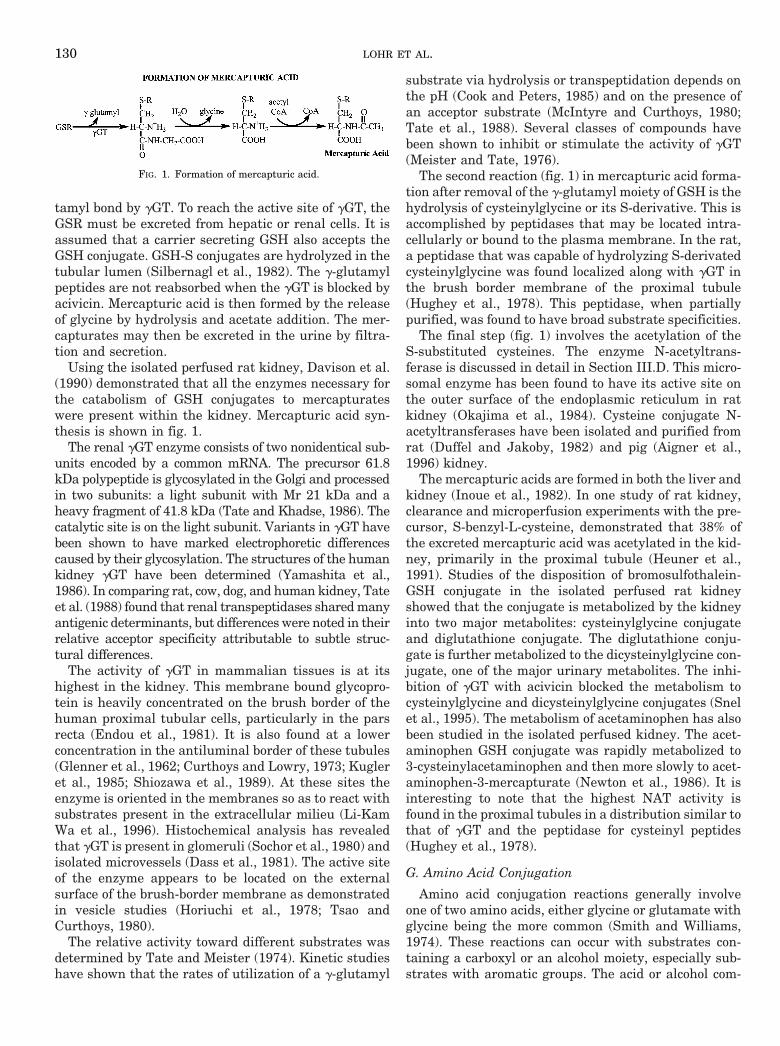
tamyl bond by gGT. To reach the active site of gGT, theGSR must be excreted from hepatic or renal cells. It isassumed that a carrier secreting GSH also accepts theGSH conjugate. GSH-S conjugates are hydrolyzed in thetubular lumen (Silbernagl et al., 1982). The g-glutamylpeptides are not reabsorbed when the gGT is blocked byacivicin. Mercapturic acid is then formed by the releaseof glycine by hydrolysis and acetate addition. The mer-capturates may then be excreted in the urine by filtra-tion and secretion.
Using the isolated perfused rat kidney, Davison et al.(1990) demonstrated that all the enzymes necessary forthe catabolism of GSH conjugates to mercapturateswere present within the kidney. Mercapturic acid syn-thesis is shown in fig. 1.
The renal gGT enzyme consists of two nonidentical sub-units encoded by a common mRNA. The precursor 61.8kDa polypeptide is glycosylated in the Golgi and processedin two subunits: a light subunit with Mr 21 kDa and aheavy fragment of 41.8 kDa (Tate and Khadse, 1986). Thecatalytic site is on the light subunit. Variants in gGT havebeen shown to have marked electrophoretic differencescaused by their glycosylation. The structures of the humankidney gGT have been determined (Yamashita et al.,1986). In comparing rat, cow, dog, and human kidney, Tateet al. (1988) found that renal transpeptidases shared manyantigenic determinants, but differences were noted in theirrelative acceptor specificity attributable to subtle struc-tural differences.
The activity of gGT in mammalian tissues is at itshighest in the kidney. This membrane bound glycopro-tein is heavily concentrated on the brush border of thehuman proximal tubular cells, particularly in the parsrecta (Endou et al., 1981). It is also found at a lowerconcentration in the antiluminal border of these tubules(Glenner et al., 1962; Curthoys and Lowry, 1973; Kugleret al., 1985; Shiozawa et al., 1989). At these sites theenzyme is oriented in the membranes so as to react withsubstrates present in the extracellular milieu (Li-KamWa et al., 1996). Histochemical analysis has revealedthat gGT is present in glomeruli (Sochor et al., 1980) andisolated microvessels (Dass et al., 1981). The active siteof the enzyme appears to be located on the externalsurface of the brush-border membrane as demonstratedin vesicle studies (Horiuchi et al., 1978; Tsao andCurthoys, 1980).
The relative activity toward different substrates wasdetermined by Tate and Meister (1974). Kinetic studieshave shown that the rates of utilization of a g-glutamyl
substrate via hydrolysis or transpeptidation depends onthe pH (Cook and Peters, 1985) and on the presence ofan acceptor substrate (McIntyre and Curthoys, 1980;Tate et al., 1988). Several classes of compounds havebeen shown to inhibit or stimulate the activity of gGT(Meister and Tate, 1976).
The second reaction (fig. 1) in mercapturic acid forma-tion after removal of the g-glutamyl moiety of GSH is thehydrolysis of cysteinylglycine or its S-derivative. This isaccomplished by peptidases that may be located intra-cellularly or bound to the plasma membrane. In the rat,a peptidase that was capable of hydrolyzing S-derivatedcysteinylglycine was found localized along with gGT inthe brush border membrane of the proximal tubule(Hughey et al., 1978). This peptidase, when partiallypurified, was found to have broad substrate specificities.
The final step (fig. 1) involves the acetylation of theS-substituted cysteines. The enzyme N-acetyltrans-ferase is discussed in detail in Section III.D. This micro-somal enzyme has been found to have its active site onthe outer surface of the endoplasmic reticulum in ratkidney (Okajima et al., 1984). Cysteine conjugate N-acetyltransferases have been isolated and purified fromrat (Duffel and Jakoby, 1982) and pig (Aigner et al.,1996) kidney.
The mercapturic acids are formed in both the liver andkidney (Inoue et al., 1982). In one study of rat kidney,clearance and microperfusion experiments with the pre-cursor, S-benzyl-L-cysteine, demonstrated that 38% ofthe excreted mercapturic acid was acetylated in the kid-ney, primarily in the proximal tubule (Heuner et al.,1991). Studies of the disposition of bromosulfothalein-GSH conjugate in the isolated perfused rat kidneyshowed that the conjugate is metabolized by the kidneyinto two major metabolites: cysteinylglycine conjugateand diglutathione conjugate. The diglutathione conju-gate is further metabolized to the dicysteinylglycine con-jugate, one of the major urinary metabolites. The inhi-bition of gGT with acivicin blocked the metabolism tocysteinylglycine and dicysteinylglycine conjugates (Snelet al., 1995). The metabolism of acetaminophen has alsobeen studied in the isolated perfused kidney. The acet-aminophen GSH conjugate was rapidly metabolized to3-cysteinylacetaminophen and then more slowly to acet-aminophen-3-mercapturate (Newton et al., 1986). It isinteresting to note that the highest NAT activity isfound in the proximal tubules in a distribution similar tothat of gGT and the peptidase for cysteinyl peptides(Hughey et al., 1978).
G. Amino Acid Conjugation
Amino acid conjugation reactions generally involveone of two amino acids, either glycine or glutamate withglycine being the more common (Smith and Williams,1974). These reactions can occur with substrates con-taining a carboxyl or an alcohol moiety, especially sub-strates with aromatic groups. The acid or alcohol com-
FIG. 1. Formation of mercapturic acid.
130 LOHR ET AL.
bines with an amino acid to form an amide bond. Thegeneral reaction is shown below:
R(CO)OH
1 NH2CH2(CO)OH → R(CO)NHCH2(CO)OH [11]
An example of amino acid conjugation is the formationof hippuric acid from benzoic acid and glycine. The re-sulting product (hippuric acid) is more polar in solutionand possesses different excretion rates than the originalsubstrate (benzoic acid). The enzymes required foramino acid conjugation are acyl-CoA synthetase andN-acyltransferase and they are present in liver and kid-ney, with some animals having higher activity in kidney(Krishna and Klotz, 1994).
Benzoic acid 1 CoA 1 ATPacyl CoA synthetaseOOOOOOOOO¡
Benzoyl CoA 1 AMP 1 Ppi [12]
Benzoyl CoA 1 glycineN-acyltransferaseOOOOOOOO¡
Hippuric acid 1 CoA [13]
In the kidney, these reactions take place in the mito-chondria of cortical cells. The first step is catalyzed bythe class of enzymes known as butyryl-CoA synthetases(EC 6.2.1.2, butyrate: CoA ligase (AMP-forming) (Aliand Qureshi, 1992). For benzoic acid conjugation, thebenzoyl CoA synthetase has been shown to have a lowerspecific activity than the enzyme for reaction (13). Thisstep may be inhibited by substances capable of formingCoA esters, such as valproic acid. The creation of thevalproate CoA ester competes with benzoic acid conju-gation for the available ATP, CoA, and enzyme (Gregus,1993a). The second reaction is catalyzed by a series ofenzymes known as glycine N-acyltransferases, whichare known to act on several endogenous and exogenoussubstances. There are two distinct types of this enzymecorresponding to two distinct classes of substrate. Thefirst of these is the arylacyl transferase (E.C.2.3.1.14),acting on arylacetic acids such as phenylacetylCoA, andthe second is arylalkyl transferase (E.C.2.3.2.13), whichacts on benzoic acid and its derivatives (Kelley and Ves-sey, 1993). There is evidence from studies in animalsthat these enzymes are more fully developed in neonatesin the liver than in the kidney; whether this is true forhumans is unknown (Ali and Qureshi, 1992).
Conjugation reactions can be limited by the amount ofglycine available in the body or by the amount of enzymeavailable to catalyze the reaction. Whereas levels ofglutamate do not seem to affect glutamate conjugationsin that all reactions show saturation above a concentra-tion of available substrate. Small organic acids such asbenzoic and salicylic will be glycinated by the kidneysinto hippuric and salicyluric acids respectively and thenexcreted. When this pathway works at maximal capac-
ity, the remainder of these drugs are glucuronidated(Vree et al., 1992).
1. Glycine conjugation. Glycine conjugation is themain metabolic pathway for salicylic acid. A small in-crease in the carbon side chain resulting in phenylaceticacid makes conjugation with glycine impossible and, inhumans, conjugation is carried out with glutamine (Wanand Riegleman, 1972). Wan et al. (1972) demonstratedthat the kidney contributed more to glycination than didthe liver. Poon and Pang (1995) compared enzymaticconstants for benzoic acid glycination in perfused ratkidney and liver and found the Vmax to be 195 versus 101nmol/min/g and the Km to be 5.3 and 12.0 mM respec-tively.
The role of glycine in establishing this maximal rate ofbenzoic acid conjugation can be established by measur-ing the increase in hippurate excretion after the admin-istration of glycine. Even at the maximum rate of hip-purate excretion, increasing the supply of glycine willresult in an increase in excretion of hippurate (Quick,1931). Gregus et al. (1993b) established that increases inthe elimination of benzoic acid after coadministration ofsodium benzoate and glycine depended upon the dose ofsodium benzoate. The supply of glycine sets a maximalconjugation rate that can be reached with an optimalconcentration of benzoate.
This is not the whole story. There are actually twofactors that limit the rates of hippuric acid synthesis,namely depletion of Coenzyme A and the availability ofglycine for conjugation. In the second reaction (13), gly-cine is consumed by conjugation, whereas CoA is regen-erated each time a molecule of hippuric acid is synthe-sized. Once glycine levels fall low enough to limit therate of reaction (13), CoA is trapped in the form ofbenzoyl CoA. This pool of activated intermediates effec-tively denies CoA to other metabolic processes; it alsohalts the production of more benzoyl CoA. In effect, thislimits the supply of benzoyl CoA for reaction (13), plac-ing a limit on the reaction rate (Gregus et al., 1992).
It was formerly believed that beyond the maximal rateof hippurate synthesis benzoic acid was shunted to an-other conjugation pathway with glucuronic acid (coveredin III. A. Glucuronidation). More accurate techniques forthe measurement of levels of excreted glucuronic acidconjugates have revealed that glucuronidation occurs ateven very low levels of benzoic acid. Glucuronic acidconjugation with benzoic acid increases proportionallywith benzoic acid concentration, showing no relationshipwith glycine availability or the resulting maximal rate ofhippuric acid formation. Glucuronidation of benzoic acidto form hippuric acid is not a reserve pathway for ben-zoic acid elimination, but rather a second pathway op-erating completely independently of glycine conjugation(Schachter and Mauro, 1958).
Glycine plays the same role in the metabolism of sal-icylic acid via conjugation to salicyluric acid. The rate ofsalicylic acid elimination is roughly 20 times slower than
RENAL DRUG METABOLISM 131

the rate for benzoic acid glycine conjugation. Liver me-tabolism is low, and practically all of the salicyluric acidis formed by conjugation in the kidney. Saturation ofsalicylic acid conjugation does not occur caused by ashortage of glycine, as occurs in benzoic acid formation,but rather caused by the saturation of the enzymesinvolved (Wan and Riegleman, 1972).
Metabolism of PABA follows two pathways, either byacetylation to p-acetamidobenzoic acid (Section III. D.)and by glycine conjugation to p-aminohippuric acid.These reactions are localized to the kidney and liver.The quantitatively more important reaction is the acet-ylation (Wan et al., 1972).
2. Glutamine conjugation. Glutamine conjugation re-actions are limited in the body to specific arylaceticacids. One such substance is phenylacetic acid, whichcan be converted in the kidney to indolacetylglutaminein various species of Old and New World monkeys and inhumans (Smith and Williams, 1974).
H. Cysteine Conjugate b-Lyase
Cysteine conjugates of aromatic drugs are metabo-lized principally to the corresponding mercapturic acid.However, cysteine conjugates may also undergo ab-elimination (equation 14 below) or a transaminationreaction catalyzed by the enzyme cysteine conjugateb-lyase (CCbL) (E.C. 4.4.1.13).
H2O 1 NH2CH(COOH)CH2SRCCbLOO¡ CH3(CO)COOH
1 :NH3 1 RSH [14]
This enzyme will also catalyze transamination reac-tions. For example S-(12-dichlorovinyl)-L-cysteine canbe transaminated to the 2-oxo-3-mercaptopropionateconjugate (Stevens et al., 1989). This enzyme also hasbeen the focus of much study because of the fact thatrather than resulting in detoxification, it may be respon-sible for the bioactivation of nephrotoxic cysteine andhomocysteine conjugates (Elfarra and Anders, 1984;Monks et al., 1990). Liver and kidney are the principalsites of activity. CCbL activity was localized to the cys-tolic and mitochondrial fraction of rat kidney cortex(Stevens, 1985) and subsequently purified from bothcystolic and mitochondrial sources (Stevens et al., 1988).
The cDNA for rat kidney CCbL has been sequencedand found to code for a protein of 48 kDa. The cDNA wastransfected into COS-1 tissue culture cells with a result-ant increase of 7- to 10-fold in cystolic b-lyase and glu-taminase K activity (Perry et al., 1993).
In human kidney, b-lyase activity was found in cyto-solic, mitochondrial, and microsomal fraction using S-(2-benzothiazolyl)-L-cysteine as the substrate, with thehighest concentration found in the cytosolic fraction(Lash et al., 1990). The b-lyase activity copurified withcytosolic glutamine transaminase K, with total Mr of 85kDa and with a 45 kDa subunit. The activity was inhib-
ited by aminooxyacetic acid, indicating that the enzymecontains pyridoxal phosphate. Human kidney CCbL wasrecently cloned and sequenced and found to have anoverall 82% similarity in amino acid sequence with thatof rat, with 90% similarity around the pyridoxal phos-phate binding site. Expression of the cDNA in COS-1cells produced a cystolic enzyme with CCbL and glu-tamine transaminase K activity (Perry et al., 1995).
Immunohistochemical studies showed that rat kidneyCCbL was mainly localized to the proximal tubule(Jones et al., 1988) with one study demonstrating anincreased concentration of enzyme in the pars recta (S3segment), which displays a greater sensitivity to damagefrom nephrotoxic cysteine conjugates (MacFarlane et al.,1989).
The substrates studied using renal CCbL have prin-cipally been certain L-cysteine conjugates of aromaticcompounds, such as S-(2-benzothiazolyl)L-cysteine, andS-(1, 2,dichlorovinyl) L-cysteine. N-acetyl-S-(12,3,44-pentachloro-13-butadienyl)-L-cysteine induced CCbLactivity in female rat kidney at low dosages, whereas athigh dosages it suppressed activity (McFarlane et al.,1993). Alpha-ketoacids stimulated rat renal CCbL activ-ity (Elfarra et al., 1987).
IV. Localization of Drug Metabolizing Enzymesin the Kidney
Those enzymes that have been localized by variousstudies using immunocytochemistry and enzyme activ-ity measurements reported in this review are summa-rized here and appear in table 5 and fig. 2. Table 5 showsthe distribution of enzymes in the kidney correspondingto fig. 2A. It appears that the cortical region is rich inmetabolizing enzymes. Only in a few instances were themedullary zones studied. As seen in B of fig. 2, theproximal tubule is active in metabolizing drugs, al-though enzymes were found in all segments of thenephron. Many of these enzymes were localized to eitherthe cytoplasmic or the microsomal fraction (C of fig. 2),whereas some appeared in both. These studies show thepredominant distribution of drug metabolizing enzymesby regional, tubular, and cellular sites but may not be
TABLE 5Distribution of drug metabolizing enzymes in kidney zones
Enzyme Cortex Medulla Outer Inner
N-acetyltransferase u u — —Alcohol dehydrogenase u u — —Aldose reductase — — u uAldehyde reductase u — — —Amino acid conjugation u — — —Cytochrome P450 ua — u —Carboxylesterase u — — —Cysteine conjugate b-lyase u — — —Glutathione S-transferase u u — —Monamine oxidase ua u — —NADPH-cytochrome c reductase ua — — uSteroid 21 hydroxylase u u — —Sulfotransferase u — u —
a Signifies greatest activity.
132 LOHR ET AL.

exclusive because all regions were not systematicallystudied.
V. Effects of Renal Metabolism
Although the kidney will generally metabolize endog-enous or exogenous chemicals to compounds with re-duced biological activity, there are several instances inwhich metabolism will produce a toxic intermediate thatmay result in mutagenesis or cell necrosis. This topichas recently been reviewed (Anders and Dekant, 1994;Dekant et al., 1994; Spahn-Langguth and Benet, 1992).
Instances of metabolism to active metabolites havebeen demonstrated and can lead to beneficial effects. Forexample, the angiotensin-converting enzyme inhibitor,enalapril, is metabolized to enalaprilat, an active andpolar dicarboxylic acid metabolite. The intrarenallyformed metabolite either re-enters the circulation orundergoes excretion into the lumen (deLannoy et al.,1990).
Investigations of renal metabolism of drugs have ledto the development of target organ-directed drug deliv-ery systems in which systemic side effects of drugs areavoided. For example, Elfarra and Hwang (1993) dem-onstrated that the high concentration of renal b-lyasefacilitated the conversion of S-(6-purinyl)-L-cysteine tothe antitumor and immunosuppressant drug 6MP bythe kidney. This permits the accumulation of muchhigher concentrations at its target site in the kidney andavoids systemic toxic effects. Another example is the useof the high concentration of g-glutamyl transpeptidasein the proximal tubule brush border to produce kidney
specific production of dopamine after the administrationof the precursor g-glutamyl dopa. The concentration ofdopamine in mouse kidney after injection of g-glutamyldopa was five times higher than that seen after anequivalent dosage of dopamine was given (Wilk et al.,1978).
Metabolism of drugs and endogenous compounds canlead to alterations in either excretion or reabsorptiondepending on the pathway followed and the activity ofthe enzyme. The metabolite will determine the directionof movement. Amino acid conjugation results in loss ofpharmacological activity and a compound more readilyexcreted into urine via tubular secretion mechanisms(Gregus, 1993a). Similarly, the formation of meperidineN-oxide contributed to a more rapid excretion of thatdrug by the isolated perfused kidney (Acara et al., 1981).If this route were not functioning as in renal failure,then other metabolites, such as normeperidine, mightaccumulate with attending effects. With the exception ofmethylation, most conjugative metabolisms lead to morepolar compounds that may be excreted from the bodyrapidly. The activity of the metabolizing enzymes alsoplays a role. Choline for example will be metabolized tobetaine, which is reabsorbed. However, when the cholineoxidase enzyme is saturated, choline itself moves in thedirection of excretion (Acara et al., 1979).
The most common ways in which the excretion of asubstance is increased are for the drugs to become ableto bind to the carriers for excretory transport and/or tobecome water soluble and filtered. Certain biotransfor-mations contribute to these changes (Ullrich et al.,
FIG. 2. Regional (A), tubular (B), and subcellular (C) localization of drug-metabolizing enzyme systems in the kidney.
RENAL DRUG METABOLISM 133

1990). An OH group inserted into a benzene ring rendersthe molecule acceptable to the r-aminohippuric acid(PAH) transporter, if an electron-attracting group(NO2
2, Cl2, Br2, HCO32) is already present in themolecule. Esterification of carboxylates decreases theiraffinity to the PAH transporter. On the other hand,amino acids and oligopeptides such as glutathione be-come substrates of the organic cation transport systemwhen they are esterified. Zwitterionic amino acids (ex-cept those that are too hydrophyllic) become pure cat-ions by esterification and pure anions by N-acetylation.However, N-acetylation of uncharged amino groups haslittle effect on affinity for the PAH transporter. Glycineconjugation increases the affinity of salicylate for thePAH transporter (Ullrich et al., 1987). The metabolismof an amine to its N-oxide leads to an increase in polarityand a drop in pKa. The semipolar N3O linkage impartsto N-oxides a salt-like character that makes themreadily soluble in water and only slightly soluble inorganic solvents. The excretion of trimethyl amines bythe renal tubule occurs predominantly as a result of theformation of an N-oxide by the renal tubular epithelialcells followed by movement into the tubule lumen.
Fig. 3 shows the possible pathways that may resultduring renal metabolism of a drug. Entry across eitherthe brush border (BBM) or the basolateral membrane(BLM) is accompanied by biotransformation of the drug(A 5 organic anion; C 5 organic cation) to a metabolite(B or D, respectively). The metabolite may then move inthe direction of reabsorption or secretion/excretion. Inthe upper section of the left panel, an organic anion suchas salicylic acid can exchange with a-ketoglutarateacross the BLM in a tertiary active transport step de-pendent on sodium a-ketoglutarate cotransport. Glyci-nation of salicylic acid to e.g., salicyluric acid (indicatedas B) provides a compound that more readily enters theurine. Other possibilities are that A remains unmetabo-
lized and is simply excreted as the administered drug; orB could be a different metabolite that is more likely to bereabsorbed. The lower left hand panel depicts reabsorp-tion of an organic anion that may occur by anion ex-change across the BBM as well as through cotransportwith sodium. For example, pyrazinoate is cotransportedinto the tubule cell with sodium and is returned to theblood as pyrazinoate. Biotransformation of A to B couldlead to a metabolite with a predominant pathway ofeither reabsorption or secretion. The right hand paneldepicts possible pathways for disposition of organic cat-ions. In the upper right of the panel an organic cationsuch as meperidine enters the renal tubule cell along itselectrochemical gradient. It may be biotransformed tomeperidine N-oxide (D), a more polar compound thatenters the tubule fluid. Unmetabolized meperidine mayexchange for a proton across the BBM or the metabolitemay follow a reabsorptive route such as would be thecase for the demethylated normeperidine. The lower halfof the right hand panel depicts an organic cation (C),such as isoproterenol that can be reabsorbed across theBBM in exchange for a proton. Upon entry into the cellcatechol-O-methyl transferase activity produces methy-lated isoproterenol that moves back into the blood. Someof the isoproterenol may itself cross the BLM and/or amore polar metabolite could enter the tubule fluid. Inthe isolated perfused rat kidney the fractional excretionof isoproterenol decreased as the amount of the 3-O-methyl metabolite increased (Szefler and Acara, 1979).
In summary, although the liver plays a dominant rolein drug metabolism, this review demonstrates that thekidney is metabolically active in the biotransformationof drugs. In some instances this role may, in fact, exceedthat of the liver. The metabolic pathways of the kidneygenerally lead to a product that is more readily excretedin the urine, but in certain instances the metabolite maybe reabsorbed and more active or toxic than the parentcompound.
The metabolic pathways of the kidney should be con-sidered in the administration of drugs, particularly topatients with renal impairment. Renal failure may af-fect the handling of drugs in several ways including drugdistribution, bioavailability, and excretion. From thisreview, it is clear that the effects of alterations in kidneyfunction on renal drug metabolism should also be takeninto account in the determination of optimum drug ther-apy. Additional studies of renal drug metabolism of spe-cific agents are needed to provide a better understand-ing of the role of the kidney in the disposition of drugs.
Acknowledgments. The authors wish to thank James Reuther andTeresa Schuster for assistance in the preparation of this manuscript.
REFERENCES
Abbott WA, Bridges RJ and Meister A (1984) Extracellular metabolism of glutathi-one accounts for its disappearance from the basolateral circulation of the kidney.J Biol Chem 259:15393–15400.
Acara M (1979) Effect of ethanol on the renal excretion and metabolism of choline inthe isolated perfused rat kidney. Drug Metab Dispos 7:113–117.
FIG. 3. Proposed interaction of transport and metabolism in the renalproximal tubule. Please see text for explanation of figure.
134 LOHR ET AL.

Acara M (1992) Renal pharmacology: Diuretics, in Textbook of Pharmacology (SmithC and Reynard A, eds) pp 554–588, W. B. Saunders, Philadelphia.
Acara M and Rennick B (1972) Renal tubular transport of choline: Modificationscaused by intrarenal metabolism. J Pharmacol Exp Ther 182:1–13.
Acara M and Rennick B (1973) Regulation of plasma choline by the renal tubule:Bidirectional transport of choline. Am J Physiol 225:1123–1128.
Acara M, Camiolo S and Rennick B (1977) Renal N-oxidation of trimethylamine inthe chicken during tubular excretion. Drug Metab Dispos 5:82–90.
Acara M, Gessner T, Greizerstein H and Trudnowski R (1981) Renal N-oxidation ofmeperidine by the perfused kidney of the rat. Drug Metab Dispos 9:75–79.
Acara M, Roch-Ramel F and Rennick B (1979) Bidirectional renal tubular transportof free choline: A micropuncture study. Am J Physiol 236:F112–F118.
Agarwal DP, Eckey R, Rudnay A, Volkens T and Goedde HW (1989) High Kmaldehyde dehydrogenase isozymes in human tissues: Constitutive and tumor-associated forms. Prog Clin Biol Res 290:119–131.
Ahmed M, Felsted RL and Bachur NR (1979) Comparison and characterization ofmammalian xenobiotic ketone reductases. J Pharmacol Exp Ther 209:12–19.
Aigner A, Jager M, Pasternak R, Weber P, Weinke D and Wolf S (1996) Purificationand characterization of cysteine-S-conjugate N-acetyltransferase from pig kidney.Biochem J 317:213–218.
Aitio A (1973) Induction of UDP glucuronyltransferase in the liver and extrahepaticorgans of the rat. Life Sci 13:1705–1713.
Aitio A (1974) UDP glucuronyltransferase activity in various rat tissues. J Biochem5:325–330.
Aitio A and Parkki MG (1978) Organ specific induction of drug metabolizing enzymes by2,3,7,8-tetrachlorodibenzo-p-dioxin in the rat. Toxicol Appl Pharmacol 44:107–114.
Aitio A, Vainio H and Hanninen O (1972) Enhancement of drug oxidation andconjugation by carcinogens in different rat tissues. FEBS Lett 24:237–240.
Aksoy S, Raftogianis R and Weinshilboum R (1996) Human histamine N-methyltransferase gene: Structural characterization and chromosomal location.Biochem Biophys Res Commun 219:548–554.
Aldridge WN (1953) Serum esterase I. Two types of esterases (A & B) hydrolyzingp-nitrophenyl acetate, proprionate and butyrate, and a method for their determi-nation. Biochem J 53:110–117.
Ali A and Kaur S (1983) Mammalian tissue acetylsalicyclic acid esterases: Identifi-cation, distribution, and discrimination from other esterases. J Pharmacol ExpTher 226:589–594.
Ali A and Qureshi IA (1992) Benzoyl-CoA ligase activity in the liver and kidneycortex of weanling guinea pigs treated with various inducers: Relationship withhippurate synthesis and carnitine levels. Dev Pharmacol Ther 18:55–64.
Ambruziak W and Pietruszko R (1993) Metabolic role of aldehyde dehydrogenase.Adv Exp Med Biol 328:5–15.
Anders MW (1980) Metabolism of drugs by the kidney. Kidney Int 18:636–647.Anders MW and Dekant W (1994) Aminocylases. Adv Pharmacol 27:431–444.Anders MW, Lash I, Dekant W, Elfarra AA and Dohn DR (1988) Biosynthesis and
biotransformation of glutathione S-conjugate to toxic metabolites. CRC Crit RevToxicol 18:311–341.
Anderson ME and Meister A (1980) Dynamic state of glutathione in blood plasma.J Biol Chem 255:9530–9533.
Armbrecht HJ, Birnbaum LS, Zenser TV, Mattamal MB and Davis BB (1979) Renalcytochrome P450’s - Electrophoretic and electron paramagnetic resonance studies.Arch Biochem Biophys 197:277–284.
Ashour MA, Moody MA, Moody DE and Hammock BD (1987) Apparent induction ofmicrosomal carboxylesterase activities in tissues of clofibrate-fed mice and rats.Toxicol Appl Pharmacol 89:361–369.
Axelrod J (1962) The enzymatic N-methylation of serotonin and other amines.J Pharmacol Exp Ther 138:28–33.
Bach AW, Lana ND, Johnson DL, Abell CW, Bembemek ME, Kwan SW, Seeburg PWand Shih C (1988) cDNA cloning of liver monoamine oxidase A and B: Molecular basisof differences in enzymatic properties. Proc Natl Acad Sci USA 85:4934–4938.
Baetge EE, Suh YH and Joh TH (1986) Complete nucleotide and deduced amino acidsequence of bovine phenylethanolamine N-methyl transferase: Partial amino acidhomology with rat tyrosine hydrolase. Proc Natl Acad Sci USA 83:5454–5458.
Baker J, Struempler A and Chaykin S (1963) A comparative study of trimethyl-N-oxide biosynthesis. Biochim Biophys Acta 71:58–63.
Barker EV, Hume R, Hallas A and Coughtrie MWH (1994) Dehydroepiandrosteronesulfotransferase in the developing human fetus: Quantitative biochemical andimmunological characterization of the hepatic, renal, and adrenal enzymes. En-docrinology 134:982–989.
Barth H, Naber K, Barthel KE, Niemyer I and Lorenz W (1975) Induction ofhistamine methyltransferase during experimental hydronephrosis. A mechanismof action for histamine homeostasis in rabbit kidney. Agents Actions 5:442–443.
Beckett GJ, Howie AF, Hume R, Matharoo B, Hiley C, Jones P and Strange RC(1990) Human glutathione S-transferases: Radioimmunoassay studies in the ex-pression of alpha-, mu-, pi- class isozymes in developing lung and kidney. BiochimBiophys Acta 1036:176–182.
Beebe L, Fornwald LW, Alworth WL, Dragnev KH and Lubet RA (1995) Effect ofdietary Aroclor 1254 exposure on lung and kidney cytochromes P450 in femalerats: Evidence for P4501A2 expression in kidney. Chem Biol Interact 97:215–227.
Beetham JK, Tian T and Hammock BD (1993) cDNA cloning and expression of asoluble epoxide hydrolase from human liver. Arch Biochem Biophys 305:197–201.
Bell DR, Bars RG and Elcombe CR (1992) Differential tissue-specific expression andinduction of cytochrome P4501VA1 and acyl-CoA oxidase. Eur J Biochem 206:979–986.
Bergmann F, Segal R and Rimon S (1957) A new type of esterase in hog kidneyextract. Biochem J 67:481–486.
Bessighir K and Roch-Ramel, F (1988) Renal excretion of drugs and other xenobiot-ics. Renal Physiol 10:221–241.
Bettuzi S, Marinellli M, Strocchi P, Davalli P, Cevolaani D and Corti A (1995)Different localization of spermidine/spermine N-aceetyltransferase and ornithinedecarboxylase transcripts in the rat kidney. FEBS Lett 377:321–324.
Bhat PV, Labrecque J, Boutin J-M, Lacroix A and Yoshida A (1995) Cloning of acDNA encoding rat aldehyde dehydrogenase with high activity for retinal oxida-tion. Gene 166:303–306.
Bhikharidis B, Mann LRB and McLeod WR (1975) Indoleamine N- methyltrans-ferase. J Neruochem 24:203–205.
Blagbrough IS, Bycroft BW, Evans DC, Green T and Shaw PN (1986) Cysteineconjugate substrates for rat renal b-lyase: An examination of the structure-activityrelationships. J Pharm Pharmacol 38:107P.
Blum M, Grant DM, McBride W, Heim M and Meyer UA (1990) Human arylamineN-acetyltransferase genes: Isolation, chromosomal localization, and functionalexpression. DNA Cell Biol 9:193–203.
Bock KW, Forster A, Schaidmeier H, Bruck M, Munel P, Schareck W, Fournel-GigleuxS and Burchell B (1993) Paracetamol glucuronidation by recombinant rat and humanphenol UDP-glucuronosyltransferases. Biochem Pharmacol 45:1809–1814.
Bock KW, Lipp HP and Bock-Hennig BS (1990) Induction of drug-metabolizingenzymes by xenobiotics. Xenobiotica 20:1101–1111.
Bocking A, Hansert J and Von Deimling O (1976) Esterase XXII. Cellular andsubcellular localization of the Es-9 esterase in mouse kidney. Histochemistry46:177–188.
Bogaards JJP, Van Ommen B and Van Bladeran PJ (1992) Purification and char-acterization of eight glutathione-S-transferase isoenzymes of hamster. Biochem J286:383–388.
Bohren KM, Bullock B, Wermuth B and Gablzy KH (1989) The aldo-keto reductasesuperfamily. J Biol Chem 264:9547–9551.
Bompart GJ, Proevot DS and Bascands JL (1990) Rapid automated analysis ofglutathione reductase, peroxidase, and S-transferase activity: Application to cis-platinum induced toxicity. Clin Biochem 23:501–504.
Booth J (1966) Acetyl transfer in arylamine metabolism. Biochem J 100:745–753.Bosron WF and Prairie RL (1972) Triphosphopyridine nucleotide-linked aldehyde
reductase. J Biol Chem 247:4480–4485.Bosron WF and Prairie RL (1973) Reduced triphosphopyridine-linked aldehyde
reductase II. Species and tissue distribution. Arch Biochem Biophys 154:166–172.Bowman RH (1978) Methodology for study of isolated perfused rat kidney in vitro, in
Methods in Pharmacology: Renal Pharmacology (Martinez-Maldonado M, ed) pp385–399, Plenum, NY.
Bowsher RR, Verburg KM and Henry DP (1983) Rat histamine N-methyltransferase.J Biol Chem 258:12215–12220.
Burchell B, Coughtrie M and Jansen P (1994) Function and regulation of UDP-glucuronyltransferase genes in health and disease. Hepatology 20:1622–1630.
Butera L, Feinfeld DA and Bhargava M (1990) Sex differences in the subunits ofglutathione-S-transferase from rat and human kidney. Enzyme 43:175–182.
Campbell DES (1960) Improved method for collecting and measuring ureteral urineflow in birds. Acta Pharmacol Toxicol 17:205–212.
Capdevila JH, Falck JR and Estabrook RW (1992) Cytochrome P450 and the ara-chidonate cascade. FASEB J 6:731–736.
Cappiello M, Franchi M, Giuliani L and Pacifici GM (1989) Distribution of 2-naph-thol sulphotransferase and its endogenous substrate adenosine 39-phosphate 59-phosphosulfate in human tissues. Eur J Clin Pharmacol 37:317–320.
Cappiello M, Giuliani L and Pacifici GM (1991) Distribution of UDP-glucuronosyl-transferase and its endogenous substrate uridine 59-diphosphoglucuronic acid inhuman tissues. Eur J Clin Pharmacol 41:345–350.
Cassidy MK and Houston JB (1980) In vivo assessment of extrahepatic conjugativemetabolism in first pass effects using the model compound phenol. J PharmPharmacol 32:57–58.
Chowdhury JR, Novikoff PM, Chowdhury NR and Novikoff AB (1985) Distribution ofUDP glucuronosyltransferase in rat tissue. Proc Natl Acad Sci 82:2990–2994.
Cojocel C, Maita K, Pasino DA, Kuo C and Hook JE (1983) Metabolic heterogeneityof the proximal and distal kidney tubules. Life Sci 33:855–861.
Cook ND and Peters TJ (1985) The effect of pH on the transpeptidation and hydro-lytic reactions of rat kidney g-glutamyltransferase. Biochim Biophys Acta 832:142–147.
Corcoran GB and Salazar DE (1988) Obesity as a risk factor in drug-induced organinjury. Increased renal cytochrome P-450 in the obese overfed rat. Drug MetabDispos 16:898–900.
Coughtrie MWH, Burchell B and Bend JR (1987) Purification and properties of ratkidney UDP glucuronosyltransferase. Biochem Pharmacol 3:245–251.
Cummings SW and Prough RA. (1983) Metabolic formation of toxic metabolites, inGlutathione Conjugation: Mechanisms and Biological Significance (Caldwell J andJakoby WB, eds) pp 415–446, Academic Press, London.
Curthoys NP and Lowry OH (1973) The distribution of glutaminase isoenzyme in thevarious structures of the nephron in normal, acidotic and alkalotic rat kidney.J Biol Chem 248:162–168.
Darmenton P, Cronenberger L and Pacheco H (1976) Purification et proprietes de lacatechol-O-methyltransferase du rein de rat. Biochimie 58:1031–1045.
Dass PD, Misra RP and Welbourne TC (1981) Presence of g-glutamyltranspeptidasein the renal microvascular compartment. Can J Biochem 59:383–386.
Davies SJ, D’Sousa R, Philips H, Mattey D, Hiley C, Hayes JD, Aber GM and StrangeR (1993) Localization of alpha, mu, and pi, class glutathione S-transferases inkidney: Comparison with CuZn superoxide dismutase. Biochim Biophys Acta1157:204–208.
Davison KL, Bakke JE and Larsen GL (1990) Metabolism of the glutathione conju-gate of propachlor by in situ perfused kidneys and livers of rats. Xenobiotica20:375–383.
Dawson JR, Norbeck K and Moldeus P (1983) The effectiveness of different sulfateprecursors in supporting extrahepatic sulfate conjugation. Biochem Pharmacol32:1789–1791.
Dees H, Masters BS, Mullereberhard U and Johnson EF (1982) Effect of 2,3,7,8-tetrachlorodibenzo-p-dioxin and phenobarbital on the occurence and distributionof four cytochrome P-450 isozymes in rabbit kidney, lung and liver. Cancer Res42:1423–1432.
Deininger M, Szumlanski CL, Otterness DM, Van Loon J, Ferber W and Wein-
RENAL DRUG METABOLISM 135

shilboum RM (1994) Purine substrates for human thiopurine methyl transferase.Biochem Pharmacol 48:2135–2138.
Deitrich RA (1966) Tissue and subcellular distribution of mammalian aldehydeoxidizing capacity. Biochem Pharmacol 15:1911–1922.
Dekant WL, Vamkakas S and Anders MW (1994) Formation and fate of nephrotoxicand cytotoxic glutathione-S-conjugates: Cysteine conjugate beta-lyase pathway.Adv Pharmacol 27:115–162.
de Lannoy IA, Hirayama H and Pang KS (1990) A physiological model for renal drugmetabolism: Enalapril esterolysis to elalaprilat in the isolated perfused rat kidney.J Pharmacokinet Biopharm 18:561–587.
DeLooze SM, Ronai. A and von Deimling OH (1985) Biochemistry and genetics ofesterase-20 (ES-20), a second trimeric carboxylesterase of the house mouse (Musmusculus). I. Purification and characterization of ES-20 from male kidney. Bio-chem Genet 23:741–757.
Del Villar E, Gaule C and Vega P (1995) Kidney drug metabolizing activities instreptozotocin diabetic rats. Gen Pharmacol 26:137–141.
Denny RM, Fritz RR, Patel NT, Widen SG and Abell CW (1983) Use of a monoclonalantibody for comparative studies of monoamine oxidase B in mitochondrial ex-tracts of human brain and peripheral tissues. Mol Pharmacol 24:60–68.
Derbel M, Igarashi T and Satoh T (1993) Differential induction of glutathione-Stransferase subunits by phenobarbital 3-methylcholanthrene and ethoxyquin inrat liver and kidney. Biochim Biophys Acta 1158:175–180.
Diamond GL and Quebbemann AJ (1981) In vivo quantification of renal sulfate andglucuronide conjugation in the chicken. Drug Metab Dispos 9:402–409.
Diamond GL, Anders MW, Tremaine LM and Quebbemann AJ (1982) Salicylateenhanced renal glucuronide conjugation and tubular excretory transfer of phenols.Drug Metab Dispos 10:573–578.
DiIlio C, Aceto A, Bucciarelli T, Angelucci S, Felaco M, Gulli A, Zezza A, Tenaglia Rand Federici G (1991) Glutathione transferase isoenzymes in normal and neoplas-tic human kidney tissue. Carcinogenesis 12:1471–1475.
Ding X, Koop DR, Crump BL and Coon MJ (1986) Immunochemical identification ofcytochrome P-450 isozymes 3a (P-450alc) in rabbit nasal and kidney microsomesand evidence for differential induction by alcohol. Mol Pharmacol 30:370–378.
Dipple KM and Crabb DW (1994) The mitochondrial aldehyde dehydrogenase generesides in an HTF island but is expressed in a tissue specific manner. BiochemBiophys Res Commun 193:420–427.
Dolphin C, Shephard EA, Povey S, Palmer CMA, Ziegler DM, Ayesh R, Smith RL andPhillips IR (1991) Cloning, primary sequence, and chromosomal mapping of ahuman flavin-containing monooxygenase (FMO1). J Biol Chem 266:12379–12385.
Duffel MW and Jakoby WB (1982) Cysteine-S-conjugate N-acetyltransferase fromrat kidney microsomes. Mol Pharmacol 21:444–448.
Ecobichon DJ (1972) Relative amounts of hepatic and renal carboxylesterases inmammalian species. Res Comm Chem Pathol Pharmacol 3:629–635.
Ecobichon DJ and Kalow W (1964) Properties and classification of the solubleesterases of human kidney. Can J Biochem 42:277–286.
Elbers R, Kampffmeyer HG and Rabes H (1980) Effects and metabolic pathway of4-dimethylaminophenol during kidney perfusion. Xenobiotica 10:621–632.
Elfarra AA and Anders MW (1984) Renal processing of glutathione conjugates.Biochem Pharmacol 33:3729–3732.
Elfarra AA and Hwang IY (1993) Targeting of 6-mercaptopurine to the kidneys:Metabolism and kidney-selectivity of S-(6-purinyl)-L-cysteine analogs in rats.Drug Metab Dispos 21:841–845.
Elfarra AA, Lash LH and Anders MW (1987) Alpha-keto acids stimulate rat renalcysteine conjugate B-lyase activity and potentiate the cytotoxicity of S-(1,2-Dichlorovinyl)-L-cysteine. Mol Pharmacol 31:208–212.
Ellin A, Jakobsson V, Schenkman JB and Orrhenius S (1972) Cytochrome P450K ofrat kidney cortex microsomes: Its involvement in fatty acid v- and (v-1)-hydroxylation. Arch Biochem Biophys 150:64–71.
Eloranta TO (1977) Tissue distribution of S-adenosylmethionine, and S-adenosylhomocysteine in the rat. Effect of age, sex, and methionine administrationon the metabolism of S-adenosylmethionine, S-adenosylhomocysteine and poly-amines. Biochem J 166:521–529.
Emslie KR, Smail MC, Calder IC, Hart SJ and Tange JD (1981) Paracetamol and theisolated perfused kidney: Metabolism and functional effects. Xenobiotica 11:43–50.
Endou H (1983a) Cytochrome P-450 monooxygenase system in the rabbit kidney: Itsintranephron localization and its induction. Jpn J Pharmacol 33:423–433.
Endou H (1983b) Distribution and some characteristics of cytochrome P-450 in thekidney. J Toxicol Sci 8:165–176.
Endou H, Shimada H, Koseki Y, Yokokura Y and Sakai Y (1981) Distribution andpossible functions of U-glutamyl transpeptidase in the kidney. Jpn J Nephrol23:981–988.
Faed EM (1984) Properties of acyl glucuronides: Implications for studies of thepharmacokinetics and metabolism of acidic drugs. Drug Metab Rev 15:1213–1249.
Feldman JM and Rocke JM (1978) Effects of aging on monoamine oxidase activity ofmouse and rabbit tissue. Exp Aging Res 4:97–107.
Felder MR, Watson G, Huff MO and Ceci J (1988) Mechanism of induction of mousekidney alcohol dehydrogenase by androgen. J Biol Chem 263:14531–14537.
Felsted RL and Bachur N (1980) Mammalian carbonylreductase. Drug Metab Rev11:1–60.
Fernandes MH and Soares-da-silva P (1990) Role of monamine oxidase A and B inthe deamination of newly formed dopamine in the rat kidney. J Neural Transm32:155–159.
Fernandes MH and Soares-da-silva P (1992) Type A and B monoamine oxidaseactivities in the human and rat kidney. Acta Physiol Scand 195:363–367.
Flynn TG (1982) Aldehyde reductases: Monomeric NADPH dependent oxidoreduc-tases with multifunctional potential. Biochem Pharmacol 31:2705–2712.
Flynn TG (1986) Aldose and aldehyde reductase in animal tissues. Metabolism35(suppl 1):105–108.
Forster RP (1948) Use of thin kidney slices and isolated renal tubules for direct studyof cellular transport kinetics. Science 108:65–68.
Forte-McRobbie CM and Pietrusko R (1986) Purification and characterization of
human liver “high Km” aldehyde dehydrogenase and its identification as glutamicT-semialdehyde dehydrogenase. J Biol Chem 261:2154–2163.
Foster JR, Elcombe CR, Boolis AR, Davies DS, Sesardie D, McQuade J, Robson RT,Hayward H and Loch EA (1986) Immunocytochemical localization of cytochromeP-450 in hepatic and extrahepatic tissues of the rat with a monoclonal antibodyagainst cytochrome P-450c. Biochem Pharmacol 35:4543–4554.
Foulkes EC (1996) Slices and sacs: Limitations on metabolic and functional studiesin kidney cortex and intestine. Proc Soc Exp Biol Med 211:155–162.
Fry JR, Wiebkin P, Kao J, Jones CA, Gwynn J and Bridges JW (1978) A comparisonof drug-metabolizing capability in isolated viable rat hepatocytes and renal tubulefragments. Xenobiotica 8:113–120.
Fujita T and Suzuoki Z (1973) Enzymatic studies on the metabolism of the tetrahy-drofurfuryl mercaptan moiety of thiamine tetrahydrofuryl disulfide. I. MicrosomalS-transmethylase. J Biochem 74:717–722.
Funae Y, Seo R and Imaoha S (1985) Multiple forms of cytochrome P-450 frommicrosomes of rat liver, kidney and lung resolved by high performance liquidchromatography. Biochem Int 11:523–531.
Gargalidis-Moudanos C, Remaury A, Pizzinet N and Parini A (1997) Predominantexpression of monoamine oxidase B isoform in rabbit renal proximal tubule:Regulation by I2 imidazoline ligands in intact cells. Mol Pharmacol 51:637–643.
Girard B, Otterness DM, Wood TC, Herchel R, Wieben ED and Weinshilboum RM(1994) Human histamine N-methyltransferase pharmacogenetics: Cloning andexpression of kidney cDNA. Mol Pharmacol 45:461–468.
Glenner GC, Folk JE and McMillan PJ (1962) Histochemical demonstration ofg-glutamyl transpeptidase-like activity. J Cytochem 19:481–489.
Goeppinger A, Riebschlager M, Ronai A and Von Diemling O (1978) Purification andcharacterization of esterase-9A of mouse kidney. Biochim Biophys Acta 525:74–86.
Goldbarg JA, Friedman OM, Pineda EP, Smith EE, Chatterji R, Stein EJ andRutenberg AM (1960) The colorimetric determination of U-glutamyl transpepti-dase with a synthetic substrate. Arch Biochem Biophys 91:61–70.
Goldstein PJ, Weinshilboum RM, Dunnett JH and Creveling CR (1980) Developmen-tal patterns of catechol-O-methyltransferase in genetically different rat strains:Enzymatic and immunochemical studies. J Neurochem 34:153–1162.
Gonzalez FJ (1989) The molecular biology of cytochrome P450’s. Pharmacol Rev40:243–275.
Grant DM, Blum M, Beer M and Meyer UA (1991) Monomorphic and polymorphichuman arylamine N-acetyltransferases: A comparison of liver isozymes and ex-pressed products of two cloned genes. Mol Pharmacol 39:184–191.
Gray PA, Park GR, Cockshott ID, Douglas EJ, Shuker B and Simmons PJ (1992)Propofol metabolism in man during the anhepatic and reperfusion phases of livertransplantation. Xenobiotica 22:105–114.
Green T and Odum J (1985) Structure/activity studies of the nephrotoxic andmutagenic action of cysteine conjugates of chloro- and fluoroalkenes. Chem BiolInteract 54:15–31.
Gregus Z, Fekete F, Varga F and Klaassen CD (1992) Availability of glycine andcoenzyme A limits glycine conjugation in vivo. Drug Metab Dispos 20:234–240.
Gregus Z, Fekete F, Varga F and Klaassen CD (1993a) Effect of valproic acid onglycine conjugation of benzoic acid. J Pharmacol Exp Ther 267:1068–1075.
Gregus Z, Fekete F, Varga F and Klaassen CD (1993b) Dependence of glycineconjugation on availability of glycine cleavage system. Xenobiotica 23:141–153.
Grimsby J, Chen K, Wang L, Lan NC and Shih JC (1991) Human monoamine oxidaseA and B genes exhibit identical exon-intron organization. Proc Natl Acad Sci88:3637–3641.
Grossman EB and Hebert SC (1989) Renal inner medullary choline dehydrogenaseactivity: Characterization and modulation. Am J Physiol 256:F107–F112.
Guengerich FP (1993) Cytochrome P450 enzymes. Am Sci 81:440–447.Guenther TM (1990) Epoxide hydrolases, in Conjugative reactions in drug metabo-
lism (Mulder E, ed) pp 365–404, Taylor and Francis, London.Gupta S, Medh RD, Leal T and Awasthi YC (1990) Selective expression of the three
classes of glutathione S-transferase isozymes in mouse tissue. Toxicol Appl Phar-macol 104:533–542.
Guthenberg C, Jenssen H, Nystrom L, Osterlund E, Tahir M and Mannervik B(1985) Isoenzymes of glutathione transferase in rat kidney cytosol. Biochem J230:609–615.
Haenick D, Boehme DH and Voel WH (1981) Monoamine oxidase in four rat andhuman tissues. Biochem Med 26:451–454.
Hahn R, Wendel A and Flohe L (1978) The fate of extracellular glutathione in the rat.Biochim Biophys Acta 539:324–337.
Handler JS and Burg MB (1992) Application of tissue culture techniques to the studyof renal tubular epithelia, in Renal Physiology, Section 8, Handbook of Physiology(Windhager E, ed) pp 385–415, Oxford University Press, New York.
Hanninen O and Aitio A (1968) Enhanced glucuronide formation in different tissuesfollowing drug administration. Biochem Pharmacol 17:2307–2311.
Hara A, Harada T, Nakagawa M, Matsuura K, Nakayama T and Sawaada H (1989)Isolation from pig lens of two proteins with dihydrodiol dehydrogenase and alde-hyde reductase activities. Biochem J 264:403–407.
Hardwick JP, Song BJ, Huberman E and Gonzalez FJ (1987) Isolation, complemen-tary DNA sequence and regulation of rat hepatic luric acid v-hydroxylase (Cyto-chrome P-450 LA omega): Identification of a new cytochrome P450 gene family.J Biol Chem 262:801–810.
Hart SGE, Cartun RW, Wyand S, Khairallah EA and Cohen SD (1995) Immunohisto-chemical localization of acetaminophen in target tissues of the CD-1 mouse: Corre-spondence of covalent binding with toxicity. Fundam Appl Toxicol 24:260–274.
Harvima RJ, Kajander EO, Harvima IT and Fraki JE (1985) Purification and partialcharacterization of rat kidney histamine N-methyltransferase. Biochim BiophysActa 841:42–49.
Hassett C, Turnblem SM, DeAngeles A and Omiecinski CJ (1989) Rabbit microsomalpoxide hydrolase: Isolation and characterization of the xenobiotic metabolizingenzyme cDNA. Arch Biochem Biophys 271, 380–389.
Hasumura S, Endou H, Kakuno K, Hojo K and Sakai F (1983) Effects of starvation
136 LOHR ET AL.

of microsomal cytochome P-450 and laurate-v-hyroxylation of rat kidney and liver.Jpn J Pharmacol 33:999–1006.
Hawke RL and Welch RM (1985) Major differences in the specificity and regulationof mouse renal cytochrome P-450-dependent monooxygenases. Mol Pharmacol27:283–289.
Hazelton GA, Hjelle JJ, Dills RL and Klaasen CD (1985) A radiometric method forthe measurement of adenosine 39-phosphate 59-phosphosulfate in rat and mouseliver. Drug Metab Dispos 13:30–34.
Hein DW, Flamming TJ, Kirlin WG, Trinidad A and Ogolla F (1987a) Acetylatorgenotype-dependent metabolic activation of carcinogenic N-hydroxyarylamine byS-acetyl coenzyme A-dependent enzymes of inbred hamster tissue cytosols: Rela-tionship to arylamine N-acetyltransferase. Carcinogenesis 8:1767–1774.
Hein DW, Kirlin WG, Ogolla F and Trinidad A (1987b) Extrahepatic expression ofN-acetylator genotype in the inbred hamster. Drug Metab Dispos 15:68–73.
Heinemann FS and Ozols J (1984) The covalent structure of hepatic microsomal epoxidehydrolase: II. The complete amino acid sequence. J Biol Chem 259:797–804.
Henderson CJ and Wolf CR (1991) Evidence that the androgen receptor mediatessexual differentiation of mouse renal cytochrome P-450 expression. Biochem J278:499–503.
Her C, Kaur GP, Athwal RS and Weinshilboum RM (1997) Human sulfotransferaseSULT 1C1: cDNA cloning, tissue-specific expression, and chromosomal location.Genomics 41:467–470.
Heuner A, DeKant W, Schwegler JS and Silbernagl S (1991) Localization andcapacity of the last step of mercapturic acid biosynthesis and the reabsorption andacetylation of cysteine S-conjugates in the rat kidney. Pfluegers Arch 417:523–527.
Heymann E (1980) Carboxylesterases and amidases, in Enzymatic Basis of Detoxi-fication (Jakoby WB, ed) pp 291–323, Academic Press, New York.
Heymann E and Iglesia J (1974) Investigation of the micro-heterogeneity and thesubunit structure of carboxylesterases by quantitative determination of the ter-minal amino acids. Hoppe-Seyler’s Z Physiol Chem 355:1112–1120.
Higuchi T, Imamura Y and Otagiri M (1993) Kinetic studies on the reduction ofacetohexamide catalyzed by carbonyl reductase from rabbit kidney. Biochim Bio-phys Acta 1158:23–28.
Higuchi T, Imamura Y and Otagiri M (1994) Chemical modification of arginine andlysine residues in coenzyme binding domain of carbonyl reductase from rabbitkidney: Indomethacin affords a significant protection against inactivation of theenzyme by phenylglyoxal. Biochim Biophys Acta 1199:81–86.
Hinchman CA and Ballatori N (1990) Glutathione-degrading capacities of liver andkidney in different species. Biochem Pharmacol 40:1131–1135.
Hjelle JT, Hazelton GA, Klaassen CD and Hjelle JJ (1986) Glucuronidation andsulfation in rabbit kidney. J Pharmacol Exp Ther 236:150–156.
Hjelle JT, Peterson DR and Hjelle JJ (1983) Drug metabolism in isolated proximaltubule cells. J Pharmacol Exp Ther 224:699–705.
Hoivik DJ, Manautou JE, Tveit A, Emeigh-Hart SG, Khairallah EA and Cohen SD(1995) Gender-related differences in susceptibility to acetaminophen-induced pro-tein arylation and nephrotoxicity in the CD-1 mouse. Toxicol Appl Pharmacol130:257–271.
Holcenberg JS and Rosen SW (1965) Enzymatic sulfation of steroids by bovinetissues. Arch Biochem Biophys 110:551–557.
Holmes RS, Courtney YR and Van de Berg JL (1986) Alcohol dehydrogenaseisozymes in baboons: Tissue distribution, catalytic properties and variant pheno-types in liver, kidney, stomach, testis. Alcohol Clin Exp Res 10:623–630.
Holmes RS, Van Oorschot RAH and Van de Berg JL (1991) Aldehyde dehydrogenase(ALDH) isozymes in the gray short-tailed opossum (monodelphis domestica): Tis-sue and subcellular distribution and biochemical genetics of ALDH3. BiochemGenet 29:163–175.
Honchel R, Aksoy I, Szumlanski C, Wood TC, Otterness DM, Wieben ED andWeinshilboum RM (1993) Human thiopurine methyltransferase: Molecular clon-ing and expression of T84 colon carcinoma cell cDNA. Mol Pharmacol 43:878–887.
Honscha W, Oesch F and Friedberg T (1991) Tissue specific expression and differ-ential inducibility of several microsomal epoxide hydrolase mRNA’s which areformed by alternative splicing. Arch Biochem Biophys 287:380–386.
Hook GER, Haseman JK and Lucier JW (1975) Induction and suppression of hepaticand extrahepatic microsomal foreign-compound-metabolizing enzyme systems by2,3,7,8-tetrochlorodibeno-p-dioxin. Chem Biol Interact 10:199–214.
Horiuchi S, Inoue M and Morino Y (1978) U-glutamyl transpeptidase: Sidedness ofits active site on renal brush border membrane. Eur J Biochem 87:425–437.
Howe JL, Back DJ and Colbert J (1992) Extrahepatic metabolism of zidovudine. Br JPharmacol 33:190–192.
Hsu LC, Bendel RE and Yoshida A (1988) Genomic structure of the human mito-chondrial aldehyde dehydrogenase gene. Genomics 2:57–65.
Hsu LC, Chang WC and Yoshida A (1989) Genomic structure of the human cytosolicaldehyde dehydrogenase gene. Genomics 5:857–865.
Hsu LC, Chang WC and Yoshida A (1994) Cloning and characterization of a cDNAencoding human ALDH7, a new member of the aldehyde dehydrogenase family.Gene 151:285–289.
Hu JJ, Lee MJ, Vapiwala M, Reuhl K, Thomas PE and Yang CS (1993) Sex-relateddifferences in mouse renal metabolism and toxicity of acetaminophen. Toxicol ApplPharmacol 122:16–26.
Hughey RP, Rankin BB, Elce JS and Curthoys NP (1978) Specificity of a particulaterat renal peptidase and its localization along with other enzymes of mercapturicacid synthesis. Arch Biochem Biophys 186:211–217.
Imamura Y, Higuchi T, Nazaki Y, Sugino E, Hibino S and Otagiri M (1993) Purifi-cation and properties of carbonyl reductase from rabbit kidney. Arch BiochemBiophys 300:570–576.
Imaoka S, Nagashima K and Funae Y (1990a) Characterization of three cytochromeP450’s purified from renal microsomes of untreated male rats and comparison withhuman renal cytochrome P450. Arch Biochem Biophys 276:473–480.
Imaoka S, Ogawa H, Kimura S and Gonzalez FJ (1993) Complete cDNA sequenceand cDNA-directed expression of CYP4A11, a fatty acid v-hydroxylase expressedin human kidney. DNA and Cell Biol 12:893–899.
Imaoka S, Yamaguchi Y and Funae Y (1990b) Induction and regulation of cyto-chrome P450 K-5 (lauric acid hydroxylase) in rat renal microsomes by starvation.Biochim Biophys Acta 1036:18–23.
Imaoka S, Yamazoe Y, Kato R and Funae Y (1992) Hormonal regulation of rat renalcytochrome P450s by androgen and the pituitary. Arch Biochem Biophys 299:179–184.
Inoue M, Okajima K and Morino Y (1982) Metabolic coordination of liver and kidneyin mercapturic acid biosynthesis in vivo. Hepatology 2:311–316.
Ioannides C and Parke DV (1990) The cytochrome P450 I gene family of microsomalhemoproteins and their role in the metabolic activation of chemicals. Drug MetabRev 22:1–85.
Izumi T, Shimuzu T, Seyama Y, Ohishi N and Takaku F (1986) Tissue distributionof leukotriene A4 hydrolase activity in guinea pig. Biochem Biophys Res Commun135:139–145.
Jakobsson SV, Okita RT, Prough RA, Mock NI, Buja LM, Graham JW, Petty CA andMasters BS (1978) Studies on the cytochrome P-450 dependent monooxygenasesystems of human liver, lung and kidney microsomes, in Industrial and Environ-mental Xenobiotics (Fouts JR and Gut I, eds) pp 71, Exerpta Medica, Amsterdam.
Johnson EF, Walker D, Griffin KJ, Clark JE, Okita RT, Muerhoff AS and Masters BS(1990) Cloning and expression of 3 rabbit cDNAs encoding lauric acid v-hydroxy-lases. Biochemistry 29:873–879.
Johnson JA, Finn KA and Siegel FL (1992) Tissue distribution of enzymic methyl-ation of glutathione S-transferase and its effects on catalytic activity. Biochem J282:279–289.
Johnston JP (1968) Some observations upon a new inhibitor of monoamine oxidasein brain tissues. Biochem Pharmacol 17:1285–1297.
Jollie DR and Maines MD (1985) Effect of cis-platinum on kidney cytochrome P-450and heme metabolism: Evidence for the regulatory role of the pituitary hormones.Arch Biochem Biophys 240:51–59.
Jones DP, Sundby GB, Ormstad K and Orrhenius S (1979) Use of isolated kidneycells for study of drug metabolism. Biochem Pharmacol 28:929–935.
Jones TW, Qin C, Schaeffer VH and Stevens JL (1988) Immunohistochemical local-ization of glutamine transaminase K, a rat kidney cysteine conjugate b-lyase andthe relationship to segment specificity of cysteine conjugate nephrotoxicity. MolPharmacol 34:621–627.
Kaneda N, Ichinose H, Kobayashi K, Oka K, Kishi F, Nakazawa Y, Fujita K andNagatsu T (1988) Molecular cloning of cDNA and chromosomal assignment of thegene for human phenylethanolamine N-methyltransferase, the enzyme for epi-nephrine biosynthesis. J Biol Chem 263:7672–7677.
Karara A, Makita K, Jacobson HR, Falck JR, Guengerich FP, DuBois RN andCapdevila JH (1993) Molecular cloning, expression and enzymatic characteriza-tion of the rat kidney-cytochrome P-450 arachidonic acid epoxygenase. J BiolChem 268:13565–13570.
Karkunen T, Tilgmann C, Ulmanen I, Julkunen I and Panula P (1994) Distributionof catechol-O-methyl transferase activity in rat tissues. J Histochem Cytochem42:1079–1090.
Kaur S and Gill SS (1985) Age related changes in activities of epoxide hydrolases indifferent tissues of mice. Drug Metab Dispos 13:711–715.
Kelley M and Vessey DA (1993) Isolation and characterization of mitochondrialacyl-CoA: Glycine N-acetyltransferases from kidney. J Biochem Toxicol 8:63–69.
Kennedy B, Bigby T and Ziegler M (1995) Nonadrenal epinephrine-forming enzymesin humans: Characteristics, distribution, regulation, and relationship to epineph-rine levels. J Clin Invest 95:2896–2902.
Ketterer B and Mulder GJ (1990) Glutathione conjugation, in Conjugative Reactionsin Drug Metabolism (Mulder GJ, ed) pp 307–364, Taylor and Francis, London.
Khani SC, Porter TD, Fujita VS and Coon MJ (1988) Organization and differentialexpression of two highly similar genes in the rabbit alcohol-inducible cytochromeP-450 subfamily. J Biol Chem 263:7170–7175.
Kim H, Kim SG, Lee MY and Novak RF (1992) Evidence for elevation of cytochromeP-4502E1 (alcohol-inducible form) mRNA levels in rat kidney following pyridineadministration. Biochem Biophys Res Commun 186:846–853.
Kim SG, Jung KH, Yang WK and Kim ND (1994) Differential expression of micro-somal epoxide hydrolase gene by azole hererocycles in rats. Biochem Pharmacol48:111–120.
Kim SG and Kim Y (1992) Gender-related expression of rat microsomal epoxidehydrolase during maturation: Post-transcriptional regulation. Mol Pharmacol 42:75–81.
Kimura S, Hanioka N, Matsunaga E and Gonzalez FJ (1989a) The rat clofibrate-inducible CYP4A gene subfamily I. Complete intron and exon sequence of theCYP4A2 genes, unique exon organization, and identification of a conserved 19-bpupstream element. DNA 8:503–516.
Kimura S, Hardwick JP, Kozak CA and Gonzalez FJ (1989b) The rat clofibrate-inducible CYP4A Subfamily II. cDNA sequence of IVA3, mapping of the Cyp4alocus to mouse chromosome 4, and coordinate and tissue-specific regulation of theCYP4A genes. DNA 8:517–525.
Knehr M, Thomas H, Arand M, Gebel T, Zeller H and Oesch F (1993) Isolation andcharacterization of a cDNA encoding rat liver cytosolic epoxide hydrolase and itsfunctional expression in Escherichia coli. J Biol Chem 268:17623–17627.
Knoll J and Magyar K (1972) Some puzzling pharmacological effects of monoamineoxidase inhibitors, in Advances in Biochemical Psychopharmaceuticals (Costa Eand Sandler M, eds) pp 393–408, Raven Press, New York.
Koster AS, Schirmer G and Bock KW (1986) Immunochemical and functional char-acterization of UDP-glucuronosyltransferases from rat liver, intestine and kidney.Biochem Pharmacol 35:3971–3975.
Krishna DR and Klotz R (1994) Extrahepatic metabolism of drugs in humans. ClinPharmacokinet 26:144–160.
Kubic VL and Anders MW (1975) Metabolism of dihalomethanes to carbon monoxide:II. In vitro studies. Drug Metab Dispos 3:104–112.
Kugler P, Wolf G and Scherberich J (1985) Histochemical demonstration of pepti-dases in the human kidney. Histochemistry 83:337–341.
Kuo C-H, Rush GF and Hook JB (1982) Renal cortical accumulation of phenobarbital
RENAL DRUG METABOLISM 137

in rats and rabbits: Lack of correlation with induction of renal microsomal mo-nooxygenases. J Pharmacol Exp Ther 220:547–551.
Kusunose E, Sawamura A, Kawashima H, Kubota I and Kusunose M (1989) Isolationof a new form of cytochrome P-450 with prostaglandin A and fatty acid v-hydrox-ylase activities from rabbit kidney cortex microsomes. J Biochem 106:194–196.
Labrecque J, Dumas F, Lacroix A and Bhat PV (1995) A novel isoenzyme of aldehydedehydrogenase specifically involved in the biosynthesis of 9-cis and all-transretinoic acid. Biochem J 305:681–684.
Lai JCK, Leung TKC and Lim L (1982) Monoamine oxidase activities in liver, heart,spleen and kidney of the rat. Organ specific changes in aging and after chronicmanganese chloride administration. Exp Gerontol 17:219–225.
Laniado-Schwartzman M and Abraham NG (1992) The renal cytochrome P-450arachidonic acid system. Pediatr Nephrol 6:490–498.
Laniado-Schwartzman M, da Silva J-L, Lin F, Nishimura M and Abraham NG (1996)Cytochrome P4504A expression and arachidonic acid omega-hydroxylation in thekidney of the spontaneously hypertensive rat. Nephron 73:652–663.
Lash LH, Nelson RM, Van Dyke RA and Anders MW (1990) Purification andcharacterization of human kidney cystolic cysteine conjugate b-lyase activity.Drug Metab Dispos 18:50–54.
LeGuellec C, LaCarelle B, Villard P-H, Point H, Catalin J and Durand A (1995)Glucuronidation of propofol in microsomal fractions from various tissues andspecies including humans: Effect of different drugs. Anesth Analg 81:855–861.
Lemoine A, Johann M and Cresteil T (1990) Evidence for the presence of distinctflavin-containing monooxygenases in human tissues. Arch Biochem Biophys 276:336–342.
Liehr JG, Hall ER, Avitts TA, Randerath E and Randerath K (1987) Localization ofestrogen-induced DNA adducts and cytochrome P-450 activity at the site of renalcarcinogenesis in the hamster kidney. Cancer Res 47:2156–2159.
Liem HH, Muller-Eberhard U and Johnson EF (1980) Differential induction by2,3,7,8-tetrachlorodibenzo-p-dioxin of multiple forms of rabbit microsomal cyto-chrome P-450: Evidence for tissue specificity. Mol Pharmacol 18:565–570.
Li-Kam Wa TC, Freestone S, Samson RR, Johnston NR and Lee MR (1996) Renalmetabolism and effects of the glutamyl derivatives of L-dopa and 5-hydroxytryto-phan in man. Clin Sci 91:177–185.
Lindell S-E and Schayer RW (1958) The renal removal of injected 14C-histaminefrom the blood in dogs. Br J Pharmacol 13:44–51.
Litterst CL, Mimnaugh EG, Reagan RL and Gram TE (1975) Comparison of in vitrodrug metabolism by lung, liver and kidney of several common laboratory species.Drug Metab Dispos 3:259–265.
Liu PT, Ioannides C, Shavila J, Symons AM and Parke DV (1993) Effects of etheranaesthesia and fasting on various cytochromes P-450 of rat liver and kidney.Biochem Pharmacol 45:871–877.
Lohr J and Acara M (1990) Effect of dimethylaminoethanol, an inhibitor of betaineproduction, on the disposition of choline in the rat kidney. J Pharmacol Exp Ther252:154–158.
Lyles GA and Shaffer C (1979) Substrate specificity and inhibitor sensitivity of mono-amine oxidase in rat kidney mitochondria. Biochem Pharmacol 28:1099–1106.
Maack T (1980) Physiologic evaluation of the isolated perfused rat kidney. Am JPhysiol 238:F71–78.
Maack T (1986) Renal clearance and isolated kidney perfusion techniques. KidneyInt 30:142–151.
MacFarlane M, Foster JR, Gibson GG, King LJ and Lock EA (1989) Cysteineconjugate b-lyase of rat kidney cytosol: Characterization, immunocytochemicallocalization and correlation with hexachlorobutadiene nephrotoxicity. Toxicol ApplPharmacol 98:185–197.
MacFarlane M, Schofield M, Parker N, Oelandt L, David M, Lock EA, King LJ,Goldfarb PS and Gibson GG (1993) Dose-dependent induction or depression ofcysteine conjugate b-lyase in rat kidney by N-acetyl-S-(1,2,3,4,-pentachloro-1,3,butadienyl)-L-cysteine. Toxicology 77:133–144.
Makita K, Falck JR and Capdevila JH (1996) Cytochrome P450, the arachidonic acidcascade and hypertension: New vistas for an old enyzme system. FASEB J 10:1456–1463.
Mannervik B, Awasthi YC, Board PG, Hayes JD, DiIlio C, Ketterer B, Listowsky I,Morganstern R, Muramatsu M and Pearson WR (1992) Nomenclature for humanglutathione transferase. Biochem J 282:305–308.
Maser E, Friebertshauser J and Mangoura SA (1994) Ontogenic pattern of carbonylreductase activity of 11b-hydroxysteroid dehydrogenase in mouse liver and kid-ney. Xenobiotica 24:109–117.
Mayer RD, Berman S, Cockett AT and Maines MD (1989) Differential effects ofcyclosporin on hepatic and renal heme, cytochrome P-450 and drug metabolism.Biochem Pharmacol 38:1001–1007.
Mazoit JC, Sandouk P, Scherrmann JM and Rocke A (1990) Extrahepatic metabo-lism of morphine occurs in humans. Clin Pharmacol & Ther 48:613–618.
McIntyre TM and Curthoys NP (1980) The inter-organ metabolism of glutathione.Int J Biochem 12:545–551.
McKay JA, Weaver RJ, Murray GI, Ewen SW, Melvin WT and Burke MD (1995)Localization of microsomal epoxide hydrolase in normal and neoplastic humankidney. J Histochem Cytochem 43:615–620.
McMartin DN, O’Connor JA and Kaminsky LS (1981) Effects of differential changesin rat hepatic and renal cytochrome P-450 concentrations on hepatotoxicity andnephrotoxicity of chloroform. Res Commun Chem Pathol Pharmacol 31:99–109.
Meister A and Tate SS (1976) Glutathione and related g-glutamyl compounds:Biosynthesis and utilization. Annu Rev Biochem 45:559–604.
Meister B, Bean AJ and Aperia A (1993) Catechol-O-methyl transferase mRNA inthe kidney and its appearance during ontogeny. Kidney Int 44:726–733.
Mellor JD and Hobkirk R (1975) In vitro synthesis of estrogen glucuronides andsulfates by human renal tissue. Can J Biochem 53:779–783.
Mohandas J, Duggin GG, Horvath JS and Tiller DJ (1981a) Metabolic oxidation ofacetaminophen (Paracetamol) mediated by cytochrome P450 mixed-function oxi-dase and prostaglandin endoperoxide synthetase in rabbit kidney. Toxicol ApplPharmacol 61:252–259.
Mohandas J, Duggin GG, Horvath JS and Tiller DJ (1981b) Regional differences inperoxidatic activation of paracetamol (acetaminophen) mediated by cytochromeP450 and prostaglandin endoperoxide synthetase in rabbit kidney. Res CommunChem Path Pharmacol 34:69–80.
Mohandas J, Marshall JJ, Duggin GG, Horvath JS and Tiller DJ (1984) Differentialdistribution of glutathione related enzymes in rabbit kidney. Possible implicationsin analgesic nephropathy. Biochem Pharmacol 33:1801–1807.
Moller E, McIntosh JF and Van Slyke DD (1928) Studies of urea excretion. II.Relationship between urine volume and the rate of urea excretion by normaladults. J Clin Invest 6:427–465.
Monks TJ, Anders MW, DeKant W, Stevens JL, Lau SS and Van Bladeren PJ (1990)Glutathione conjugate mediated toxicities. Toxicol Appl Pharmacol 106:1–19.
Moody DE, Lowry DN and Hammock BD (1987) Immunochemical determination ofmicrosomal epoxide hydrolase (preneoplastic antigen) in extrahepatic tissue. Bio-chem Pharmacol 36:570–572.
Moreland T and Hewick CS (1975) Studies on a ketone reductase in human and ratliver and kidney using warfarin as a substrate. Biochem Pharmacol 24:1953–1957.
Morgan EW, Yan B, Greenway D, Peterson DR and Parkinson A (1994) Purificationand characterization of two rat liver microsomal carboxylesterases (hydrolase Aand B). Arch Biochem Biophys 315:495–512.
Moser K, Papenberg J and Von Wartburg JP (1968) Heterogenitat und organvertei-lung der alkoholdehydrogenase bei verschiedenen spezies. Enzymol Biol Clin9:447–458.
Mottino AD, Guibert EE and Rodriguez EA (1991) Effect of spironolactone andphenobarbital administration on bilirubin glucuronidation in hepatic and extra-hepatic rat microsomes. Biochem Pharmacol 41:1075–1077.
Mouelhi ME, Beck S and Bock KW (1993) Stereoselective glucuronidation of (R)- and(S)-naproxen by recombinant rat phenol UDP-glucuronosyltransferase (UGT1A1)and its human ortholog. Biochem Pharmacol 46:1298–1300.
Mozier NM and Hoffman JL (1990) Biosynthesis and urinary excretion of methylsulfonium derivatives of the sulfur mustard analog, 2-chloroethyl ethyl sulfide,and other thioethers. FASEB J 4:3329–3333.
Mulder GJ (1992) Glucuronidation and its role in regulation of biological activity ofdrugs. Annu Rev Pharmacol Toxicol 32:25–49.
Mulder GJ, Brouwer S, Weitering JG, Scholtens E and Pang KS (1985) Glucuronida-tion and sulfation in the rat in vivo: The role of the liver and the intestine in thein vivo clearance of 4-methylumbelliferone. Biochem Pharmacol 34:1325–1329.
Mulder GJ and Jokoby WB (1990) Sulfation, in Conjugation Reactions in DrugMetabolism (Mulder GJ, ed) pp 107–162, Taylor and Francis, London.
Mulder GJ, Weitering JG, Scholtens E, Dawson JR and Pang SK (1984) Extrahepaticsulfation and glucuronidation in the rat in vivo. Biochem Pharmacol 34:3081–3087.
Muse KE, Oberly TD, Sempf JM and Oberly LW (1994) Immunolocalization ofantioxidant enzymes in adult hamster kidney. Histochem J 26:734–753.
Nakagawa M, Matsura M, Hara A, Sawada H, Bunai Y and Ohya I (1989) Dimericdihydrodiol dehydrogenase in monkey kidney: Substrate specificity, stereospeci-ficity of hydrogen transfer and distribution. J Biochem 106:1104–1109.
Nakamura M, Imaoka S, Miura K, Tanaka E, Misawa S and Funae Y (1994)Induction of cytochrome P-450 isozymes in rat renal microsomes by cyclosporin A.Biochem Pharmacol 48:1743–1746.
Nelson DR, Kamataki T, Waxman DJ, Guengerich FP, Estabrook RW, Feyereisen R,Gonzalez FJ, Coon MJ, Gunsalus IC, Gotoh O, Okuda K and Nebert DW (1993)The P450 superfamily: Update on new sequences, gene mapping, accession num-bers, early trivial names of enzymes, and nomenclature. DNA Cell Biol 12:1–51.
Nelson DR, Koymans L, Kamataki T, Stegeman JJ, Feyereisen R, Waxman DJ,Waterman MR, Gotoh O, Coon MJ, Estabrook RW, Gunsalus IC and Nebert DW(1996) P450 superfamily: Update on new sequences, gene mapping, accessionnumbers and nomenclature. Pharmacogenetics 6:1–42.
Newton JF, Hoefle F, Gemborys MW, Mudge GH and Hook JB (1986) Metabolismand excretion of a glutathione conjugate of acetaminophen in the isolated perfusedkidney. J Pharmacol Exp Ther 237:519–524.
Nicoll-Griffith DA, Gupta N, Twa SP, Williams H, Trimble CA and Yergey JA (1995)Verkulast (MK-0679) conjugation with glutathione by rat liver and kidney cytosolsand excretion in the bile. Drug Metab Dispos 23:1085–1093.
Nishiitsutsuji-Uwo JM, Ross BD and Krebs HA (1967) Metabolic activities of theisolated perfused rat kidney. Biochem J 103:852–862.
Nissinen E (1984) The site of O-methylation by membrane-bound catechol-O-methyltransferase. Biochem Pharmacol 33:3105–3108.
Nizet A (1978) The isolated perfused kidney: Possibilities, limitations and results.Kidney Int 7:1–11.
Nousaianen V, Torronen R and Hanninen O (1984) Differential induction of variouscarboxylesterases by certain polycyclic aromatic hydrocarbons in the rat. Toxicol-ogy 32:243–251.
Nouws JFM, Mevius D, Vree TB, Baakman M and Degen M (1988) Pharmacokinet-ics, metabolism and renal clearance of sulfadiazine, sulfamerazine, and sulfameth-azine and of their N4-acetyl and hydroxy metabolites in calves and cows. Am J VetRes 49:1059–1065.
Oesch F, Glatt H and Schmassman H (1977) The apparent ubiquity of epoxidehydralase in rat organs. Biochem Pharmacol 26:603–706.
Oesch F and Schmassman NH (1978) Species and organ specificity of the trans-stilbene oxide induced effects on epoxide hydratase and benzo(a)pyrene monoox-ygenase activity in rodents. Biochem Pharmacol 28:171–176.
Ohishi N, Imaoka S and Funae Y (1994) Changes in content of P-450 isozymes inhepatic and renal microsomes of the male rat treated with cis-diamminedichloro-platinum. Xenobiotica 24:873–880.
Okajima K, Inoue M, Morino Y and Itoh K (1984) Topological aspects of microsomalN-acetyltransferase, an enzyme responsible for the acetylation of cysteine-S-conjugates of xenobiotics. Eur J Biochem 142:281–286.
Okita JR, Casstle PJ and Okita RT (1993) Characterization of cytochromes P-450 inliver and kidney of rats treated with Di-2-ethylhexylphthalate. J Biochem Toxicol8:135–144.
O’Leary KA, McQuiddy P and Kasper CB (1996) Transcriptional regulation of the
138 LOHR ET AL.

TATA-less NADPH cytochrome P450 oxidoreductase gene. Arch Biochem Biophys330:271–280.
Oliw EH (1994) Oxygenation of polyunsaturated fatty acids by cytochrome P-450monooxygenases. Prog Lipid Res 33:329–354.
Opperman V, Maser E, Mangoura S and Netter K (1991) Heterogeneity of carbonylreduction in subcellular fractions and different organs in rodents. Biochem Phar-macol 42:5189–5195.
Orrhenius S, Ellin A, Jakobsson SV, Thor H, Cinti D, Schenkman J and EstabrookRW (1973) The cytochrome P-450 containing monooxygenase system of rat kidneycortex microsomes. Drug Metab Dispos 1:350–357.
Pacifici GM, Bencini C and Rane A (1986) Acetyltransferase in humans: Develop-ment and tissue distribution. Pharmacology 32:283–291.
Pacifici GM, Franchi M, Bencini C, Repetti F, DiLascio N and Muraro GB (1988a) Tissuedistribution of drug-metabolizing enzymes in humans. Xenobiotica 18:849–856.
Pacifici GM, Franchi M, Colizzi C, Giuliani L and Rane A (1988b) Sulfotransferasein humans: Development and tissue distribution. Pharmacology 36:411–419.
Pacifici GM, Kubrich M, Giuliani L, DeVries M and Rane A (1993) Sulphation andglucuronidation of ritodrine in fetal and adult tissues. Eur J Clin Pharmacol44:259–264.
Pacifici GM and Rane A (1982) Distribution of UDP-glucuronyl transferase in dif-ferent human fetal tissues. Br J Clin Pharmacol 13:732–735.
Pacifici GM, Santerini S and Giuliani L (1991b) Methylation of captopril in humanliver, kidney and intestine. Xenobiotica 21:1107–1112.
Pacifici GM, Vannuci L, Bencini C, Tusini G and Mosca F (1991a) Sulfation ofhydroxybiphenyls in human tissues. Xenobiotica 21:1113–1118.
Palmer CN, Richardson TH, Griffin J, Hsu MH, Muerhoff AS, Clark E and JohnsonEF (1993) Characterization of a cDNA encoding a human kidney, cytochrome P4504A fatty acid omega-hydroxylase and the cognate enzyme expressed in Escherichiacoli. Biochim Biophys Acta 1172:161–166.
Paolini M, Meserca R, Pozzetti L, Sapone A and Cantelli-Forte G (1996) Molecularnon-genetic biomarkers related to fenarimol cocarcinogenesis: Organ-and sex-specific CYP induction in rat. Cancer Lett 101:171–178.
Pares X, Cederlund E, Moreno A, Saubi N, Hoog J-O and Jorvall H (1992) Class IValcohol dehydrogenase (the gastric enzyme). FEBS Lett 303:69–72.
Parkinson A (1996) Biotransformation of xenobiotics, in Casarett and Doull’s Toxi-cology: The Basic Science of Poisons (Klaasen CD, ed) pp 113–186, McGraw-Hill,New York.
Paul D, Standifer KM, Inturrisi CE and Pasternak GW (1989) Pharmacologicalcharacterization of morphine 6b-glucuronide: A very potent morphine metabolite.J Pharmacol Exp Ther 251:477–483.
Pelkonen P, Lang M and Pasanen M (1994) Tissue and sex-dependent differences inCYP2A activities in hamsters. Arch Toxicol 68:416–422.
Perry S, Harries H, Scholfield C, Lock T, King L, Gibson G and Goldfarb P (1995)Molecular cloning and expression of a cDNA for human kidney cysteine conjugateb-lyase. FEBS Lett 360:277–280.
Perry SJ, Schofield MA, MacFarlane M, Lock EA, King LJ, Gibson GG and GoldfarbPS (1993) Isolation and expression of a cDNA coding for rat kidney cytosoliccysteine conjugate b-lyase. Mol Pharmacol 43, 660–665.
Phillips IR, Dolphin CT, Clair P, Hadley MR, Hutt AJ, McCombie RR, Smith RL andShephard EA (1995) The molecular biology of the flavin-containing monooxygen-ases of man. Chem Biol Interact 96:17–32.
Piatkowski TS, Day WW and Weiner M (1993) Increased renal drug metabolism intreadmill-exercised Fischer-344 male rats. Drug Metab Dispos 21:474–479.
Pickett CB and Lu AYH (1989) Glutathione-S-transferases: Gene structure, regula-tion and biologic function. Annu Rev Biochem 58:501–508.
Pinot F, Grant DF, Spearlow JL, Parker AG and Hammock BD (1995) Differentialregulation of soluble epoxide hydrolase by clofibrate and sexual hormones in theliver and kidneys of mice. Biochem Pharmacol 50:501–508.
Pond AL, Chambers HW and Chambers JE (1987) Organophosphate detoxificationpotential of various rat tissues via A-esterase and aliesterase activities. ToxicolLett 78:361–369.
Poon K and Pang S (1995) Benzoic acid glycine conjugation in the isolated perfusedrat kidney. Drug Metab Dispos 23:255–260.
Porter TD, Beck TW and Kasper CB (1986) Complementary DNA and amino acidsequence of rat liver microsomal, xenobiotic epoxide hydrolase. Arch BiochemBiophys 248:121–129.
Powis G, Moore DJ, Wilke TJ and Santone KS (1987) A high-performance liquidchromatography assay for measuring integrated biphenyl metabolism by intact cells:Its use with rat liver and human liver and kidney. Anal Biochem 167:191–198.
Pritchard JB and Miller DS (1993) Mechanisms mediating renal secretion of organicanions and cations. Physiol Rev 73:765–796.
Quebbemann AJ and Anders MW (1973) Renal tubular conjugation and excretion ofphenol and p-nitrophenol in the chicken: Differing mechanisms of renal transfer.J Pharmacol Exp Ther 181:696–708.
Quebbemann AJ and Rennick BR (1969) Effects of structural modifications of cat-echolamines on renal tubular transport in the chicken. J Pharmacol Exp Ther166:52–62.
Quick AJ (1931) The conjugation of benzoic acid in man. J Biol Chem 92:65–85.Qulali M, Dipple KM and Crabb DW (1993) Modulation of hepatic and renal alcohol
dehydrogenase activity and mRNA by steroid hormones in vivo. Adv Exp Med Biol328:581–589.
Qulali M, Ross RA and Crabb DN (1991) Estradiol induces class I alcohol dehydro-genase activity and mRNA in kidney of female rats. Arch Biochem Biophys 288:406–413.
Redegeld FA, Hofman GA and Noordhoek J (1988) Conjugative clearance of 1-naph-thol and disposition of its glucuronide and sulfate conjugates in the isolatedperfused rat kidney. J Pharmacol Exp Ther 244:263–267.
Redegeld FAM and Noordhoek J (1986) Active tubular secretion of glucuronide in theisolated perfused rat kidney. Drug Metab Dispos 14:622–624.
Remy CN (1963) Metabolism of thiopyrimidines and thiopurines. J Biol Chem238:1078–1084.
Rennick BR (1981) Renal excretion of drugs: Tubular transport and metabolism.Annu Rev Pharmacol 12:141–156.
Rennick B and Quebbemann A (1970) Site of excretion of catechol and cat-echolamines: Renal metabolism of catechol. Am J Physiol 218:1307–1312.
Roberts BJ, Shoaf SE, Jeong K-S and Song BJ (1994) Induction of CYP2E1 in liver,kidney, brain and intestine during chronic ethanol administration and with-drawal: Evidence that CYP2E1 possesses a rapid phase half-life of 6 hours or less.Biochem Biophys Res Commun 205:1064–1071.
Roch-Ramel F, Besseghir K and Murer H (1992) Renal excretion and tubular trans-port of organic anions and cations, in Handbook of Physiology, Section 8: RenalPhysiology (Windhager E, ed) pp 2189–2262, Oxford University Press, New York.
Roerig S, Fujimoto JM, Wang RIH, Pollock SJ and Lange D (1976) Preliminarycharacterization of enzymes for reduction of naloxone and naltrexone in rabbit andchicken liver. Drug Metab Dispos 4:53–58.
Ront VK, Elkington JSH and Holmes RS (1987) Developmental changes in humantissues: Constitutive and tumor-associated forms. Prog Clin Biol Res 290:119–131.
Ross B, Tange J, Emslie K, Hart S, Smail M and Calder I (1980) Paracetamolmetabolism by the isolated perfused rat kidney. Kidney Int 18:562–570.
Rozell B, Hansson HA, Gutenberg C, Kalina-Tahir M and Mannervik B (1993)Gluttathione transferases of classes a, m and p show selective expression indifferent regions of the kidney. Xenobiotica 23:835–849.
Rush GF and Hook JB (1984) Characteristics of renal UDP-glucuronyltransferase.Life Sci 35:145–153.
Ryan R, Grimm S, Kedzie K, Halpert J and Philpot R (1993) Cloning, sequencing,and functional studies of phenobarbital-inducible forms of cytochrome P-450 2Band 4B expressed in rabbit kidney. Arch Biochem Biophys 304:454–463.
Sandberg M and Meijer J (1996) Structural characterization of the human solubleepoxide hydrolase gene (EPHX2). Biochem Biophys Res Commun 221:333–339.
Sasano H, White P, New M and Sasano N (1998) Immunohistochemistry of cyto-chrome P-450 11-hydroxylase: Microscopic examination of the enzyme in thebovine adrenal cortex and kidney. Endocrinology 122:291–295.
Satoh T and Hosokawa M (1995) Molecular aspects of carboxylesterase isoforms incomparison with other esterases. Toxicol Lett 82/83:439–445.
Schachter D and Mauro JG (1958) Salicylate and salicyluric conjugation; fluoromet-ric estimation, biosynthesis, and renal excretion in man. J Clin Invest 37:800–807.
Schladt L, Worner W, Setiabudi F and Oesch R (1986) Distribution and inducibilityof cytosolic epoxide hydrolase in male Sprague-Dawley rats. Biochem Pharmacol35:3309–3316.
Schlondorff D (1986) Isolation and use of specific nephron segments and their cellsin biochemical studies. Kidney Int 30:201–207.
Schuetz EG, Schuetz JD, McLean-Grogan W, Naray-Fejes-Toth G, Ravcy J, GuzeliaP, Gineola K and Watlington CO (1992) Expression of cytochrome P-450 3A inamphibian, rat, and human kidney. Arch Biochem Biophys 294:206–214.
Schwenk M and Locher M (1985) 1-naphthol conjugation in isolated cells from liver,jejunum, ileum, colon and kidney of the guinea pig. Biochem Pharmacol 34:697–701.
Scott MC, Guerciolini R, Szumlanski C and Weinshilboum RM (1991) Mouse kidneyhistamine methyltransferase: Assay conditions, biochemical properties and strainvariation. Agents Actions 32:194–209.
Sewer MB, Koop DR and Morgan ET (1997) Differential inductive and suppressiveeffects of endotoxin and particulate irritants on hepatic and renal cytochromeP-450 expression. J Pharmacol Exp Ther 280:1445–1454.
Shaw PN and Blagbrough IS (1989) Cysteine conjugate b-lyase, II: Isolation, prop-erties and structure-activity relationships, in Sulphur-Containing Drugs and Re-lated Organic Compounds 2: Analytical, Biochemical and Toxicological Aspects ofSulphur Xenobiochemistry (Damani LA, ed) pp 135–155, Ellis Horwood Limited,Chichester, UK.
Sheehan JE, Pitot HC and Kasper CB (1991) Transcription and localization of thetissue-specific induction of epoxide hydrolase by lead acetate in rat kidney. J BiolChem 266:5122–5127.
Shieber A, Frank R and Ghisla S (1992) Purification and properties of prostaglandin9-ketoreductase from pig and human kidney. Eur J Biochem 206:491–502.
Shimojo N, Ishizaki T, Imaoka S, Funae Y, Fujii S and Okuda K (1993) Changes inamounts of cytochrome P450 isozymes and levels of catalytic activities in hepaticand renal microsomes of rats with streptozocin-induced diabetes. Biochem Phar-macol 46:621–627.
Shiozawa M, Yamashita S, Aiso S and Yasuda K (1989) A monoclonal antibodyagainst human kidney gamma-glutamyl transpeptidase preparation, immuno-chemical, and immunohistochemical characteristics. J Histochem Cytochem 37:1053–1061.
Siest G, Antoine B, Fournel S, Magdalou J and Thomassin J (1987) The glucurono-syltransferases: What progress can pharmacologists expect from molecular biologyand cellular enzymology? Biochem Pharmacol 36:983–989.
Silbernagl S (1992) Tubular transport of amino acids and small peptides, in Hand-book of Physiology: Renal Physiology (Windhager E, ed) pp 1937–1976, OxfordUniversity Press, New York.
Silbernagl S, Curthoys NP and Shapiro R (1982) The role of renal tubular enzymesin metabolism and absorption of glutathione and cysteinyl-glycine, in Biochemistryof Kidney Functions (Morel F, ed) pp 429–437, Elsevier, Amsterdam.
Simmons DL and Kasper CB (1989) Quantitation of mRNA’s specific for the mixedfunction oxidase system in rat liver and extrahepatic tissues during development.Arch Biochem Biophys 271:10–20.
Singh SV, Leal T, Ansari GAS and Awasthi YC (1987) Purification and character-ization of glutathione-S-transferase of human kidney. Biochem J 246:179–186.
Smith HW (1956) Principles of Renal Physiology, Oxford University Press, NewYork.
Smith J, Rush G and Hook J (1986) Induction of renal and hepatic mixed functionoxidases in the hamster and guinea pig. Toxicology 38:209–218.
Smith PF, Gandolfi AJ, Krumdieck CL, Putnam CW, Uoski CF, Davic WM andBrendel K (1985) Dynamic organ culture of precision liver slices for in vitrotoxicology. Life Sci 36:1367–1375.
RENAL DRUG METABOLISM 139
Smith RL and Williams RT (1974) Comparative metabolism of drugs in man andmonkeys. J Med Primatol 3:138–152.
Smolen TN, Brewer JA and Weber WW (1993) Testosterone modulation of N-acetylation in mouse kidney. J Pharmacol Exp Ther 264:854–858.
Snel CA, Moons MM, Russel FG and Mulder GJ (1995) Disposition of the bromosul-fophthalein-glutathione conjugate in the isolated perfused rat kidney. J Pharma-col Exp Ther 273:1300–1306.
Snyder SH and Axelrod J (1965) Sex difference and hormonal control of histaminemethyltransferase activity. Biochim Biophys Acta 111:416–421.
Sochor M, EL-Sheik OK and McLean P (1980) g-glutamyltranspeptidase in glomer-uli and tubules of rat kidney: Effect of experimental diabetes. Enzyme 25:205–208.
Spahn-Langguth H and Benet LZ (1992) Acyl glucuronides revisited: Is the glucu-ronidation process a toxification as well as a detoxification mechanism? DrugMetab Rev 24:5–48.
Sperber I (1946) A new method for the study of renal tubular secretion in birds.Nature 158:131.
Stevens JL (1985) Cysteine conjugate b-lyase activities in rat kidney cortex: Subcel-lular localization and relationship to the hepatic enzyme. Biochem Biophys ResCommun 129:499–504.
Stevens JL, Ayoubi N and Robbins JD (1988) The role of mitochondrial matrixenzymes in the metabolism and toxicity of cysteine conjugates. J Biol Chem263:3395–3401.
Stevens JL and Bakke JE (1990) S-methylation in conjugation reactions, in DrugMetabolism (Mulder GJ, ed) pp 251–272, Taylor and Francis, London.
Stevens JL, Hatzinger PB and Hayden PJ (1989) Quantitation of multiple pathwaysfor the metabolism of nephrotoxic cysteine conjugates using selective inhibitors ofL-alpha-hydroxy acid oxidase (L-amino acid oxidase) and cysteine conjugateb-lyase. Drug Metab Dispos 17:297–309.
Stevenson IH and Dutton GJ (1962) Glucuronide synthesis in kidney and gastroin-testinal tract. Biochem J 82:330–340.
Stromstedt M, Hayashi S-L, Zaphiropoulos PG and Gustafsson J-A (1990) Cloningand characterization of a novel member of the cytochrome P450 subfamily IVA inrat prostate. DNA Cell Biol 9:569–577.
Sullivan JP, McDonnell L, Hardiman OM, Farrell MA, Phillips JP and Tipton KF(1986) The oxidation of tryptamine by the two forms of monoamine oxidase inhuman tissues. Biochem Pharmacol 35:3255–3260.
Sundseth S and Waxman D (1992) Sex-dependent expression and clofibrate inducibilityof cytochrome P-450 4A fatty acid v-hydroxylases. J Biol Chem 267:3915–3921.
Sutherland L, Ebner T and Burchell B (1993) The expression of UDP-glucuronosyl-transferases of the UGT1 family in human liver and kidney and in response todrugs. Biochem Pharmacol 45:295–301.
Szefler SJ and Acara M (1979) Isoproterenol excretion and metabolism in theisolated perfused rat kidney. J Pharmacol Exp Ther 210:295–300.
Tachibana T, Taniguchi S, Imamura S, Fujiwara M and Hayashi H (1988) Effects ofdrugs on the activity of histamine-N-methyltransferase from guinea pig skin.Biochem Pharmacol 37:2872–2876.
Takemura M, Tanaka T, Taguchi Y, Imamura I, Mizuguchi H, Kuroda M, Fukui H,Yamatodani A and Wada H (1992) Histamine N-methyltransferase from rat kid-ney. J Biol Chem 267:15687–15691.
Tate SS and Khadse V (1986) Renal g-glutamyl transpeptidases influence of glyco-sylation on the electrophoretic behavior and molecular weights of their subunits.Biochem Biophys Res Commun 141:1189–1194.
Tate SS, Khadse V and Wellner D (1988) Renal gamma-glutamyl transpeptidases:Structural and immunological studies. Arch Biochem Biophys 262:397–408.
Tate SS and Meister A (1974) Interaction of gamma-glutamyl transpeptidase withamino acids, dipeptides, and derivatives and analogs of glutathione. J Biol Chem249:7593–7602.
Tateoka N, Tsuchide S, Soma Y and Sato K (1987) Purification and characterizationof glutathione S-transferase in human kidney. Clin Chim Acta 166:207–218.
Temellini A, Castiglione M, Guiliani L, Mussi A, Guilianotti P, Pietrabissa A,Angeletti C, Mosca F and Pacifici GM (1995) Glutathione conjugation with1-chloro-2,4-dinitrobenzene (CDNB): Interindividual variability in human liver,lung, kidney, and intestine. Int J Clin Pharmacol Ther Toxicol 33:493–503.
Tenhunen J and Ulmanen I (1993) Production of rat soluble and membrane-boundcatechol-O-methyltransferase forms from bifunctional mRNAs. Biochem J 296:595–600.
Terubayashi R, Sato S, Nishimura C, Kador PF and Kinoshita JH (1989) Localiza-tion of aldose and aldehyde reductase in the kidney. Kidney Int 36:843–851.
Thakker DR and Creveling CR (1990) O-methylation, in Conjugation Reactions inDrug Metabolism (Mulder GJ, ed) pp 193–232, Taylor and Francis, London.
Thomas PE, Bandiera S, Maines LS, Ryan DE and Levin W (1987) Regulation ofcytochrome P-450j, a high-affinity N-nitrosodimethylamine demethylase, in rathepatic microsomes. Biochemistry 26:2280–2289.
Toback FG, Havener LJ and Spargo BH (1977a) Stimulation of renal phospholipidformation during potassium depletion. Am J Physiol 233:E212–E218.
Toback FG, Havener LJ, Dodd RC and Spargo BH (1977b) Phospholipid metabolismduring renal regeneration after acute tubular necrosis. Am J Physiol 232:E216–E222.
Toback FG, Smith PD and Llowenstein LM (1974) Phospholipid metabolism in theinitiation of renal compensatory growth after acute reduction of renal mass. J ClinInvest 54:91–97.
Toftgard R and Nilsen O (1982) Effects of xylene and xylene isomers on cytochromeP-450 and in vitro enzymatic activities in rat liver, kidney and lung. Toxicology23:197–212.
Toretti J and Weiner IM (1976) The renal excretion of drugs, in Methods in Phar-macology (Martinez-Maldonade M ed) pp 357–379, Plenum, New York.
Tremaine LM, Diamond GL and Quebbemann AJ (1984) In vivo quantification ofrenal glucuronide and sulfate conjugation of 1-naphthol and p-nitrophenol. Bio-chem Pharmacol 33:419–427.
Tremaine LM, Diamond GL and Quebbemann AJ (1985) Quantitative determinationof organ contribution to excretory metabolism. J Pharmacol Methods 13:9–35.
Tsao B and Curthoys NP (1980) The absolute asymmetry of orientation of g-glu-tamyltranspeptidase and aminopeptidase on the external surface of the rat renalbrush border membrane. Biol Chem 255:7708–7711.
Tsuda H, Moore M, Asamoto M, Inooue T, Fukushima S, Ito N, Satoh K, Amelizad.A and Oesch F (1987) Immunohistochemically demonstrated altered expression ofcytochrome P-450 molecular forms and epoxide hydrolase in N-ethyl-N-hydroxyethylnitrosamine-induced rat kidney and liver lesions. Carcinogenesis8:711–717.
Tsujita T, Miyada T and Okuda H (1988) Purification of rat kidney carboxylesteraseand its comparison with other tissue esterases. J Biochem 103:327–331.
Tsujita T and Okuda H (1993) Palmitoyl-coenzyme A hydrolyzing activity in ratkidney and its relationship to carboxylesterases. J Lipid Res 34:1773–1781.
Tucker GT (1981) Measurement of the renal clearance of drugs. Br J Clin Pharmacol12:71–770.
Tussey L and Felder MR (1989) Tissue specific genetic variation in the level of mousealcohol dehydrogenase is controlled transcriptionally in kidney and post-transcriptionally in liver. Proc Natl Acad Sci USA 86:5903–5907.
Tuteja N, Gonzalez FJ and Nebert DW (1985) Developmental and tissue-specificdifferential regulation of the mouse dioxin-inducible P1–450 and P3–450 genes.Dev Biol 112:177–184.
Ueng T-H, Friedman FK, Miller H, Park SS, Gellboin HV and Alvares AP (1987)Studies on ethanol-inducible cytochrome P450 in rabbit liver, lungs, and kidney.Biochem Pharmacol 36:2689–2691.
Ullrich KJ, Rumrich G and Kloss S (1987) Contraluminal para-aminohippurate(PAH) transport in the proximal tubule of the rat kidney: III. Specificity: Mono-carbolylic acids. Pfluegers Arch 409:547–554.
Ullrich KJ, Rumrich G, Gemborys M and DeKant W (1990) Transformation andtransport: How does metabolic transformation change the affinity of substrates forthe renal contraluminal anion and cation transporters? Toxicol Lett 53:19–27.
Van Loon JA, Szumlanski CL and Weinshilboum RM (1992) Human kidney thiopu-rine methyltransferase. Photoaffinity labelling with S-adenosylmethionine. Bio-chem Pharmacol 44:775–785.
Van Loon JA and Weinshilboum RM (1990) Thiopurine methyltransferase isozymesin human renal tissue. Drug Metab Dispos 18:632–638.
Vasilou V and Marselos M (1989) Tissue distribution of inducible aldehyde dehydro-genase activity in the rat after treatment with phenobarbital or methylcholan-threne. Pharmacol & Toxicol 64:39–42.
Vatsis KP and Weber WW (1994) Human N-acetyltransferases, in Conjugation-deconjugation reactions in Drug Metabolism and Toxicity (Kauffman FC, ed) pp109–130, Springer-Verlag, Berlin.
Vickers AEM, Fischer V, Connors MS, Biggi WA, Heitz F, Baldeck J-P and BrendelK (1996) Biotransformation of the antiemetic 5-HT3 antagonist tropisetron in liverand kidney slices of human, rat and dog with a comparison to in vivo. Eur J DrugMetab Pharmacokinet 21:43–50.
Vieira-Coehlo MA and Soares-da-Silva P (1996) Ontogenic aspects of liver andkidney catechol-O-methyl transferase sensitivity to tolcapone. Br J Pharmacol117:516–520.
Vogel WH, Gentile NT, Menduke H and Boehme DH (1983) Inter and intraindividualdifferences in monoamine oxidase A and B activities in human central and periph-eral tissues. Biochem Med 29:392–397.
Vree TB, Hekster YA and Anderson PG (1992) Contribution of the human kidney tothe metabolic clearance of drugs. Ann Pharmacother 26:1421–1428.
Wan SH and Riegleman SM (1972) Renal contribution to overall metabolism ofdrugs: I. Conversion of benzoic acid to Hippuric acid. J Pharm Sci 61:1285–1287.
Wan SH, Von Lehman B and Riegleman S (1972) Renal contribution to overallmetabolism of drugs: III. Metabolism of p-aminobenzoic acid. J Pharm Sci 61:1288–1292.
Warner DR, Mozier NM, Pearson JD and Hoffman JL (1995) Cloning and basesequence analysis of a cDNA encoding mouse lung thioether S-methyltransferase.Biochim Biophys Acta 1246:160–166.
Watrous WM and Fujimoto JM (1971) Inhibition of morphine metabolism by catecholin the chicken kidney. Biochem Pharmacol 20:1479–1491.
Watson G and Paigen K (1990) Progressive induction of mRNA synthesis for andro-gen responsive genes in mouse kidney. Mol Cell Endocrinol 68:67–74.
Waxman DJ, Sundeth SS, Srivastava PK and Lapenson DP (1992) Gene-specificoligonucleotide probes for alpha, mu, pi, and microsomal rat glutathione-S-transferase analysis of liver transferase expression and its modulation by hepaticenzyme inducers and platinum anticancer drugs. Cancer Res 52:5797–5802.
Webb SJ, Xiao G-H, Geoghegan TE and Prough RA (1996) Regulation of CYP4Aexpression in rat by dehydroepiandrosterone and thyroid hormone. Mol Pharmacol49:276–287.
Weber WW and Glowinski IB. (1980) Acetylation, in Enzymatic Basis of Detoxifica-tion (Jakoby WD, ed) pp 169–186, Academic Press, New York.
Weinshilboum RM (1978) Human erythrocyte catechol-O-methyltransferase: Corre-lation with lung and kidney activity. Life Sci 22:625–630.
Weinshilboum RM, Otterness DM, Aksoy IA, Wood TC, Her C and Raftogianis RB(1997) Sulfotransferase molecular biology: cDNAs and genes. FASEB J 11:13–14.
Weinshilboum RM, Raymond FA and Frohnauer M (1979) Monogenic inheritance ofcatechol-O-methyltransferase activity in the rat: Biochemical and genetic studies.Biochem Pharmacol 28:1239–1247.
Wermuth B, Bohren KM and Ernst E (1993) Autocatalytic modification of humancarbonyl reductase by 2-oxocarboxylic acids. FEBS Lett 335:151–154.
Wermuth B, Bohren KM, Heineman G, Von Wartburg JP and Gabbay KH (1988)Human carbonyl reductase: Nucleotide sequence analysis and amino acid se-quence of the encoded protein. J Biol Chem 263:16185–16188.
Wermuth B, Platts KL, Seidel A and Oesch F (1986) Carbonyl reductase provides theenzymatic basis of quinone detoxification in man. Biochem Pharmacol 35:1277–1282.
Weyler W, Hsu YP and Brakefield XO (1990) Biochemistry and genetics of mono-amine oxidase. Pharmacol Ther 46:391–417.
Wienker T, Forsgren K and Von Diemling O (1973) Esterase XI: Histochemical, electro-
140 LOHR ET AL.
phoretic and quantitative observations on the sex-specific pattern of substrate cleav-age by the non-specific kidney esterase of mice. Histochemie 37:275–286.
Wilk S, Mizoguchi H and Orlowski M (1978) g-glutamyl dopa: A kidney specificdopamine precursor. J Pharmacol Exp Ther 206:227–232.
Williams DE, Silder-Masters BS, Lech JJ and Buhler DR (1986) Sex differences incytochrome P-450 isozyme composition and activity in kidney microsomes of ma-ture rainbow trout. Biochem Pharmacol 35:2017–2023.
Williams RT (1967) The biogenesis of conjugation and detoxication products, inBiogensis of Natural Compounds (Bernfeld P, ed) pp 589–639, Pergamon Press,Oxford.
Wilson J, Miller H, Gelboin H and Friedman F (1990) Variation in inducibility ofcytochrome P-450c and aryl hydrocarbon hydroxylase in rat liver, lung, kidney,pancreas and nasopharynx. Pharmacology 41:256–262.
Wirth H and Wermuth B (1992) Immunohistochemical localization of carbonylreductase in human tissues. J Histochem Cytochem 40:1857–1863.
Wislocki PG, Miwa GT and Lu YH (1980) Reactions catalyzed by the cytochromeP450 system, in Enzymatic Basis of Detoxification, (Jakoby WD, ed) pp 136–182,Academic Press, New York.
Wixtrom RM and Hammock BD (1985) Membrane bound insoluble fraction epoxidehydrolases, in Biochemical Pharmacology and Toxicology (Zakim D and VesseyDA, eds) pp 1–93, Wiley Interscience, New York.
Wong KP (1977) Measurement of nanogram quantities of UDP-glucuronic acid intissues. Anal Biochem 82:559–53.
Woodson LC and Weinshilboum RM (1983) Human kidney methyltransferase. Pu-rification and biochemical properties. Biochem Pharmacol 32:819–826.
Yamashita K, Hitoi A, Matsuda Y, Miura T, Katunama N and Kobota A (1986)Structure of sugar chains of human kidney g-glutamyltranspeptidase. J Biochem99:162–168.
Yan B, Yang D, Brady M and Parkinson A (1994) Rat kidney carboxylesterase.Cloning, sequencing, cellular localization, and relationship to rat liver hydrolase.J Biol Chem 269:29688–29696.
Yasunami M, Chen C-S and Yoshida A (1991) A human alcohol dehydrogenase gene(ADHG) encoding an additional class of isozyme. Proc Natl Acad Sci USA 88:7610–7614.
Yokota H, Fukuda T and Yuasa A (1991) Differential effects of phospholipids on two
similar forms of UDP-glucuronyltransferase purified from rat liver and kidneymicrosomes. J Biochem 110:50–53.
Yokota H, Ohgiya N, Ishihara G, Ohta K and Yuasa A (1989) Purification andproperties of UDP-glucuronyltransferase from kidney microsomes of b-napthofla-vone-treated rats. J Biochem 106:248–252.
Yokota H and Yuasa A (1990) Increase of a form of UDP-glucuronyltransferaseglucuronizing various phenolic xenobiotics and the corresponding translatablemRNA in 3-methylcholanthrene-treated rat liver. J Biochem 107:92–96.
Yoshimura R, Yoshimura N, Kusunose E, Matsui S, Hamashima T, Kishimoto T,Oka T, Kusunose M and Maekawa M (1989) Role of kidney microsomal cytochromeP-450 in cyclosporin-induced nephropathy. Transplant Proc 21:934–936.
Yoshimura R, Yoshimura N, Nakatani T, Kusunose E, Yamaguchi T, Oka T andKishimoto T (1993a) The effects of cyclosporin on renal microsomal cytochromeP-450 systems. Clin Nephrol 40:339–345.
Yoshimura R, Yoshimura N, Ohyama A, Kusunose E, Nakatani T, Kahan BD,Stepkowski SM and Kishimoto T (1993b) Effects of rapamycin on renal microsomalP-450 systems in the rat. Transplant Proc 25:750–751.
Yue Q, Odar-Cederlof O, Svensson J-O and Sawe J (1988) Glucuronidatio of mor-phine in human kidney microsomes. Pharmacol Toxicol 63:337–341.
Yue Q, Von Bahr C, Odar-Cederlof I and Sawe J (1990) Glucuronidation of codeineand morphine in human liver and kidney microsomes: Effect of inhibitors. Phar-macol Toxicol 66:221–226, 1990.
Zelikovic I and Chesney RW (1989) Sodium-coupled amino acid transport in renaltubule. Kidney Int 36:351–359.
Zenser TV, Mattammal MB and Davis BB (1978) Differential distribution of themixed-function oxidase activities in rabbit kidney. J Pharmacol Exp Ther 207:719–725.
Zenser TV, Mattammal MB and Davis BB (1979) Demonstration of separate path-ways for the metabolism of organic compounds in rabbit kidney. J Pharmacol ExpTher 208:418–421.
Zerilli A, Lucas D, Amet Y, Beauge F, Volant A, Floch HH, Berthou F and Menez J-F(1995) Cytochrome P-450 2E1 in rat liver, kidney and lung microsomes afterchronic administration of ethanol either orally or by inhalation. Alcohol Alcohol30:357–365.
RENAL DRUG METABOLISM 141