repetitive mild traumatic brain injury in a mouse model ... repetitive mtbi relevant to human brain...
TRANSCRIPT

Repetitive Mild Traumatic Brain Injury in a MouseModel Produces Learning and Memory Deficits
Accompanied by Histological Changes
Benoit Mouzon,1–3 Helena Chaytow,1,4 Gogce Crynen,1,3 Corbin Bachmeier,1–3 Janice Stewart,5
Michael Mullan,1–3 William Stewart,5,6 and Fiona Crawford1–3
Abstract
Concussion or mild traumatic brain injury (mTBI) represents the most common type of brain injury. However, in contrast
with moderate or severe injury, there are currently few non-invasive experimental studies that investigate the cumulative
effects of repetitive mTBI using rodent models. Here we describe and compare the behavioral and pathological conse-
quences in a mouse model of single (s-mTBI) or repetitive injury (r-mTBI, five injuries given at 48 h intervals) admin-
istered by an electromagnetic controlled impactor. Our results reveal that a single mTBI is associated with transient motor
and cognitive deficits as demonstrated by rotarod and the Barnes Maze respectively, whereas r-mTBI results in more
significant deficits in both paradigms. Histology revealed no overt cell loss in the hippocampus, although a reactive gliosis
did emerge in hippocampal sector CA1 and in the deeper cortical layers beneath the injury site in repetitively injured
animals, where evidence of focal injury also was observed in the brainstem and cerebellum. Axonal injury, manifest as
amyloid precursor protein immunoreactive axonal profiles, was present in the corpus callosum of both injury groups,
though more evident in the r-mTBI animals. Our data demonstrate that this mouse model of mTBI is reproducible, simple,
and noninvasive, with behavioral impairment after a single injury and increasing deficits after multiple injuries accom-
panied by increased focal and diffuse pathology. As such, this model may serve as a suitable platform with which to
explore repetitive mTBI relevant to human brain injury.
Key words: behavior; immunohistochemistry; in vivo studies; models of injury; TBI
Introduction
It is estimated that 1,700,000 traumatic brain injuries (TBI)
occur annually in the United States with *75% of these cate-
gorized as mild TBI (mTBI) or concussion.1 Since the 1970s,
several studies have reported a relationship between repetitive
brain injury and progressive neurological deterioration.2–7 How-
ever, it is only recently that mTBI has become a major health issue,
in part because of increased media attention surrounding the fre-
quency and outcome of repetitive mTBI (r-mTBI) in military per-
sonnel and professional athletes.8–14 One of the earliest
descriptions of the neurological consequences of brain injury
characterized the neuropathological features associated with box-
ing in the condition originally termed ‘‘dementia pugilistica.’’2
More recently, similar pathology, now referred to as chronic trau-
matic encephalopathy (CTE), has been reported in athletes who
sustain repeated mild concussions.15,16 There is growing recogni-
tion that for both repetitive and single moderate to severe injuries,
the adverse effects of TBI may continue for many years after the
original event, with trauma representing the strongest environ-
mental risk factor for developing neurodegenerative disorders, in-
cluding Alzheimer’s disease.12,17–19
Studying mild brain injury in humans is challenging, as it is
restricted to cognitive assessment and brain imaging to evaluate
such injuries. Although the full complexity of human brain injury
cannot be completely addressed in laboratory models, they offer the
ability to investigate molecular and neurophysiological changes
from minutes to days following nonfatal injury. Different animal
models that have been studied to characterize the consequences of
TBI include pigs,20–22 and primates,23 but primarily these studies
1Roskamp Institute, Sarasota, Florida.2James A. Haley Veterans Administration Medical Center, Tampa, Florida.3The Open University, Department of Life Sciences, Milton Keynes, United Kingdom.4University of Cardiff, School of Biosciences, Cardiff, United Kingdom.5Department of Neuropathology, Institute of Neurological Sciences, Southern General Hospital, Glasgow, United Kingdom.6University of Glasgow, Department of Neuropathology, Glasgow, United Kingdom.
JOURNAL OF NEUROTRAUMA 29:2761–2773 (December 10, 2012)ª Mary Ann Liebert, Inc.DOI: 10.1089/neu.2012.2498
2761

have been performed using rodents.24–31 This report characterizes
the neuropathological and neurobehavioral consequences of both
s-mTBI and r-mTBI in a novel closed head injury (CHI) mouse
model of concussion; our choice of a murine model driven by the
future potential to study this mTBI model in mice genetically
modified at loci of relevance to TBI such as APOE or Tau. The CHI
model used in this study is more representative of mTBI/concussive
conditions than invasive brain injury models such as fluid percus-
sion injury (FPI) or controlled cortical impact (CCI) that require a
craniectomy, which itself can confer profound proinflammatory
and behavioral damages.32 It is also amenable to the study of re-
peated concussions and, in terms of reproducibility, incorporates an
electromagnetic (EM) coil-based delivery device, which delivers
consistent strike velocities.33,34
To date, published mouse models of CHI have typically inves-
tigated the effect of a single mTBI or two mTBIs with an inter-
injury interval of 24 h.24,26,31,35 A single mTBI was shown to cause
subtle and transient behavioral and immunohistochemical abnor-
malities, whereas two such injuries 24 h apart worsened the out-
come24,26,27,31 Unfortunately, little is known about the cumulative
consequence of more than two brain injuries at multiple time in-
tervals, as investigations of this nature are scarce and have pre-
dominantly employed the weight drop model.25,28,36,37 In this
study, we addressed this void by examining mice subjected to a
s-mTBI or to a total of five r-mTBIs. Moreover, our interconcussive
interval was 48 h, a temporal window during which the mouse brain
is known to be vulnerable to subsequent injuries27 in order to mimic
human situations (combat or sports) in which additional injuries are
sustained prior to full recovery from the previous injury. The pur-
pose of these studies was to develop a reliable and robust mouse
model of mTBI/concussion to investigate the neurobehavioral and
neuropathological consequences of single and r-mTBI, and provide
a platform for investigation of the long-term consequences of
mTBI, particularly r-mTBI.
Methods
Animals
Male, C57BL/6J mice (10 weeks, 24–30g, Jackson Laboratories,Bar Harbor, ME) were singly housed under standard laboratoryconditions (23�C – 1�C, 50% – 5% humidity, and 12 h light/darkcycle) with free access to food and water throughout the study. Micewere allowed to adapt to the vivarium for 1 week prior to experi-mental procedures. All procedures involving mice were performed
under Institutional Animal Care and Use Committee approval andin accordance with the National Institute of Health Guide for theCare and Use of Laboratory Animals.
Injury groups and schedule
For the behavioral analyses, a total of 48 mice were randomlyassigned to one of four groups: single injury, single sham (anes-thesia alone), repetitive injury (total of five hits with an inter-concussion interval of 48 h), and repetitive sham (five anesthesias,48 h apart). The behavior analysis began 24 h after the sole/lastmTBI/anesthesia for each group (Fig. 1). Behavior outcomes wereassessed by an experimenter blinded to group assignment.
For histological examination, a total of 35 mice were randomlyassigned to one of five treatment groups: single injury, single sham,repetitive injury, repetitive sham (all euthanized at 24 h post sole/last mTBI/anesthesia), and single injury euthanized at 10 days postmTBI (s-mTBI-10D). The s-mTBI-10D treatment group was in-cluded to enable comparison of the consequences of one hit versusfive hits at the same time point after the first injury.
Injury protocol
Mice were anesthetized with 1.5 L/min of oxygen and 3% iso-flurane and, after its head was shaved, each mouse was transferredinto a stereotaxic frame ( Just For Mice� Stereotaxic, Stoelting,Wood Dale, IL) mounted with an EM controlled impact device(Impact OneTM Stereotaxic Impactor, Richmond, IL). The animalswere placed on a heating pad to maintain their body temperature at37�C and noninvasive rubber pads were adjusted in height to levelthe skull. The head holders were positioned such that lateralmovements would not occur when the head was impacted. A 5 mmblunt metal impactor tip was retracted and positioned above thesagittal suture midway before each impact (Fig. 2). The injury wastriggered using the myNeuroLab controller at a strike velocity of5 m/sec, strike depth of 1.0 mm, and dwell time of 200 ms; theseparameters selected after pilot experiments revealed that a strikevelocity > 5 m/sec and/or strike depth > 1.0 mm resulted in skullfracturing. Based on the manufacturer’s instructions, the forceapplied to the mouse head at the time of impact is 72 N under theseconditions. At the end of the procedure, the animal was removedfrom the stereotaxic table and allowed to recover on a heating padand, upon becoming ambulatory, was returned to its home cage. Forthe r-mTBI group, additional injuries were administered at days 3,5, 7, and 9 after the original injury. Sham injured animals under-went the same procedures and anesthesia duration on each occa-sion, but did not receive a hit, in order to control for the effects ofrepeated anesthesia.
FIG. 1. Outline of experimental schedule. Color image is available online at www.liebertonline.com/neu
2762 MOUZON ET AL.

Assessment of righting reflex and traumatic apnea
Immediately after termination of anesthesia of both impactedand sham groups, each animal was placed on its back and thelatency period to return to an upright position was recorded. Apneaduration was visually measured after each injury as the time toreturn to spontaneous breathing. The time taken by injured andsham animals to return to an upright position and the traumaticrespiration depression following each injury was recorded as anindicator of restoration of neurological function.
Assessment of motor function
The rotarod apparatus (Ugo Basile, Varese, Italy) was used toassess motor performance following the injury paradigm, withperformance evaluated by measuring the latency to remain on anelevated rotating accelerating rod (3 cm in diameter). Three accli-mation trials were performed at a fixed speed of 5 revolutions perminute (RPM) with a 3 min duration. Rotarod pre-training (to es-tablish a performance baseline) was conducted at linear acceler-ating speed of 5–50 RPM for three trials per day for 3 consecutivedays. Each trial lasted a maximum of 5 min with a 3 min rest in-terval to avoid fatigue. Rotarod testing started 24 h following thesole/last mTBI or anesthesia. For each trial, latency to fall wasrecorded by an experimenter blinded to group assignment. The trialwas also terminated if the animal spun around the rod through threecomplete revolutions. Testing occurred on days 1, 3, 5, and 7 afterthe sole/last mTBI/anesthesia.
Assessment of cognitive function
After the final day of rotarod testing, cognitive function wasevaluated using the Barnes Maze (BM). Ethovision XT (Noldus,Wageningen, the Netherlands) was used to track and record the
movement of each animal. Mice were given 90 sec to locate andenter the target box, and they were required to remain in the targetbox for 30 sec prior to retrieval, regardless of success. For a periodof 6 days, four trials were given per day, with mice starting fromone of four cardinal points on each trial. On the 7th day, a singleprobe trial lasting 60 sec was performed with the mouse startingfrom the center of the maze and the target box removed. AnEthovision XT system was used to continually record the positionof the mouse and measure the distance from the target box 30 timesper second for the duration of each trial. The sum of that value wasexpressed as cumulative distance from target hole. Escape latencywas measured as the time taken for the mouse to enter the box.
Histology
All mice were euthanized 24 h after their last injury/sham injuryexcept for one group that was euthanized 10 days after a singlemTBI. All mice were anesthetized with isoflurane perfused trans-cardially with heparinized PBS, pH-7.4 followed by PBS con-taining 4% paraformaldehyde. After perfusion, the brains werepostfixed in a solution of 4% paraformaldehyde at 4�C for 48 h. Theintact brains were then blocked and processed in paraffin usingTissue-Tek VIP (Sakura, USA). Sagittal (n = 4 brains/group) andcoronal (n = 3 brains/group) 6 lm sections were cut with a micro-tome (2030 Biocut, Reichert/Leica, Germany) and mounted onpositively charged glass slides (Fisher, Superfrost Plus). Prior tostaining, sections were deparaffinized in xylene, and rehydrated inan ethanol to water gradient. For each group, sets of sagittal (lateral0.2–0.4 mm) and coronal ( - 1.5 and - 3.0mm relative to bregma)sections were cut. Each slide was visualized with a bright field mi-croscope (Leica, Germany) and digital images were scannedwithout zoom and with a resolution of 16896 · 26624 pixels forfurther analysis with a NanoZoomer (Hamamatsu, Japan). Theslides were viewed using Slidepath Digital Image Hub software.
FIG. 2. Stereotaxic location of the 5mm impactor tip on the head surface. Three-dimensional view (A) (image adapted withpermission from Stephen Larson http://wholebraincatalog.org), sagittal view (B), and coronal view (C) (Images adapted from Franklinet al., 2001).38 Color image is available online at www.liebertonline.com/neu
NEUROBEHAVIOR AND NEUROPATHOLOGY IN AN r-mTBI MODEL 2763

Sections were stained with hematoxylin and eosin (H&E) andcombined Luxol fast blue and cresyl violet (LFB/CV) using stan-dard histological protocols. Sets of adjacent sections were stainedfor glial fibrillary acid protein (GFAP, 1:20,000; Dako, Glostrup,Denmark, ZO334), ionized calcium binding adaptor molecule 1(Anti-Iba1. 1:5000; Abcam, Cambridge, MA, ab5076), or amyloidprecursor protein (APP, 1:40,000; Millipore, Billerica, MA,MAB348). As a negative control, one section was incubated withall reagents except the primary antibody. Tissue sections weresubjected to antigen retrieval with either heated tris-ethylenedia-minetetraacetic acid (EDTA) buffer (pH-8.0) or modified citratebuffer (Dako, Glostrup, Denmark, S1699) under pressure. En-dogenous peroxidase activity was quenched with a 15 min H2O2
treatment (3% in water). Each section was rinsed and incubatedwith the appropriate blocking buffer (ABC Elite kit, MOM kit,Vector Laboratories, CA) for 20 min, before applying the appro-priate primary antibody overnight at 4�C. Then, the diluted bioti-nylated secondary antibody from the ABC Elite Kit was applied oneach glass slide. Antibodies were detected using the avidin-per-oxidase complex, and labeling was revealed after incubating thesections in 3,3¢-diaminobenzidine (DAB) peroxidase solution(0.05% DAB - 0.015% H2O2 in 0.01M PBS, pH 7.2) for 6–7 minand counterstained with hematoxylin.
Immunohistochemical quantification
For each animal, sets of sagittal (n = 4) and coronal (n = 3)sections were stained and analyzed by an observer blinded to ex-perimental conditions using ImageJ software (US National In-stitutes of Health, Bethesda, MD). Using this software, images wereseparated into individual color channels (hematoxylin counter stainand DAB chromagen) using the color deconvolution algorithm.39
Two non-overlapping areas of 200 lm2 in the CA1 region and twonon-overlapping areas of 150 lm2 in the corpus callosum (CC)were randomly selected within which the area of GFAP immuno-reactivity was calculated and expressed as a percentage of the fieldof view. Two non-overlapping areas of 200 lm2 in the cortex un-derlying the impact site and three non-overlapping areas of100 lm2 in the CC were randomly selected within which the area ofanti-Iba1 immunoreactivity was calculated and expressed as apercentage of the field of view. APP-immunohistochemistry wasquantified in discrete areas, namely the CC and the brainstem (BS)(lateral 0.2–0.4 mm). The numbers of APP-positive profiles werecounted in three non-overlapping areas of 100 lm2 within the CC,and two non-overlapping areas of 200 lm2 within the BS. Im-munoreactive axonal profile counts from the four sections fromeach animal (n = 4) were then averaged, and all were combined for
each group to determine a mean value. Overt cell death wasdetermined by assessing the density of degenerating neurons(acidophilic with shrunken perikarya and pyknotic nuclei) per lm2
in H&E stained sections (lateral 0.2–0.4mm). The percentage ofdegenerating neurons in CA1 regions in the ipsilateral and con-tralateral hippocampus was then averaged across four sections toevaluate histological changes in each group.
Statistical analysis
All data were analyzed using JMP 8.0 (SAS, Cary, NC). Datawere tested for normality using the Shapiro–Wilk W test; when notnormally distributed, the data were transformed using square rootor natural log transformation. If the data were still not normal aftertransformation, they were analyzed using nonparametric methods.Normally distributed data were analyzed using parametric methodssuch as analysis of variance (ANOVA) and t-test. Repeated mea-sures ANOVA was used to compare performance between cohortsfor both rotarod and BM experiments. Only p values < 0.05 wereconsidered to be statistically significant (indicated by an asterisk inthe figures).
Results
Acute neurological responses
Following CHI, all injured animals demonstrated a period of
apnea ranging from 3 to 30 sec followed by a prolonged period of
unresponsiveness (3–6 min). There were no significant differences
among impact apneas across injury groups, although the time of
apnea did decrease with each mTBI in the r-mTBI group (repeated
measures ANOVA). In addition, there were no significant differ-
ences in righting between sham and injured groups. Similar to
apnea, the righting reflex recovery duration slightly decreased with
each mTBI, although this was not significant (repeated measures
ANOVA). Therefore, regardless of the number of injuries, the
traumatic apnea and the time required for recovery of the righting
reflex was similar, and did not increase with increasing number
of injuries.
Sensorimotor function
Motor function was evaluated 24 h following the sole/last mTBI/
anesthesia. Overall, all animals exhibited a decreased rotarod per-
formance when compared to their last day of pre-training (Fig. 3).
By the last day of rotarod, the s-mTBI and r-mTBI fall latency was
FIG. 3. Effect of mild traumatic brain injury (mTBI) on rotarod performance. Values were recorded on 7 separate days with three 5 minaccelerating trials. Results are the mean – SEM of the time animals remained on the rotarod before falling. There was a gradation ofimpairment with multiply injured animals remaining less time on the rod between days 1 and 7 after sole/last mTBI or anesthesia (s-mTBI vs.s-sham [**p = 0.005]; r-mTBI vs. r-sham [**p < 0.001]; [n = 12 per group]). Color image is available online at www.liebertonline.com/neu
2764 MOUZON ET AL.

shorter than that of their sham controls, 9% and 19% less, respec-
tively (s-mTBI, p = 0.005; r-mTBI, p < 0.001; repeated measures
ANOVA). Mice with a single mTBI improved over the testing
period from 90 – 13% of pre-injury level on day 1 to 95 – 12% on
day 7 (interaction ‘‘Injury and test day’’; p < 0.05; repeated mea-
sures ANOVA). In contrast, the r-mTBI mice did not return to an
uninjured baseline level; 84 – 9% of pre-injury level on day 1 to
82 – 15% on day 7 (interaction ‘‘Injury and test day’’; p > 0.05;
repeated measures ANOVA). Rotarod testing was also conducted
at 24 and 30 days after the final injury, but by this extended time
point the performance of singly or repetitively injured animals was
not different from that of controls, demonstrating the transient
nature of the motor deficits.
Cognitive deficit after single and repetitive injury
Acquisition deficits were observed in brain-injured mice rela-
tive to their sham controls (Fig. 4A and B). Again, we found that
the repetitively injured animals were performing worse than the
singly injured mice. Although both sham and injured mice were
able to find the correct hole by the last day of the acquisition
period, both singly and repetitively injured mice had a greater
cumulative distance from the hole than their sham controls
(52% and 73% further, respectively) (s-mTBI, p < 0.005; r-mTBI,
p < 0.0001; repeated measures ANOVA). Their ability to find a
previously learned location of the escape hole was also assessed
and all groups showed improvement in escape latency, but with
the r-mTBI group performing worse than controls and the s-mTBI
group. Only day 6 from this dataset passed normality testing, and
analyzing that day alone, the r-mTBI group was found to be sig-
nificantly different from the other three groups ( p < 0.0001). The
velocity across each group was similar, (s-sham, 6.3 – 0.34 cm/s;
r-sham, 6.6 – 0.60 cm/s; s-mTBI, 5.9 – 0.34 cm/s; r-mTBI, 6.4 –0.31 cm/s) indicating that differences in BM performance were
not a function of motor deficits.
The probe trial analysis of the average time to reach the target
zone, (defined by the target escape hole and its adjacent north and
south holes) revealed that the r-mTBI mice performed the worst,
requiring on average 15 sec to reach the target zone, followed by the
s-mTBI (8.3 sec), the r-sham (4.8 sec), and the s-sham (2.9 sec). For
both injury groups, the time to reach the target or adjacent holes was
significantly longer than for their sham controls ( p < 0.02 for both
injury groups; ANOVA) (Fig. 4C). As with the acquisition trials,
the average velocity was not significantly different across all four
groups ( p > 0.05).
Pathology of single and repetitive injury
Macroscopic examination of fixed brains revealed focal archi-
tectural disturbance with evidence of hemorrhage ( < 1mm2) in the
inferior surface of the cerebellum in all animals subjected to
r-mTBI. These were also evident, but to a lesser extent, in every
singly injured mouse at 10 days after injury. Otherwise, there were
no focal macroscopic lesions in any of the animals subjected to
injury. Specifically, there were no skull fractures, cerebral hem-
orrhages, or contusions identified using this injury model. Further,
examining sections stained for H&E or LFB/CV revealed no evi-
dence of focal structural pathology in the cortex, hippocampus, or
hilus of the dentate gyrus (Fig. 5), with quantification of hippo-
campal neurons demonstrating no overt cell loss in either the
ipsi- or contralateral hippocampi of the injured groups. Animals
subjected to s-mTBI displayed no evidence of cerebellar injury at
24 h post- injury (Fig. 5F). However, at 10 days after single injury, a
focal, superficial region of architectural disruption associated with
a glial response was observed in the inferior surface of the cere-
bellum (Fig. 5I). In the r-mTBI group, this lesion was extensive and
extended into underlying white matter (Fig. 5O).
FIG. 4. Evaluation of learning (acquisition) and spatial memoryretention (probe) using the Barnes maze on days 8–14 after sole/last mild traumatic brain injury (mTBI). Mice were tested in theBarnes maze for their ability to locate a black box at the targethole. During the acquisition testing, both injured groups traveled agreater cumulative distance before escaping to the target holecompared with their respective anesthesia controls, with ther-mTBI groups versus the r-sham group showing a greater signifi-cant difference than the s-mTBI groups versus the s-sham group(**s-mTBI, p < 0.005; r-mTBI, ***p < 0.0001) (A). The mean timeto escape to the target hole was longer for the r-mTBI group. On day6 of the acquisition trial, the r-mTBI group spent significantly moretime escaping to the target hole than did their sham control, r-sham(***p < 0.0001) (B). For the probe trial (1 day following the 6 daysof acquisition testing), the target box was removed and mice wereplaced in the middle of the table for a single, 60 sec trial. Althoughboth mTBI groups had a greater latency to reach the target zone thantheir respective shams (*p < 0.02), there was a clear trend for ther-mTBI mice to take longer to reach the target zone (C). The meanvelocity for acquisition phase and probe test was similar across allgroups (data not shown). Data are presented as mean – SEM. Colorimage is available online at www.liebertonline.com/neu
NEUROBEHAVIOR AND NEUROPATHOLOGY IN AN r-mTBI MODEL 2765

APP immunostaining
Numerous APP-immunoreactive axonal profiles were identi-
fied in sections from injured animals (Fig. 6A and B). These APP-
immunoreactive axonal profiles were observed as either granular
or more elongated, fusiform swellings in the white matter of the
parasagittal cortex, the CC, and the spinal trigeminal tracts of the
BS. Unique to the r-mTBI group, there was also evidence of
cytoplasmic staining in neurons of the primary and secondary
motor cortex (Fig. 6C). APP-immunoreactive axonal profiles
were observed 24 h post-injury in the CC of the s-mTBI (Fig. 7G)
and r-mTBI groups (Fig. 7S) but not in their controls (Fig. 7C–O).
The numbers of APP-immunoreactive profiles in the CC of the
s-mTBI was greater than in the r-mTBI group (s-mTBI group
14.4 – 2.26 vs. r-mTBI 6.0 – 0.8 axonal profiles/100lm2;
p < 0.001; Fig. 8A). Axonal damage in the BS was minimal in the
s-mTBI, whereas greater numbers of punctate immunoreactive
swellings were present in the r-mTBI group (s-mTBI 0.4 – 0.2 vs.
r-mTBI 5.2 – 1 axonal profiles/200 lm2; p < 0.001; Fig. 8B). By
10 days post-mTBI, no immunoreactive profiles were observed
after s-mTBI (Fig. 7J and K).
Glial fibrillary acidic protein immunostaining
For mice subjected to r-mTBI, immunostaining for GFAP re-
vealed evidence of a mild reactive astrogliosis in regions of the
cortex underlying the impact site (Fig. 9R), the CC (Fig. 9S), and
the hippocampus (Fig. 9T). In contrast, no gliosis was observed in
the cortex at the impact site in shams (Fig. 9B) or 24 h after a single
injury (Fig. 9F). In the CC, the r-mTBI (Fig. 9S) and s-mTBI-10D
(Fig. 9K) groups showed a notable increase in the area of GFAP
immunoreactivity compared with their respective sham controls,
with the magnitude of this increase greater in the r-mTBI than in the
s-mTBI-10D group (s-sham 0% vs. s-mTBI-10D 1.8 – 0.7%;
p < 0.0001; r-sham 0.6.1 – 0.2% vs. r-mTBI 5.0 – 0.7%; p < 0.0001;
Fig. 10A). In the CA1 region, there was no evidence of increased
immunoreactivity in mice subjected to s-sham (Fig. 9D), or in the
singly injured animals at either time point (Fig. 9H–L), (s-sham
1.5 – 0.2% vs. s-mTBI 1.2 – 0.2%, vs. s-mTBI-10D 1.9 – 0.3%;
p > 0.05). By contrast, increased GFAP immunoreactivity was ob-
served in the CA1 region of the r-mTBI group (Fig. 9T) compared
with its sham (Fig. 9P), (r-mTBI 5.7 – 0.4% vs. r-sham 3.8 – 0.5%;
Fig.10B; p < 0.005). Interestingly, we also observed an increase in
FIG. 5. Luxol fast blue/cresyl violet staining revealed no overt abnormalities in the cerebral cortex of the injured or non-injured mice.Coronal sections of mouse brain at *1.9 mm posterior to bregma (A, D, G, J, M). The red box indicates the region of interest, which isshown at a higher magnification (B, E, H, K, N). Sagittal sections of the cerebellum (0.26 mm lateral to midline) (C, F, I, L, O). A focalregion of architectural disturbance in the inferior surface of the cerebellum was observed in the r-mTBI group (O), which was lessextensive in the s-mTBI-10D group (I). Color image is available online at www.liebertonline.com/neu
2766 MOUZON ET AL.

GFAP-immunoreactivity in the CA1 region of the r-sham animals
when compared with the s-mTBI and the s-sham animals (Fig. 10B,
p < 0.01).
Iba1 immunostaining
In the sham animals, none of the regions of interest showed cells
with structural characteristics of activated microglia (hypertrophic
and bushy morphology); (Fig. 11A–C; J–L). In the singly injury
animals, microglial activation in the BS and in the cortex was not
noticeable at either 24 h or 10 days post injury (Fig. 11D–I).
However, in the CC, the s-mTBI and the s-mTBI-10D showed a
notable increase in Iba1-immunoreactivity (s-sham 1.34 – 0.2% vs.
s-mTBI 2.5 – 0.3%, vs. s-mTBI-10D 2.9 – 0.9%; p < 0.005;
Fig.11E–H and Fig. 12A). For mice subjected to r-mTBI, im-
munostaining for anti-Iba-1 revealed clusters of activated micro-
glias in the BS (Fig. 11M), the CC (r-sham 1.5 – 0.3% vs. s-mTBI
8.5 – 0.8%; p < 0.0001; Fig. 11N), and microglia, with a bushy
morphology in the region of the cortex underlying the impact site
(r-sham 0.3 – 0.09% vs. s-mTBI 4.0 – 1.1%; p < 0.0001; Fig. 11O).
Discussion
We have developed and characterized a new mouse model of
CHI produced by an EM controlled impact device. The purpose in
developing this model is to establish an easily implemented, robust,
and highly replicable animal model to address the consequences
of mTBI and understand the cumulative and chronic effects of
repetitive mild injury. Moreover, to our knowledge, this is the first
study of its kind to examine the consequences of more than two
repetitive hits at an interconcussive interval of 48 h. Our results
demonstrate that animals exposed to s-mTBI have short-term be-
havioral abnormalities that manifest as transient deficits in motor
function and spatial memory, which are accompanied by reactive
astrocytosis and sparse APP-immunoreactive axonal pathology in
the CC. By contrast, animals subjected to r-mTBI with an inter-
injury interval of 48 h demonstrated greater cognitive impairment,
microglial activation, more widespread and marked reactive as-
trocytosis, and multifocal axonal pathology, as well as focal injury
in the cerebellum (see summary in Table 1).
The main limitation of this model is a function of interspecies
physiological differences. The murine skull has a greater deform-
ability than that of the rat, and its relatively small mass compared to
that of humans complicates biomechanical studies, as it cannot
simulate a true human rotational/angular injury induced by the brain
inertia.40,41 Consequently, the lack of head movement is a common
issue of brain concussion in subprimate animals, as this is incon-
sistent with human brain concussion. Nonetheless, the key elements
of brain trauma neuropathology remain similar across species. For
example, the pathological consequences of axonal injury (axonal
varicosities, axonal bulbs) have been observed in humans, pigs, rats,
and mice.17,22,31,42 The other advantages of mouse models (genetic
manipulability, ease of protocol implementation, and cost) make this
an appropriate platform in which to identify molecular consequences
of repetitive versus single injury for exploration in higher mammals.
FIG. 6. Photomicrographs of sagittal sections stained with APP. Immunoreactive axonal profiles were observed as either granular (A)or more elongated, fusiform (B) swellings in both s-mTBI and r-mTBI groups (only the r-mTBI group is displayed). APP immuno-reactive neurons were only present in the r-mTBI group, and occasionally observed in the cortex underneath the impact site (C). No APPstaining was observed in the sham animals (D). Tissue sections were counterstained with hematoxylin. Scale bar, 100 lm. Color imageis available online at www.liebertonline.com/neu
NEUROBEHAVIOR AND NEUROPATHOLOGY IN AN r-mTBI MODEL 2767
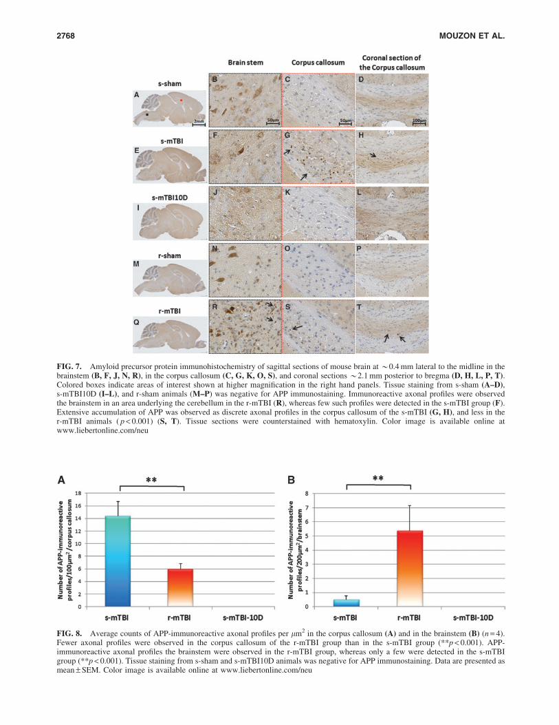
FIG. 7. Amyloid precursor protein immunohistochemistry of sagittal sections of mouse brain at *0.4 mm lateral to the midline in thebrainstem (B, F, J, N, R), in the corpus callosum (C, G, K, O, S), and coronal sections *2.1 mm posterior to bregma (D, H, L, P, T).Colored boxes indicate areas of interest shown at higher magnification in the right hand panels. Tissue staining from s-sham (A–D),s-mTBI10D (I–L), and r-sham animals (M–P) was negative for APP immunostaining. Immunoreactive axonal profiles were observedthe brainstem in an area underlying the cerebellum in the r-mTBI (R), whereas few such profiles were detected in the s-mTBI group (F).Extensive accumulation of APP was observed as discrete axonal profiles in the corpus callosum of the s-mTBI (G, H), and less in ther-mTBI animals ( p < 0.001) (S, T). Tissue sections were counterstained with hematoxylin. Color image is available online atwww.liebertonline.com/neu
FIG. 8. Average counts of APP-immunoreactive axonal profiles per lm2 in the corpus callosum (A) and in the brainstem (B) (n = 4).Fewer axonal profiles were observed in the corpus callosum of the r-mTBI group than in the s-mTBI group (**p < 0.001). APP-immunoreactive axonal profiles the brainstem were observed in the r-mTBI group, whereas only a few were detected in the s-mTBIgroup (**p < 0.001). Tissue staining from s-sham and s-mTBI10D animals was negative for APP immunostaining. Data are presented asmean – SEM. Color image is available online at www.liebertonline.com/neu
2768 MOUZON ET AL.

Consistent with other mouse models of r-mTBI and evidence
from athletes who have experienced repeated mild concussive in-
juries, our model shows acute postural equilibrium impairment
post-injury as assessed by the rotarod motor task.37,43 Whereas the
performance of the s-mTBI group improved over time, the r-mTBI
group was unable to recover their sensorimotor function up to 7
days post-injury, which was the latest day tested in our studies.
Over this time period, each injury group performed worse than their
respective shams with a rank order of performance of r-mTBI <s-mTBI < r-sham = s-sham. When tested for cognitive performance
in the BM, both injury groups showed cognitive impairment con-
sistent with findings in previous brain injury studies.25,30,31,42,44,45
There was a worsening trend for r-mTBI compared with s-mTBI
during the acquisition trials as well as on time to zone in the probe
trial, and on the final day of acquisition training the r-mTBI group
performed significantly worse than the other three groups.
From a neuropathological perspective, the only macroscopic
abnormality observed was a focal contusion injury on the inferior
surface of the cerebellum away from the site of injury in the r-mTBI
and s-mTBI-10D groups. In addition, we observed an evolving
cerebellar injury in the s-mTBI-10D group that is likely the result
of secondary injury processes that develop over a period of days
after the initial trauma. Aside from this observation, there was no
evidence of structural damage to the brain based on routine
FIG. 9. Glial fibrillary acid protein (GFAP) immunohistochemistry of sagittal sections of the mouse brain at *0.4mm lateral tomidline in the cortex (B, F, J, N, R), corpus callosum (C, G, K, O, S), and the CA1 subregion of the hippocampus (D, H, L, P, T).Colored boxes indicate areas of interest shown at higher magnification in the right panels. There were no changes in the s-mTBI group(E–H) compared with their respective sham group (A–D) at 24 h post-injury. An increase in the area of GFAP staining was observed inthe corpus callosum at 10 days post s-mTBI (K). An increased area of GFAP immunoreactivity was observed in the CA1 region of ther-sham group (P). GFAP immunohistochemistry shows extensive staining in the r-mTBI at 24 h after final injury in the cortex, corpuscallosum, and hippocampus (R–T). Tissue sections were counterstained with hematoxylin. Color image is available online atwww.liebertonline.com/neu
FIG. 10. Quantitative analysis of GFAP staining intensity in two 150 lm2 areas of the corpus callosum (A) and two 200 lm2 areas ofthe CA1 region (B) (n = 4). Data are presented as mean – SEM, (*p < 0.05; **p < 0.001; ***p < 0.0001). Color image is available onlineat www.liebertonline.com/neu
NEUROBEHAVIOR AND NEUROPATHOLOGY IN AN r-mTBI MODEL 2769
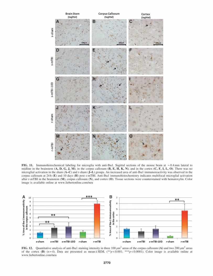
FIG. 11. Immunohistochemical labeling for microglia with anti-Iba1. Sagittal sections of the mouse brain at *0.4 mm lateral tomidline in the brainstem (A, D, G, J, M), in the corpus callosum (B, E, H, K, N), and in the cortex (C, F, I, L, O). There was nomicroglial activation in the sham (A–C) and r-sham (J–L) groups. An increased area of anti-Iba1 immunoreactivity was observed in thecorpus callosum at 24 h (E) and 10 days (H) post s-mTBI. Anti-Iba1 immunohistochemistry indicates multifocal microglial activationafter r-mTBI in the brainstem (M), corpus callosum (N), and cortex (O). Tissue sections were counterstained with hematoxylin. Colorimage is available online at www.liebertonline.com/neu
FIG. 12. Quantitative analysis of anti-Iba1 staining intensity in three 100 lm2 areas of the corpus callosum (A) and two 200 lm2 areasof the cortex (B) (n = 4). Data are presented as mean – SEM, (**p < 0.001; ***p < 0.0001). Color image is available online atwww.liebertonline.com/neu
2770

morphological assessment with H&E and LFB/CV staining, in-
cluding no signs of neuronal loss. Axonal injury following trauma
results in swollen and tortuous axons that can be detected by the
accumulation of APP as early as 35 min after brain injury,46 and in
our model, a single impact resulted in APP-immunoreactive axons
localized to the CC, evident at 24 h after injury. By 10 days post-
injury (s-mTBI-10D) there was no evidence for APP immunore-
activity, consistent with the current literature for animal models in
that strong APP staining is present at 24 h post-injury,22,30,47 which
diminishes by 14 days post-injury.42,46 In the r-mTBI group, more
widespread damage was observed, with immunoreactive axons also
evident in the spinal trigeminal tracts of the BS and beneath the
impact site in the form of APP immunoreactive neuronal perikar-
yas. However, in the r-mTBI group, the APP immunoreactive ax-
onal swellings were less frequent and less robustly stained in the
CC than those observed at 24 h after s-mTBI. These observations
may be consistent with a deleterious effect of repetitive versus
single injury on the ability of the brain to efficiently catabolize and
clear APP, facilitating amyloidogenic processing and contributing
to later evolution of amyloid plaque pathology.17,48 More work is
required to explain why no increase in APP immunoreactivity was
observed in r-mTBI versus s-mTBI. Nevertheless, the comparison
of r-mTBI with s-mTBI-10D (where both groups are matched for
time since the first/single injury) clearly demonstrates that repeti-
tive injury sustains an APP immunoreactive axonal profile over the
10-day period following the initial injury.
In addition to the white matter damage, the r-mTBI group was
associated with a notable reactive astrocytosis, with a distinct
pattern of GFAP-positive reactive astrocytes localized to the me-
dium and deep layers of the cortex beneath the impact site. As
observed in the s-mTBI-10D group, our model produced a mea-
surable reactive astrocytosis at 10 days after initial mTBI. These
results are consistent with other studies that showed astrocytosis
peaking between 10 and 14 days following concussion in
rodents.42,49,50 This increase in gliosis is also believed to coincide
with macrophage accumulation, which has also been shown to
peak at 10 days post- injury. Our data demonstrate that repetitive,
noninvasive mTBI in the mouse resulted in a graded injury re-
sponse, with the extent of reactive astrocytosis increased in animals
exposed to repetitive impacts. Whereas a single impact produced
moderate reactive astrocytosis at 10 days post-injury and was iso-
lated to the CC, five impacts over 9 days showed increased staining
in the CC as well as cortical and hippocampal involvement.
As well as axonal injury and astrogliosis, microgliosis was also
detected in the CC of injured animals as early as 24 h and up to at
least 10 days post-mTBI, whereas the r-mTBI group revealed
clusters of activated microglia in multiple brain regions (CC, BS,
and cortex). The time course of microglial activation observed in
this study was similar to that noted in previous reports.31,51–53
Depending mainly upon injury severity and the duration of the
ensuing inflammatory cascade, microgliosis can be either beneficial
or detrimental with respect to tissue preservation.54 Known as the
first line of defense in both focal and diffuse TBI,55 microglial
activation has been reported to play a beneficial role by secreting
brain-derived neurotrophic growth factors and insulin-like growth
factor.56–58 They have also been shown to have a detrimental role
at an acute time point by secreting inflammatory cytokines such
as interleukin (IL)-1b and tumor necrosis factor (TNF)-a.59,60
Moreover, in several TBI models, minocycline, which has anti-
inflammatory properties, has been shown to reduce histopatho-
logical consequences after mTBI in mice.61,62 Of particular interest
is a recent study by Siopi and collaborators that demonstrated that
reducing neuroinflammation post-injury attenuated memory im-
pairment in a mouse model of CHI;63,64 however, more work is
required to address the impact of microglial activation on cognitive
function after TBI.
Conclusion
In conclusion, we have developed a simple and reproducible
mouse model of mTBI, which induces pathological and behavioral
features comparable to those observed in the human condition.
Moreover, by recapitulating the pathological alterations observed
in human TBI patients, this model is also suitable for studying
various mechanisms of post-traumatic injury such as axonal injury,
cell death, or apoptosis. Moving forward, this model of mTBI can
be used to investigate the long term consequences of r-mTBI, the
influence of different numbers of injuries and different inter-injury
intervals, and the impact of r-mTBI in mice genetically modified at
loci of relevance to human TBI such as Tau or APOE, aspects
currently lacking in the literature. An increased understanding of
mTBI and its cumulative effects will enable identification of mo-
lecular targets specific to mTBI and ultimately the development of
novel, effective therapeutics, which are desperately needed.
Acknowledgments
This research was funded by a Department of Defense award
(W81XWH-10-1-0759) to Dr. Fiona Crawford and by the Roskamp
Foundation.
Author Disclosure Statement
No competing financial interests exist.
References
1. Faul, M., Xu, L., Wald, M.M., and Coronado, V.G. (2010). TraumaticBrain Injury in The United States: Emergency Department Visits, Hos-pitalizations, and Deaths 2002-2006. Centers for Disease Control andPrevention, National Center for Injury Prevention and Control: Atlanta.
2. Corsellis J.A., Bruton, C.J., Freeman-Browne, D. (1973). The after-math of boxing. Psychol. Med. 3, 270–303.
3. Guskiewicz, K.M., McCrea, M., Marshall, S.W., Cantu, R.C., Ran-dolph, C., Barr, W., Onate, J.A., and Kelly, J.P. (2003). Cumulative
Table 1. Summary of the Location and Relative
Intensity of Immunohistochemical Data for Amyloid
Precursor Protein (A), Glial Fibrillary Acid
Protein (G), and Anti-Iba1 (M)
s-sham s-mTBI s-mTBI-10D r-sham r-mTBI
Cortex - - - - A- - - G G G G- - - - M M
Corpus callosum - A A A - - AA- - G G G G G GM M M M M M M M M
CA1 - - - - -G G G G G G G GM M M M M
Brainstem - A - - A A- - - - -- - - - M
Present (X), moderate staining (XX), and intense staining (XXX).s-mTBI, single mild traumatic brain injury; r-mTBI, repetitive mild
traumatic brain injury.
NEUROBEHAVIOR AND NEUROPATHOLOGY IN AN r-mTBI MODEL 2771

effects associated with recurrent concussion in collegiate footballplayers: the NCAA concussion study. JAMA 290, 2549–2555.
4. Guskiewicz, K.M., Marshall, S.W., Bailes, J., McCrea, M., Cantu,R.C., Randolph, C., and Jordan, B.D. (2005). Association betweenrecurrent concussion and late-life cognitive impairment in retiredprofessional football players. Neurosurgery 57, 719–726.
5. Guskiewicz, K.M., Marshall, S.W., Bailes, J., McCrea, M., Harding,H.P., Matthews, A., Mihalik, J.R., and Cantu, R.C. (2007). Recurrentconcussion and risk of depression in retired professional footballplayers. Med. Sci. Sports Exerc. 39, 903–909.
6. Omalu, B.I., DeKosky, S.T., and Minster, R.L. (2005). Chronic trau-matic encephalopathy in a national football league player. Neurosur-gery 57, 128–134.
7. Omalu, B.I, DeKosky, S.T., and Hamilton, R.L. (2006). Chronictraumatic encephalopathy in a national football league player: part II.Neurosurgery 59, 1086–1092.
8. Omalu, B.I., Hamilton, R.L., Kamboh, M.I., DeKosky, S.T., andBailes J. (2010). Chronic traumatic encephalopathy in a nationalfootball league player: case report and emerging medico-legal practicequestions. J. Forensic Nurs. 6, 40–46.
9. Omalu, B.I., Fitzsimmons, R.P., Hammers, J., and Bailes, J. (2010).Chronic traumatic encephalopathy in a professional American wres-tler. J. Forensic Nurs. 6, 130–136.
10. Omalu, B., Bailes, J., Hamilton, R.L., Kamboh, M.I., Hammers, J.,Case, M., and Fitzsimmons, R. (2011). Emerging histomorphologicphenotypes of chronic traumatic encephalopathy in American athletes.Neurosurgery 69, 173–183.
11. Broglio, S.P., Eckner, J.T., Martini, D., Sosnoff, J.J., Kutcher J.S., andRandolph, C. (2011). Cumulative head impact burden in high schoolfootball. J. Neurotrauma 28, 2069–2078.
12. Gavett, B.E., Stern, R.A., Cantu, R.C., Nowinski, C.J., and McKee,A.C. (2010). Mild traumatic brain injury: a risk factor for neurode-generation. Alzheimers Res. Ther. 2, 18.
13. Gavett, B.E., Stern, R.A., and McKee, A.C. (2011). Chronic traumaticencephalopathy: a potential late effect of sport-related concussive andsubconcussive head trauma. Clin. Sports Med. 30, 179–188.
14. Stern, R.A., Riley, D.O., Daneshvar, D.H., Nowinski, C.J., Cantu,R.C., and McKee, A.C. (2011). Long-term consequences of repetitivebrain trauma: chronic traumatic encephalopathy. PM R 3, Suppl. 2,S460–467.
15. McKee, A.C., Cantu, R.C., Nowinski, C.J., Hedley–Whyte, E.T.,Gavett, B.E., Budson, A.E., Santini,V.E., Lee, H.S., Kubilus, C.A.,and Stern, R.A. (2009). Chronic traumatic encephalopathy in athletes:progressive tauopathy after repetitive head injury. J. Neuropathol.Exp. Neurol. 68, 709–735.
16. Costanza, A., Weber, K., Gandy, S., Bouras, C., Hof, P.R., Gianna-kopoulos, P., and Canuto, A. (2011). Contact sport-related chronictraumatic encephalopathy in the elderly: clinical expression andstructural substrates. Neuropathol. Appl. Neurobiol. 37, 570–584.
17. Johnson, V.E., Stewart, W., and Smith, D.H. (2010). Traumatic braininjury and amyloid-b pathology: a link to Alzheimer’s disease? Nat.Rev. Neurosci. 11, 361–370.
18. Van Den Heuvel, C., Thornton, E., and Vink, R. (2007). Traumaticbrain injury and Alzheimer’s disease: a review. Prog. Brain Res, 161,303–316.
19. Forstl, H., Haass, C., Hemmer, B., Meyer, B., and Halle, M. (2010).Boxing-acute complications and late sequelae: from concussion todementia. J. Forensic Nurs. 6, 40–46.
20. Raghupathi, R., and Margulies, S.S. (2002). Traumatic axonal injuryafter closed head injury in the neonatal pig. J. Neurotrauma 19, 843–853.
21. Raghupathi, R., Mehr, M., Helfaer, M.A., and Margulies, S.S. (2004).Traumatic axonal injury is exacerbated following repetitive closedhead injury in the neonatal pig. J. Neurotrauma 21, 307–316.
22. Browne, K.D., Chen, X.H., Meaney, D.F., and Smith, D.H. (2011).Mild traumatic brain injury and diffuse axonal injury in swine.J. Neurotrauma 28, 1747–1755.
23. Gennarelli, T.A., Thibault, L.E., Adams, J.H., Graham, D.I., Thomp-son, C.J., and Marcincin, R.P. (1982). Diffuse axonal injury andtraumatic coma in the primate. Ann. Neurol. 12, 564–574.
24. Laurer, H.L., Bareyre, F.M., and Lee, V.M. (2001). Mild head injuryincreasing the brain’s vulnerability to a second concussive impact.J. Neurosurg. 95, 859–870.
25. DeFord, S.M., Wilson, M.S., and Rice, A.C. (2002). Repeated mildbrain injuries result in cognitive impairment in B6C3F1 mice.J. Neurotrauma 19, 427–438.
26. Uryu, K., Laurer, H., McIntosh, T., Pratico, D., Martinez, D., Leight,S., Lee, V.M., and Trojanowski, J.Q. (2002). Repetitive mild braintrauma accelerates Abeta deposition, lipid peroxidation, and cognitiveimpairment in a transgenic mouse model of Alzheimer amyloidosis.J. Neurosci. 22, 446–454.
27. Longhi, L., Saatman, K.E., Fujimoto, S., Raghupathi, R., Meaney,D.F., Davis, J., McMillan, B.S., Conte, V., Laurer, H.L., Stein, S.,Stocchetti, N., and McIntosh, T.K. (2005).Temporal window of vul-nerability to repetitive experimental concussive brain injury. Neuro-surgery 56, 364–374.
28. Creeley, C.E., Wozniak, D.F., Bayly, P.V., Olney, J.W., and Lewis,L.M. (2004). Multiple episodes of mild traumatic brain injury result inimpaired cognitive performance in mice. Acad. Emerg. Med. 11, 809–819.
29. Nakajima, Y., Horiuchi, Y., Kamata, H., Yukawa, M., Kuwabara, M.,and Tsubokawa, T. (2010). Distinct time courses of secondary braindamage in the hippocampus following brain concussion and contusionin rats. Tohoku J. Exp. Med. 221, 229–235.
30. Prins, M.L., Hales, A., Reger, M., Giza, C.C., and Hovda, D.A.(2010). Repeat traumatic brain injury in the juvenile rat is associatedwith increased axonal injury and cognitive impairments. Dev. Neu-rosci. 32, 510–518.
31. Shitaka, Y., Tran, H.T., Bennett, R.E., Sanchez, L., Levy, M.A.,Dikranian, K., and Brody, D.L. (2011). Repetitive closed-skulltraumatic brain injury in mice causes persistent multifocal axonalinjury and microglial reactivity. J. Neuropathol. Exp. Neurol. 70,551–567.
32. Cole, J.T., Yarnell, A., Kean, W.S., Gold, E., Lewis, B., Ren, M.,McMullen, D.C., Jacobowitz, D.M., Pollard, H.B., O’Neill, J.T.,Grunberg, N.E., Dalgard, C.L., Frank, J.A., and Watson, W.D. (2011).Craniotomy: true sham for traumatic brain injury, or a sham of asham? J. Neurotrauma 28, 359–369.
33. Brody, D.L., Mac Donald, C., Kessens, C.C., Yuede, C., Parsadanian,M., Spinner, M., Kim, E., Schwetye, K.E., Holtzman, D.M., andBayly, P.V. (2007). Electromagnetic controlled cortical impact devicefor precise, graded experimental traumatic brain injury. J. Neuro-trauma 24, 657–673.
34. Albert-Weissenberger, C., and Siren, A.L. (2010). Experimentaltraumatic brain injury. Exp. Transl. Stroke Med. 2, 16.
35. Conte, V., Uryu, K., Fujimoto, S., Yao, Y., Rokach, J., Longhi, L.,Trojanowski, J. Q., Lee, V. M.-Y., McIntosh, T. K., and Pratico, D.(2004). Vitamin E reduces amyloidosis and improves cognitivefunction in Tg2576 mice following repetitive concussive brain injury.J. Neurochem. 90, 758–764.
36. Yoshiyama, Y., Uryu, K., Higuchi, M., Longhi, L., Hoover, R., Fu-jimoto, S., McIntosh, T., Lee, V.M., and Trojanowski, J.Q. (2005).Enhanced neurofibrillary tangle formation, cerebral atrophy, andcognitive deficits induced by repetitive mild brain injury in a trans-genic tauopathy mouse model. J. Neurotrauma 22, 1134–1141.
37. Kane, M.J., Angoa–Perez, M., Briggs, D.I., Viano, D.C., Kreipke,C.W., and Kuhn, D.M. (2012). A mouse model of human repetitivemild traumatic brain injury. J. Neurosci. Methods 203, 41–49.
38. Franklin, K.B.J., and Paxinos, G. (2001). The Mouse Brain in Ste-reotaxic Coordinates, 2nd ed. Elsevier/Academic Press: San Diego,London.
39. Ruifrok, A.C., and Johnston, D.A. (2001). Quantification of histo-chemical staining by color deconvolution. Anal. Quant. Cytol. Histol.23, 291–299.
40. Park, H.K., Fernandez, I.I., Dujovny, M., and Diaz, F.G. (1999). Ex-perimental animal models of traumatic brain injury: medical andbiomechanical mechanism. Crit. Rev. Neurosurg. 9, 44–52.
41. LaPlaca, M.C., Simon, C.M., Prado, G.R., and Cullen, D.K. (2007).CNS injury biomechanics and experimental models. Prog. Brain Res.161, 13–26.
42. Huh, J.W., Widing, A.G., and Raghupathi, R. (2008).Midline braininjury in the immature rat induces sustained cognitive deficits, bihe-mispheric axonal injury and neurodegeneration. Exp. Neurol. 213, 84–92.
43. Guskiewicz, K.M. (2011). Balance assessment in the management ofsport-related concussion. Clin. Sports Med. 30, 89–102.
44. Shultz, S.R., Bao, F., Omana, V., Chiu, C., Brown, A., and Cain, D.P.(2012). Repeated mild lateral fluid percussion brain injury in the ratcauses cumulative long-term behavioral impairments, neuroin-flammation, and cortical loss in an animal model of repeated con-cussion. J. Neurotrauma 29, 281–294.
2772 MOUZON ET AL.

45. Eakin, K., and Miller, J.P. (2012). Mild traumatic brain injury is as-sociated with impaired hippocampal spatiotemporal representation inthe absence of histological changes. J. Neurotrauma 29, 1180–1187.
46. Pierce, J.E., Trojanowski, J.Q., Graham, D.I., Smith, D.H., andMcIntosh, T.K. (1996). Immunohistochemical characterization ofalterations in the distribution of amyloid precursor proteins and beta-amyloid peptide after experimental brain injury in the rat. J. Neurosci.16, 1083–1090.
47. Huh, J.W., Widing, A.G., and Raghupathi, R. (2007). Basic science;repetitive mild non-contusive brain trauma in immature rats exacer-bates traumatic axonal injury and axonal calpain activation: a pre-liminary report. J. Neurotrauma 24, 15–27.
48. Sivanandam, T.M., and Thakur, M.K. (2012). Traumatic brain injury:a risk factor for Alzheimer’s disease. Neurosci. Biobehav. Rev. 36,1376–1381.
49. Hamberger, A., Viano, D.C., Saljo, A., and Bolouri, H. (2009). Con-cussion in professional football: morphology of brain injuries in theNFL concussion model—part 16. Neurosurgery 64, 1174–1182.
50. Hellewell, S.C., Yan, E.B., Agyapomaa, D.A., Bye, N., and Morganti–Kossmann, M.C. (2010). Post-traumatic hypoxia exacerbates braintissue damage: analysis of axonal injury and glial responses. J. Neu-rotrauma 27, 1997–2010.
51. Kelley, B.J., Lifshitz, J., and Povlishock, J.T. (2007). Neuroin-flammatory responses after experimental diffuse traumatic brain in-jury. J. Neuropathol. Exp. Neurol. 66, 989–1001.
52. Chen, S., Pickard, J.D., and Harris, N.G. (2003). Time course ofcellular pathology after controlled cortical impact injury. Exp. Neurol.182, 87–102.
53. Haselkorn, M.L., Shellington, D.K., Jackson, E.K., Vagni, V.A.,Janesko–Feldman, K., Dubey, R.K., Gillespie, D.G., Cheng, D., Bell,M.J., Jenkins, L.W., Homanics, G.E., Schnermann, J., and Kochanek,P.M. (2010). Adenosine A1 receptor activation as a brake on themicroglial response after experimental traumatic brain injury in mice.J. Neurotrauma 27, 901–910.
54. Neumann, H., Kotter, M.R., and Franklin, R.J. (2009). Debris clear-ance by microglia: an essential link between degeneration and re-generation. Brain. 132 (Pt. 2), 288–295.
55. Morganti–Kossmann, M.C., Rancan, M., Stahel, P.F., and Kossmann,T. (2002). Inflammatory response in acute traumatic brain injury: adouble–edged sword. Curr. Opin. Crit. Care 8, 101–105.
56. Neumann, J., Gunzer, M., Gutzeit, H.O., Ullrich, O., Reymann, K.G.,and Dinkel, K. (2006). Microglia provide neuroprotection after is-chemia. FASEB J. 20, 714–716.
57. Lalancette–Hebert, M., Gowing, G., Simard, A., Weng, Y.C., andKriz, J. (2007). Selective ablation of proliferating microglial cellsexacerbates ischemic injury in the brain. J. Neurosci. 27, 2596–2605.
58. Thored, P., Heldmann, U., Gomes–Leal, W., Gisler, R., Darsalia, V.,Taneera, J., Nygren, J.M., Jacobsen, S.E.W., Ekdahl, C.T., Kokaia, Z.,and Lindvall, O. (2009). Long-term accumulation of microglia withproneurogenic phenotype concomitant with persistent neurogenesis inadult subventricular zone after stroke. Glia 57, 835–849.
59. Lloyd, E., Somera–Molina, K., Van Eldik, L.J., Watterson, D.M., andWainwright, M.S. (2008). Suppression of acute proinflammatory cy-tokine and chemokine upregulation by post-injury administration of anovel small molecule improves long-term neurologic outcome in amouse model of traumatic brain injury. J. Neuroinflammation 30, 28.
60. Schmidt, O., Heyde, C., Ertel, W., and Stahel, P. (2005). Closed headinjury–an inflammatory disease? Brain Res. Rev. 48, 388–399.
61. Homsi, S., Piaggio, T., Croci, N., Noble, F., Plotkine, M., Marchand–Leroux, C., and Jafarian–Tehrani, M. (2010). Blockade of acute mi-croglial activation by minocycline promotes neuroprotection and re-duces locomotor hyperactivity after closed head injury in mice: atwelve-week follow-up study. J. Neurotrauma 27, 911–921.
62. Siopi, E., Cho, A.H., Homsi, S., Croci, N., Plotkine, M., Marchand–Leroux, C., and Jafarian–Tehrani, M. (2011). Minocycline restoressAPPa levels and reduces the late histopathological consequences oftraumatic brain injury in mice. J. Neurotrauma 28, 2135–2143.
63. Siopi, E., Calabria, S., Plotkine, M., Marchand–Leroux, C., andJafarian–Tehrani, M. (2012). Minocycline restores olfactory bulbvolume and olfactory behavior after traumatic brain injury in mice.J. Neurotrauma 29, 354–361.
64. Siopi, E., Llufriu–Daben, G., Fanucchi, F., Plotkine, M., Marchand–Leroux, C., and Jafarian–Tehrani, M. (2012). Evaluation of late cog-nitive impairment and anxiety states following traumatic brain injuryin mice: the effect of minocycline. Neurosci. Lett. 511, 110–115.
Address correspondence to:
Benoit Mouzon, M.S.
Roskamp Institute
2040 Whitfield Avenue
Sarasota, FL 34243
E-mail: [email protected]
NEUROBEHAVIOR AND NEUROPATHOLOGY IN AN r-mTBI MODEL 2773