replacing the first-generation dentition in …replacing the first-generation dentition in...
TRANSCRIPT
Replacing the first-generation dentition in pufferfishwith a unique beakGareth J. Frasera,1, Ralf Britzb, Andie Hallb, Zerina Johansonc, and Moya M. Smithd
aDepartment of Animal and Plant Sciences, University of Sheffield, Sheffield S10 2TN, United Kingdom; bZoology Department and cPalaeontologyDepartment, The Natural History Museum, London SW7 5BD, United Kingdom; and dDepartment of Craniofacial Development and Stem Cell Biology,Dental Institute, King’s College London, London SE1 9RT, United Kingdom
Edited by David B. Wake, University of California, Berkeley, CA, and approved April 9, 2012 (received for review November 29, 2011)
Teleost fishes comprise approximately half of all living verte-brates. The extreme range of diversity in teleosts is remarkable,especially, extensive morphological variation in their jaws anddentition. Some of the most unusual dentitions are found amongmembers of the highly derived teleost order Tetraodontiformes,which includes triggerfishes, boxfishes, ocean sunfishes, andpufferfishes. Adult pufferfishes (Tetraodontidae) exhibit a distinc-tive parrot-like beaked jaw, forming a cutting edge, unlike in anyother group of teleosts. Here we show that despite novelty in thestructure and development of this “beak,” it is initiated by formationof separate first-generation teeth that line the embryonic pufferfishjaw, with timing of development and gene expression patternsconserved from the last common ancestor of osteichthyans. Mostof these first-generation larval teeth are lost in development. Con-tinuous tooth replacement proceeds in only four parasymphysealteeth, as sequentially stacked, multigenerational, jaw-length den-tine bands, before development of the functional beak. These datasuggest that dental novelties, such as the pufferfish beak, candevelop later in ontogeny through modified continuous toothaddition and replacement. We conclude that even highly derivedmorphological structures like the pufferfish beak form via a con-served developmental bauplan capable of modification duringontogeny by subtle respecification of the developmental module.
evolutionary developmental biology | morphological novelty |tooth development | replacement dentition | phenotypic diversity
Vertebrates offer an impressive range of morphological di-versity, especially in dentitions. Morphological diversity in
teleost fishes is unparalleled among vertebrates, exemplified bythe bizarre forms assigned to the order Tetraodontiformes. Somemembers of this teleost order, the Gymnodontes, are known fortheir unusual jaws, superficially resembling the beak of a parrot(Fig. 1) (1). The members of one family of Gymnodontes, thepufferfishes (Tetraodontidae), possess a unique oral dentitionof four teeth, two in the upper jaws and two in the lower jaws,extending laterally from the midline (Fig. 1 C and D). These teethform paired opposing beak-shaped toothplates that can crush orslice prey items, different from most other teleost dentitions(Fig. 2 H and N) (2, 3). The ontogenetic and developmentalmechanisms that form the unique tetraodontid dentition havebeen little studied; only a brief mention of larval teeth in puf-ferfishes (4) and descriptions of the adult dentition (5) have beenpublished to date.However, when dental development is examined in detail, this
adult morphological novelty shows greater similarity and struc-tural conservation in initial development to that of osteichthyansthan was previously appreciated (Fig. 2). The pufferfishes are amorphologically derived group of teleosts with numerous re-ductive characteristics, including a lack of pelvic fins, ribs, andlower pharyngeal jaws, a reduced number of vertebrae, and ab-sence of various cranial bones (6–9). Moreover, members of thefamily Tetraodontidae possess some of the most concise verte-brate genomes known (10–13). In the present study, we focusedon the southern Asian freshwater pufferfish genus Monotrete and
examined embryos of several closely related species (M. abei,M. cochinchinensis,M. leiurus, andM. suvattii) (Fig. 1). Our initialhypothesis was that the pufferfish “beak” (Fig. 2 I, N) representsa unique dental structure from an unknown developmental ge-netic bauplan. We expected this genetic bauplan to be unique notonly among teleosts, but also among vertebrates.To test this hypothesis, we investigated how this highly derived
beak-like pufferfish dentition forms developmentally. Specifi-cally, we examined how the spatial and temporal pattern of geneexpression unfolds, as related to tooth initiation and develop-ment, during sequential ontogenetic stages of the embryonic andhatchling dentitions (Fig. 2). Gene expression associated withdevelopmental phases during formation of the pufferfish denti-tion has received little attention so far (14). Thus, we have takenadvantage of this unique dentition to address more generalquestions regarding genetic control related to the developmentalorigins of teleost morphological diversity and the evolution ofthese patterns. Here we document the morphogenetic progressionfrom initial stages of formation of the first-generation dentitionthrough to transitional stages of beak initiation.
ResultsWe examined the expression of a subset of highly conserved genes(expressed similarly across many taxa) known to be active duringall similarly studied stages of tooth development in several tele-osts, reptiles, and mammals (15–17) for comparison. We chosethe genes bmp4, pax9, pitx2, and shh for this study because theyinclude some of the most studied gene representatives across taxafor tooth development from fish to mammals, allowing for gen-eralizations across vertebrates (15, 16, 18–24). For this study, wegenerated riboprobes from recently developed genomic resourcesfor closely related pufferfishes, Tetraodon (http://www.genoscope.cns.fr/externe/tetranew/) and Fugu (http://www.fugu-sg.org/), toexamine temporal and spatial expression patterns by whole-mount in situ hybridization (Figs. 3 and 4) and monitor how geneexpression changed during ontogenetic formation of the beak. Weaimed to test whether developmental mechanisms common toother teleost dentitions were present in the pufferfish dentition.
Pufferfish First-Generation Dentition Originates from a ConservedOsteichthyan Pattern. Timed development of the earliest Mono-trete dentition can be seen in cleared and double-stained skeletalpreparations (Fig. 2) and also before morphological emergencevia gene expression patterns (Fig. 3). Both of these developmentalpatterns show that a row of separate first-generation teeth formsin a timed sequence along the jaw margin, quite unlike in the
Author contributions: G.J.F., R.B., Z.J., and M.M.S. designed research; G.J.F., R.B., A.H., Z.J.,and M.M.S. performed research; G.J.F., R.B., Z.J., and M.M.S. analyzed data; and G.J.F., R.B.,Z.J., and M.M.S. wrote the paper.
The authors declare no conflict of interest.
This article is a PNAS Direct Submission.1To whom correspondence should be addressed. E-mail: [email protected].
This article contains supporting information online at www.pnas.org/lookup/suppl/doi:10.1073/pnas.1119635109/-/DCSupplemental.
www.pnas.org/cgi/doi/10.1073/pnas.1119635109 PNAS | May 22, 2012 | vol. 109 | no. 21 | 8179–8184
DEV
ELOPM
ENTA
LBIOLO
GY
Dow
nloa
ded
by g
uest
on
May
20,
202
0

adult beak and before any stages of beak initiation. These firststages of tooth development reiterate those of other teleostsstudied to date. Importantly, the timing and spacing of toothinitiation along the jaw follows the same pattern that is wellconserved across osteichthyan fishes (Fig. 3) (15, 18, 19). As inother bony fishes, initial tooth germs on the lower jaw appear in
a temporal sequence not matched by tooth position along thejaw. The first tooth germ develops in position 2, followed first byposition 3, then position 1 (roman numerals in Fig. 3 representthe positional order along the jaw), and then by successiveproximal addition of tooth germs in positions 4–9 (Figs. 2 and 3).There is a different arrangement of separate teeth in the upperjaw comprising an oblique anteroposterior row with fewer teeth(between four and six; Fig. 2 C–F), compared with a total of 14–18 on the lower jaw. Comparable data have been reported in therainbow trout Oncorhynchus mykiss (25, 26). Later in both jaws,two prominent parasymphyseal teeth at tooth position 1 in eachquadrant (Fig. 2 E, H, and M) are retained for, and involved in,subsequent continuous tooth replacement (see below) duringbeak formation (Fig. 2 H, I, M, and N).The first indication of tooth initiation is in restricted gene
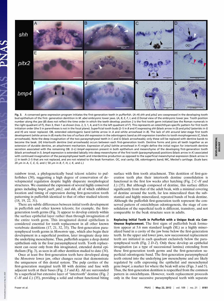
expression in Monotrete spp. at 6 d postfertilization, localized toa dentally competent epithelial strip, the odontogenic bandcoexpressing pitx2 and shh (Fig. 3 A–G). Underlying this epi-thelial expression are aggregated mesenchymal cells showingrestricted expression of well-known “odontogenic-related” genesbmp4 (Fig. 3 I–L) and pax9 (Fig. S1) in underlying tooth fields,presumed to be reciprocal activity (18, 19, 27–29). However, pax9is later restricted to those mesenchymal cells surrounding eachtooth unit (Fig. S1), following the conserved gene expressiondomains of other teleosts (15, 18, 22). For example, identicalpatterns of gene expression have been documented in the
Fig. 1. Adult freshwater pufferfish, (A)Monotrete abeimale guarding eggson the substrate. (B) Lateral view of the M. abei head showing the mouthwith a partly exposed beak; the large lips cover most of the beak. (C) Lateralview of a typical pufferfish skull of tetraodon lineatus (skeletal preparation;scale bar: 2 cm) and (D) frontal view showing the extensive beak tissue fusedwith the bone of the articulated jaws. (Scale bar: 1 cm.)
Fig. 2. Developmental sequence from a conserved pattern of initial teeth to replacement dentine bands during the formation of the unique pufferfish beak.(A–D, F–H, M, and N) Frontal views (into the mouth) of the developing dentition (3.6 mm NL to 10.3 mm SL) of a pufferfish (Monotrete suvattii). (E, I, and L)Lateral views. (K) Medial view. A–M show specimens cleared and double- stained with alizarin red (staining calcium-rich tissues, e.g., bone and dentine) andalcian blue (staining mucopolysaccharides in cartilage). The first-generation dentition in pufferfish is composed of individual teeth with acrodin (enameloid)caps identical to those of other actinopterygians (A–H). From the youngest stage with 2 teeth in the lower jaw (LJ) (A, black arrowheads, T1), separate teethare added along the jaws [B, 4 teeth plus a developing tooth (black box) in the LJ, 2 teeth plus a developing tooth in the upper jaw (UJ), denoted by anasterisk] with up to 14 (C) to 16 teeth (D) in the LJ and up to 6 teeth in the UJ (C and D). First-generation teeth and superficial intertooth dentine are retaineduntil worn (H, asterisk). Strongly mineralized jaw length bands of dentine form from individual replacement teeth, stacked below the first-generation teethin the UJ and LJ, increasing in number with size from one band in D to up to four bands in H and N, the largest stage that we studied (10.3 mm SL). Thesestacks of dentine bands (R1–R4) form as multigeneration replacement teeth of only the four most medial teeth (D–H). H shows a frontal view of the lower jawbeak, showing the four generations of replacement bands (R1–R4) of stacked dentine that will form the adult beak. The asterisk denotes the retained first-generation teeth at the beak surface; black arrows denote the symphysis between the left and right halves of the LJ (D, G, and H ). Dentine tubules from livingcells are present in first-generation teeth (J) and in replacement dentine bands (L, black arrowheads). (N) Optical projection tomography image of the juvenileM. suvattii beak in frontal view, showing the pink fluorescent bands of stacks of replacement dentine bands forming the beak. (Scale bar: 200 μm.)mc, Meckel’s cartilage; mx, maxillary; pmx, premaxillary. Lengths are provided as either NL or standard length SL in mm of embryonic and juvenile M. suvattii(A, 3.6 mm NL; B, 3.75 mm NL; C, 4.8 mm SL; D–F, 5.6 mm SL; G, 8.5 mm SL; H, I, and N, 10.3 mm SL; J–M, 5.6 mm SL).
8180 | www.pnas.org/cgi/doi/10.1073/pnas.1119635109 Fraser et al.
Dow
nloa
ded
by g
uest
on
May
20,
202
0
rainbow trout, a phylogenetically basal teleost relative to puf-ferfishes (30), suggesting a high degree of conservation of de-velopmental regulation despite highly disparate morphologicalstructures. We examined the expression of several highly conservedgenes including bmp4, pax9, pitx2, and shh, all of which exhibitedlocation and timing of expression during first-generation toothpatterning in pufferfish identical to that of other studied teleosts(18, 19, 22, 31).There are subtle differences between initial tooth development
in pufferfish and other known teleosts; for example, the first-generation tooth germs (Fig. 3) appear to develop entirely withinthe surface epithelial layer rather than through invagination ofthe entire tooth germ. This invaginated dental epithelium isconsidered essential for tooth replacement in all other knownvertebrate dentitions (17, 21, 32, 33). The first-generation para-symphyseal tooth germs in Monotrete spp., which also begin theirdevelopment in a superficial position, later invaginate deep intothe underlying mesenchyme. Thus, pufferfish acquire a deep dentalepithelium only in the four parasymphyseal teeth. Tooth replace-ment can occur only from this invaginated, extended dental epi-thelium (Fig. 3), as seen in all other dentition studied to date (34).Once at least five first-generation teeth have developed along
the Monotrete lower jaw, other changes occur that demonstratethe uniqueness of this dental system. The pulp cavities of thefirst-generation marginal teeth fill with dentine and fuse withadjacent teeth at their bases (Fig. 2 J and K). All are surroundedby a superficial but extensive layer of “intertooth” dentine (Fig. 2C–H and L) (35), providing a solid and robust functional biting
surface with firm tooth attachment. This dentition of first-gen-eration teeth plus their intertooth dentine consolidation isfunctional in the first few weeks after hatching (Fig. 2 C–H andL) (35). But although composed of dentine, this surface differssignificantly from that of the adult beak, with a minimal coveringof dentine around the teeth, far less mineralized than the sub-stantial and highly mineralized, repeated bands of beak dentine.Although the pufferfish first-generation teeth represent the con-served pattern of osteichthyan odontogenesis, the stage of con-solidation of the superficial teeth is different, transitory, and notcomparable to the beak structure seen in adults.
Replacing Initial Teeth in Pufferfish with a Unique Beak via Con-tinuous Replacement. The first signs of pufferfish beak forma-tion appear at 5.6 mm standard length (SL) as a highly miner-alized band in a cavity of the jaw bone below the first-generationteeth. In the upper and lower jaws, new successional replacementteeth are initiated in each quadrant exclusively below the para-symphyseal teeth (Fig. 2 D–I). Only these develop an epithelialinvagination (as a type of successional lamina) extending fromthese first-generation tooth germs and the first-generation su-perficial odontogenic band. The first-generation parasymphysealteeth extend into the underlying jaw mesenchyme and are likelyregulated by cells expressing the epithelial and mesenchymalgene bmp4, a marker for where the beak will arise (Figs. 3 and 5).Thus, the first-generation dentition is respecified from the commonpattern in osteichthyans. However, tooth replacement proceedsonly in the four successive parasymphyseal teeth with separate
Fig. 3. A conserved gene expression program initiates the first-generation teeth in pufferfish. (A–H) shh and pitx2 are coexpressed in the developing toothbud epithelium of the first- generation dentition in M. abei embryonic lower jaws. (A, B, E, F, I, and J) Dorsal view of the embryonic lower jaw. Tooth positionnumber along the jaw (B) does not reflect the time order in which the teeth develop; position 2 is the first tooth germ initiated (see the Roman numerals inthe right quadrant of F), then 3, then 1 as shown (nos. 2, 3, 1, 4, and 5 in the left quadrant of F). This represents an osteichthyan-specific pattern for first toothinitiation order (the 5 in parentheses is not in the focal plane). Superficial tooth buds (2–5 in B and F) expressing shh (black arrow in D) and pitx2 (asterisks in Gand H) are never replaced. OB, extended odontogenic band (white arrow in A and white arrowhead in B). The lack of shh around later-stage first toothdevelopment (white arrow in B) marks the loss of surface shh expression in the odontogenic band as shh expression transfers to tooth morphogenesis (C, blackarrowheads). Note the deep invagination of the two parasymphyseal teeth in C and G (black arrowheads); only these will be replaced with dentine bands tobecome the beak. (H) Intertooth dentine (red arrowheads) occurs between each first-generation tooth. Dentine forms and joins all teeth together as anextension of durable dentine, an attachment mechanism. Expression of pitx2 (white arrowhead in F) might define the initial region for intertooth dentinesecretion associated with the remaining OB. (I–L) bmp4 expression present in both epithelium and mesenchyme of the developing first-generation tooth(black arrowhead in I). bmp4 expression is extended labially into deep mesenchyme of the first tooth (parasymphyseal) positions (black arrow in K) associatedwith continued invagination of the parasymphyseal teeth and interdentine production as opposed to the superficial mesenchymal expression (black arrow inL) in teeth 2–5 that are not replaced, and are not related to the beak formation. OC, oral cavity; OB, odontogenic band; MC, Meckel’s cartilage. (Scale bars:20 μm in A, C, E, G, and I; 50 μm in B, F, H, J, K, and L.)
Fraser et al. PNAS | May 22, 2012 | vol. 109 | no. 21 | 8181
DEV
ELOPM
ENTA
LBIOLO
GY
Dow
nloa
ded
by g
uest
on
May
20,
202
0

cusps of acrodin, below the first ones (Fig. 2M, arrows). For allother first-generation tooth positions (from position 2 along thejaw proximally), the process of tooth replacement is inhibited.Another unique feature is the extension of dentine proximallyfrom these parasymphyseal replacement teeth in a continuousgrowth mode for each tooth to form the distinctive jaw quadrantlength, stacked dentine bands characteristic of phylogeneticallyderived pufferfishes such as Monotrete (Figs. 2 D–I and N and5 C and D). These vertical stacks of dentine bands serve as thereplacement tissue for the first-generation tooth set embeddedin superficial intertooth dentine. Each dentine band representsone replacement tooth with proximally extended growth (Fig. 4)occurring simultaneously in each quadrant. New bands are addedcontinuously throughout life, and through the process of weareach dentine band eventually becomes the functional biting(tritural) surface as many stacked dentine bands form to replacea single tooth position (a system known as many-for-one re-placement). As noted earlier, the replacement teeth are initiatedwhere coexpression of key regulators of tooth initiation, shh,pitx2 (Fig. 4), and bmp4 (Fig. S2), are redeployed to form thesedentine bands during beak morphogenesis.
DiscussionOur developmental and gene expression data obtained at earlyontogenetic stages of Monotrete spp. demonstrate that the first-generation set of marginal teeth is induced in a spatiotemporalseries that represents the typical teleost and likely a general
osteichthyan pattern (Fig. 3 A–C), the latter being conservedsince the actinopterygian/sarcopterygian split at least 416 Mya(36). Thus, the pufferfish beak is not developed de novo, butrather emerges ontogenetically as a modification of the programfor tooth replacement. Our new data show how, after the con-served tooth program has unfolded in time and space as in mostosteichthyan developmental programs (Figs. 2 and 3), the dentalmodule is modified during the first replacement phase viarespecification of continuous rounds of tooth addition and re-placement to produce a unique beak capable of crushing or slicingprey (Figs. 1, 2, 4, and 5). Only the four parasymphyseal teethform replacement teeth, and through the activity of odontoblastsextend dentine growth to form the beak within cavities (intra-medullary bone spaces) of both the upper and lower jaws (Figs. 2D–I and M and 5 B and C). As a new testable hypothesis, wesuggest that despite their highly diverse and divergent dentalstructure, teleost dentitions will be initiated in ontogeny with thesame conserved program, and that dental diversity and novelty willbe manifested only during cyclical replacement. This offers insightinto another question: Is a complete initial tooth row necessary forcorrect pufferfish beak formation? We suggest that pufferfish re-quire only the formation of two parasymphyseal teeth (on theupper and lower jaws) in the correct position for the beak toinitiate and form continuous replacement tooth bands for eachjaw. It is in this medial location that the dental epithelium extends(invaginates) ventrally from the superficial tooth germs to initiateparasymphyseal replacement teeth. This parasymphyseal location
Fig. 4. The unique pufferfish beak develops as a result of replacement from only the four parasymphyseal teeth. (A and E) Frontal whole-mount views oflower jaw beak. (B–D and F–H) Sagittal thin sections. A–C and E–G show epithelial expression of shh and pitx2, respectively (white arrowheads). These genesare redeployed for replacement tooth morphogenesis, forming the extended dentine bands that provide units parallel to the oral surface that compose thebeak in M. abei. (A and E) Frontal view of gene expression in the beak after in situ hybridization. S, symphyseal suture (black arrow). (B) Epithelial (labial)downgrowth (with green dotted outline, white arrow, and black dashes marking the dentine beak boundary) provides odontogenic epithelial cells forcontinued replacement bands (R1, white arrowhead; upper and lower beak sections together). (D) H&E-stained coronal section through the early beak. Seenare the developing dentine band (R2, white arrowhead) and underlying cluster of odontogenic cells for the newest successive dentine band (R3, white arrow)forming within the bone cavity (BC) of the beak. Medial first teeth are still present as T1 (black arrowheads in A–C and E–G). (H) Schematic drawing of thepufferfish beak in sagittal section, showing pale-green oral epithelium and labial downgrowth (as in B), acrodin cap (red) with dentine of a functional tooth(T1, dark blue), and bone of the cavity wall (purple). Expression of shh, pitx2, and bmp4 would be within the dental epithelium (dark green) surrounding thesuccessive developing dentine bands R1 (already showing mineralized acrodin in red and dentine in dark blue) and R2 bud stage with condensing mesen-chyme (pale blue), where bmp4 expression would occur (Fig. S2), underlying the dental epithelium (dark green), where shh, pitx2, and bmp4 would beexpressed. The cavity mesenchyme is in light gray. (Scale bar in A: 200 μm.)
8182 | www.pnas.org/cgi/doi/10.1073/pnas.1119635109 Fraser et al.
Dow
nloa
ded
by g
uest
on
May
20,
202
0

is essential for both the sutured bony linkage of the two upper andtwo lower jaw halves and for the addition of their replacementdentine bands for a functional beak. Thus, it appears that no teethof the first-generation dentition except the four parasymphysealteeth play an obvious role in the development of the adult dentalstructure. However, these teeth are required for earlier larvalfeeding and are lost, essentially through the process of wear, asthey become the first trituration (biting) surface of the jaw.The beak-like dentition in the Monotrete pufferfish (Fig. 1)
differs from that of other Gymnodontes, including the phyloge-netically basal family Triodontidae (4). In the Triodontidae, thebeak results from the coalescence of numerous separate toothunits, whereas the Monotrete beak develops from compaction ofconsecutive replacement dentine bands (5) that continue to formvia modified tooth replacement established in the second gen-eration of tooth development. In contrast, in the Triodontidaethe separate tooth units develop within separate small cavities onthe lateral surface of the jaw bones. This indicates that toothreplacement has shifted to a cavity inside the jaw bones (37, 38)during pufferfish evolution, although replacement mechanismshave undergone further significant changes with diversification ofthe group.Our results indicate that the highly modified dentition of puf-
ferfishes is derived by tinkering with a developmental plan ofa generalized osteichthyan dentition, itself retained over 400million
years, highlighting the gradual nature of evolutionary change andthe principle of natura non facit saltus (“nature does not makejumps”). Future research should consider how highly distinctivevertebrate morphological structures are derived developmentallyand how individual developmental modifications facilitate evo-lution away from a conserved bauplan. We have demonstratedhow respecification of continuous tooth replacement can be reor-ganized to form a specialized, unique mechanism for dental tissues.The unique morphological structure of pufferfishes should promotetheir use as a new evo-devo model for the study of developmentalmodification and evolutionary novelty.
Materials and MethodsFish Husbandry. Embryos and larvae of Monotrete pufferfish were raised tothe required stage in a recirculating aquarium system at 20–23 °C at theNatural History Museum, London. Lengths given refer to either notochordlength (NL) before flexion or SL after flexion of larvae.
cDNA and Riboprobes. Cloned cDNA sequences used to generate digoxigenin-labeled antisense riboprobes from Monotrete spp. were identified throughpartial genome assemblies of Fugu and Tetraodon spp. (http://www.genoscope.cns.fr/externe/tetranew/; http://www.fugu-sg.org/) and cloned from M. abeicDNA libraries. The following primers were used to generate the M. abeicDNA fragments for cloning and riboprobe synthesis from exact matchingsequences from the foregoing databases: bmp4 forward, CCTTAGCAGCA-TTCCAGAGG; bmp4 reverse, CCCAGGCTCTTGGTGTAAAG; pax9 forward,
Fig. 5. Conserved developmental origins in pufferfishwith teeth in order before appearance of the unique beakrepresented by schematic drawings of the transitionalstages forming the beak constructed via tooth replacement(representing only the lower jaw dentition). (A) In stage A,first-generation superficial tooth development exemplifiesthe osteichthyan pattern. The order of initiation is differ-ent from the order along the jaw in a conserved pattern:T2, T3, and T1, with the number referring to position alongthe jaw from most proximal (parasymphyseal), T1. In ad-dition, expression of key “tooth” genes (bmp4, shh, andpitx2) is conserved during this phase of development. Allteeth form a cap of acrodin (red), but parasymphysealteeth (T1) are larger. (B) In stage B, development of theparasymphyseal (medial, T1) teeth progresses to providea deep extension (arrow) of dental epithelium from thetooth germ for initiation of beak morphogenesis. At thistime, new intertooth dentine (purple) develops to sur-round first-generation teeth and join bases together fora functional surface, the “first bite.” (C) In stage C, re-stricted replacement teeth form (R1) only for the para-symphyseal teeth. Proximal tooth positions (e.g., T2, T3)are lost (X) and never replaced. Replacement teeth (R1) arestructurally very different from the first- generation teeth,as growth extends to the length of the jaw and eachdentine band forms in the cavity within the bone of thejaws. First and future generations of replacement teeth areformed of single bands of dentine for each replacementround, as successive events within the bone. Expression ofpitx2, shh, and bmp4 continues to be redeployed for fur-ther morphogenesis of the replacement teeth into thecharacteristic bands of dentine (dark blue) to form thebeak structure. The medial tips of each replacement band(stage D, R1–R3) contain an acrodin cap (red). (D) In stageD, further rounds of replacement (R1–R3) continue beforethe stacked dentine bands become a fully functional andnovel structure as the beak. Each quadrant of the beak isseparated by a complex sutured symphysis (S, dashed line).Dental epithelium (green) expresses bmp4, pitx2, and shhduring both tooth initiation and continued replacementand initiation of dentine bands. Dental mesenchyme (lightblue) expresses bmp4 at all stages of tooth initiation andreplacement band initiation and development.
Fraser et al. PNAS | May 22, 2012 | vol. 109 | no. 21 | 8183
DEV
ELOPM
ENTA
LBIOLO
GY
Dow
nloa
ded
by g
uest
on
May
20,
202
0

GCACATTCGGACATACAAGC; pax9 reverse, TTGGAGGTCGGGTAGGAGTA; pitx2forward, TTGGTTCAAGAACCGGAGAG; pitx2 reverse, AGTGCTGCTTGGCTTT-CAGT; shh forward, GTCGGTCTCCTCTGCTTGTC; shh reverse, CACCGGTGT-TCTCTTCATCC.
In Situ Hybridization. To ensure that the embryos of comparison were ofequivalent stages (especially during gene expression comparisons), speci-mens used from the same brood were stage-matched based on externalfeatures, including pectoral and caudal fin development and eye de-velopment and maturity. Specimens for whole-mount in situ hybridizationwere anesthetized in tricaine methanesulfonate (MS-222; Argent ChemicalLaboratories) and fixed overnight in 4% (wt/vol) paraformaldehyde (PFA)in PBS at 4 °C. Whole-mount in situ hybridization experiments were basedon published protocols (15), modified as follows. Embryos were transferredto methanol for dehydration and stored at −20 °C. Specimens were rehy-drated through to PBS with Tween-20 and digested with 4–10 μg/mL ofproteinase K. After hybridization, embryos were washed in 10 mM NaCl,10 mM Tris-HCl, and Tween-20 in diethylpyrocarbonate-treated H2O.During the color reaction stage of the protocol, all embryos were allowedto fully develop the color. Thus, embryos were continuously transferredinto fresh NBT/BCIP solution (Roche) in NTMT until full staining was com-plete; this was determined after multiple regions of known expressionbecame positive. All in situ hybridization experiments were performedwith multiple specimens (with multiple individuals fixed at regular inter-
vals, within single broods, then repeated at least twice with alternativebroods), to fully characterize the expression patterns. After color reaction(NBT/BCIP; Roche), embryos were washed in PBS and fixed again in 4%(wt/vol) PFA in PBS before whole-mount imaging using a Leica Micro-systems M205 stereomicroscope. Embryos were embedded in gelatin andchick albumin with 2.5% gluteraldehyde. The gelatin–albumin blocks werepostfixed in 4% PFA before sectioning. Thin sections (15–25 μm) were cutwith a Leica Microsystems VT1000 vibratome.
Skeletal Staining. The embryonic and juvenile stages of pufferfishes werecleared and double-stained with alizarin red (bone and dentine) and alcianblue (cartilage) according to the protocol of Taylor and Van Dyke (39).Specimens were photographed with a Zeiss Discovery V20 stereomicroscopeusing Zeiss Axiovision z-stacking software.
ACKNOWLEDGMENTS. We thank Alasdair Edgar for the optical projectiontomography imaging and Rolf Ericsson, Patricia Dyal, Juan Galindo, Chris Healy,and Liam Rasch for laboratory assistance. We also thank Greg Edgecombe,George Mattox, and Robert Knight for their comments on earlier drafts of themanuscript; Noboru Tazoe for providing a copy of Fujita’s 1962 hard-to-findpaper (4); Ai Nonaka for translating relevant sections of Fujita’s paper; andPeter Konstantinidis for preserving some of the embryonic stages used in thisstudy. This work was funded by the Natural History Museum, London (Z.J. andR.B.), the University of Sheffield (G.J.F.), and the Leverhulme Trust (G.J.F.).
1. Cuvier G (1805) Leçons d’Anatomie Comparee: La Première Partie des Organes de laDigestion (Crochard, Paris), Vol III.
2. Konstantinidis P, Harris MP (2011) Same but different: Ontogeny and evolution of themusculus adductor mandibulae in the Tetraodontiformes. J Exp Zoolog B Mol DevEvol 316:10–20.
3. Turingan RG (1994) Ecomorphological relationships among Caribbean tetraodonti-form fishes. J Zool (Lond) 233:493–521.
4. Fujita S (1962) Studies on the life history and culture of common pufferfishes in Japan.Nagasaki Prefectural Research Station Report Vol 2 (NPRS, Japan), pp 1–121.
5. Andreucci RD, Britski HA, Carneiro J (1982) Structure and evolution of tetraodontoidteeth: An autoradiographic study (Pisces, Tetraodontiformes). J Morphol 171:283–292.
6. Amores A, et al. (2004) Developmental roles of pufferfish Hox clusters and genomeevolution in ray-fin fish. Genome Res 14:1–10.
7. Brainerd EL, Patek SN (1998) Vertebral columnmorphology, C-start curvature, and theevolution of mechanical defenses in Tetraodontiform fishes. Copeia (4):971–984.
8. TanakaM, et al. (2005) Developmental genetic basis for the evolution of pelvic fin lossin the pufferfish Takifugu rubripes. Dev Biol 281:227–239.
9. Tyler JC (1980) Osteology, Phylogeny, and Higher Classification of the Fishes of theOrder Plectognathi (Tetraodontiformes) (National Oceanic and Atmospheric Admin-istration, National Marine Fisheries Service, Washington, DC) NOAA Tech Rep NMFSCirc 434:1–422.
10. Venkatesh B, Gilligan P, Brenner S (2000) Fugu: A compact vertebrate reference ge-nome. FEBS Lett 476:3–7.
11. Brenner S, et al. (1993) Characterization of the pufferfish (Fugu) genome as a com-pact model vertebrate genome. Nature 366:265–268.
12. Elgar G, et al. (1996) Small is beautiful: Comparative genomics with the pufferfish(Fugu rubripes). Trends Genet 12:145–150.
13. Brainerd EL, Slutz SS, Hall EK, Phillis RW (2001) Patterns of genome size evolution intetraodontiform fishes. Evolution 55:2363–2368.
14. Kawasaki K, Suzuki T, Weiss KM (2005) Phenogenetic drift in evolution: The changinggenetic basis of vertebrate teeth. Proc Natl Acad Sci USA 102:18063–18068.
15. Fraser GJ, Graham A, Smith MM (2004) Conserved deployment of genes duringodontogenesis across osteichthyans. Proc Biol Sci 271:2311–2317.
16. Thesleff I, Sharpe P (1997) Signalling networks regulating dental development. MechDev 67:111–123.
17. Fraser GJ, Berkovitz BK, Graham A, Smith MM (2006) Gene deployment for toothreplacement in the rainbow trout (Oncorhynchus mykiss): A developmental model forevolution of the osteichthyan dentition. Evol Dev 8:446–457.
18. Fraser GJ, Bloomquist RF, Streelman JT (2008) A periodic pattern generator for dentaldiversity. BMC Biol 6:32.
19. Fraser GJ, Graham A, Smith MM (2006) Developmental and evolutionary origins ofthe vertebrate dentition: Molecular controls for spatio-temporal organisation oftooth sites in osteichthyans. J Exp Zoolog B Mol Dev Evol 306:183–203.
20. Handrigan GR, Richman JM (2010) Autocrine and paracrine Shh signaling are neces-sary for tooth morphogenesis, but not tooth replacement in snakes and lizards(Squamata). Dev Biol 337:171–186.
21. Handrigan GR, Richman JM (2010) A network of Wnt, hedgehog and BMP signalingpathways regulates tooth replacement in snakes. Dev Biol 348:130–141.
22. Jackman WR, Draper BW, Stock DW (2004) Fgf signaling is required for zebrafishtooth development. Dev Biol 274:139–157.
23. Dassule HR, Lewis P, Bei M, Maas R, McMahon AP (2000) Sonic hedgehog regulatesgrowth and morphogenesis of the tooth. Development 127:4775–4785.
24. Chen Y, et al. (2000) Conservation of early odontogenic signaling pathways in Aves.Proc Natl Acad Sci USA 97:10044–10049.
25. Berkovitz BK (1977) The order of tooth development and eruption in the Rainbowtrout (Salmo gairdneri). J Exp Zool 201:221–226.
26. Berkovitz BK (1978) Tooth ontogeny in the upper jaw and tongue of the rainbowtrout (Salmo gairdneri). J Biol Buccale 6:205–215.
27. Peters H, Balling R (1999) Teeth: Where and how to make them. Trends Genet 15:59–65.
28. Peters H, Neubüser A, Balling R (1998) Pax genes and organogenesis: Pax9 meetstooth development. Eur J Oral Sci 106(Suppl 1):38–43.
29. Peters H, Neubüser A, Kratochwil K, Balling R (1998) Pax9-deficient mice lack pha-ryngeal pouch derivatives and teeth and exhibit craniofacial and limb abnormalities.Genes Dev 12:2735–2747.
30. Nelson JS (2006) Fishes of the World (Wiley, Hoboken, NJ), p 601.31. Fraser GJ, et al. (2009) An ancient gene network is co-opted for teeth on old and new
jaws. PLoS Biol 7:e31.32. Handrigan GR, Leung KJ, Richman JM (2010) Identification of putative dental epi-
thelial stem cells in a lizard with life-long tooth replacement. Development 137:3545–3549.
33. Buchtová M, et al. (2008) Initiation and patterning of the snake dentition are de-pendent on sonic hedgehog signaling. Dev Biol 319:132–145.
34. Smith MM, Fraser GJ, Mitsiadis TA (2009) Dental lamina as source of odontogenicstem cells: Evolutionary origins and developmental control of tooth generation ingnathostomes. J Exp Zoolog B Mol Dev Evol 312B:260–280.
35. Britski HA, Andreucci RD, Menezes NA (1985) Coalescence of teeth in fishes. Rev Brasilde Zool 2:459–484.
36. Hurley IA, et al. (2007) A new time-scale for ray-finned fish evolution. Proc Biol Sci274:489–498.
37. Huysseune A, Thesleff I (2004) Continuous tooth replacement: The possible in-volvement of epithelial stem cells. Bioessays 26:665–671.
38. Trapani J (2001) Position of developing replacement teeth in teleosts. Copeia 2001:35–51.
39. Taylor WR, van Dyke GC (1985) Revised procedures for staining and clearing smallfishes and other vertebrates for bone and cartilage study. Cybium 9:107–119.
8184 | www.pnas.org/cgi/doi/10.1073/pnas.1119635109 Fraser et al.
Dow
nloa
ded
by g
uest
on
May
20,
202
0