report of the workshop on sexual maturity staging of ... reports/expert group report/aco… · 1...
TRANSCRIPT

WKMSCEPH REPORT 2010 ICES ADVISORY COMMITTEE
ICES CM 2010/ACOM:49
Report of the Workshop on Sexual Maturity Staging of Cephalopods (WKMSCEPH)
8-11 November 2010
Livorno, Italy

International Council for the Exploration of the Sea Conseil International pour l’Exploration de la Mer
H. C. Andersens Boulevard 44–46 DK-1553 Copenhagen V Denmark Telephone (+45) 33 38 67 00 Telefax (+45) 33 93 42 15 www.ices.dk [email protected]
Recommended format for purposes of citation:
ICES. 2010. Report of the Workshop on Sexual Maturity Staging of Cephalopods, 8-11 November 2010, Livorno, Italy. ICES CM 2010/ACOM:49. 97 pp.
For permission to reproduce material from this publication, please apply to the Gen-eral Secretary.
The document is a report of an Expert Group under the auspices of the International Council for the Exploration of the Sea and does not necessarily represent the views of the Council.
© 2010 International Council for the Exploration of the Sea

WKMSCEPH Report 2010 | i
Contents Executive summary ................................................................................................................ 1
1 Introduction .................................................................................................................... 2
1.1 Opening of the meeting and adoption of the Agenda ..................................... 2 1.2 Terms of Reference ............................................................................................... 4
1.3 Scientific justification and aims .......................................................................... 4
1.4 Data Collection before the Workshop ................................................................ 5
2 Results of the workshop ............................................................................................... 6
2.1 Octopoda Session (Octopus vulgaris, Eledone cirrhosa, Eledone moschata) ................................................................................................................. 8 2.1.1 Calibration exercise ................................................................................. 9 2.1.2 Proposal for a common macroscopic maturity scale for
Octopoda ................................................................................................. 11 2.2 Teuthida Session (Loligo vulgaris, Loligo forbesii, Illex coindetii,
Todaropsis eblanae) ............................................................................................... 14 2.2.1 Calibration exercise ............................................................................... 14 2.2.2 Proposal for a common macroscopic maturity scale for
Teuthida .................................................................................................. 16
2.3 Sepiida session (Sepia officinalis) ........................................................................ 18 2.3.1 Calibration exercise ............................................................................... 18 2.3.2 Proposal for a common macroscopic scale for Sepiida ..................... 20
2.4 Histological analysis in cephalopods ............................................................... 22 2.4.1 Histological Techniques ........................................................................ 22 2.4.2 Protocol and terminology to classify the oogenic stages .................. 23 2.4.3 Histological scale proposed .................................................................. 24
2.5 Conversion tables ............................................................................................... 25
3 Conclusions .................................................................................................................. 29
4 References ..................................................................................................................... 30
Annex 1: List of participants............................................................................................... 33
Annex 2: Agenda ................................................................................................................... 36
Annex 3: WKMSCEPH Terms of Reference for the next meeting ............................... 39
Annex 4: Recommendations ............................................................................................... 40
Annex 5: Abstracts of Working Documents( WD) ......................................................... 41
Annex 6: Maturity stage scales currently in use ............................................................. 48
Annex 7: Octopoda gonad reference photos .................................................................... 66
Annex 8: Teuthida gonad reference photos ..................................................................... 73

ii | WKMSCEPH Report 2010
Annex 9: Sepiida gonad reference photos ....................................................................... 81
Annex 10: Histological reference photos .......................................................................... 83
Annex 11: The ICES (International Council for the Exploration of the Sea) management divisions in the Northeast Atlantic .................................................. 91
Annex 12: The GFCM (General Fisheries Commission for the Mediterranean) Geographical Subareas (GSA) ..................................................... 92
Annex 13: The CECAF area (FAO Division 34) ............................................................... 93

1 WKMSCEPH Report 2010
Executive summary
The Workshop on Sexual Maturity Staging of Cephalopod, WKMSCEPH (Octopoda: Octopus vulgaris, Eledone cirrhosa, Eledone moschata; Teuthida: Loligo vulgaris, Loligo forbesii, Illex coindetii, Todaropsis eblanae; Sepiida: Sepia officinalis), took place in Livorno, Italy, from 8 to 11 November 2010, chaired by Paola Belcari (Pisa University, Italy, and University College London and London Centre for Nanotechnology, UK), Danila Cuccu (University of Cagliari, Italy) and Paolo Sartor (Centro Interuniversi-tario di Biologia Marina ed Ecologia Applicata, Livorno, Italy). A total of 21 partici-pants from 5 countries attended the meeting; five colleagues could not join, mainly due to lack of funds, but one of them sent us scientific material to be presented and, therefore, a 6th country was included in the list.
The workshop had been proposed by the Planning Group on Commercial Catches, Discards and Biological Sampling and by the Planning Group for the Mediterranean (PGCCDBS & PGMed 2009) under the Data Collection Framework (DCF). The main goal was to review the maturity scales currently in use and to agree on the adoption of common scales, which should provide a biological background consistent with the objectives of DCF. As a matter of fact, different scales are frequently adopted for the same species and, even when the same scale is adopted, discrepancies among differ-ent laboratories and even within scientists of the same laboratory may occur.
The workshop was carried out in 3 sessions (Octopoda, Teuthida and Sepiida). A to-tal of 19 working documents, including pictures of all maturity stages at macroscopic and microscopic level, were presented. In each working session, specimens of the species under consideration were used to perform a calibration exercise in order to point out possible discrepancies in the definition of maturity stages and to reach a common agreement on the new scales proposed.
After a plenary discussion based on the working documents presented, on the macro-scopic and microscopic descriptions of the gonads and on the calibration exercises, all the participants agreed to split the MEDITS maturity scale, currently in use in the Mediterranean for the DCF, into three scales, one for each order. However, it was also taken into consideration the recommendation of maintaining the new scales as simi-lar as possible to the existing ones, in order to avoid the impact on maturity historical series. Therefore, the new maturity scales proposed maintain the same number of stages of the MEDITS scales currently in use (1, 2a, 2b, 3a, 3b), but consider males and females separately, thus allowing a more extensive and thorough description of the characteristics of each stage at a macroscopic level.
A collection of pictures at macroscopic and microscopic levels was organized; its use, instrumental in solving interpretation’s problems, is highly recommended for ease of reference. Histology proved to be an essential key to support the macroscopic identi-fication and its use should be extended.
Conversion tables between the scales currently in use in the different laboratories and the proposed WKMSCEPH maturity scales were established, providing a common tool for exchanging data and scientific information.
In order to verify the suitability of the new scales and to discuss the potential prob-lems that might arise, it is highly desirable that workshops of this kind be periodi-cally organized. Furthermore, the maturity ogive estimation is a point that still requires a discussion and a thorough investigation of an appropriate strategy and implementation methods.

2 | WKMSCEPH Report 2010
1 Introduction
1.1 Opening of the meeting and adoption of the Agenda
The welcome and the opening remarks were pronounced by the Chair Paola Belcari, who firstly thanked the colleagues for their participation to the workshop though, re-gretfully, five of them could not join mainly due to lack of funds. The Chair high-lighted the important role played by the EU, the Italian Ministry for Agricultural, Alimentary and Forestry Policies (MiPAAF) and ICES in supporting the workshop. A special acknowledgment was addressed to the Director General of the MiPAAF - PEMAC VI, Francesco Saverio Abate, to Almuth Janisch, Assisting Secretary of the Advisory Programme of ICES and to Paolo Carpentieri (MiPAAF delegate to the DCF Commission). Finally, the Universities of Pisa and Cagliari and the CIBM (Centro In-teruniversitario di Biologia Marina ed Ecologia Applicata of Livorno) were thanked for their contribution to the organization of the workshop.
During the opening, the Chair pointed out how expanding knowledge on the matura-tion process is vital in understanding the life cycles of the considered species and in-strumental in any study of fishery science. She also underlined the fact that when the maturation process is divided into stages, artificial discontinuities are imposed onto what is in reality a continuous process. Nevertheless, maturity scales are widely used in population analysis and maturation and size at maturity are basic information in any fishery management model.
After the opening remarks, the agenda of the workshop was showed by the co-Chairs Danila Cuccu and Paolo Sartor and was agreed and adopted by all the participants.
Subsequently, Angel Gonzalez delivered a keynote speech focused on the historical evolution of cephalopod sexual maturity scales. Angel Gonzalez is very active and well known in the field of cephalopod biology and ecology. He works at the CSIC (Consejo Superior de Investigaciones Cientificas) in Vigo, Spain, as Head of the Ecol-ogy and Marine Biodiversity Group.
The complete list of participants, the workshop Agenda and the abstracts of the working documents presented can be found in Annexes 1, 2 and 5 respectively.
The table below (Tab. 1) shows the list of Institutes/groups of research involved in the workshop.

3 WKMSCEPH Report 2010
Table 1 - List of Institutes/groups of research involved in the WKMSCEPH.
Country - City Institute - Company
Greece, Athens Hellenic Centre for Marine Research, Institute of Marine Biological Resources (HCMR - IMBC)
Italy, Bari University of Bari (UNIBA) Department of Animal and Environmental Biology
Italy, Cagliari University of Cagliari (UNICA) Department of Animal Biology and Ecology
Italy, Genova University of Genova (UNIGE) Dipartimento per lo studio del Territorio e delle sue risorse (Dip.Ter.Ris.)
Italy, Livorno Centro Interuniversitario di Biologia Marina ed Ecologia Applicata (C.I.B.M.)
Italy, Livorno Agenzia Regionale per la Protezione Ambientale della Toscana - area mare (ARPAT-RIBM)
Italy, Messina Institute for Coastal Marine Environment, National Research Council (IAMC -CNR)
Italy, Pisa University of Pisa (UNIPI) Dipartimento di Scienze dell’Uomo e dell’Ambiente
Malta, Marsaxlokk Agriculture and Fisheries Regulation Department (MMRA)
Portugal, Lisboa INRB - IPIMAR and Center of Oceanography / Guia Marine Laboratory (FCUL)
Slovenia, Ljubljana Fisheries Research Institute of Slovenia (FRIS)
Spain, Cádiz Instituto Español de Oceanografía (IEO) Centro Oceanografico de Cádiz
Spain, Esporles (Majorca) Instituto Mediterraneo de Estudios Avanzados (IMEDEA – CSIC – UIB) Department of Natural Resources
Spain, Santa Cruz de Tenerife Instituto Español de Oceanografía (IEO) Centro Oceanografico de Canarias
Spain, Vigo Institut de Ciències del Mar (CSIC) Instituto de Investigaciones Marinas de Vigo

4 | WKMSCEPH Report 2010
1.2 Terms of Reference
a) Compare the macroscopic maturity scales used in the different laborato-ries for Octopoda (Octopus vulgaris, Eledone cirrhosa, Eledone moschata), Teuthida (Loligo vulgaris, Loligo forbesii, Illex coindetii, Todaropsis eblanae), Sepiida (Sepia officinalis);
b) Standardize the criteria to classify each maturity stage to be used for DCF and discuss the existing maturity scales;
c) Validate the macroscopic maturity stages according to the common stan-dardized scales, possibly using histological evidences and gonadal indices (e.g. gonadosomatic index, Hayashi index), as well as some macroscopical analyses on preserved specimens;
d) Propose a common scale, with common classification criteria, to be used by all laboratories;
e) Organize a collection of photos to identify, for the different species, the peculiar aspects of each maturity stage;
f) Formulate conversion rules to make it possible the correspondence be-tween the locally used scales and the common ones.
1.3 Scientific justification and aims
Knowledge of the maturation process is vital in understanding the life cycle of organ-isms, in recognising spawning populations and in identifying possible control factors. Thus, a correct and widely applicable maturity scale may have a great value in any study of fishery science. The concept of maturity scale may be easily defined as a “se-quence of observable sets of primary and secondary sexual characteristics that are synchronous in time and which the organism in question passes through in sequence, during its sexual maturation” (Juanico, 1983).
Actually, when we divide the maturation process into stages we do impose artificial discontinuities onto what is in reality a continuous process. On the other hand, the maturity stage is a crucial biological parameter, and maturation and size at maturity are basic information in the study of reproductive cycles and management models. As a matter of fact, the continuity of the process makes the number and type of phases described in different species very variable, and the differentiation into dis-tinct maturity stages quite subjective. Frequently, different scales are adopted for the same species and, even when the same scale is adopted, discrepancies among differ-ent laboratories and even within scientists of the same laboratory may occur.
The scale should provide the biological background to the research, and must have distinct stages only for the part of the maturity process at which the research is aimed.
The need of a common standardized system for the identification and the macro-scopic classification of maturity stages by the laboratories involved in WKMSCEPH has to be considered an important priority in the assessment of the fishery resources.
Therefore, the main goal of the workshop was to review the previous scales and to

5 WKMSCEPH Report 2010
agree on the adoption of common maturity scales, which should provide a biological background consistent with the objectives of DCF. This objective could be reached through the comparison of the scales in use in the different laboratories and through the standardization of maturity defining criteria based on the macroscopic observa-tion, validated by histological analysis and gravimetric indices. Furthermore, a con-version table should be supplied, to make it possible a correspondence between old and new scales.
An important aim of the workshop was also to organize a collection of photographs at a macroscopic and microscopic level, displaying the different steps in the matura-tion process for every considered species. Jointly with the direct observation of the gonads, the collection would help the participants to point out major sources of dis-agreements and should afterwards be used in every laboratory for ease of reference.
1.4 Data Collection before the Workshop
Before the workshop, each participant was invited to anticipate his contribution to the workshop and to provide a Working Document (WD) with a brief summary on the available methodology currently in use at his own laboratory. Information should regard:
• maturity scales used at present and/or in the past, by species or major taxa; • validation of maturity stages according to histological analysis and/or gra-
vimetric indices, when available; • estimators (means, medians, size at 50% of maturity) adopted to discrimi-
nate life phases (juveniles and adults); • doubts/problems/potential discrepancies between scientists using the exist-
ing maturity scales; • comments or suggestions to improve the definition of the existing maturity
stages.
In addition, each participant should collect macroscopic and, if possible, microscopic photos of different species of the three orders object of the workshop.

6 | WKMSCEPH Report 2010
2 Results of the workshop
The workshop was carried out in 3 sessions (Octopoda, Teuthida and Sepiida). A to-tal of 19 working documents, including pictures of all maturity stages at macroscopic and microscopic level was presented, as resumed (by author and species) in the fol-lowing section. The abstracts of the Working Documents are available in the Annex 5. Full presentations are available on the ICES Share point web page http://groupnet.ices.dk/WKMSCEPH2010/default.aspx.
In each working session, specimens of the species under consideration were used to perform a calibration exercise in order to point out possible discrepancies in the defi-nition of maturity stages and to reach a common agreement on the new scales pro-posed. Details of the existing maturity scales are provided in Annex 6.
At the beginning of the first session, Paolo Sartor provided a brief illustration of the cephalopod maturity scale currently used by the Mediterranean GSAs in the DCF biological sampling and experimental trawl surveys (Tab. 2). This scale, elaborated in the framework of the EU MEDITS international experimental trawl survey (Relini et al., 2008), attributes maturity stages to the target species of cephalopods belonging to the orders Octopoda (Octopus vulgaris; Eledone cirrhosa and E. moschata), Teuthida (Illex coindetii; Loligo vulgaris) and Sepiida (Sepia officinalis). Paolo Sartor explained that the scale consists in 5 stages (immature/virgin, developing, maturing, mature and spent) for males and females. The main characteristics of each stage were de-scribed as well as some morphometric aspects (e.g. the size of the eggs). The presen-tation triggered comments and criticisms on some aspects of the scale, in order to highlight issues to be discussed in the subsequent sessions.
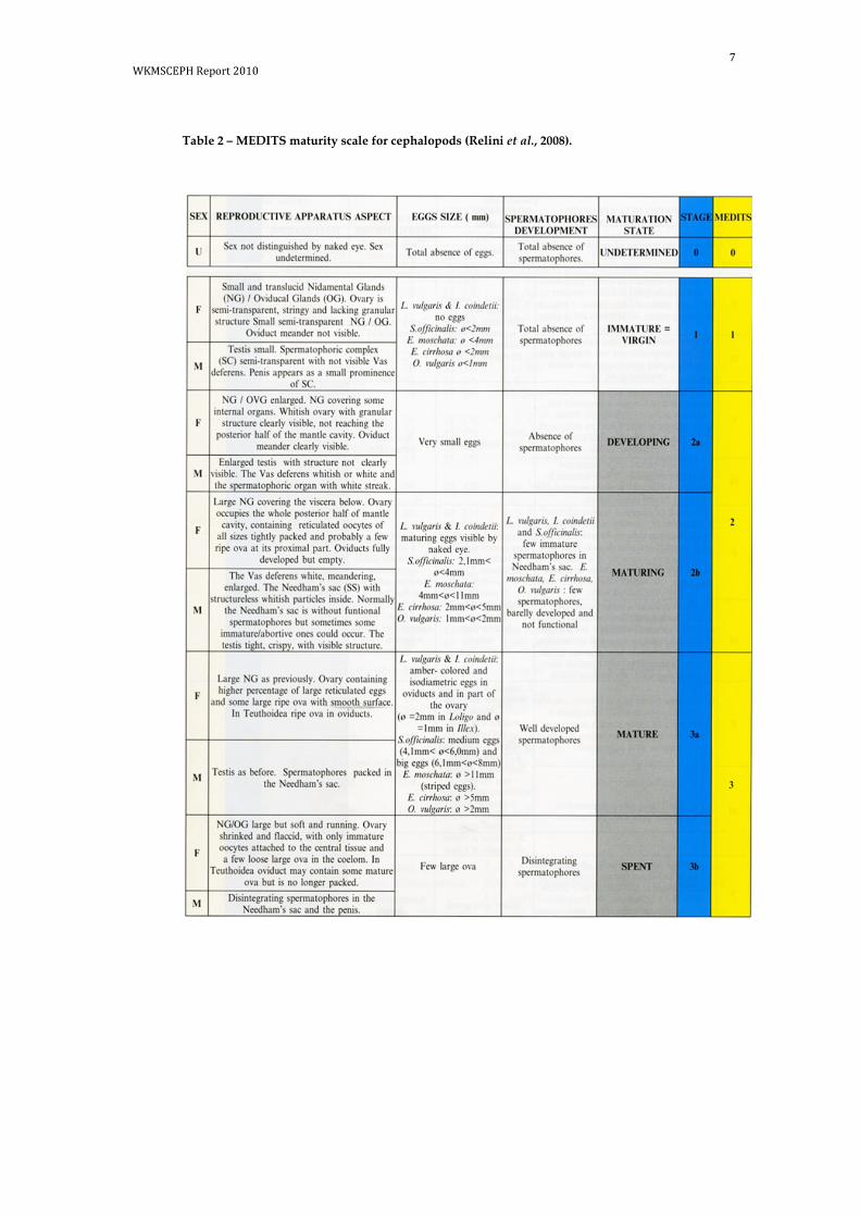
7 WKMSCEPH Report 2010
Table 2 – MEDITS maturity scale for cephalopods (Relini et al., 2008).

8 | WKMSCEPH Report 2010
2.1 Octopoda Session (Octopus vulgaris, Eledone cirrhosa, Eledone mo-schata)
Danila Cuccu (University of Cagliari – GSA11) showed some macroscopic photos of males and females of Eledone cirrhosa and E. moschata from the Sardinian waters, at different maturity stages using the MEDITS scale previously described. In particular, for the species Octopus vulgaris, she made an oral presentation in which a six stage (immature, developing, maturing, mature, spawning and spent) macroscopic scale was used, validated by microscopic and gravimetric analysis performed during a specific study on that species in GSA11 (see WD1 in Annex 5). Danila Cuccu also highlighted the need to split the current MEDITS scale in three different scales, ac-cording to the three Orders of Cephalopoda (Octopoda, Teuthida and Sepiida). In particular for the Octopoda scale, on the basis of the results obtained for O. vulgaris, she proposed some amendments to the current MEDITS scale: a clearer description of the reproductive system in both sexes in the different stages and the insertion of the characteristics of the female oviducal glands to better describe the stages “mature” and “maturing”. Another suggestion consisted in proposing the use of oocytes’ total length (measured along their major axis), instead of the diameter, due to the fact that the oocytes in Octopoda species are not isodiametric.
Silvia Lourenco (INRB-IPIMAR/FCUL – Portugal) showed the maturity scale in use at her laboratory for O. vulgaris, described by Goncalves (1993) (four stages for males and five stages for females). She reported some difficulties in discriminating between males in stage II and males in stage III, because the testis often had characteristics of a stage III but the spermatophoric sac was still half full (stage II). She also highlighted the difficult assessment of the stage IV in females and the discrepancies between the macroscopic analysis and the gravimetric indices at this stage. As the problem had led to subjective interpretations of the maturity stage, often with different results in calibration exercises between technicians, she suggested a histological approach. She also presented the results of a recent study regarding the different reproductive strategies adopted by the Octopus vulgaris populations of different coastal areas of Portugal (see WD2 in Annex 5).
Paola Belcari (University of Pisa – GSA9) made an oral presentation on E. cirrhosa (see WD3 in Annex 5) highlighting the importance of the gravimetric indices in the validation of maturity stages in this species. The study was based on individuals col-lected on a monthly basis and classified according to two maturity scales: a three-stage scale, according to Mangold-Wirz (1963) and Moriyasu (1988), and a five-stage scale according to MEDITS. It was shown that the monthly variations of the gona-dosomatic index and the Hayashi index perfectly reflected the evolution of the ma-turity process in this species. Moreover, each index showed a well-defined mean value when computed per maturity stage and sex, particularly in females. A discus-sion on the use of gravimetric indices in octopods arose, and their adoption as a use-ful tool for validating maturity stages was confirmed.
Luis Silva Caparro (IEO – C.O. Cádiz) presented the descriptions of different matur-ity scales used for cephalopod species sampled in different areas of the Mediterra-nean and the Atlantic Ocean (see WD4 in Annex 5). As regards O. vulgaris, he noticed the formation of a denticulate apical region in the oviducal glands of developing fe-males, before the appearance of the brown ring. He proposed to introduce this aspect as an additional criterion for a better identification of females belonging to this stage.

9 WKMSCEPH Report 2010
Alejandro Sancho Rafel (IEO – C.O. Canarias) showed the maturity scale at present in use in his laboratory for O. vulgaris (Annex 6) when assessing maturity stages through the macroscopic examination of the gonads. The description of each stage was based on a general scale defined by Dia (1988), except for the stage IV described by Guerra (1975) and Perales-Raya (2001). He underlined that the appearance of the thin brown horizontal band at the base of the oviducal gland was instrumental in de-fining the maturity stage in females. He presented some macroscopic photographs of the gonads at different maturity stages and the variations of the gonadosomatic index (see WD8 in Annex 5).
Daniela Giordano (IAMC – CNR – GSA 10b) showed some macroscopic photo-graphs of Octopoda species at different maturity stages assessed according to the cur-rent MEDITS scale.
Roberta Mifsud (MRRA – GSA 15) reported that her team in Malta is using the MEDITS scale. She showed some photographs of E. moschata at different maturity stages.
2.1.1 Calibration exercise
The calibration exercise was carried out on 32 specimens of E. cirrhosa by five opera-tors: three followed the Macroscopic MEDITS maturity scale (Relini et al., 2008), while the other two followed the Spanish ICES maturity scale for O. vulgaris (according to Guerra, 1975) or the IPIMAR maturity scale for O. vulgaris (according to Gonçalves, 1993).
The results of the calibration exercise are shown in Table 3.
The maturity staging as determined by the three operators using the MEDITS scale showed a satisfactory degree of concordance: for the majority of the specimens, the maturity stages assigned by the different operators closely agreed.
Most of the differences concerned the immature/developing specimens, which in some cases were classified as belonging to the stage 1, while in others to the stage 2a.
It has to be underlined that most of the specimens showed gonads in transition from the stage 1 to the stage 2a, so the little difference detected in the maturity interpreta-tion by different operators could be attributed to these aspects.
When the maturity stages obtained according to the ICES or IPIMAR scale were translated into those of the MEDITS scale, a substantially good agreement was achieved with the stages previously assigned. Also in this case, most of the discrep-ancies were detected in the maturity stages assigned to the immature or developing specimens.

10 | WKMSCEPH Report 2010
Table 3 – Results of the calibration exercise on maturity staging of Octopoda (E. cirrhosa).

11 WKMSCEPH Report 2010
2.1.2 Proposal for a common macroscopic maturity scale for Octopoda
After a plenary discussion based on the working documents presented, on the macro-scopic and microscopic descriptions of the gonads and on the calibration exercise, all the participants agreed to split the MEDITS maturity scale currently in use into three scales, one for each taxonomic group under consideration. The decision was mainly based on the fact that a maturity scale should accurately describe the stages precisely, avoiding any ambiguity, and this goal is difficult to achieve if the three Orders (Octo-poda, Teuthida and Sepiida) are kept together. However, it was also taken into con-sideration the recommendation of maintaining the new scales as similar as possible to the existing ones, in order to avoid the impact on maturity historical series. Moreover, all the participants agreed that the main aim of using the macroscopic assignments is mainly to estimate the maturity ogives and the timing of the spawning season.
Therefore, for Octopoda, a new maturity scale was proposed for the species under consideration (O. vulgaris, E. cirrhosa and E. moschata). The scale maintains the same number of stages of the MEDITS scale currently in use in the Mediterranean in the DCF, but considers males and females separately, thus allowing a better description of the characteristics of each stage at a macroscopic level (see Tables 4 and 5). A few ex-amples:
a ) Using the length taken along the major axis of the oocyte as the oocyte to-tal length (OTL). OTL size ranges were proposed according to the different species and maturity stages. It was suggested to choose the OTL which re-fers to the majority of the oocytes inside the ovary.
b ) Introducing the description of oviducal glands’ features from stage 2a. c ) Considering spawning individuals (males and females) belonging to stage
3a, even though the spawning has already begun.

12 | WKMSCEPH Report 2010
Table 4 – Proposed macroscopic maturity scale for females of O. vulgaris, E. cirrhosa and E. mo-schata.
PROPOSED STAGE
MATURATION STAGE
REPRODUCTIVE APPARATUS ASPECT OOCYTES SIZE (mm) OTL*
0 UNDETERMINED Sex not distinguished with the naked eye. -
1 IMMATURE = VIRGIN
Ovary semi-transparent, stringy and lacking a granular structure. Oviducal Glands (OG) small and translucent. Oviduct meander not visible.
Oocytes not visible to the naked eye
2a DEVELOPING
Whitish-creamy ovary with granular structure clearly visible, not reaching the posterior half of the mantle cavity. Developing OG: in Octopus vulgaris, a white thin denticulate apical region may appear. Oviduct meander clearly visible.
E. moschata: OTL <4 mm E. cirrhosa: OTL <2 mm Octopus vulgaris: OTL <1 mm
2b MATURING
Creamish-yellowish ovary occupies the whole posterior half of mantle cavity, containing reticulated oocytes of different sizes. Creamy/amber in color OG: in Octopus vulgaris, the denticulate apical region is followed by a light brown ring. Oviducts fully developed but empty.
E. moschata: 4 mm < OTL < 11 mm E. cirrhosa: 2 mm < OTL < 5 mm Octopus vulgaris: 1 mm < OTL < 2 mm
3a MATURE / SPAWNING
Light yellow ovary containing a high percentage of large reticulated oocytes. Well-developed OG. In Octopus vulgaris the denticulate apical region is larger and easily identifiable, the ring is enlarged and brown. Oviducts as before, they can contain oocytes.
E. moschata: OTL > 11 mm E. cirrhosa: OTL > 5mm Octopus vulgaris: OTL >2 mm
3b SPENT Shrunken, flaccid ovary, with only immature oocytes attached to the central tissue and a few loose large oocytes in the coelom.
Few large oocytes
*OTL: Oocytes Total Length, taken along its major axis. Sizes refer to Mediterranean specimens.
OTL refers to the majority of the oocytes inside the ovary.

13 WKMSCEPH Report 2010
Table 5 – Proposed macroscopic maturity scale for males of Octopus vulgaris, Eledone cirrhosa, Eledone moschata.
PROPOSED STAGE
MATURATION STATE
REPRODUCTIVE APPARATUS ASPECT
SPERMATOPHORES DEVELOPMENT AND OCCURRENCE IN THE NEEDHAM’SAC
0 UNDETERMINED Sex not distinguished with the naked eye. -
1 IMMATURE = VIRGIN
Testis small, thin and translucent. Spermatophoric Complex (SC) with vas deferens not visible. Absence of
spermatophores 2a DEVELOPING Developing and whitish/ivory testis. SC transparent with
visible vas deferens; a white streak may appear.
2b MATURING
Enlarged and spherical testis. The vas deferens is white, meandering, enlarged. Needham’s Sac (SS) may contain few spermatophores partially developed (visible as whitish particles) and/or few fully developed spermatophores.
Few spermatophores partially or fully developed
3a MATURE / SPAWNING
Testis white to grayish/brownish, with large and white vas deferens. Large Needham’ Sac full of packed spermatophores.
Plenty of well developed spermatophores
3b SPENT Testis flaccid. SS empty or with few spermatophores. None or few spermatophores

14 | WKMSCEPH Report 2010
2.2 Teuthida Session (Loligo vulgaris, Loligo forbesii, Illex coindetii, To-daropsis eblanae)
Miguel Cabanellas Reboredo (IMEDEA-CSIC-UIB) presented the maturity scale applied to Loligo vulgaris from the NW Mediterranean. Basically, the five-stage mac-roscopic maturity scale described in Boyle & Ngoile (1993) is being used in his labora-tory (Annex 6). Histological analyses of the gonads were carried out in order to validate the maturation stages (see WD5 in Annex 5).
Paola Belcari made an oral presentation on behalf of Eugenia Lefkaditou (HCMR-IMBC – GSA20&22) on the reproductive biology of L. vulgaris and Illex coindetii from the Thracian Sea (NE Mediterranean). She described eight macroscopic stages vali-dated through histological analyses (see WD6 in Annex 5 and Annex 6).
Danila Cuccu (University of Cagliari – GSA11) showed some macroscopic photos of males and females of I. coindetii, L. vulgaris, Loligo forbesii and Todaropsis eblanae from the Sardinian waters, at different maturity stages, attributed using the MEDITS scale. She proposed some amendments to be applied to the current MEDITS scale, regard-ing a better description of the reproductive apparatus in both sexes at different stages. Taking into account that Teuthida are intermittent spawners, she also pro-posed to merge fully mature and spawning individuals (stage: mature/ spawning) in the same maturity stage, separating them from the specimens at the end of the matur-ity process (spent).
Daniela Giordano (IAMC – CNR – GSA 10b) showed some macroscopic pictures of the reproductive apparatus of different species belonging to this order. She high-lighted the difficulty, in some cases, to identify the spawning individuals when the spawning process has already begun.
Alejandro Sancho Rafel (IEO – C.O. Canarias) reported that, in his laboratory, for L. vulgaris the maturity stage was assessed through the macroscopic examination of the gonads according to the squid universal scale of Lipinsky (1979). He highlighted the difficulty to discern the nidamental glands in small size females. He also underlined that, as a consequence of the two peaks of reproduction occurring for this species in the CECAF area, it is difficult to single out a cohort in the catches and, consequently, adults of both sexes with small sizes and juveniles with large sizes are often present contemporarily. He showed some macroscopic pictures of the gonads of this species in different maturation stages (see WD8 in Annex 5).
2.2.1 Calibration exercise
The calibration exercise was carried out on 41 specimens of I. coindetii and 10 of L. vulgaris by six operators: three of them followed the MEDITS macroscopic maturity scale (Relini et al., 2008), two of them used the five-stage macroscopic maturity scale described in Boyle & Ngoile (1993) in use at IPIMAR and in several Spanish Mediter-ranean institutes and the last operator used the scale proposed by Lipinsky (1979), used by CECAF & ICES (Annex 6).
The results of the calibration exercise are shown in Tab. 6. It is possible to detect a close agreement in the maturity staging coming from the three operators who used the MEDITS maturity scale. The only differences are related to consecutive stages (e.g. 2a and 2b), probably due to the subjectivity of assessing very close maturity stages. When the maturity stages obtained according to the CECAF-ICES or IPIMAR scales were translated into those of the MEDITS scale, a good agreement was achieved.
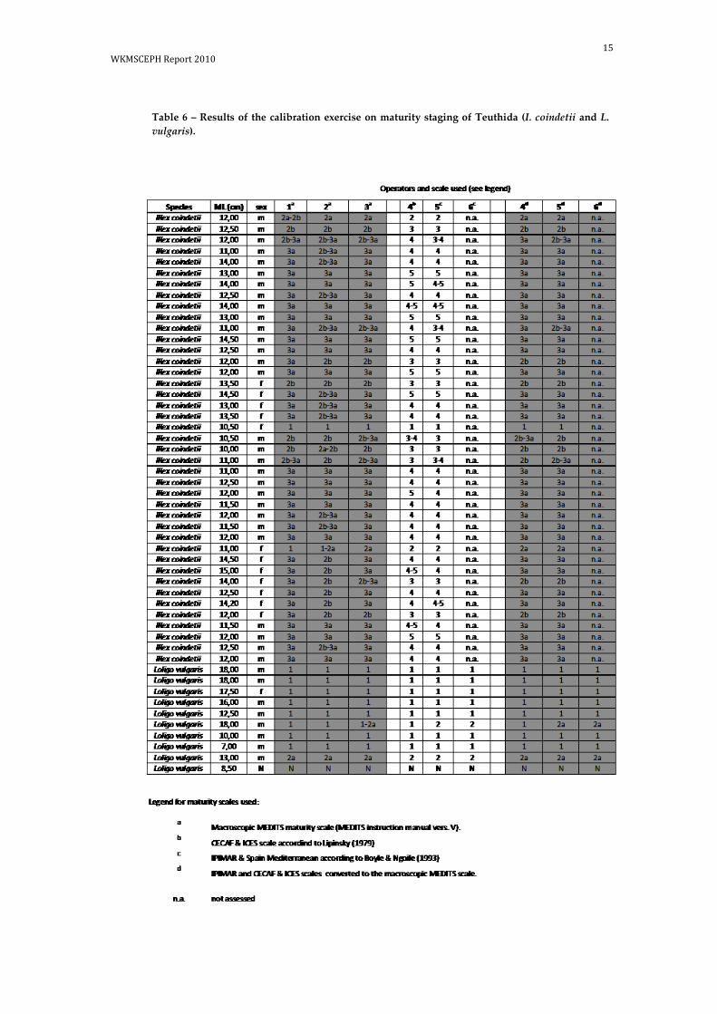
15 WKMSCEPH Report 2010
Table 6 – Results of the calibration exercise on maturity staging of Teuthida (I. coindetii and L. vulgaris).

16 | WKMSCEPH Report 2010
2.2.2 Proposal for a common macroscopic maturity scale for Teuthida
The scale maintains the same number of stages of the MEDITS scale currently in use in the Mediterranean in the DCF, but considers males and females separately, thus allowing a better description of the characteristics of each stage at a macroscopic level (see Tab. 7 and 8). Spawning specimens were considered belonging to the stage 3a in both sexes, even when the spawning process has already begun.
Table 7 – Proposed macroscopic maturity scale for females of Loligo vulgaris, Loligo forbesii, Illex coindetii and Todaropsis eblanae.
PROPOSED STAGE
MATURATION STATE
REPRODUCTIVE APPARATUS ASPECT OOCYTE SIZE (mm) *Ø
0 UNDETERMINED Sex not distinguished with the naked eye. -
1 IMMATURE = VIRGIN
Ovary is semi-transparent, stringy and lacking granular structure. Small and translucent Nidamental Glands (NG) and Oviducal Glands (OG). Oviduct meander not visible.
Oocytes not visible to the naked eye
2a DEVELOPING
Whitish ovary with visible granular structure, not reaching the posterior half of the mantle cavity. Developing NG/OG. NG enlarged, covering some internal organs. Oviduct meander clearly visible.
Very small oocytes visible to the naked eye.
2b MATURING
Ovary occupies the whole posterior half of the mantle cavity, containing tightly packed oocytes. Large NG covering the viscera below. Oviducts fully developed but empty.
Very small isodiametric oocytes visible to the naked eye.
3a MATURE / SPAWNING Ovary containing a higher percentage of large oocytes. Enlarged and turgid NG/OG. Plenty of oocytes in the oviducts.
Amber-coloured oocytes in the oviducts Ø >= 2 mm in Loligo spp.; Ø >= 1 mm in I. coindetii
3b SPENT Flaccid ovary with strikingly loose disorderly aspect. Few oocytes, which may be attached to the central tissue. Flaccid NG/OG.
Few oocytes which may be attached to the central tissue
*Ø refers to the majority of the oocytes inside the ovary. Sizes refer to Mediterranean specimens.

17 WKMSCEPH Report 2010
Table 8 – Proposed macroscopic maturity scale for males of Loligo vulgaris, Loligo forbesii, Illex coindetii and Todaropsis eblanae.
PROPOSED STAGE
MATURATION STATE REPRODUCTIVE APPARATUS ASPECT
SPERMATOPHORES DEVELOPMENT
AND OCCURRENCE IN THE NEEDHAM’
SAC
0 UNDETERMINED Sex not distinguished with the naked eye. -
1 IMMATURE = VIRGIN Small and translucent testis. Thin and semitransparent Spermatophoric
Complex (SC) with not visible vas deferens. Absence of
spermatophores 2a DEVELOPING
Whitish-grey testis, filling almost ½ of the posterior part of the mantle cavity. SC whitish with visible vas deferens. Penis appears as a small
prominence of SC.
2b MATURING
Whitish-grey testis, filling the posterior part of mantle cavity, with visi-ble structure. The vas deferens is white, meandering and enlarged. The Needham’s Sac (SS) may contain few partially developed spermatopho-res (visible as whitish particles) and/or few fully developed spermato-
phores
Few spermatophores partially or fully devel-
oped
3a MATURE / SPAWNING Yellowish well-developed testis with large and white vas deferens.
Spermatophores packed in the Needham’s Sac; spermatophores may occur in the penis.
Plenty of well devel-oped spermatophores
3b SPENT Testis flaccid. SS empty or with few spermatophores. None or few spermato-phores

18 | WKMSCEPH Report 2010
2.3 Sepiida session (Sepia officinalis)
Daniela Giordano (IAMC – CNR – GSA 10b) made an oral presentation on maturity and fecundity of Sepia officinalis in the Southern Tyrrhenian Sea, Central Mediterra-nean (see WD7 in Annex 5). As regards some critical points of the current MEDITS scale, she suggested to better define the stage 2b in males and to clarify at which stage small oocytes are visible in females.
Alejandro Sancho Rafel (IEO – C.O. Canarias) explained that, in his laboratory, the maturity of S. officinalis and S.hierredda was assessed through the macroscopic exami-nation of the gonads based on Bakhayokho (1980) and Perales-Raya (2001) (see WD8 in Annex 5 and Annex 6).
Macrocopic photos of some maturity stages of Sepia officinalis were also presented by Danila Cuccu (University of Cagliari – GSA11) who underlined that the presence of smooth oocytes in the females’ oviducal glands could be the key to understand when the individuals are fully mature, even though this feature is not detectable when spawning has already begun.
2.3.1 Calibration exercise
The calibration exercise was carried out on 37 specimens of S. officinalis by six opera-tors: three of them followed the MEDITS macroscopic maturity scale (Relini et al., 2008), two of them used the scale proposed for this species by Bakhayoko (1980) and Perales-Raja (2001), used by CECAF & ICES and the last operator used the IPIMAR simplified scale. The results of the calibration exercise are shown in Tab 9. For this species, the closest agreement was achieved by the three operators who used the MEDITS maturity scale. Similarly, when the maturity stages obtained according the CECAF-ICES or IPIMAR scales were translated into those of the MEDITS scale, a good agreement was achieved.

19 WKMSCEPH Report 2010
Table 9 – Results of the calibration exercise on maturity staging of Sepiida (S. officinalis).

20 | WKMSCEPH Report 2010
2.3.2 Proposal for a common macroscopic scale for Sepiida
The scale maintains the same number of stages of the MEDITS scale currently in use in the Mediterranean in the DCF, but considers males and females separately, thus allowing a better description of the characteristics of each stage at a macroscopic level (see Tab. 10 and 11). Spawning specimens were considered belonging to the stage 3a in both sexes, even when the spawning process has already begun.
Table 10 - Proposed macroscopic maturity scale for females of Sepia officinalis.
PROPOSED STAGE
MATURATION STATE
REPRODUCTIVE APPARATUS ASPECT OOCYTE SIZE (mm) *Ø
0 UNDETERMINED Sex not distinguished with the naked eye. -
1 IMMATURE = VIRGIN Translucent ovary, small, with granular structure. Small and translucent Nidamental Glands (NG) and Oviducal Glands (OG). Oviduct meander not visible. Very small oocytes visible to the
naked eye. Oocytes Ø < 2 mm
2a DEVELOPING
Creamy ovary, enlarged but not reaching the posterior half of the mantle cavity. Developing and white NG/OG. NG covering some internal organs Oviduct meander clearly visible.
2b MATURING
Pale-yellow ovary, occupying the whole posterior half of the mantle cavity and containing only reticulated oocytes. Large NG and OG; NG covering the viscera below. Oviduct fully developed but empty.
Oocytes 2 mm < Ø < 4 mm.
3a MATURE / SPAWNING Amber-coloured and gelatinous ovary, containing reticulated and smooth oocytes. Enlarged and turgid NG/OG. Oocytes may occur in the oviduct.
Oocytes Ø > 4 mm.
3b SPENT Flaccid ovary with strikingly loose disorderly aspect. Few oocytes, which may be attached to the central tissue. Flaccid NG/ OG.
Few oocytes which may be attached to the central tissue.
*Ø refers to the majority of the oocytes inside the ovary. Sizes refer to Mediterranean specimens.

21 WKMSCEPH Report 2010
Table 11 - Proposed macroscopic maturity scale for males of Sepia officinalis.
PROPOSED STAGE
MATURATION STATE REPRODUCTIVE APPARATUS ASPECT
SPERMATOPHORES DEVELOPMENT AND OCCURRENCE IN THE
NEEDHAM’ SAC
0 UNDETERMINED Sex not distinguished with the naked eye. -
1 IMMATURE = VIRGIN Small, white and clearly visible testis. Semitransparent Spermato-
phoric Complex (SC) with not visible vas deferens. Absence of
spermatophores 2a DEVELOPING
Testis increased in volume but not reaching the posterior half of the mantle cavity. SC white with visible vas deferens. Penis appears as
a small prominence of SC.
2b MATURING
Testis filling the posterior half of the mantle cavity. Vas deferens white, meandering and enlarged. The Needham’s Sac (SS) may
contain few spermatophores partially developed (visible as whitish particles) and/or few fully developed spermatophores.
Few spermatophores par-tially or fully developed
3a MATURE / SPAWNING Well-developed testis with large and white vas deferens. Sper-
matophores packed in the Needham’s Sac and sometimes present in the penis.
Plenty of well developed spermatophores
3b SPENT Testis flaccid. SS empty or with few spermatophores. None or few spermatopho-
res

22 | WKMSCEPH Report 2010
2.4 Histological analysis in cephalopods
Cristina Porcu (University of Cagliari – GSA11) made an oral presentation on the use of histological analyses as a valid tool in the study of sexual maturity of cephalo-pods (see WD9 in Annex 5). In particular, she showed the methodology in use in her laboratory, focusing the attention on the most important phases of the analysis of cephalopod species. She also presented a study carried out on O. vulgaris, describing several microscopic stages of oogenesis and spermatogenesis. She also highlighted the importance of the histological observation of other reproductive organs (in addi-tion to the ovary, e.g. the oviducal glands), because it may reflect crucial changes dur-ing the sexual maturation of the common octopus.
2.4.1 Histological Techniques
Key aspects concerning the histological methodologies applied by the workshop par-ticipants are hereunder presented.
GSA11 – Octopus vulgaris
1 ) Histological techniques require the utilization of fresh specimens; 2 ) Gonads were wholly extracted; 3 ) Gonads were immediately fixed in 5% formaldehyde 0.1 M phosphate
buffer (pH 7.4); 4 ) Small fragments of gonads and oviducal glands were cut in transverse sec-
tions; 5 ) Fragments were extensively washed in fresh water for one day and then
stored in 70% ethanol; 6 ) Tissues were dehydrated through an alcohol series and embedded in resin
(2-idrossiethylmetacrilate; glycol-methacrylate method; Technovit 7100); 7 ) Sections were cut at 3.5 microns with a rotary microtome; 8 ) Sections were stained with hematoxylin and eosin or with Masson’s
Trichrome to better identify details of the ovary histological structures (e.g. yolk droplets, chorion and follicle epithelium);
9 ) Sections were covered with a synthetic mounting media; 10 ) Histological slides were observed with an optical microscope and, for each
cell type, sizes were measured at the maximum diameter (along the long axis).
GSA20 & 22- L. vulgaris and I. coindetii
1 ) Small sections of ovaries had been fixed on board in formalin solution 10% and stored for at least two months;
2 ) Sections were dehydrated in alcohol solutions of density increasing from 30% to 100% and transferred to absolute xylol to improve tissue transpar-ency;
3 ) Sections were then embedded in paraffin; 4 ) Thin sections of 5-7 µm thickness were cut; 5 ) The thin sections, after their fitting on glass slides, were dewaxed and hy-
drated through xylol and through descending grades of alcohol in water; 6 ) Sections were stained with eosin and mounted on slides with balsam of
Canada, following the methodology described by Bullock (1989).

23 WKMSCEPH Report 2010
2.4.2 Protocol and terminology to classify the oogenic stages
GSA11 – Octopus vulgaris
Stage 1 – Oogonia (OO): oogonia are small round cells of 7.0-16.8 µm diameter, without visible cytoplasm. These cells are attached to the germinal epithelium.
Stage 2 – Early Primary Oocyte (EPO): at this stage oocytes are associated with one or several follicles. They have an oval shape, are smaller than the oocytes and are lo-cated on the connective tissue. At this stage oocytes range from 23.8-85.2 µm.
Stage 3 – Late Primary Oocyte (LPO): oocytes are surrounded by a layer of flat folli-cle cells. The oocyte diameters vary between 140.2 and 220.4 µm. The portion of the cell occupied by the cytoplasm is larger than the one occupied by the nucleus. It is possible to observe some lipid inclusions in the cytoplasm.
Stage 4 – Previtellogenic Oocyte (PVO): the follicle epithelium initiates the oocyte embedding by intensive multiplication of the follicle cells (two layers of follicular cells, the inner one consisting of cuboidal cells, the outer one of flat follicular cells), with a subsequent displacement of the nucleus at the polar zone of the cell. In a few oocytes the follicle cells form a syncytium (folds). These folds can penetrate deeply inside the cell and encroach the cytoplasm. Oocyte size increases (190.4-648.0 µm). There is an initiation of nucleoli degeneration and the first production of yolk glob-ules. The lipid inclusions multiply and become bigger.
Stage 5 – Vitellogenic Oocyte (VO): this stage is characterized by a very strong oo-cyte diameters increase. The follicular epithelium is active in vitellogenesis and in the formation of a chorion. The follicular folds are displaced towards the periphery of the oocytes by the formation of the yolk. Oocyte sizes range from 876.5 to 1396.3 µm. In some oocytes the nuclei in the polar zone are still visible.
Stage 6 – Advanced Vitellogenic Oocyte (AVO): these oocytes reach the maximum size (879-3980 µm). The cytoplasm is filled with yolk granules; the whole is sur-rounded by the chorion (well developed).
Stage 7 – Ripe Oocytes (RO): oocytes are issued by the preovulatory follicle. They are dissolved in the ovarian cavity and in the proximal part of the oviduct. The cyto-plasm is filled by yolk granules and the folds are completely reabsorbed, involved and protected by the chorion. At the end, they have a distal chorionic acellular long filament (peduncle). These oocytes are ready for ovulation. The oocytes (with pedun-cle) ranged in size between 1884 and 3998 µm.
Stage 8 – Post-Ovulatory Follicle (POF): after ovulation, the follicular epithelium generates the post-ovulatory follicle. This follicle shows an irregular contour and a star-shaped lumen, which contains fibrillar material and highly amorphous and ba-sophils bodies. On the wall of the follicle, collagen fibres are present, as well as blood vessels and clusters of follicular cells whose nuclei are pyknotic or degenerating.
Stage 9 – Atretic Oocyte (AO): the atretic oocytes show disorganized follicular epi-thelium. The fibrillar connective tissue was replaced by collagen fibres. The chorion is disorganized and disintegrates into fragments. The proportion of all these stages and the predominance of each type allow us to identify several macrostages.

24 | WKMSCEPH Report 2010
2.4.3 Histological scale proposed
GSA 11 – Octopus vulgaris
Phases of the ovary maturation:
Stage 1 (immature=virgin): the germinal epithelium shows oogonia (7.0-16.8 µm) and early primary oocytes (23.8-85.2 µm).
Stage 2a (developing): germline is characterized by the formation of late primary oo-cytes (140.2-220.4 µm) and previtellogenic oocytes (190.4-648.0 µm) with a double layer of follicular cells. In this stage is also possible to observe oogonia and early pri-mary oocytes. Oviducal glands have two glandular compartments and the sper-mothecae may contain a few spermatozoa.
Stage 2b (maturing): abundant vitellogenic oocytes are observed (876.5-1396.3 µm). The follicular epithelium is active in vitellogenesis and in the formation of a chorion. In addition, it is possible to observe previtellogenic oocytes, primary oocytes and oo-gonia. In oviducal glands, two glandular regions are well separated by a basal lam-ina. Spermothecae may contain abundant sperm with the heads inserted within the mucosa.
Stage 3a (mature/spawning): abundant presence of Advanced Vitellogenic Oocytes (AVO, 879-3980 µm) and Ripe Oocytes (RO, 1884-3998 µm, free in the ovarian lumen and in the proximal part of the oviduct). All types of oocytes are observed, including not previously identified primary oocytes or oogonia. Histological description of ovi-ducal glands as before.
Stage 3b (spent): ovarian tissue appears empty and only Post-Ovulatory Follicles (POF) and Atretic Oocytes (AO) are observed. In oviducal glands, the glandular component appears less compact with wide lumens among tubular components and spermothecae almost empty, but spermatozoa still visible.
Phases of the testis maturation:
Stage 1 (immature=virgin): seminiferous tubules are well defined but small and empty. Only Spermatogonia are present in low numbers.
Stage 2a (developing): in seminiferous tubules, spermatogonia, numerous primary and secondary spermatocytes and spermatids and few spermatozoa (not in all tu-bules) are observed.
Stage 2b (maturing): seminiferous tubules are clearly defined along the testis. Sper-matogonia, primary and secondary spermatocytes and spermatids are present. Sper-matozoa are more evident and visible in all seminiferous tubules.
Stage 3a (mature/spawning): seminiferous tubules are large and well defined. There are not empty spaces between cells. All types of cells are present with abundant spermatozoa in the central lumen.
Stage 3b (spent): only few primary and secondary spermatocytes, spermatids and spermatozoa are present, dispersed throughout the seminiferous tubules.

25 WKMSCEPH Report 2010
2.5 Conversion tables
The new proposed scales were compared with the existing maturity scales derived from the participants’ presentations and from literature. Discrepancies and similari-ties were highlighted and a plenary discussion took place to formulate conversion rules and make it possible the correspondence between the existing scales and the agreed ones, reported below. (Tab. 12-13-14).

26 | WKMSCEPH REPORT 2010
Table 12 – Octopoda maturity scale proposed by WKMSCEPH and maturity scales used by different Institutes.
Maturity scale
Scale proposed by WKMSCEPH
Maturation state
MEDITS Current
Scale
MEDITS Old
Scale FRIS
Spain ICES according to Guerra, 1975
IPIMAR according to Goncalves,
1993
IPIMAR according to Goncalves,
1993
Spain CECAF according to
Dia, 1988; Guerra, 1975 and Perales-
Raya, 2001
Spain ICES according
to Moriyasu,
1988
Spain ICES according to Ezzeddine-Najai, 1997
Species Octopoda Cephalopoda Octopus vulgaris Eledone cirrhosa
Eledone moschata
Sex F & M F & M F & M F & M F & M F M F & M F & M F & M
Stages
0 Undetermined 0 0 1*
2**
1 Immature = Virgin 1 1 3 1 1 1
1 1 1
2a Developing 2a 2 4 2 2
2
2b Maturing 2b 2 3 2 2
3a Mature / Spawning 3a 3 5 3 3
3 4 3 3 4
3b Spent 3b 6 4 5 4 5 4 4
* ignored; ** undetermined.

27 WKMSCEPH Report 2010
Table 13 – Teuthida maturity scale proposed by WKMSCEPH and maturity scales used by different Institutes.
Maturity scale Scale proposed by WKMSCEPH
Maturation state
MEDITS Current Scale
MEDITS Old Scale
FRIS Spain CECAF &
ICES according to Lipinski, 1979
IPIMAR & Spain Mediterranean ac-cording to Boyle &
Ngoile, 1993
Species Teuthoidea Cephalopoda Loligo vulgaris
Sex F & M F & M F & M F & M F & M F & M F & M
Stages
0 Undetermined 0 0 1*
2**
1 Immature = Virgin 1 1 3 1 1
2a Developing 2a 2 4
2 2
2b Maturing 2b 3 3
3a Mature / Spawning
3a 3
5 4 4 5 5
3b Spent 3b 6 6
* ignored; ** undetermined.

28 | WKMSCEPH REPORT 2010
Table 14 – Sepiida maturity scale proposed by WKMSCEPH and maturity scales used by different Institutes.
Maturity scale Scale proposed by WKMSCEPH
Maturation state
MEDITS Current Scale
MEDITS Old Scale
FRIS
Spain CECAF & ICES ac-cording to
Bakhayako, 1980 and Perales-Raja, 2001
IPIMAR simplified
scale
Species Sepioidea Cephalopoda Sepioidea Sepia spp.
F M
Stages
0 Undetermined 0 0 1*
2**
1 Immature = Virgin 1 1 3 1
1 1
2a Developing 2a 2 4 2 2
2b Maturing 2b 2
3a Mature / Spawning 3a 3
5 3 + 4 3 3
3b Spent 3b 6 5 4
* ignored; ** undetermined.

29 WKMSCEPH Report 2010
3 Conclusions
The identification and macroscopic classification of maturity stages can play a key-role in the assessment of fishery resources, therefore the urgent need of improving the quality of these estimates by means of reliable information on the maturity pa-rameters has been universally recognized. The workshop on maturity staging had the aim to agree on the adoption of common scales based on the standardization of ma-turity defining criteria; as a general conclusion, it is possible to affirm that this goal and all the expectations of the TOR’s were fulfilled.
Through the analysis of the MEDITS scales and of those in use in the different labora-tories, the direct observation of the samples’ gonads and of the macroscopic and mi-croscopic pictures, a thorough discussion arose, and brought to the definition of the new scales.
It was taken into account the fact that a maturity scale should accurately describe the stages precisely as well as the recommendation to maintain the new scales as similar as possible to the existing ones, in order to avoid the impact on maturity historical se-ries, Therefore, the participants agreed to split the MEDITS maturity scale currently in use into three scales, one for each taxonomic group under consideration. Moreover, for the sake of clarity and handiness, in each scale males and females are presented separately.
Number of stages and their classifications are maintained as in the MEDITS scale cur-rently in use (1, 2a, 2b, 3a, 3b), but a more extensive and thorough description has been added, in order to avoid any ambiguity.
The calibration exercise was very useful for identifying sources of discrepancies and as a test for the agreed scales. The collection of pictures at macroscopic and micro-scopic levels was instrumental in solving interpretation’s problems and could be used afterwards in every laboratory for ease of reference.
Histology proved to be an essential key to support the macroscopic identification and the gonadosomatic and Hayashi indices were recognized to be important tools to clarify doubts.
The conversion from the existing scales to the proposed WKMSCEPH maturity scales provides a common tool for exchanging data and scientific information.

30 | WKMSCEPH REPORT 2010
4 References
Arkhipkin A.I., 1992. Reproductive System Structure, Development and Function in Cephalo-pods with a New General Scale for Maturity Stages. Journal of Northwest Atlantic Fishery Science, 12: 63-74.
Arnold J.M. & Williams-Arnold L.D., 1977. Cephalopoda. Decapoda. In: A.C. Giese & J.S. Pearce (eds.), Reproduction of Marine Invertebrates. Academic Press Inc., New York, vol. IV, 243-290.
Avila-Poveda O.H., Colin-Flores R.F. & Rosas C., 2009. Gonad development during the Early Life of Octopus maya (Mollusca: Cephalopoda). Biological Bulletin, 216: 94-102.
Bakhayokho M., 1980. Peche et Biologie des Cephalopodes exploites sur les cotes du SENEGAL (12º 20’ N- 16º03’ N). Thèse de Docteur. Universite de Bretagne Occidentale, 120 pp.
Bakhayokho M., 1983. Biology of the cuttlefish Sepia officinalis hierredda. In: J.F. Caddy (ed.), Advanced in assessment of world cephalopods resources. FAO Fisheries Technical Papers, 231: 204-264.
Bhattacharya C.G., 2002. A simple method of resolution of a distribution into Gaussian compo-nents. Biometrics, 23(1): 115-135.
Boavida-Portugal J., Moreno A., Gordo L. & Pereira J., 2010. Environmentally adjusted repro-ductive strategies in females of the commercially exploited common squid Loligo vulgaris. Fisheries Research, 106: 193-198.
Boyle P.R. & Knobloch D., 1983. The female reproductive cycle of the octopus, Eledone cirrhosa. Journal of the Marine Biological Association of the United Kingdom, 63: 71-83.
Boyle P.R. & Ngoile M.A.K., 1993. Assessment of Maturity state and seasonality of reproduc-tion in Loligo forbesi (Cephalopoda: Loliginidae) from Scottish waters. In: T. Okutani, R. K. O’Dor & T. Kubodera (eds.), Recent advances in Fisheries Biology. Tokai University Press, Tokyo, 37-48.
Bullock A. M., 1989. Laboratory Methods. In: R. J. Roberts (ed.), Fish Pathology. Baillière Tindall, 374-405.
Coelho M.L. & O’Dor R.K., 1993. Maturation, spawning patterns, and mean size at maturity in the short-finned squid Illex illecebrosus. In: T. Okutani, R. K. O. Dor & T. Kubodera (eds.), Recent Advances in Cephalopod Fisheries Biology. Tokai University Press, Tokyo, 81-92.
Di Cosmo A., Di Cristo C. & Paolucci M., 2001. Sex steroid hormone fluctuations and morpho-logical changes of the reproductive system of the female of Octopus vulgaris throughout the annual cycle. Journal of Experimental Zoology, 289: 33-47.
Dia M.A., 1988. Biologie et exploitation du poulpe Octopus vulgaris (Cuvier, 1797) des côtes Mauritaniennes. Ph.D. thesis, University of West Brittany, 164 pp.
Ezzedine-Najai S., 1985. Fecundity of the cuttlefish, Sepia officinalis L. (Mollusca: Cephalopoda) from the gulf of Tunis. Vie et Milieu, 35(3–4): 283-284.
Gabr H.R., Hanlon R.T., Hanafy M.H. & El-Etreby S.G., 1998. Maturation, fecundity and sea-sonality of reproduction of two commercially valuable cuttlefish, Sepia pharaonis and S. dollfusi, in the Suez Canal. Fisheries Research, 36(2): 99-115.
Gonçalves J.M., 1993. Octopus vulgaris Cuvier, 1797 (polvo-comum): Sinopse da biologia e exploração. Trabalho de síntese a apresentar para as Provas de Aptidão Pedagógica e capacidade científica, para acesso à categoria de assistente da Carreira Docente Universitário. Universidade dos Açores, Departamento de Oceanografia e Pescas, 470 pp.
Guerra A., 1975. Determinacíón de las diferentes fases del desarrollo sexual de Octopus vulgaris Lamarck, mediante un índice de madurez. Investigación Pesquera, 39: 397-416.

31 WKMSCEPH Report 2010
Hayashi Y., 1970. Studies on the maturity conditions of the common squid. I. A method of ex-pressing maturity conditions by numeric values. Bullettin of the Japanese Society of Scientific Fisheries, 36: 995-999.
Juanico M., 1983. Squid maturity scales for population analysis. In: G. F. Caddy (ed.), Advances in assessment of world cephalopod resources. FAO Fisheries Technical Paper, 231: 341-378.
Khallahi O.M.F. & Inejih C.A., 2002. Proposition d'une échelle macroscopique de maturité sexuelle des femelles de poulpe Octopus vulgaris (Cuvier, 1797). Bulletin du Centre National de Recherches Oceanographiques et des Pechês, 23: 51-57.
Khallahi O.M.F., 2001. Etude de la gamétogenèse chez le poulpe Octopus vulgaris (Cuvier, 1797). Bulletin du Centre National de Recherches Oceanographiques et des Pechês, 28: 45-53.
Knipe J.H. & R.D. Beernan., 1978. Histological observations on oogenesis in Loligo opalescens. California Department of Fish and Game’s Fish Bulletin, 169: 23-33.
Laptikhovsky V. & Arkhipkin A., 2001. Oogenesis and gonad development in the cold water loliginid squid Loligo gahi (Cephalopoda: Myopsida) on the Falklandshelf. Journal of Mol-luscan Studies, 67: 475-482.
Lefkaditou, E., 2006. Taxonomy and biology of Cephalopods in the North Aegean Sea. PhD thesis, University of Patras, Patras, Greece, 298 pp + Annexes (in Hellenic, English ab-stract).
Lipinski M., 1979. Universal maturity scale for the commercially-important squids (Cephalo-poda: Teuthoidea).The results of maturity classification of the Illex illecebrosus Leseur, (1821) Population for years 1973-1977. International Commission for the Northwest Atlantic Fisheries, ICNAF Res. Doc. 79/II/38, 40 pp.
Lopes S.S., Coelho M.L., Andrade J.P., 1997. Analysis of Oocyte Development and Potential Fe-cundity of the Squid Loligo vulgaris from the Waters of Southern Portugal. Journal of the Marine Biological Association of the United Kingdom, 77: 903-906.
Mangold Wirz, K., 1963. Biologie des Céphalopodes benthiques et nectoniques de la Mer Cata-lane. Vie et Milieu, 13 (suppl.): 285 pp.
Mangold K., 1989. Cephalopodes. In: P.P.Grassé (ed.), Traité de Zoologie. Anatomie, systématique, biologie. Masson, Paris, tome V(4), 804 pp.
Moreno A., Cunha M.M. & Pereira J.M.F., 1994. Population biology of veined squid (Loligo forbesi) and European squid (Loligo vulgaris) from the Portuguese coast. Fisheries Research, 21: 71-86.
Moreno A., Pereira J., Arvanitidis C., Robin J.P., Koutsoubas D., Perales-Raya C., Cunha M.M., Balguerias E. & Denis V., 2002. Biological variation of Loligo vulgaris (Cephalopoda: Lo-liginidae) in the eastern Atlantic and Mediterranean. Bulletin of Marine Science, 71(1): 515-534.
Moriyasu M., 1988. Analyse de la maturation sexuelle d’Eledone cirrosa (Cephalopoda: Octo-poda) du Golfe de Lion. Aquatic Living Resources, 1: 59-65.
Olivares A.P., Zamora M.C., Portilla P.R. & Zúñiga O.R., 2001. Estudio histológico de la ovogénesis y maduración ovárica en Octopus mimus (Cephalopoda: Octopodidae) de la II Región de Chile. Estudios Oceanologicos, 20:13-22.
Onsoy B. & Salman A., 2005. Reproductive biology of the common cuttlefish Sepia officinalis L. (Sepiida:Cephalopoda) in the Aegean Sea. Journal of Veterinary and Animal Sciences, 29: 613-619.
Perales-Raya, C. 2001. Determinación de la edad y estudio del crecimiento del choco (Sepia hierredda), el calamar (Loligo vulgaris) y el pulpo (Octopus vulgaris) de la costa noroccidental africana. Tesis Doctoral. Universidad de La Laguna, 192 pp.

32 | WKMSCEPH REPORT 2010
Pereira J.M.F., 1999. Control of the Portuguese artisanal octopus fishery. In: C.P. Nolan (ed.), Proceedings of the International Conference on Integrated Fisheries Monitoring. FAO, Rome, 369-378.
Relini G., Carpentieri P. & Murenu M. (eds), 2008. Medits Instruction Manual Version 5 rev. Biologia Marina Mediterranea, 15 (suppl. 2): 1-78.
Richard A., 1971. Contribution à l’ètude experimentale de la croissance et de la maturation sexuelle de Sepia officinalis L. (Mollusque Céphalopode). Ph.D. Thesis, University Lille, France, 264 pp.
Roper C.F.E. & Voss G.L., 1983. Guidelines for taxonomic descriptions of Cephalopod species. Memoirs of the National Museum Victoria, (44): 48-63.
Rodríguez-Rúa A., Pozuelo I., Prado M.A., Gómez M.J. & Bruzón M.A., 2005. The gametogenic cycle of Octopus vulgaris (Mollusca: Cephalopoda) as observed on the Atlantic coast of Andalusia (south of Spain). Marine Biology, 147: 927-933.
Sauer W.H.H. & Lipinski M.R., 1990. Histological validation of morphological stages of sexual maturity in chokker squid Loligo vulgaris reynaudii D'Orb (Cephalopoda: Loliginidae). Afri-can Journal of Marine Science, 9: 189-200.
Takahashi N. & Yahata T., 1973. Histological studies on the maturation of the ovary in the squid, Todarodes pacificus. Bulletin of the Faculty of Fisheries Hokkaido University, 24:63-68.
Wells M. J. & Wells J., 1977. Cephalopoda: Octopoda. In: A. Giese and J. S. Pearse (eds.), Repro-duction of marine invertebrates, Molluscs: Gastropods and Cephalopods. Academic press, Lon-don, vol. 4, 291-337.
Zamora M.C. & Olivares A.P., 2004. Histological and biochemical variations produced during the reproductive event of female Octopus mimus (Mollusca: Cephalopoda). International Journal of Morphology, 22(3): 207-216.
33 WKMSCEPH Report 2010
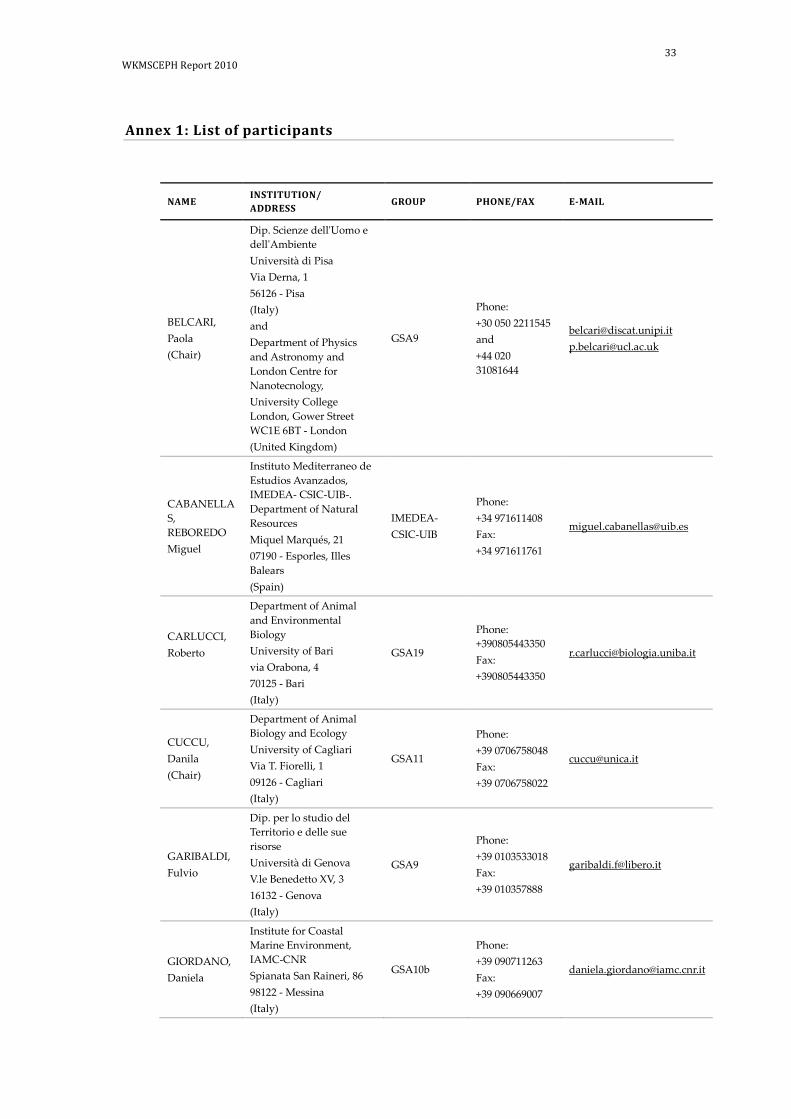
Annex 1: List of participants
NAME INSTITUTION/ ADDRESS
GROUP PHONE/FAX E-MAIL
BELCARI, Paola (Chair)
Dip. Scienze dell'Uomo e dell'Ambiente Università di Pisa Via Derna, 1 56126 - Pisa (Italy) and Department of Physics and Astronomy and London Centre for Nanotecnology, University College London, Gower Street WC1E 6BT - London (United Kingdom)
GSA9
Phone: +30 050 2211545 and +44 020 31081644
[email protected] [email protected]
CABANELLAS, REBOREDO Miguel
Instituto Mediterraneo de Estudios Avanzados, IMEDEA- CSIC-UIB-. Department of Natural Resources Miquel Marqués, 21 07190 - Esporles, Illes Balears (Spain)
IMEDEA- CSIC-UIB
Phone: +34 971611408 Fax: +34 971611761
CARLUCCI, Roberto
Department of Animal and Environmental Biology University of Bari via Orabona, 4 70125 - Bari (Italy)
GSA19
Phone: +390805443350 Fax: +390805443350
CUCCU, Danila (Chair)
Department of Animal Biology and Ecology University of Cagliari Via T. Fiorelli, 1 09126 - Cagliari (Italy)
GSA11
Phone: +39 0706758048 Fax: +39 0706758022
GARIBALDI, Fulvio
Dip. per lo studio del Territorio e delle sue risorse Università di Genova V.le Benedetto XV, 3 16132 - Genova (Italy)
GSA9
Phone: +39 0103533018 Fax: +39 010357888
GIORDANO, Daniela
Institute for Coastal Marine Environment, IAMC-CNR Spianata San Raineri, 86 98122 - Messina (Italy)
GSA10b
Phone: +39 090711263 Fax: +39 090669007

34 | WKMSCEPH REPORT 2010
NAME INSTITUTION/ ADDRESS
GROUP PHONE/FAX E-MAIL
GONZALEZ, Angel
Institut de Ciències del Mar, CSIC Instituto de Investigaciones Marinas de Vigo Eduardo Cabello, 6 ES-36208 - Vigo (Pontevedra) (Spain)
CSIC Phone: +34 986231930
LANTERI, Luca
Dip. per lo studio del Territorio e delle sue risorse Università di Genova V.le Benedetto XV, 3 16132 - Genova (Italy)
GSA9
Phone: +39 0103533018 Fax: +39 010357888
LIGAS, Alessandro
Centro Interuniversitario di Biologia Marina (CIBM) Viale Nazario Sauro, 4 57128 - Livorno (Italy)
GSA9
Phone: +39 0586260723 Fax: +39 0586809149
LOURENCO, Silvia
INRB - IPIMAR and Center of Oceanography/Guia Marine Laboratory (FCUL) Avenida de Brasília 1449-006 - Lisboa (Portugal)
INRB- IPIMAR/FCUL
Phone: +351 965692225 Phone: +351 213027113
MANNINI, Alessandro
Dip. per lo studio del Territorio e delle sue risorse Università di Genova V.le Benedetto XV, 3 16132 - Genova (Italy)
GSA9
Phone: +39 0103533015 Fax: +39 010357888
[email protected] [email protected]
MEREU, Marco
Department of Animal Biology and Ecology University of Cagliari Via T. Fiorelli, 1 09126 - Cagliari (Italy)
GSA11
Phone: +39 0706758042 Fax: +39 0706758022
MIFSUD, Roberta
MRRA Fort San Lucjan BBG 1283 - Marsaxlokk (Malta)
GSA15
Phone: +356 22293315 Fax: +356 21659380
MODIC, Tomaz
Fisheries Research Institute of Slovenia Sp. Gameljne, 61a SI-1211 - Ljubljana-Šmartno Slovenia
GSA17
Phone: +386 12443409 Fax: +386 12443 405

35 WKMSCEPH Report 2010
NAME INSTITUTION/ ADDRESS
GROUP PHONE/FAX E-MAIL
PORCU, Cristina
Department of Animal Biology and Ecology University of Cagliari Via T. Fiorelli, 1 09126 - Cagliari (Italy)
GSA11
Phone: +39 0706758042 Fax: +39 0706758022
RIA, Michela
ARPAT-AREA MARE Via Marradi, 114 57127 - Livorno (Italy)
GSA9
Phone: +39 586263456 Fax: +39 0585263476
ROSSETTI, Ilaria
Centro Interuniversitario di Biologia Marina (CIBM) Viale Nazario Sauro, 4 57128 - Livorno (Italy)
GSA9
Phone: +39 0586260723 Fax: +39 0586809149
SANCHO, Alejandro Rafel
Instituto Espanol de oceanografia Centro Ocanografico de Canarias General Gutierrez, 3 38003 - Santa Cruz de Tenerife (Spain)
IEO – C.O. Canarias
Phone: +34 922 549400 Fax: +34 922 549554
SARTOR, Paolo (Chair)
Centro Interuniversitario di Biologia Marina (CIBM) Viale Nazario Sauro, 4 57128 - Livorno (Italy)
GSA9
Phone: +39 0586260723 Fax: +39 0586809149
CAPARRO, Luis Silva
Instituto Espanol de oceanogradia, C.O. de Cadiz Puerto Pesquero s/n, Muelle de Levante 11006 - Cadiz (Spain)
IEO – C.O. Cádiz
Phone: +34 956294189 Fax: +34 956294232
VOLIANI, Alessandro
ARPAT- Via Mameli, 114 57126 - Livorno (Italy)
GSA9
Phone: +39 0586263461 Fax: +39 0586263477

36 | WKMSCEPH REPORT 2010
Annex 2: Agenda
Monday, 8 November
14:30 Registration of participants
Opening Session
Chair: Paola Belcari
15:30 Welcome and Opening Remarks – Paola Belcari
15:50 Address by the responsible of the Livorno Aquarium
16:10 Adoption of the Agenda – Paolo Sartor
16:30 Keynote speech - Angel Gonzalez
Short review on the historical evolution of sexual maturity scales used in cephalopod species
17:10 Discussion
17:30 Visit to the Aquarium
18:30 -19:30 Participants’ welcome cocktail
Tuesday, 9 November
Chair: Paola Belcari
09:00 Presentation of the MEDITS cephalopod maturity scales - Paolo Sartor
09:30 Maturity studies presentation on Octopoda
• Octopus vulgaris:
o Danila Cuccu
o Ana Moreno / Silvia Laurenco
• Eledone cirrhosa:
o Case study: Validation of maturity stages by go-nadal and gravimetric indices - Paola Belcari
10:40 Coffee break
11:00 Octopoda (Octopus vulgaris, Eledone cirrhosa, Eledone

37 WKMSCEPH Report 2010
moschata): Participants’ presentations of own reference collec-tions of gonad pictures and of the maturity scales and esti-mators used.
12:00 Reviewing, comparing and discussing differ-ences/similarities in the existing maturity scales and criteria used to assess the maturity for Octopoda
13:00 Lunch break
14:30 Analysis of octopod specimens in laboratory: maturity stage calibration exercise.
17:00 Coffee break
17:20 Proposal of standard classification criteria of octopod matur-ity stages to be used in each laboratory involved in the EU Data Collection of Fishery (DCF).
18:30 End of session
20:00 Dinner
Wednesday, 10 November
Chair: Paolo Sartor
09:00 Teuthida (Loligo vulgaris, Loligo forbesii, Illex coindetii, Todarop-sis eblanae): participants’ presentations of own reference col-lections of gonad pictures and of the maturity scales and estimators used.
09:30 Maturity studies presentation on Teuthida
• Loligo vulgaris:
o Miguel Cabanellas
o Eugenia Lefkaditou
• Illex coindetii:
o Eugenia Lefkaditou
10:00 Reviewing, comparing and discussing differ-ences/similarities in the existing maturity scales and criteria used to assess maturity for Teuthida.
10:30 Coffee break
10:50 Analysis of squid specimens in laboratory: maturity stage calibration exercise.
13:00 Lunch break
14.30 Proposal of standard classification criteria of squid maturity stages to be used in each laboratory involved in the EU Data Collection of Fishery (DCF).

38 | WKMSCEPH REPORT 2010
15:40 Sepiida (Sepia officinalis): participants’ presentations of own reference collections of gonad pictures and of the maturity scales and estimators used.
16.15 Maturity studies presentation on Sepiida
• Sepia officinalis: Daniela Giordano
16.30 Reviewing, comparing and discussing differ-ences/similarities in the existing maturity scales and criteria used to assess maturity for Sepia officinalis.
17.00 Coffee break
17:20 Analysis of Sepia officinalis specimens in laboratory: maturity stage calibration exercise.
18.30 End of session
Thursday, 11 November
Chair: Danila Cuccu
9:00 Proposal of standard classification criteria of Sepia officinalis maturity stages to be used in each laboratory involved in the EU Data Collection of Fishery (DCF).
10:15 Histological studies presentation:
• Cristina Porcu
10:30 Coffee break
10:50 Harmonising the different sections and proposing a fol-low-up for problems and uncertainties.
12:00 Elaborating the final report
13.00 Lunch break
14:30 Elaborating and discussing the final report
16:00 Adoption of the report
17:30 End of meeting

39 WKMSCEPH Report 2010
Annex 3: WKMSCEPH Terms of Reference for the next meeting
The terms of reference for the next Working Group on Cephalopod Maturity Staging for Octopoda (Octopus vulgaris, Eledone cirrhosa, Eledone moschata), Teuthida (Loligo vulgaris, Loligo forbesii, Illex coindetii, Todaropsis eblanae), Sepiida (Sepia officinalis) are:
a) Verify the suitability of the new scales and discuss the potential problems arose.
b) Verify the agreement on the standard measurement for cephalopods.
c) Discuss and propose an optimal strategy to estimate accurate maturity ogives.
d) Compare the histological studies that should be carried out on the consid-ered species.
e) Improve the collection of microscopic and macroscopic pictures.

40 | WKMSCEPH REPORT 2010
Annex 4: Recommendations
The WKMSCEPH makes the following recommendations to the scientists involved in researches carried out within the framework of the EU Data Collection Framework, covering both the ICES Divisions and the GFCM Geographical Subareas:
a ) It is recommended that this type of workshops be held on a routine basis. b ) The application of the proposed scales (both on fresh and frozen speci-
mens) by all laboratories is highly advised, in order to check their suitabil-ity.
c ) It is also recommended that potential discrepancies in maturity staging be-tween scientists of the same laboratory and within laboratories be investi-gated. Therefore, calibration exercises with fresh and/or frozen specimens should be carried out regularly.
d ) The collection of both macroscopic and microscopic photos should be in-creased and directed to a higher number of species of concern. There should be an exchange of them between institutes in order to calibrate the maturity identifications.
e ) More histology studies should be done to validate the macroscopic matur-ity key, as histology is an important tool to achieve a consensus on matur-ity stage description/classification.
f ) Histological analyses from different structures, such as oviducal and nidamental glands, are also desirable.
g ) Histology should be carried out only on fresh specimens. h ) A general agreement on the cephalopod size measure is mandatory. Gen-
erally, the dorsal mantle length represents the standard measure. As a mat-ter of fact, some institutes (the vast majority) collect the dorsal mantle length, others the ventral mantle length, probably due to a misleading fig-ure in the MEDITS Instructions Manual (Relini et al., 2008). A discussion on this point in the next MEDITS meeting is highly recommended.

41 WKMSCEPH Report 2010
Annex 5: Abstracts of Working Documents( WD)
WD1: Cuccu Danila, Mereu Marco, Porcu Cristina & Cau Angelo, 2010 – Depart-ment of Animal Biology and Ecology – University of Cagliari (GSA11), Cagliari, Italy.
Sexual maturity of common octopus Octopus vulgaris Cuvier, 1797 from the Sardin-ian waters (Central Western Mediterranean): The sexual maturity process of the common octopus Octopus vulgaris Cuvier, 1797, from the Sardinian waters was stud-ied from monthly samples obtained principally by traps –fishery but also by trawl surveys (MEDITS) and landing data from 2008 to 2009. For each fresh specimens dor-sal mantle length (ML mm), weight (TW), sex and maturity stage were recorded. For both sexes, 6 sexual maturity stages (immature, developing, maturing, mature, spawning and spent) were described on the base of macroscopic observations vali-dated by histological analysis. For each stage some estimators (Gonad-Somatic index, Hayashi index, Oviduct gland index and Spermatophoric Complex index) were also calculated. The morphological features of the oviducal glands (with a white denticu-late apical region and a brown ring) were considered the key to understand when the females are fully mature instead for the males it was the presence of fully developed spermatophores inside the Needham's sac. Specimens in spawning were character-ized by the presence of a reddish area in the spermatophoric complex in males and by ripe eggs in the oviducts in females. The sizes at maturity were 520 g of TW and 120 of ML in females and 320 g of TW and 70 mm ML in males.
WD2: Lourenço Sílviaa,b, Moreno Anab & Pereira Joãob, 2010 – aOceanography Cen-tre/Guia Marine Laboratory, Sciences Faculty of Lisbon, Cascais, Portugal; bMarine Resources and Sustainability Unit, IPIMAR, Lisboa, Portugal.
Maturity on Octopus vulgaris: Octopus vulgaris is one of the most important species captured in the Portuguese coast, both in landings, 9 200 ton per year, and commer-cial value, 4€ per Kg (INE, 2010), being captured mainly by the small-scale fleet (Pereira, 1999). The collection of biological data regarding the common octopus Octo-pus vulgaris has been carried out since 1996, gathering information from both the bot-tom trawl surveys and the commercial fleet landings from three ports, Matosinhos and Peniche in the Northwest coast, and Olhão in the south coast. In the biological sampling conducted in the laboratory for each individual we collect the weight, dor-sal mantle length, sex and maturity stage, digestive gland weight and stomach full-ness index. For males, the testis and Needham’s complex are weighed separately, and for females ovary and oviducal gland are also weighed separately. The maturity scale adopted for the Octopus vulgaris is described by Gonçalves (1993). This is a four-stage maturity scale for males (I: immature, II: maturing, III: spawning, IV: post-spawning) and a five-stage one for females (I: immature, II: maturing, III: pre-spawning, IV: spawning, V: post-spawning). By the sole evaluation of the macroscopic characteris-tics of the different reproductive structures, frequently we observe that, in the same animal, different structures give different maturity stage indications. This problem leads to a subjective interpretation of the maturity scale often with different results if calibration exercises are attempted between technicians:
a ) It is especially difficult to establish a frontier between males II and males III. In this case, the testis has often characteristics of a stage III but the spermatophoric complex and spermatophoric sac are still half full (stage II);

42 | WKMSCEPH REPORT 2010
b ) Normally, due to the size and appearance of the oviducal gland and size and appearance of the ovary (biggest organ in the mantle cavity, heteroge-neous with yellow visible eggs), we tend to attribute a maturity stage IV, although the gravimetric analysis done in some samples of ovaries of fe-males in that condition shows that the oocytes are not fully developed at that stage.
Presently, we need to validate some aspects of the common octopus maturity staging, focusing on independent functional aspects of gonads and accessory structures such as the Needham’s complex and oviducal gland. A histological approach seems to be the most appropriate, although the histological processes described for fish in general seem to be inadequate for these invertebrates due to the form and the structure of the reproductive system of cephalopods.
WD3: Belcari Paola, 2010 – Dipartimento di Scienze dell'Uomo e dell'Ambiente - Università di Pisa (GSA9b), Italy and Department of Physics and Astronomy and London Centre for Nanotechnology, University College London, UK.
Validation of maturity stages by gonadal and gravimetric indices in Eledone cir-rhosa (Cephalopoda: Octopoda): The variations of two maturity indices have been investigated in order to validate the maturity stages assigned to the lesser octopus Eledone cirrhosa by the macroscopic analysis of the gonads. The specimens were col-lected on a monthly basis in the frame of DCF in the Geographic Sub Area 9, North-ern Tyrrhenian Sea, (Western Mediterranean) from March 2005 to June 2006. A total of 1389 individuals was analyzed. A single cohort was singled out, following the pro-gression of the size frequency distributions (Bhattacharia, 1967). Maturation stages were assessed through the macroscopic observation of the gonads. Two different ma-turity scales were used: a three-stage scale according to Mangold-Wirz (1963) and Moriyasu (1988) and a five-stage scale according to MEDITS (Relini et al., 2008). Through the assessment of the relative weights of the reproductive system’s compo-nents in the two sexes, two indices were computed, the Gonadosomatic Index (GSI) (Moriyasu, 1988) and the Hayashi Index (HI) (Hayashi, 1970) modified for octopuses according to Guerra (1975). Their mean monthly variations reflected the peculiarity of E. cirrhosa life cycle, the maturation process occurring in spring-summer and more precociously in males with respect to females. Moreover, in each maturity scale, the mean value of the two indices was calculated per maturity stage and sex. In males, the mean GSI values enable only two phases to be distinguished, a problem already brought up by previous authors and which is due to the easy delivery of spermato-phores in mature individuals. In all the other cases, well-defined mean values corre-spond to each maturity stage, thus showing that both the three-stage scale and the five-stage scale are suitable to describe the evolution of the maturation process in this organism.
WD4: Caparro Luis Silva1, Aguilar María González2, Alejandro Rafel Sancho3 & Casas Esther Abad4, 2010 – 1Instituto Español de Oceanografía IEO, Centro Ocean-ográfico de Cádiz, Cádiz, Spain; 2Centro Oceanográfico de Málaga, Málaga, Spain; 3Centro Oceanográfico de Canarias, Canarias, Spain; 4Centro Oceanográfico de Vigo, Vigo, Spain.

43 WKMSCEPH Report 2010
Biological sampling by the Spanish Institute of Oceanography (IEO) under the EU Data Collection Framework (DCF): The Spanish National Sampling Programme (PNDB, in their Spanish abbreviation) has been carried out since 2002, according to the multiannual Community programmes (Commission Regulation nº 1639/2001; Commission Decision 2008/949/EC) pursuant to Council Regulation (EC) 1543/2000 and (EC) 199/2008 establishing a Community framework for the collection (DCF), management and use of data in the fisheries sector and a support for scientific advice regarding the common fisheries policy. Following this Community Programme, dif-ferent cephalopod species have been chosen, based on criteria such as their impor-tance in landings, in order to estimate different biological parameters (sex-ratio, maturity, length-size relationship and fecundity). These biological samplings are car-ried out in all Area/Stock showing species and the sampling frequency is different in each Area/Stock. The Area/Stock sampled by the Spanish Institute of Oceanography correspond to ICES Division VIIIc+IXa, split in VIIIc +IXa–north and IXa-south, Gulf of Cádiz; Mediterranean Sea: Medit 1.1, which include GSA 1, 5 and 6; and CECAF Area, currently with samples from Mauritanian and Guinea-Bissau waters. Besides, these biological samplings are carried out in the laboratories as well as in research surveys involved in the DCF. Within these surveys are the Western IBTS 4th quarter, including demersal survey (ICES VIIIc+ IXa North) and ARSA Survey ( ICES IXa - South) in Atlantic waters and the MEDITS survey (Medit 1.1) in Mediterranean wa-ters. Different maturity scales are utilized in the maturity staging by species. How-ever, the maturity scales used in some species can be different according to the sampling area or the sampling site (laboratory/ survey). In general, the samplings are always carried out onboard in survey and in the laboratory in VIIIc + IXa-north and CECAF area, respectively. This working document presents the sampled cephalopod species by area/stock, indicating the sampling site and the number of maturity stages used by sex. The descriptions of the different maturity scales are included and they are described by species for each laboratory.
WD5: Cabanellas-Reboredo, M. 1 & Grau, A.2, 2010 – 1Instituto Mediterráneo de Es-tudios Avanzados, IMEDEA (CSIC-UIB), Islas Baleares, Spain; 2LIMIA, Dirección Ge-neral de Pesca, Govern de les Illes Balears, Port d’Andratx, Spain.
Scale applied to determine the maturity stages in Loligo vulgaris from NW Mediter-ranean: The European squid Loligo vulgaris is one of the most important species tar-geted by commercial fisheries in the NW Mediterranean. The declared captures by these professional fleets amount to 119.5 tons/year in Balearic Islands. The study by Morales-Nin et al. (2005) reported that the recreational fishery is one of the main lei-sure activities in Mallorca Island, being Loligo vulgaris one of the most important spe-cie targeted by this sector. Moreover, this species represents a high socio-economical value, rising like an icon of the traditional fishing. In this sense, the studies on Loligo vulgaris population dynamics are necessary in order to obtain the scientific bases for the managements and the sustainable exploitation of this important fishery. Management of a species demands knowledge of its biology. One of the main bio-logical aspects is the reproduction of these cephalopod. Therefore, the maturity stages of 1080 L. vulgaris individuals (90 individuals per month during one year) were de-termined applying the five-stage macroscopic maturity scale described in Boyle & Ngoile (1993). Moreover, the gonads samples of the squids were fixed. The histologi-cal analysis of the gonads will permit us to corroborate the accurate determination of the maturity stages. Both, the macroscopic and microscopic analysis are important and essential to determine the maturity stage of the cephalopods species.

44 | WKMSCEPH REPORT 2010
WD6: Lefkaditou Eugenia, 2010 – Hellenic Centre for Marine Research, Institute of Marine Biological Resources, Athens, Greece.
Teuthoidea. Validation of Maturity Stages: The reproductive biology of Loligo vul-garis and Illex coindetii was studied on samples collected on a monthly basis from the Thracian Sea (NE Mediterranean). Eight maturity stages were determined based on macroscopic observation of both female and male gonads. In respect to the 5 maturity stages determined for the on-board classification during the MEDITS trawl surveys, stages 1, 3a and 3b were split into two stages, based on a more detailed examination of the maturation process carried out in the laboratory. Thus, in juveniles of both sexes, immature (1a) individuals with translucent hardly visible gonad parts were distinguished with the naked eye from the developing immature (1b) individuals with whitish parts. Mature (3a) females were distinguished from fully mature ones (3b) by the fullness of the oviducts, whereas partly spent (4a) individuals were dis-tinguished from spent (4b) ones by the general aspect of the ovary. Mature (3a) and fully mature (3b) males were distinguished by the close or open penis tip of the packed Needham’s Sac, whereas partly spent (4a) and spent (4b) individuals were as-sessed through the spermatophore’s aspect.
In females, macroscopically determined maturity stages have been validated through histological preparations. Small sections of ovaries, which had been fixed in formalin solution 10% on board of fishing vessels and stored for at least two months, were used. They were transferred to successive ethyl alcohol solutions of density increas-ing between 30% and 100%, in order to dehydrate the tissue, and to absolute xylol to increase tissue transparency. They were then embedded to paraffin and thin sections of thickness 5-7μm were cut. The thin sections after their fitting on glass slides, were dewaxed, hydrated through xylol and descending grades of alcohol in water and fi-nally stained with eosin and mounted on slides with balsam of Canada, following the methodology described by Bullock (1989).
The ovary maturation process was also studied by means of the oocyte size frequency analysis. Three samples were taken from distal, medium and proximal part of ovaries and oviducts and stored in 10% formalin solution. Ovary samples’ weights ranged between 0.2-0.5 g for I. coindetii and between 0.5-1.0 for L. vulgaris. Oocytes were separated with the aid of a dissecting needle, classified in four oogenetic stages (1: round primary oogonia, 2: opaque cuboical oocytes with invading follicular epithe-lium, 3: semitransparent cuboical oocytes with reticulated surface, 4: transparent oo-cytes without follicular folds) and counted by stage. Then the longest diameter of about 25 oocytes per oogenetic stage was measured. The oocyte size composition for each ovary was obtained based on all three ovary samples, through a Fecundity Da-tabase specially designed in HCMR (Lefkaditou, 2006). Intermittent terminal spawn-ing pattern was concluded for both species. The higher percentage of ripe oocytes (~3.5% in I. coindetii, ~1% in L. vulgaris) in combination with the lower percentage of medium size developing oocytes, in the ovaries of mature I. coindetii, indicate a faster maturation process and spawning, in comparison to that of L. vulgaris.
WD7: Giordano D., Pirrera L., Bottari T., Profeta A., Forzano R., Perdichizzi A., Rinelli P , 2010 – IAMC-CNR (GSA10b), Messina, Italy.
Maturity and fecundity of Sepia officinalis in the Southern Tyrrhenian Sea (Central Mediterranean): The reproductive biology of the common cuttlefish Sepia officinalis (Linnaeus, 1758) of the Mediterranean Sea is well known. Mangold Wirtz (1963, 1966) assessed the maturity stages of S. officinalis in the Mediterranean Sea by means of the size and appearance of the eggs and of the presence or absence of spermatophores in

45 WKMSCEPH Report 2010
Needham’s sac in males. In the English Channel Richard (1967) determined the sex-ual maturity stages of females based on the colour of the accessory nidamental glands. A total of 170 common cuttlefish (85 males and 85 females) were examined in order to determine some biological features of this species in the Southern Tyrrhenian Sea (Central Mediterranean). In all specimens, mantle length (ML) was measured within 1 mm and body weight was estimated to the nearest 1 g. Specimens were dis-sected to determine sex and maturity stage. Five maturity stages for each sex were determined using MEDITS 2008 maturity scale. The observed total oocyte stock was assumed to represent potential fecundity (PF). Oocytes in samples were measured along the major axis to the nearest 0.01 mm and the mean oocytes length was calcu-lated for each stage. For mature males, the spermatophoric complex was dissected and the spermatophores were counted and measured using a stereomicroscope equipped with a micrometer ocular. Indices of reproductive status for males and fe-males were calculated. In females and males, the Gonadosomatic Index (GSI) and the Hayashi Index (HI) increased according to the different maturity stages, from stage I to IIIa. In females, the Nidamental Gland Index (NGI) and the Nidamental Gland Length Index (NGLI) increased in relation to the different maturity stages, from stage I to IIIa. The Spermatophoric Complex Index (SCI) in relation to the different matur-ity stages increases from stage I to stage IIIa in males. Oocytes length ranged from 1.02 mm (86 mm ML) to 7.78 mm (165 mm ML), with a mean value of 4.91 ± 3.08. PF ranged from 77 (82 mm ML) to 444 (125 mm ML) oocytes with a mean value of 263 ± 108.44. Spermatophores length ranged from 8.46 mm (90 mm ML) to 11.84 mm (150 mm ML) with a mean value of 9.92 ± 0.84. PF in males ranged from 655 (100 mm ML) to 2298 (150 mm ML) spermatophores with a value of 1759 ± 390.9. Our results are in accordance with Mangold-Wirz (1963), Ezzedine-Najai (1985) and Önsoy & Salman (2005).
WD8: Sancho Rafel, A. 1, Jurado Ruzafa, A.1, Duque Nogal, V.1, Carrasco Henarejos, Mª N.1, Pascual Alayón, P.1, Hernández Rodríguez, E.1 and García Santamaría, Mª T.1, 2010 – 1 Instituto Español de Oceanografía IEO, Centro Oceanográfico de Cana-rias, Tenerife, Spain.
Biological sampling of Cephalopods (Octopus vulgaris, Sepia officinalis, Sepia hierredda and Loligo vulgaris) in the CECAF Area (FAO Division 34): Since 1963, the Spanish cephalopod-targeted trawl fishery has been developing in the Saharan Bank. At the beginning, this fishery was carried out by bottom trawling fleets, and catches were landed fresh. Nowadays, this fishery is carried out by freezer trawlers using bottom trawl gear. Since the mid-70’s, researchers from the Centro Oceanográfico de Canarias (COC), which belongs to the Instituto Español de Oceanografía (IEO) based in Tenerife, carried out biological studies and assessed the state of the resources of these cephalopods. In 2003, the European Basic Data Collection Programme (Regula-tion (EC) 1543/200) was established in order to carry out this line of research. Every fishing trip lasts between one and two months. The average number of trips per year is 107. This fishery is subject to a two-month biological rest period (September and October). However, the Mauritanian Government established an additional biological rest period in 2008 (April and May) and in 2009 (May and June). The minimum mesh size is 70 mm. The number of vessels has decreased over the last years, from 55 in 1999 to 23 in 2010. The species targeted by Spanish cephalopod fishery in the CECAF Area are: octopus (Octopus vulgaris Cuvier, 1797), cuttlefishes (Sepia hierredda Rang, 1835 and Sepia officinalis Linnaeus, 1758) and squid (Loligo vulgaris Lamarck, 1798).

46 | WKMSCEPH REPORT 2010
This fishery is carried out under the Community licenses, sometimes shared between different fishing grounds, in the territorial waters of Mauritania and Guinea-Bissau, and, before expiration, in Senegal (2006) and Guinea Conakry (2009) as well. Until 2008, most of the fishing was carried out by freezer trawlers using the port of Puerto de la Luz y de Las Palmas (in Gran Canaria, Canary Islands) as a base and landing port. Nowadays, most of the landings take place at Nouadhibou Port (Mauritania) and carried afterwards to Gran Canaria by freighter. Rarely, it is the port of Vigo (Galicia, NW Spain) that is used for both functions. The working document presents the sampling methodology used in this laboratory, describing the different maturity scales by sex and species.
WD9: Porcu Cristina, Cuccu Danila, Mereu Marco & Cau Angelo, 2010 – Depart-ment of Animal Biology and Ecology – University of Cagliari (GSA11), Cagliari, Italy.
Histological analysis on cephalopods: maturity scales on cephalopods can be more or less detailed. Indeed, criteria for dividing the process of sexual development are based on dimension and count of eggs, on weights of gonads to evaluate different in-dices, on morphological descriptions and finally on histology. The development of a maturity scales should involve the establishment of a precise and logical correspon-dence between the changes occurring in the gonad (reflecting the development of the gametes during maturation observed by microscopic investigation) and their gross morphology. Macroscopic analysis alone may lead to some errors in the interpreta-tion of the processes occurring inside the gonads, and for this reasons before the defi-nition of a maturity scale, it would be useful to consider the histological analysis as a useful tool to validate morphological changes in sexual development. In particular, on the basis of studies on sexual development carried out on the Octopoda Octopus vulgaris by the Department of Animal Biology and Ecology in Sardinian waters, a his-tological methodology is proposed, highlighting some peculiar aspects as the utiliza-tion of fresh specimens, of alternative staining methods (Masson’s trichrome) and of the transversal cut of the sample. Histological sections of the ovary and testis of the common octopus allowed the identification of several microscopic stages of the oogenesis (oogonia, early primary oocytes, late primary oocytes, previtellogenic oo-cytes, vitellogenic oocytes, advanced vitellogenic oocytes, ripe oocytes, atretic oocytes and follicles post-ovulatory) and spermatogenenis (spermatogonia, primary and sec-ondary spermatocytes, spermatidia and spermatozoa), respectively. In particular, his-tological analysis was also necessary to individuate specific ovarian stages as ripe oocytes (present only during the spawning phase), that showed a long distal chori-onic acellular filament (peduncle) not visible to the naked eye. The proportion of all these stages and the predominance of each type allowed to identify several mac-rostages. Moreover, the microscopic observation of the serial transverse sections of the oviducal gland allowed to divide it into two regions. The former stretched from the proximal part of the oviduct to the central cavity. It contained the spermothecae embedded in compact connective tissue that were surrounded by a glandular com-partment divided into an inner and outer region with respect to the lumen of the ovi-duct. The latter stretched from the central cavity to the distal part of the oviduct. Within this region, the spermothecae were lacking and the glandular components converged and opened with ducts into the lumen of the central cavity. The observa-tion of the glandular compartment in different maturity stages highlighted some changes during the maturation. In the developing phase, two regions (inner and outer) were not separated by a basal lamina, while spermatozoa were sometimes visible inside the spermothecae. From maturing to spawning phases, a thick basal lamina separated the inner from the outer region and spermothecae were completely

47 WKMSCEPH Report 2010
filled with spermatozoa. In spent oviducal gland, the glandular component appeared less compact with wide lumens among tubular components and spermothecae were almost empty, but spermatozoa were still visible. This study showed the importance of a histological approach on gonads, but also of other reproductive traits as oviducal glands.

48 | WKMSCEPH REPORT 2010
Annex 6: Maturity stage scales currently in use
Current MEDITS cephalopods maturity scale used by GSA9, GSA10b, GSA11, GSA15, GSA17, GSA20&22.

49 WKMSCEPH Report 2010
Maturity scale used by the GSA11 (Italy) for Octopus vulgaris.
SEX REPRODUCTIVE APPARATUS ASPECT EGGS SIZE (mm) SPERMATOPHORES DEVELOPMENT
MATURATION
STATE STAGE
U Sex not distinguished by naked eye. Sex undeter-mined.
Absence of eggs Absence of sper-matophores UNDETERMINED 0
F Ovary is semi-transparent, stringy and lacking
granular structure. Small and translucid Oviducal Glands (OG). Oviduct meander not visible. Eggs not distin-
guished by naked eye.
Absence of sper-matophores
IMMATURE = VERGIN 1
M
Small, thin and flat testis. Thin, flat and semi-transparent Spermatophoric Complex (SC) with not visible vas deferens. Penis appears as a small
prominence of SC.
F
Whitish-cream ovary with granular structure clearly visible, not reaching the posterior half of
the mantel cavity. Developing and cream in color OG. Oviduct meander clearly visible. TL ≤ 1 mm Absence of sper-
matophores DEVELOPING 2a
M Developing and ivory testis. SC semitransparent with visible vas deferens.
F
Yellowish ovary, occupying the whole posterior half of mantel cavity and containing reticulated
oocytes at different sizes. Cream in color OG dis-playing a thin whitish denticulate apical region
followed by a light brown ring. Oviducts fully de-veloped but empty. TL ≤ 2 mm
No or few spermato-phores, barely devel-
oped and not functional
MATURING 2b
M
Ivory and spherical testis. The vas deferens are white, meandering and enlarged. The Needam’s
Sac (SS) with structureless whitish particles inside; sometimes it could contain some immature/ abor-
tive spermatophores.
F
Ligth yellow ovary containing a higher percentage of large reticulated oocytes. Well developed OG:
denticulate apical region larger and easily identifi-able; ring enlarged and brown. Oviducts as before.
TL ≤ 4 mm Well developed
spermatophores MATURE 3a
M
Well developed testis with large and white vas deferens.
Spermatophores packed in the SS.
F Ovary as before in which some free ripe eggs can
be observed. OG as before. Proximal oviducts with some ripe eggs. TL≤ 4 mm
Well developed
spermatophores SPAWNIG 3b
M Greyish testis and SC. SS showing reddish areas and packed spermatophores inside.
F
Shrunken, flaccid and brown ovary , with only immature oocytes attached to the central tissue
and a few loose large ova in the coelom. OG soft and running. Few large smooth ova
No or few disinte-grating spermatopho-
res SPENT 3c
M Flaccid testis. SS without or with some disinte-grated spermatophores.
TL= total length
Adults specimens

50 | WKMSCEPH REPORT 2010
Maturity scale used by the HCMR-IMBC (GSA20&22), Athens (Greece) for Teuthida males.
Reproductive Apparatus Aspect Maturity Stage
definition
Stage
code
MEDITS
New code
Small and membranous testis. Spermatophoric Complex (SC) hardly visible.
Immature 1a 1
Enlarged whitish testis and spermatophoric complex. Penis appears as a small prominence of SC.
Immature devel-oping
1b 1
Large testis with structure not clearly visible. The Vas deferens (VD) whitish or white and the spermatophoric organ with white streak (TS:
tentative spermatophore). Preparatory 2a 2a
The Vas deferens white, meandering, enlarged. The Needham’s sac (N) with structureless whitish particles inside, but without formed
spermatophores. The testis tight, crispy, with visible structure. Maturing 2b 2b
Spermatophores packed in the Needham’s sac. Penis tip closed, with-out spermatophores.
Mature 3a 3a
Spermatophores in the Needham’s sac and the tip of Penis, which is open to the mantle cavity.
Functionally Ma-ture
3b 3a
More functional intact spermatophores in the penis than in the Needham’s sac. Spermatophores in the Needham’s sac may be disin-
tegrating. Partly Spent 4a 3b
Only disintegrating spermatophores in the Needham’s sac and the pe-nis.
Spent 4b 3b

51 WKMSCEPH Report 2010
Maturity scale used by HCMR-IMBC (GSA20&22), Athens (Greece) for Teuthida females.
Reproductive Apparatus Aspect Maturity Stage
definition Stage code
MEDITS new code
Small and translucid Nidamental Glands (NG) / Oviducal Glands (OG). Ovary is transparent, stringy and lacking granular structure.
Immature 1a 1
Small but opaque NG / OG. Ovary semitransparent with granular structure not visible with naked eye. Oocytes of equal size Oviduct me-
ander not visible
Immature develop-ing
1b 1
NG / OVG enlarged. NG covering some internal organs. Whitish ovary with granular structure clearly visible, not reaching the posterior half of
the mantle cavity. Oviduct meander clearly visible. Preparatory 2a 2a
Large NG covering the viscera below. Ovary occupies the whole poste-rior half of mantle cavity, containing reticulated oocytes of all sizes
tightly packed and probably a few ripe ova at its proximal part. Ovi-ducts fully developed but empty.
Maturing 2b 2b
Large NG as previously. Ovary containing higher percentage of large reticulated eggs and some large ripe ova with smooth surface. Some
ripe ova at the distal part of oviducts. Mature 3a 3a
NG/OG large. Ovary containing reticulated oocytes of all sizes tightly packed and a higher percentage of ripe ova accumulated in the coelom.
Oviducts meander densely packed with mature smooth oocytes.
Functionally Ma-ture
3b 3a
NG/OG large and elastic. Ovary eroded, with loose large ova in the coelom. Oviducts packed with mature oocytes as before in I. coindetii,
but no longer packed and frequently with jelly at its proximal part in L. vulgaris.
Partly Spent 4a 3b
NG/OG large but soft and running ovary shrinked and flaccid, with only immature oocytes attached to the central tissue and a few loose
large ova in the coelom. Oviduct may contain some mature ova but is no longer packed.
Spent
(rarely met) 4b 3b

52 | WKMSCEPH REPORT 2010
Maturity scale used by the IEO C.O. Canaries (Spain) for Octopus vulgaris (based on a general scale defined by Dia, 1988, Guerra, 1975 and Perales-Raya, 2001).

53 WKMSCEPH Report 2010
Maturity scale used by the IEO C.O. Canaries (Spain) for Loligo vulgaris (based on a general scale defined by Lipinski, 1979).
SEX MATURITY
STAGE VISUAL IDENTIFICATION
Males
Juvenile (I) Transparent sexual organs, very hard to find with the naked eye.
Immature (II) Translucent sexual organs. Clearly visible SO.
Preparatory (III) Whitish sexual organs. Visible SO’s helical internal structure.
Maturing or mature (IV)
Presence of some spermatophores in the SS; winding internal structure of the spermatophoric duct clearly visible.
Spawning or fully-mature (V) SS (S-shaped) packed with spermatophores.
Post-spawning (VI) SS without spermatophores or with some of them burst. SO with numerous mem-branous structures. The animal’s general condition is weakened.
Females
Juvenile (I) The sexual organs are very hard to find with the naked eye. The oviducts and NG
appear (if at all) as very thin transparent strips. The ovary is translucent, mem-branous.
Immature (II)
The sexual organs are translucent or whitish. The oviducts and NG form clearly visible translucent or whitish strips. The oviduct meander visible. NG small; all viscera behind them can be easily observed. The ovary clearly visible, in most
cases without structures observable with the naked eye.
Preparatory (III) The sexual organs are not translucent. Meander of the oviduct is extended. The
NG enlarged, covering some internal organs. The structures inside the ovary (immature ova) clearly visible.
Maturing or mature (IV)
The NG are large, also covers kidneys and distal part of the liver; the external glandular oviducts are fleshy and swollen. Plenty of eggs in the oviducts; the me-anders hardly noticeable. The eggs not transparent (roughly 95%) and are pressed
together at least in the proximal part of the oviduct. There may or may not be many different stages of eggs in the distal part of the oviduct.
Spawning or fully-mature (V)
As above, but eggs are translucent (more then 60%) at least in the proximal part of the oviduct. Cut open, the NG secretes a viscous substance.
Post-spawning (VI) NG small and flaccid, oviduct almost without eggs. The animal’s general condi-tion is weakened.
SO= spermatophoric organ; SS= spermatophoric sac; NG= nidamental glands.

54 | WKMSCEPH REPORT 2010
Maturity scale used by the IEO C.O. Canaries (Spain) for Sepia officinalis and Sepia hierredda (based on a general scale defined by Bakhayokho, 1980 and Perales-Raya, 2001).
Sex Maturity
Stage Visual Identification
Males
Immature (I) SS small and without spermatophores or sub-stancial structure.
Maturing (II) SS contains some spermatophores.
Spawning (III)
SS swollen and full of spermatophores.
Post-spawning (IV)
After the spermatophores transfer, the SS be-comes flaccid and almost without spermato-
phores inside.
Gonads appearance Eggs appearance Eggs size
Females
Immature (I) Small and white-yellowish ovaries, attached to the dorsal side of the ink sac. NG hardly
visible and ANG not formed. Small and yellow eggs. Less than 2mm
Mature (II) Yellow, medium-sized gonads. Medium-sized
eggs visible individually in whole or part of the gonad. Slightly visible, red ANG.
Small eggs. Less than 2mm
Visible and medium-sized ovoid eggs, and with a reticulated ap-pearance or uniformly
whitish.
2 to 4 mm
Pre-spawning (III)
Large and yellow gonads occupying the whole concave posterior part of the cuttle
bone.No gelatinous eggs grouped in clusters. Size of eggs increasing from the inside to the outside. A gelatinous greenish substance oc-cupies the posterior part of the gonad. ANG
visible.
Small eggs. Less than 2mm
Medium-sized eggs. 2 to 4 mm
Large, round and yellow eggs with a reticulated
appearance. 4 to 6 mm
Spawning (IV)
Same as maturity stage III but the eggs are flat and outside the gonad, or stored in the ovi-
duct. Often displaying yellow and white rip-ples on the posterior part of the gonad. Red
ANG.
Small eggs. Less than 2mm
Medium-sized eggs. 2 to 4 mm
Large eggs. 4 to 6 mm
Very large eggs. Smooth, translucent and gelati-
nous. 5 to 9 mm
Post-spawning (V)
Small, white-yellowish, flaccid and empty gonad.
No eggs were observed in the ovary.
SS= spermatophoric sac; NG= nidamental glands; ANG= accessory nidamental glands.

WKMSCEPH Report 2010 | 55
Maturity scale used by the IEO (Spain) for Octopus vulgaris. C.O. Cádiz
1 INMATURE 2 MATURING 3 MATURE 4 POST-SPAWNING Male • Small testicle
without sperm in the Needham`s sac.
• Testícle large whithout sperm in the Needham`s sac. • No spermatophores.
•Testicle large with sperm in the Needham`s sac. • Spermatophores present
ICES VIIIc + IXa-North (Cantabrian+Galicia) Female • Ovary of an
uniform white colour.
• Ovary with median volume of white colour and granular appearance. • Oviductal glands developed with a white band. • Wide and white oviducts
• Large ovary with white- ivory colour and thick granular appearance. • Oviductal glands developed with three bands. • Wide and white oviducts.
Male •Small whitish coloured testicle. Needham’s sac is not clearly differentiated. •Without spermatophores in Needham´s sac.
•Larger sized and granular testicle. The Needham’s sac and its related glands are quite developed. •Needham’s sac has some developing spermatophores and is a soft texture and with empty spermatophoric sacs. Fine cream-white deferent canals having some spermatic elements.
•Voluminous ivory-cream testicle.The Needham’s sac have some developed spermatophores of rigid texture and with brownish-reddish tones in the most mature ones. In most cases, the spermatophores are present in the terminal diverticulum and in the penis. •Deferent canal is thick and cream coloured.
•Flaccid and smaller sized testicle of straw-ochre colour. It is clearly separable from its accessory glands. •There are some spermatophores in Needham´s sac and in the penis.
ICES IXa-South (Gulf of Cádiz)
Female •Small and homogeneous whitish coloured ovary. •Very small oviduct glands without any designs on its surface. •Very fine and semi-transparent oviduct glands.
•Larger sized ovary of whitish or ivory colour, which offers a fine granular aspect. •Greater oviduct glands which have a white denticulate band in the area near the ovary that sometimes have a second brownish of pale rose band. •White thick oviducts, which are much wider.
•Ovary of great size, yellowish to straw yellow in colour, with a thick, granular and heterogeneous aspect. •The glands of the oviduct are very developed having a marked white denticulate band in the distal region. Subsequently, a second brownish band appears which reaches the gland half-way. The second half of the gland adopts a cream colour, in which more mature females have longitudinal bands. •The oviducts are white and thick, with a greater lumen in the area near the ovary, especially in the females about to spawn, which allow the visualisation of developed eggs in the proximal region of the oviduct as well as in some areas of the ovary.
•Smaller and flaccid ovary, which contain some chestnut or wine red coloured tissues. •Some mature eggs are still present. •The oviduct glands are smaller having the three characteristic bands of a darker tone in each region. •Ivory-coloured and wide oviducts.
Male •No sperm or spermatophores in the Needham’s sac.
•Sperma in the Needham’s sac, no spermatophores yet.
•Spermatophores in the Needham’s sac. •Needham’s sac still bulky, but partially or totally empty, clear exhaustion symptoms. Extremely unfrequent.
Mediterranean waters
Female •Ovary whitish and smooth (without granulation at first sight). •Eggs very small.
•Eggs growing progressively. At the end of this stage the eggs reach their maximum size (variable follong the species) but are still reticulated. •Withish-evory and granular ovary. •Eggs growing progressively.
•Very big ovary, yellowish, eggs reach their maximun size. •Oviductal glands with a dark band. •The eggs are smooth (not reticulated).
•Oviducal glands shorter and darker. •Ovary with no oocytes, residual tissue Extremely unfrequent.

56 | WKMSCEPH Report 2010
1 INMATURE 2 MATURING 3- MATURE 4- SPAWNING 5- POST-SPAWNING Male •Transparent and small
testicles (diameter varies between 0.5 and 3 cm) indistinguishable from the spermatophoric sacs, which is small and without spermatophores.
•Creamy-white testicles and distinguishable from the SS (which is fully formed), which has some spermatophores, some of them in formation and some others already visible.
•Homogeneous-looking creamy-white testicles. Numerous •Spermatophores found in both the spermatophoric sacs and in the spermatophoric duct. No expulsion of liquid occurs from the penis when pressing the SS.
•Very Large testicles. SS full of spermatophores. •Expulsion of liquid occurs from the penis when pressing the spermatophoric sacs.
•Heterogeneous and flaccid testicles, without spermatophores or with few spermatophores stored.
CECAF Canary Islands
Female •Small, homogeneous and white ovaries. •Oviductal gland barely distinguishable from the oviducts, which are transparent.
•More voluminous ovary, ivory in colour and fine-granular appearance. •White oviductal gland, with white longitudinal lines at mid-proximal.
•More voluminous ovary, creamy in colour and thick-granular appearance. •Oviductal gland displaying a brown ring in the proximal part. The rest is white.
•Very large, developed and creamy-yellow ovaries. Oocytes highly developed and arranged in clusters. •Oviductal gland with maximum development and 2 light-brown rings on the outer surface, separated by a central area with white lines.
•Small and flaccid, dark-coloured ovaries with no eggs inside. •Oviductal gland similar than in maturity stage IV but smaller; heterogeneous appearance and darker colour.

WKMSCEPH Report 2010 | 57
Maturity scale used by the IEO (Spain) for Eledone cirrhosa. C.O. Cádiz
1- INMATURE 2- MATURING 3- MATURE 4 POST-SPAWNING Male • Small testicle without
sperm in the Needham`s sac
• Testícle large, whihout sperm in the Needham`s sac. • No spermatophores.
• Small testícle with sperm in the Needham`s sac. • Presece of spermatophores.
ICES VIIIc + IXa-North (Cantabrian+Galicia) Female •Ovary small white and
smooth apparence. • Granular ovary. It`s possible to see eggs. • Both gonad and oviductal glands with uniform white colour.
• Ovarian great with eggs clearly visible and well developed. • Eggs white to yellowish depending on the development stage. • Oviductal glands ivory with brownish band at the base.
Male •No sperm I the deferent canal. •No spermatophores in the Needhan` sac. •Testicule with white colour.
•Sperm present in the deferent canal. •Testicle whit a colour ivory and easily disticted of the gonoduct
• Spermatophores in the Needhan` sac and some in the penis. • It is possible to observe a decreasing in the size of the testicle
•Testicle very flaccid. •Needhan`sac without spermatophores.
ICES IXa-South (Gulf of Cádiz)
Female •Ovary very small and white colour. •Ovidultal glands are also small. •Size of the egg < 1,7-2 mm.
•Ovary more voluminous and yellow. •Oviductal glands voluminous. •Egg sizes between 2-7 mm
•Ovary big and ivory. • Ovidultal gland in contact whit the membrane of the ovary. It` possible to see sperm in the ovary. • Oviductal glands with a dark band. •Eggs sizes > 7 mm.
•Ovary small. • Some small eggs in the ovary.
Male •No sperm or spermatophores in the Needham’s sac.
•Sperma in the Needham’s sac, no spermatophores yet.
•Spermatophores in the Needham’s sac.
•Needham’s sac still bulky, but partially or totally empty, clear exhaustion symptoms. Extremely unfrequent.
Mediterranean waters
Female •Ovary whitish and smooth (without granulation at first sight). •Eggs very small.
•Eggs growing progressively. At the end of this stage the eggs reach their maximum size (variable follong the species) but are still reticulated. •Withish-evory and granular ovary. •Eggs growing progressively.
•Very big ovary, yellowish, eggs reach their maximun size. •Oviductal glands with a dark band. •The eggs are smooth (not reticulated).
•Oviducal glands shorter and darker. •Ovary with no oocytes, residual tissue. Extremely unfrequent.

58 | WKMSCEPH Report 2010
Maturity scale used by the IEO (Spain) for Eledone moschata. C.O. Cádiz
1- INMATURE 2- MATURING 3- MATURE 4 POST-SPAWNING Male •Very small testicle.
•Transparent Needham’s complex without spermatophores. No sperm in the deferent canal.
•Small whitish testicle. •Needham's complex without spermatophores or with some empty and transparent spermatophoric sacs. •Sperm present in the deferent canal.
•Large and voluminous testicle. •Thick deferent canal of cream-white color. •Needham’s complex with spermatophores clearly visible •Spermatophores in the penis.
•Flaccid testicle. •The Needham's sac maintains its volume but is totally or partially empty of spermatophores.
ICES IXa-South (Gulf of Cádiz)
Female •Small ovary, almost transparent containing small ovocytes of similar diameter with sizes less than 4 mm. •Very reduced oviduct glands.
•Very developed ovary and oviduct glands, which are much larger. •Filiform oviducts and clearly reticular oocytes with a mean size range between 4 and 8.5 mm.
•Large yellowish ovary. •Very developed globular oviduct glands that have a brownish band. •Thick and wide oviducts. •Large oocytes slightly reticular of sizes greater than 9 mm.
•Much smaller flaccid ovary without large oocytes.
Male •No sperm or spermatophores in the Needham’s sac.
•Sperma in the Needham’s sac, no spermatophores yet.
•Spermatophores in the Needham’s sac.
•Needham’s sac still bulky, but partially or totally empty, clear exhaustion symptoms. Extremely unfrequent.
Mediterranean waters
Female •Ovary whitish and smooth (without granulation at first sight). •Eggs very small.
•Eggs growing progressively. At the end of this stage the eggs reach their maximum size (variable follong the species) but are still reticulated. •Withish-evory and granular ovary. •Eggs growing progressively.
•Very big ovary, yellowish, eggs reach their maximun size. •Oviductal glands with a dark band. •The eggs are smooth (not reticulated).
•Oviducal glands shorter and darker. •Ovary with no oocytes, residual tissue Extremely unfrequent.

WKMSCEPH Report 2010 | 59
Maturity scale used by the IEO (Spain) for Loligo forbesii. C.O. Cádiz
1- JUVENILE 2- INMATURE 3-PREPARATORY 4-MATURE 5- SPAWNING
OR FULLY MATURE
6- POST-SPAWNING
Male •Transparent sexual organs, very hard to find with the naked eye.
•Translucent sexual organs. Clearly visible spermatophoric organ..
•Whitish sexual organs. Visible spermatophoric organ’s helical internal structure. Vas deferent full of sperm and creamy white colour. •No spematophores
•Presence of some spermatophores in the SS; winding internal structure of the spermatophoric sac clearly visible.
•SS (S-shaped) packed with a lot of spermatophores. They are present in the penis.
•SS without spermatophores or with some of them burst. •Spermatophoric organ with numerous membranous structures. •The animal’s general condition is weakened.
ICES IXa-South (Gulf of Cádiz) CECAF Canary Islands Mediterranean waters (Laboratory) (SS: Spermatophoric sac; NG: Nidamental gland)
Female •The sexual organs are very hard to find with the naked eye. The oviducts and NG appear (if at all) as very thin transparent strips. •The ovary is translucent, membranous.
•The sexual organs are translucent or whitish. The oviducts and NG form clearly visible translucent or whitish strips. The oviduct meander visible. NG small; all viscera behind them can be easily observed. The ovary clearly visible, in most cases without structures observable with the naked eye.
•The sexual organs are not translucent. Meander of the oviduct is extended. •The NG enlarged and opaque, covering some internal organs. The structures inside the ovary (immature ova) clearly visible.
•Ovary with eggs of differents sizes. •The NG are large, also covers the distal part of the digestive gland; the external glandular oviducts are fleshy and swollen. Plenty of eggs in the oviducts; the meanders hardly noticeable. The eggs not transparent (less than 40-50%) and are pressed together at least in the proximal part of the oviduct. There may or may not be many different stages of eggs in the distal part of the oviduct.
•As stage 4, but eggs are translucent (more then 60%) at least in the proximal part of the oviduct. Cut open, the NG secretes a viscous substance.
•NG small and flaccid, oviduct almost without eggs. •The animal’s general condition is weakened
1. INMATURE 2- MATURING 3- MATURE
Male •Testis small, whitish-translucent . No spermatophores.
• Testis large, opaque white. Sperm in Nedhan`sac. No spermatophores yet.
• Testis large, ivory white. •Meandering espermiduct. • Spermatophores present.
Mediterranean waters (Survey) ICES VIIIc + IXa-North (Cantabrian+Galicia)
Female •Ovary whitish-translucent. Eggs very small.
• Developed ovary white with granular appearance. •Eggs growing
• Large ovary with eggs clearly visible, with smooth eggs 8not reticulated). • Nidamental glands large with ivory- white colour.

60 | WKMSCEPH Report 2010
Maturity scale used by the IEO (Spain) for Illex coindetii and Todaropsis eblanae. C.O. Cádiz
1- INMATURE 2- MATURING 3- MATURE Male •Testis small, whitish-
translucent • Testis large, opaque white. • Testis large, ivory white.
•Meandering espermiduct. • Spermatophores present
ICES VIIIc + IXa-North (Cantabrian+Galicia)
Female • Small ovary whitish-translucent
• Developed ovary white with granular appearance. • Nidamental glands developed..
• Large ovary with eggs clearly visible. • Nidamental glands large with ivory- white colour.
Male •No sperm or spermatophores in the Needham’s sac.
•Sperma in the Needham’s sac, no spermatophores yet.
•Spermatophores in the Needham’s sac. Mediterranean waters
Female •Ovary whitish and smooth (without granulation at first sight). •Eggs very small.
•Eggs growing progressively. At the end of this stage the eggs reach their maximum size (variable according to species) but are still reticulated. •Withish-evory and granular ovary.
•Very big ovary, yellowish, eggs reach their maximun size. •Oviductal glands with a dark band. •The eggs are smooth (not reticulated).

WKMSCEPH Report 2010 | 61
Maturity scale used by the IEO (Spain) for Sepia officinalis and Sepia hierredda. C.O. Cádiz
Male 1- INMATURE 2- MATURING 3-MATURE 4- POST-SPAWNING Female 1- INMATURE 2- MATURING 3-PRE-SPAWNING 4- SPAWNING 5- POST-SPAWNING
Male •SS and penis without spermatophores
•Spermatophores developing in SS and/or penis
•Spermatophores developed in SS and/or penis •After release of spermatophores
ICES IXa-South (Gulf of Cádiz)
Female •Small yellowish-white gonad placed on the upper side of the ink pouch. NG hardly visible. •Small yellowish eggs less than 2 mm
•Medium-sized yellowish gonad, medium-sized well separated eggs on all or part of the outside of the gonad. ANG slightly visible. •Small eggs (<2 mm), clearly visible, medium-sized reticulated or uniformly whitish ovoid eggs (2-4 mm)
•Large yellow gonad occupies the concave rear part of the cuttlebone, egg clusters increasing in size from the interior to the exterior. ANG visible and formed. •Greenish jelly-like mass occupies the rear part of the gonad. Small (<2 mm), medium-sized (2-4 mm) and large yellow round and reticulated eggs (4-6 mm)
•Appearance same as stage II, but with smooth eggs free in the gonad sheath or engaged in the oviduct. •Yellow and white areas frequently on the rear part of the gonad. Small (<2 mm), medium-sized (2-4 mm), large (4-6 mm), and very large, smooth and transparent eggs (5-9 mm)
•Small yellowish-white gonad in an almost empty flaccid sac. •Small (<2 mm), medium-sized and large eggs (2-6 mm)
Male •SS is small and without spermatophores or substancial structures.
•SS contains some spermatophores.
•SS swollen and full of spermatophores. •After the spermatophores transfer, the SS becomes flaccid and almost without spermatophores inside.
CECAF Canary Islands
Female •Small and white-yellowish ovaries, attached to the dorsal side of the ink sac. NG hardly visible and ANG not formed. •Small and yellow eggs, less than 2mm
•Yellow, medium-sized gonads. Medium-sized eggs visible individually in whole or part of the gonad. Slightly visible, red ANG. •Small eggs (<2mm).Visible, and medium-sized eggs, ovoid and with a reticulated appearance or uniformly whitish (2-4mm)
•Large and yellow gonads occupying the whole concave posterior part of the cuttle bone. •No gelatinous eggs grouped in cluster. Size of eggs increases from the inside to the outside. A gelatinous greenish substance occupies the posterior part of the gonad. ANG visible •Small eggs (<2mm), Medium-sized eggs (2-4mm). Large, round and yellow eggs with a reticulated appearance. (4-6mm).
•Same as maturity stage III but the eggs are flat and outsite the gonad, or stored in the oviduct. Often displaying yellow and white ripples on the posterior part of the gonad. Red ANG • Small eggs (<2 mm), Medium-sized eggs (2-4 mm) and large eggs (4-6 mm). Smooth, translucent and very large gelatinous eggs (5-9 mm).
•Small white-yellowish flaccid and empty gonad. • No eggs were observed in
he ovary.
SS= spermatophoric sac; ANG= accessory nidamental gland; NG= nidamental gland 1- INMATURE 2- MATURING 3-MATURE
Male •No sperm or spermatophores in the Needham’s sac. •Sperma in the Needham’s sac, no developed functional spermatophores yet.
•Well devepoped spermatophores in the Needham’s sac.
Mediterranean waters Female •Ovary whitish and smooth (without granulation at first
sight). •Eggs very small. •Small and translucid nidamental glands.
•Eggs growing progressively. At the end of this stage the eggs reach their maximum size (variable according to species) but are still reticulated. •Withish-evory and granular ovary. •Large nidamental glands covering the viscera below.
•Large and well developed nidamental glands. •Very big ovary, yellowish, eggs reach their maximun size. •It`s possible to see eggs of different sizes, smooth (not reticulated) and reticulated.

62 | WKMSCEPH Report 2010
Maturity scale used by the IPIMAR (Portugal) for Octopus vulgaris (Gonçalves, 1993).

WKMSCEPH Report 2010 | 63
Maturity scale used by the IPIMAR (Portugal) and IMEDEA (Balearic Islands, Spain) for Loligo vulgaris and Loligo forbesii (Boyle & Ngoile, 1993).

64 | WKMSCEPH Report 2010
Maturity scale used by the IPIMAR (Portugal) for Illex coindetti and Todaropsis eblanae (Lipin-ski, 1979).

WKMSCEPH Report 2010 | 65
Maturity scale used by the IPIMAR (Portugal) for Sepia officinalis.

66 | WKMSCEPH Report 2010
Annex 7: Octopoda gonad reference photos
Octopus vulgaris: immature male (stage 1 - GSA11) Octopus vulgaris: developing male (stage 2a - GSA11)
Octopus vulgaris: maturing male (stage 2b - GSA11) Octopus vulgaris: mature male (stage 3a - GSA11)
Octopus vulgaris: spawning male (stage 3a - GSA11) Octopus vulgaris: spent male (stage 3b - GSA11)

WKMSCEPH Report 2010 | 67
Octopus vulgaris: immature female (stage 1 - GSA11) Octopus vulgaris: developing female (stage 2a - GSA11)
Octopus vulgaris: developing female (stage 2a - I.S.
Caparro - Cádiz) Octopus vulgaris: maturing female (stage 2b - GSA11)
Octopus vulgaris: maturing female (stage 2b - I.S.
Caparro - Cádiz) Octopus vulgaris: mature female (stage 3a - GSA11)
Octopus vulgaris: spawning female (stage 3a - GSA11) Octopus vulgaris: spent female (stage 3b - GSA11)

68 | WKMSCEPH Report 2010
Octopus vulgaris: immature female (stage 1 - GSA11) Octopus vulgaris: developing female (stage 2a - GSA11)
Octopus vulgaris: developing female (stage 2a - I.S. Caparro - Cádiz)
Octopus vulgaris: maturing female (stage 2b - GSA11)
Octopus vulgaris: maturing female (stage 2b - I.S.
Caparro - Cádiz) Octopus vulgaris: mature female (stage 3a - GSA11)
Octopus vulgaris: spawning female (stage 3a - GSA11) Octopus vulgaris: spent female (stage 3b - GSA11)
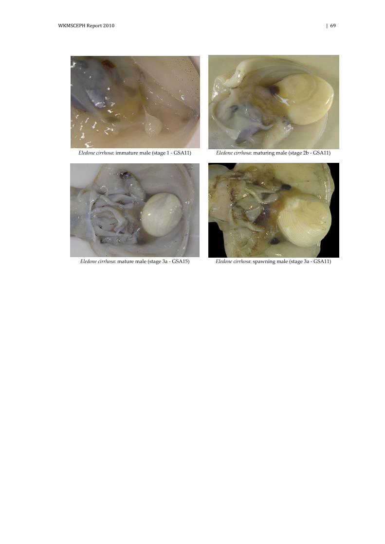
WKMSCEPH Report 2010 | 69
Eledone cirrhosa: immature male (stage 1 - GSA11) Eledone cirrhosa: maturing male (stage 2b - GSA11)
Eledone cirrhosa: mature male (stage 3a - GSA15) Eledone cirrhosa: spawning male (stage 3a - GSA11)

70 | WKMSCEPH Report 2010
Eledone cirrhosa: immature female (stage 1 - GSA11) Eledone cirrhosa: developing female (stage 2a - GSA11)
Eledone cirrhosa: maturing female (stage 2b - GSA11) Eledone cirrhosa: mature female (stage 3a - GSA11)
Eledone cirrhosa: mature female (stage 3a - M. González -
Málaga) Eledone cirrhosa: spent female (stage 3b - GSA11)

WKMSCEPH Report 2010 | 71
Eledone moschata: immature male (stage 1 - GSA11) Eledone moschata: developing male (stage 2a -
GSA11)
Eledone moschata: spawning male (stage 3a - GSA11)

72 | WKMSCEPH Report 2010
Eledone moschata: immature female (stage 1 -
GSA11) Eledone moschata: developing female (stage 2a -
GSA10b)
Eledone moschata: maturing female (stage 2b -
GSA11) Eledone moschata: mature female (stage 3a - GSA11)
Eledone moschata: mature female (stage 3a - I.S. Caparro - Cádiz)

WKMSCEPH Report 2010 | 73
Annex 8: Teuthida gonad reference photos
Loligo vulgaris: immature male (stage 1 - GSA11) Loligo vulgaris: developing male (stage 2a - GSA11)
Loligo vulgaris: developing male (stage 2a -
INRB-IPIMAR/FCUL - Portugal) Loligo vulgaris: maturing male (stage 2b - GSA10b)
Loligo vulgaris: mature male (stage 3a - GSA11) Loligo vulgaris: spawning male (stage 3a - IMEDEA-
CSIC-UIB - Balearic Islands)
Loligo vulgaris: spent male (stage 3b - GSA11) Loligo vulgaris: spent male (stage 3b - GSA11)

74 | WKMSCEPH Report 2010
Loligo vulgaris: immature female (stage 1 - GSA11) Loligo vulgaris: immature female (stage 1 - IEO C.O.
- Canaries)
Loligo vulgaris: developing female (stage 2a -
GSA11) Loligo vulgaris: maturing female (stage 2b - IEO C.O.
- Canaries)
Loligo vulgaris: maturing female (stage 2b -
INRB-IPIMAR/FCUL - Portugal) Loligo vulgaris: mature female (stage 3a - IMEDEA-
CSIC-UIB - Balearic Islands)
Loligo vulgaris: spawning female (stage 3a - GSA11) Loligo vulgaris: spent female (stage 3b - GSA11)

WKMSCEPH Report 2010 | 75
Loligo forbesi: immature male (stage 1 - GSA11) Loligo forbesi: developing male (stage 2a - GSA11)
Loligo forbesi: maturing male (stage 2b - GSA10b) Loligo forbesi: spawning male (stage 3a - GSA11)

76 | WKMSCEPH Report 2010
Loligo forbesi: immature female (stage 1 - GSA11) Loligo forbesi: developing female (stage 2a - GSA10b)
Loligo forbesi: spawning female (stage 3a - GSA11)

WKMSCEPH Report 2010 | 77
Illex coindetii: immature male (stage 1 - GSA11) Illex coindetii: developing male (stage 2a - GSA11)
Illex coindetii: maturing male (stage 2b - GSA9b) Illex coindetii: mature male (stage 3a - GSA9b)
Illex coindetii: spawning male (stage 3a - GSA10b)

78 | WKMSCEPH Report 2010
Illex coindetii: immature female (stage 1 - GSA11) Illex coindetii: immature female (stage 1 - GSA11)
Illex coindetii: developing female (stage 2a -
GSA20&22) Illex coindetii: maturing female (stage 2b - GSA11)
Illex coindetii: spawning female (stage 3a - GSA9b) Illex coindetii: spent female (stage 3b - GSA11)

WKMSCEPH Report 2010 | 79
Todaropsis eblanae: immature male (stage 1 - GSA11) Todaropsis eblanae: developing male (stage 2a -
GSA11)
Todaropsis eblanae: maturing male (stage 2b - GSA11) Todaropsis eblanae: mature male (stage 3a - GSA11)
Todaropsis eblanae: spawning male (stage 3a - GSA11)

80 | WKMSCEPH Report 2010
Todaropsis eblanae: immature female (stage 1 -
GSA11) Todaropsis eblanae: developing female (stage 2a -
GSA10b)
Todaropsis eblanae: maturing female (stage 2b -
GSA20&22) Todaropsis eblanae: spawning female (stage 3a -
GSA11)

WKMSCEPH Report 2010 | 81
Annex 9: Sepiida gonad reference photos
Sepia officinalis: immature male (stage 1 - GSA10b) Sepia officinalis: developing male (stage 2a - I.S.
Caparro - Cádiz)
Sepia officinalis: maturing male (stage 2b - IEO C.O. -
Canaries) Sepia officinalis: mature male (stage 3a - GSA11)
Sepia officinalis: spawning male (stage 3a - GSA9b)

82 | WKMSCEPH Report 2010
Sepia officinalis: immature female (stage 1 - GSA11) Sepia officinalis: developing female (stage 2a - GSA11)
Sepia officinalis: developing female (stage 2a - IEO C.O. -
Canaries) Sepia officinalis: developing female (stage 2a - IEO C.O. -
Canaries)
Sepia officinalis: maturing female (stage 2b - GSA9b) Sepia officinalis: spawning female (stage 3a - GSA11)
Sepia officinalis: spawning female (stage 3a - I.S. Caparro -
Cádiz) Sepia officinalis: spent female (stage 3b - IEO C.O. -
Canaries)

WKMSCEPH Report 2010 | 83
Annex 10: Histological reference photos Octopus vulgaris: males microscopic scale.
Octopus vulgaris: immature male (stage
1 - GSA11).
Octopus vulgaris: immature male (stage 1 - GSA11). Spermatogonia (SPG) inside the
tubules.
Octopus vulgaris: developing male
(stage 2a - GSA11). All stages are pre-sent and few spermatozoa (no in all tu-
bules).
Octopus vulgaris: developing male (stage 2a - GSA11). Spermatogonia (SPG); Primary
spematocytes (SPC I), secondary spemato-cytes (SPC II); spermatids (SPD) and sper-
matozoa (SPZ).
Octopus vulgaris: maturing male (stage 2b - GSA11). All stages are present and spermatozoa are more evident in all tu-
bules.
Octopus vulgaris: maturing male (stage 2b - GSA11). Spermatogonia (SPG); Primary spe-matocytes (SPC I), secondary spematocytes (SPC II); spermatids (SPD) and spermatozoa
(SPZ).
SPG
SPG
SPG
SPZ
SPC I
SPC II
SPD
SPG
SPZ
SPD
SPC I
SPC II

84 | WKMSCEPH Report 2010
Octopus vulgaris: mature male (stage 3a
- GSA11). Mature stage (3a). The seminiferous tubules are large and well defined. There are no empty space be-
tween cells. All type of cells are present with abundant spermatozoa in the cen-
tral lumen.
Octopus vulgaris: mature male (stage 3a - GSA11). Spermatogonia (SPG); Primary
spematocytes (SPC I), secondary spemato-cytes (SPC II); spermatids (SPD) and sper-
matozoa (SPZ), flagella (F).
Octopus vulgaris: spawning male (stage 3a - GSA11). Empty spaces exist, Sper-
matogina, primary and secondary spermatocytes, spermatids and sper-
matozoa are still present.
Octopus vulgaris: spawning male (stage 3a - GSA11). Spermatozoa (SPZ).
Octopus vulgaris: spent male (stage 3a - GSA11). Empty spaces are bigger. Few spermatocytes, spermatids and sper-
matozoa are present.
Octopus vulgaris: spent male (stage 3a - GSA11). Spermatozoa (SPZ).
SPZ
SPZ
SPG
SPD
SPZ
F
SPC II
SPC I

WKMSCEPH Report 2010 | 85
Octopus vulgaris: females microscopic scale.
Octopus vulgaris: immature female
(stage 1 - GSA11). Germinative epithe-lium (GE); oogonia (OO).
Octopus vulgaris: immature female (stage 1 - GSA11). Early
primary oocytes (EPO); fol-licular cell (FC); nucleus (N).
Octopus vulgaris: developing fe-male (stage 2a - GSA11). Late
primary oocytes (LPO) and pre-vitellogenic oocytes (PVO).
Octopus vulgaris: developing female (stage 2a - GSA11). Late primary oo-
cytes; follicular cell (FC).
Octopus vulgaris: developing female (stage 2a - GSA11).
Previtellogenic oocytes; fol-licular cell (FC); lipidic inclu-
sion (LI); fold (F).
Octopus vulgaris: maturing female (stage 2b - GSA11). Vitellogenic oocytes (VO); follicular cell (FC);
lipidic inclusion (LI).
Octopus vulgaris: maturing female (stage
2b - GSA11). Vitellogenic oocytes; fol-licular cell (FC); lipidic inclusion(LI);
yolk droplets (Y).
Octopus vulgaris: mature fe-male (stage 3a - GSA11). Ad-vanced Vitellogenic oocytes
(AVO); fold (F).
Octopus vulgaris: mature female (stage 3a - GSA11). Advanced
Vitellogenic oocytes; fold (F); yolk (Y); corion (C).
FC
OO GE
EPO
N
LPO
PVO
FC
LI
LI
N
VO
F
LI
Y
AVO
F
Y
C FC
FC
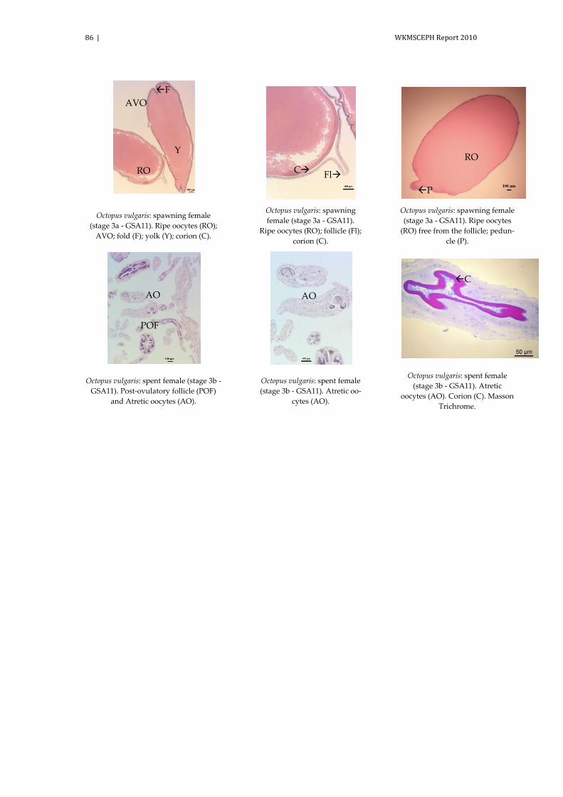
86 | WKMSCEPH Report 2010
Octopus vulgaris: spawning female
(stage 3a - GSA11). Ripe oocytes (RO); AVO; fold (F); yolk (Y); corion (C).
Octopus vulgaris: spawning female (stage 3a - GSA11).
Ripe oocytes (RO); follicle (Fl); corion (C).
Octopus vulgaris: spawning female (stage 3a - GSA11). Ripe oocytes
(RO) free from the follicle; pedun-cle (P).
Octopus vulgaris: spent female (stage 3b - GSA11). Post-ovulatory follicle (POF)
and Atretic oocytes (AO).
Octopus vulgaris: spent female (stage 3b - GSA11). Atretic oo-
cytes (AO).
Octopus vulgaris: spent female (stage 3b - GSA11). Atretic
oocytes (AO). Corion (C). Masson Trichrome.
F
Y
C
RO
Fl
AVO
RO
P
POF
AO AO C

WKMSCEPH Report 2010 | 87
Octopus vulgaris: oviductal gland at different microscopic maturity stage.
Octopus vulgaris: oviducal gland in the developing female (stage 2a -
GSA11).
Octopus vulgaris: spermothecae in the oviducal gland in developing female
(stage 2a - GSA11). Spermatozoa (SPZ).
Octopus vulgaris: spermatozoa (SPZ) inside spermothecae in the oviducal gland in devel-
oping female (stage 2a - GSA11).
Octopus vulgaris: spermotheca in
the oviducal gland in maturing fe-male (stage 2b - GSA11). Spermato-
zoa (SPZ).
Octopus vulgaris: spermatozoa into the spermothecae (arrow: heads of sper-
matozoa) (stage 2b - GSA11).
Octopus vulgaris: two regions of the oviducal gland in maturing female (stage 2b - GSA11).
Outer region (OR) and Inner Region (IR).
Octopus vulgaris: two regions of the oviducal gland in mature/ spawn-
ing female (stage 3a - GSA11). Outer region (OR) and Inner Re-
gion (IR).
Octopus vulgaris: spermotheca in the oviducal gland in mature/ spawning female (stage 3a - GSA11). Spermato-
zoa (SPZ).
Octopus vulgaris: two regions of the oviducal gland in spent female (stage 3c - GSA11). Outer
region (OR) and Inner Region (IR).
SPZ
SPZ
OR
IR
SPZ
OR
OR
IR
IR
SPZ

88 | WKMSCEPH Report 2010
Octopus vulgaris: Spermotheca in the oviducal gland in spent female (stage 3c - GSA11). Spermatozoa
(SPZ).
SPZ

WKMSCEPH Report 2010 | 89
Loligo vulgaris: males microscopic scale.
Loligo vulgaris: immature male (stage 1 - IMEDEA-CSIC-UIB -
Balearic Islands) Loligo vulgaris: developing male (stage 2a - IMEDEA-CSIC-UIB -
Balearic Islands)
Loligo vulgaris: developing male (stage 2a - IMEDEA-CSIC-UIB -
Balearic Islands) Loligo vulgaris: maturing male (stage 2b - IMEDEA-CSIC-UIB -
Balearic Islands)
Loligo vulgaris: mature/ spawning male (stage 3a - IMEDEA-
CSIC-UIB - Balearic Islands) Loligo vulgaris: mature/ spawning male (stage 3a - IMEDEA-
CSIC-UIB - Balearic Islands)
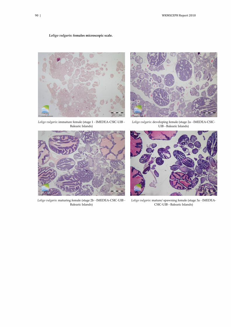
90 | WKMSCEPH Report 2010
Loligo vulgaris: females microscopic scale.
Loligo vulgaris: immature female (stage 1 - IMEDEA-CSIC-UIB -
Balearic Islands) Loligo vulgaris: developing female (stage 2a - IMEDEA-CSIC-
UIB - Balearic Islands)
Loligo vulgaris: maturing female (stage 2b - IMEDEA-CSIC-UIB -
Balearic Islands) Loligo vulgaris: mature/ spawning female (stage 3a - IMEDEA-
CSIC-UIB - Balearic Islands)

WKMSCEPH Report 2010 | 91
Annex 11: The ICES (International Council for the Exploration of the Sea) management divisions in the Northeast Atlantic
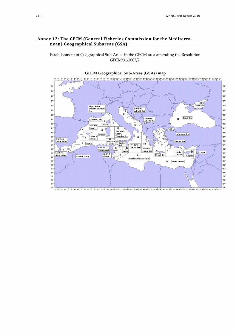
92 | WKMSCEPH Report 2010
Annex 12: The GFCM (General Fisheries Commission for the Mediterra-nean) Geographical Subareas (GSA)
Establishment of Geographical Sub-Areas in the GFCM area amending the Resolution GFCM/31/2007/2.
GFCM Geographical Sub-Areas (GSAs) map

WKMSCEPH Report 2010 | 93
Annex 13: The CECAF area (FAO Division 34)