reproduction in animals: asexual v - carnes ap bio · web viewthe four levels of protein structure...
TRANSCRIPT
AP Biology Notes Outline Chapter 5: The Structure and Function of Macromolecules
CONCEPT 1: Monomers, Polymers, and Macromolecules
Monomers: are repeating units that serve as building blocks for polymers. Polymers: are long molecule consisting of many similar or identical building blocks linked by COVALENT bonds. Macromolecules: are LARGE groups of polymers covalently bonded – 4 classes of organic macromolecules to be studied:
1. carbohydrates2. lipids3. nucleic acids4. proteins
CONCEPT 2: Building and Breaking Polymers
Review the following biological animations as you study this section of notes: http://nhscience.lonestar.edu/biol/dehydrat/dehydrat.html
How do monomers link up to form polymers? Monomers link up to form polymers in a process called condensation reaction (specifically, dehydration synthesis): two molecules covalently bond and lose a water molecule in the process. THIS TAKES ENERGY TO DO!
How do polymers break back into monomers? Hydrolysis : polymers are disassembled to monomers by adding a water molecule back (i.e. digestion of food).
As each monomer is added, a water molecule is removed – DEHYDRATION REACTION.
This is the reverse of dehydration is HYDROLYSIS…it breaks bonds between monomers by adding water molecules.

AP Biology Notes Outline Chapter 5: The Structure and Function of Macromolecules
CONCEPT 3: Organic Compounds and Building Blocks
Carbohydrates – made up of monosaccharides (glucose/sugars) joined by a glycosidic bond.
Lipids - CATEGORY DOES NOT INCLUDE POLYMERS (the grouping is based on insolubility) made up of a glycerol and 3 fatty acids joined by an ester bond. There are three types:
1. Triglycerides (glycerol and 3 fatty acids)2. Phospholipids3. Steroids
Proteins – made up of amino acids joined by a peptide bond.
Nucleic Acids – made up nucleotides
CONCEPT 4: Carbohydrates
Review the following biological animations as you study this section of notes:http://bcs.whfreeman.com/thelifewire/content/chp03/0302002.html
Carbohydrates include sugars & their polymers. They exist as three types:1. monosaccharides2. disaccharides3. polysaccharides (macromolecule stage)
Made up of Carbon, Hydrogen, and Oxygen in a 1:2:1 ratio (CnH2nOn). They have a carbonyl group (C=O) and multiple hydroxyl groups (-OH).
The size of carbon skeleton determines category. Sugars can also be classified based on size of the carbon skeleton – ranges from 3 to 7 carbons long.
• HEXOSES – 6 carbon sugars (glucose, fructose, etc.)
• TRIOSES – 3 carbon sugars• PENTOSES – 5 carbon sugars
Notice: carbonyl group (C=O)Notice: multiple hydroxyl groups (-OH)
The location of carbonyl group determines if is an aldose (aldehyde sugar) or a ketose (ketone sugar).

AP Biology Notes Outline Chapter 5: The Structure and Function of Macromolecules
Monosaccharides:Monosaccharide carbohydrates are major sources of energy for cells (i.e. glucose for cellular respiration)! Monosaccharides are simple enough to serve as raw materials for synthesis of other small organic molecules such as amino and fatty acids. The most common: glucose, fructose, galactose. Sugar molecules are generally not immediately used in these ways – but most often are incorporated as monomers into disaccharides and polysaccharides.
Glucose:• made during photosynthesis• main source of energy for plants and animals
Fructose:• found naturally in fruits• is the sweetest of monosaccarides
Galactose:• found in milk• is usually in association with glucose or fructose
All three have SAME MOLECULAR FORMULA but differ structurally so they are ISOMERS! C6H12O6 – formula for glucose, fructose, galactose.
Disaccharides:Disaccharides consist of two monosaccharides joined by a GLYCOSIDIC LINKAGE – a covalent bond resulting from dehydration synthesis. Examples:
• Maltose – 2 glucoses joined (C12H22O11) – table sugar.• Sucrose – glucose and fructose joined (C12H22O11) – malt sugar.• Lactose – glucose and galactose joined (C12H22O11) – sugar found in milk.

AP Biology Notes Outline Chapter 5: The Structure and Function of Macromolecules
The bonding of 2 glucose units forms maltose – the glycosidic link joins the number 1 carbon of one glucose to the number 4 carbon of the second glucose…JOINING THE GLUCOSE MONOMERS IN A DIFFERENT WAY WOULD RESULT IN A DIFFERENT DISACCHARIDE THAN MALTOSE!
Polysaccharides:These are the polymers of sugars – the true macromolecules of the carbohydrates. They serve as storage material that is hydrolyzed as needed in the body or as structural units that support bodies of organisms.
These are polymers with a few hundred to a few thousand monosaccharides joined by glycosidic linkages.
STARCH AND GLYCOGEN are storage polysaccharides.• Starch: storage for plants – plants store starch as granules within cellular structures called plastids.
o Made up of glucose monomers in alpha configuration (see fig. 5.7 pg. 67)o Has a helical shape that can be unbranched (amylose) or branched (amylopectin)o You can find starch in potatoes and grainso Alpha versus beta configuration of starch carbohydrates indicates the bodies ability to
effectively break down the polysaccharide. We do not have enzymes that will break down sugars formed in the beta configuration (such as cellulose).
• Glycogen: storage for animals – animals store glycogen mainly in the liver and muscle cells and this storage is usually depleted in a single day.
o extensively branched group of glucose units
CELLULOSE and CHITIN are structural polysaccharides.• Cellulose: found in cell wall of PLANTS and is the most abundant organic compound on Earth
o Cellulose is a polymer of glucose, but all glucose molecules are in the beta configuration thus, cellulose is always straight, and this provides for strength (i.e. lumber).
o Few organisms possess the enzymes to digest cellulose. Cellulose passes through the digestive tract and is eliminated in feces, BUT, the fibrils of cellulose abrade the wall of the digestive tract and stimulate secretion of mucus which is necessary for smooth food passage – so though cellulose is not nutritious, it is necessary.
o Organisms that can digest cellulose include: cows (with help of bacteria), termites (with help of microbes), some fungi.

AP Biology Notes Outline Chapter 5: The Structure and Function of Macromolecules
• Chitin: found in cell wall of FUNGI and the exoskeleton of ARTHROPODSo Another structural polysaccharide - used by arthropods to build their exoskeletons.o Pure chitin is leathery, but when encrusted with calcium carbonate it hardens into shell form.o Also used by fungi in their cell walls (instead of cellulose) - similar to cellulose, but the
glucose monomer has a nitrogen containing appendage.
CONCEPT 5: Lipids
Review the following biological animations as you study this section of notes:http://bcs.whfreeman.com/thelifewire/content/chp03/0302002.html
This group does not include polymers – only grouped together based on trait of little or no affinity for water: h ydrophobic (water fearing). Hydrophobic nature is based on molecular structure – consist mostly of hydrocarbons! REMEMBER – hydrocarbons are insoluble in water because of their non-polar C—H bonds!
Lipids are smaller than true polymeric macromolecules. They are insoluble in water, but soluble in organic solvents. They serve as energy storage molecules andcan act as chemical messengers within and between cellsInclude waxes and certain pigments. Our focus will be on (1) fats, (2) phospholipids, and (3) steroids.
Fats (Triglycerides):Fats are made of two kinds of smaller molecules – glycerol and fatty acids (one glycerol to three fatty acids). Dehydration synthesis hooks these up – 3 waters produced for every one triglyceride. ESTER linkages bond glycerol to the fatty acid tails – bond is between a hydroxyl group and a carboxyl group.
Glycerol is an alcohol with three carbons, each one with a hydroxyl group. Fatty acid has a long carbon skeleton:
• at one end is a carboxyl group (thus the term fatty “acid”)
• the rest of the molecule is a long hydrocarbon chain
The hydrocarbon chain is not susceptible to bonding, so the H-bonds of water bind to another water and excludes the fats (insoluble).
• One glycerol & 3 fatty acid molecules
• One H2O is removed for each fatty acid joined to glycerol by an ester linkage
• Result is a fat

AP Biology Notes Outline Chapter 5: The Structure and Function of Macromolecules
Saturated v. Unsaturated FatsRefers to the structure of the hydrocarbon chains of the fatty acids: no double bonds between the carbon atoms of the chain means that the max # of hydrogen atoms is bonded to the carbon skeleton (saturated). THESE ARE THE BAD ONES!!! – they can cause atherosclerosis (plaque develop, get less flow of blood, hardening of arteries)!
If one or more double bonds is present, then it is unsaturated and these tend to kink up and prevent the fats from packing together.
Most animal triglycerides are saturated (i.e. lard and butter). These are solid at room temperature (saturated fats). Plants and fish have unsaturated triglycerides, so they are liquid at room temperature – oil (i.e. vegetable oil, etc).
PhospholipidsPhospholipids are lipids that hve only two fatty acid tails! The third hydroxyl group of glycerol is joined to a phosphate group (negatively charged). These are ambivalent to water – tails are hydrophobic, heads are hydrophilic. When added to water, phospholipids self-assemble into aggregates that shield their hydrophobic portions from water (called a micelle). At a cell surface, this forms a double layer arrangement – phospholipid bilayer. Hydrophilic head of molecules are on outside of the bilayer, in contact with aqueous solutions inside & outside the cell. Hydrophobic tails point toward interior of membrane, away from water. Phospholipid bilayer forms boundary between cell & its external environment – EXAMPLE OF FORM & FUNCTION!
At room temperature, the molecules of a saturated fat are packed closely together, forming a solid.
At room temperature, the molecules of an unsaturated fat cannot pack together closely enough to solidify because of the kinks in their fatty acid tails.

AP Biology Notes Outline Chapter 5: The Structure and Function of Macromolecules
Steroids:Steroids are characterized by carbon skeleton consisting of four fused rings. Differences depend on the functional groups attached to the ring ensemble.
Many hormones are steroids. Cholesterol is the molecule from which other steroids, including sex hormones, are synthesized.
Steroids vary in the functional groups attached to their four interconnected rings (shown in gold):
CONCEPT 6: Nucleic Acids
Review the following biological animations as you study this section of notes:http://bcs.whfreeman.com/thelifewire/content/chp03/0302002.html
Nucleic acids are polymers of information (i.e. DNA and RNA). The building blocks of nucleic acids are nucleotides. All nucleotides consist of (1) a phosphate group, (2) a pentose sugar, and (3) a nitrogenous base A, T, C, G, or U.
There are two categories of nigrogenous bases:
1. Pyrimidines: smaller, have a six-membered ring of carbon and nitrogen atoms (C , U, T)
2. Purines: larger, have a six- and a five-membered ring fused together (A, G)

AP Biology Notes Outline Chapter 5: The Structure and Function of Macromolecules
Nucleic acids exist as 2 types: DNA and RNA*DNA -- *double stranded (entire code) with DEOXYRIBOSE sugar
*never leaves nucleus*bases are A,T,C,G*involved in replication and protein synthesis
*RNA -- *single stranded (partial code) with RIBOSE sugar*mobile – nucleus and cytoplasm*bases are A,U,C,G*involved in Protein Synthesis
Summary of flow of genetic information in living organisms:
DNA → transcription into RNA → translation into protein
Transcription – occurs in the nucleus of cell; opens up DNA double helix, copies section needed for protein manufacture (i.e. reads the gene), this makes messenger RNA (mRNA).
Translation – mRNA travels out of nucleus to cytoplasm to a ribosome (site of protein manufacture); ribosomal RNA (rRNA) anchors the transcript in the ribosome, transfer RNA (tRNA) brings in correct amino acid by reading 3 amino acids at a time (codon).
CONCEPT 7: Proteins
Review the following biological animations as you study this section of notes:http://bcs.whfreeman.com/thelifewire/content/chp03/0302002.html
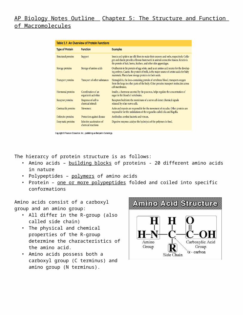
Proteins are the most structurally sophisticated molecules known and they account for more than 50% of the dry weight of cells. They are polymers of amino acids linked together by peptide bonds. There are 20 known amino acids. Functions of proteins in living systems include:

AP Biology Notes Outline Chapter 5: The Structure and Function of Macromolecules
The hierarcy of protein structure is as follows:
• Amino acids – building blocks of proteins - 20 different amino acids in nature• Polypeptides – polymers of amino acids• Protein – one or more polypeptides folded and coiled into specific conformations
Amino acids consist of a carboxyl group and an amino group:
• All differ in the R-group (also called side chain)• The physical and chemical properties of the R-
group determine the characteristics of the amino acid.
• Amino acids possess both a carboxyl group (C terminus) and amino group (N terminus).
Amino acids join as the Carboxyl group of one is adjacent to amino group of another, dehydration synthesis occurs, forms a covalent bond: PEPTIDE BOND.
When repeated over and over, we get a polypeptide:– On one end is an N-terminus (amino end);– On other is a C-terminus (carboxyl end)
• Note: dehydration synthesis.• Note: carboxyl group of one end
attaches to amino group of another.• Note: peptide bond is formed.

AP Biology Notes Outline Chapter 5: The Structure and Function of Macromolecules
• Note: repeating this process builds a polypeptide.
A PROTEIN’S FUNCTION DEPENDS ON ITS CONFORMATION:• Functional proteins consist of one or more polypeptides twisted, folded, and coiled into a unique shape• Amino acid sequence determines shape• 2 big categories – 1. Globular AND 2. Fibrous
Function of a protein depends on its ability to recognize and bind to some other molecule. CONFORMATION IS KEY (i.e. antibodies bind to particular foreign substances that invade the body…binds to it (based on shape)…and there is a lock and key fit!)
The four levels of protein structure are: (1) primary, (2) secondary, (3) tertiary, and (4) quaternary.
1. Primary Structure: unique sequence of amino acids (long chain). Notice the amino end and the carboxyl end. These are held together by peptide bonds!
2. Secondary Structure: segments of polypeptide chain that repeatedly coil or fold in patterns that contribute to overall configuration of the protein – they are the result of hydrogen bonds at regular intervals along the polypeptide backbone.
• BOTH PATTERNS HERE DEPEND ON HYDROGEN BONDING BETWEEN C=O and N-H groups along the polypeptide backbone.
• Alpha Helix – delicate coil held together by H-bonding between every fourth amino acid
• Beta pleated sheet – two or more regions of the polypeptide chain lie parallel to one another. H-bonds form here, and keep the structure together.
• NOTE – only atoms of backbone are involved, not the amino acid side chains!

AP Biology Notes Outline Chapter 5: The Structure and Function of Macromolecules
3. Tertiary Structure: superimposed on secondary structure; irregular contortions from interactions between side chains (R-groups) of amino acids:
• nonpolar side chains end up in clusters at the core of a protein – caused by the action of water molecules which exclude nonpolar substances...this is a hydrophobic interaction.
• van der Waals interactions, H-bonds, and ionic bonds all add together to stabilize tertiary structure
• may also have disulfide bridges form …when amino acids with 2 sulfhydryl groups are brought together – these bonds are much stronger than the weaker interactions mentioned above
4. Quaternary Structure: the overall protein structure that results from the aggregation of many polypeptide subunits
• Ex. collagen – structural• Ex. hemoglobin – globular
REVIEW OF FOUR LAYERS OF PROTEIN STRUCTURE:Review the following biological animations as you study this section of notes:https://mywebspace.wisc.edu/jonovic/web/proteins.html
• Primary Structure: sequence of amino acids in a polypetide.
AP Biology Notes Outline Chapter 5: The Structure and Function of Macromolecules
• Secondary Structure: bending and hydrogen bonding of a polypeptide backbone to form repeating patterns.
• Tertiary Structure: overall shape of a polypeptide…as reinforced by interactions between the side chains (R-groups) of amino acids.
• Quaternary Structure: the association between two or more polypeptides that make up a protein.
THINGS TO KNOW ABOUT PROTEINS:Polypeptide chain of given amino acid sequence can spontaneously arrange into 3-D shape
• Configuration also depends on physical and chemical conditions of protein’s environment• if pH, salt [ ], temp, etc. are altered, protein may unravel and lose native conformation –
DENATURATION• Denatured proteins are biologically inactive!• Anything that disrupts protein bonding can denature a protein!

AP Biology Notes Outline Chapter 5: The Structure and Function of Macromolecules
Denatured proteins can often renature when environmental conditions improve!HOW proteins fold is not always clear – may be several intermediate states on the way to stable conformation, but there are a few ways to track, though –
• chaperonins: protein molecules that assist the proper folding of other proteins.• computer simulations – “Blue Gene”, a supercomputer able to generate the 3-D structure of any protein
starting from its aa sequence (medical uses)