reptile scale paradigm: evo-devo, pattern formation and ...cmchuong/2009 reptile scale ijdb 53...
TRANSCRIPT
Reptile scale paradigm:
Evo-Devo, pattern formation and regeneration
CHENG CHANG1,2, PING WU1, RUTH E. BAKER3, PHILIP K. MAINI3,4, LORENZO ALIBARDI*,5
and CHENG-MING CHUONG*,1
1Department of Pathology, Keck School of Medicine, University of Southern California, Los Angeles, California, USA, 2School of LifeScience, Lanzhou University, Lanzhou, Gansu, China, 3Centre for Mathematical Biology, Mathematical Institute, University of Oxford,4Oxford Centre for Integrative Systems Biology, Department of Biochemistry, University of Oxford, UK and 5Dipartimento di Biologia
Evoluzionistica Sperimentale, University of Bologna, Bologna, Italy
ABSTRACT The purpose of this perspective is to highlight the merit of the reptile integument as
an experimental model. Reptiles represent the first amniotes. From stem reptiles, extant reptiles,
birds and mammals have evolved. Mammal hairs and feathers evolved from Therapsid and
Sauropsid reptiles, respectively. The early reptilian integument had to adapt to the challenges of
terrestrial life, developing a multi-layered stratum corneum capable of barrier function and
ultraviolet protection. For better mechanical protection, diverse reptilian scale types have
evolved. The evolution of endothermy has driven the convergent evolution of hair and feather
follicles: both form multiple localized growth units with stem cells and transient amplifying cells
protected in the proximal follicle. This topological arrangement allows them to elongate, molt and
regenerate without structural constraints. Another unique feature of reptile skin is the exquisite
arrangement of scales and pigment patterns, making them testable models for mechanisms of
pattern formation. Since they face the constant threat of damage on land, different strategies
were developed to accommodate skin homeostasis and regeneration. Temporally, they can be
under continuous renewal or sloughing cycles. Spatially, they can be diffuse or form discrete
localized growth units (follicles). To understand how gene regulatory networks evolved to
produce increasingly complex ectodermal organs, we have to study how prototypic scale-forming
pathways in reptiles are modulated to produce appendage novelties. Despite the fact that there
are numerous studies of reptile scales, molecular analyses have lagged behind. Here, we
underscore how further development of this novel experimental model will be valuable in filling
the gaps of our understanding of the Evo-Devo of amniote integuments.
KEY WORDS: stratum corneum, skin, hair follicle, skin regeneration, Turing, reaction-diffusion
Introduction
Among amniotes, stem reptiles were basal to extant reptiles,birds and mammals. When reptiles established themselves onland, their integuments had to adapt to the challenges of terres-trial life by developing barriers which can prevent water loss(Alibardi, 2003), mechanisms which can protect against ultravio-let (UV) irradiation and mechanical shields which can provideprotection against the rigors of terrestrial life. Starting from thesebasic needs, different types of reptilian scales evolved in theMesozoic period to serve different functions (Chuong et al., 2002)and adapt diverse species to different niches. As amniotes
Int. J. Dev. Biol. 53: 813-826 (2009)doi: 10.1387/ijdb.072556cc
THE INTERNATIONAL JOURNAL OF
DEVELOPMENTAL
BIOLOGYwww.intjdevbiol.com
*Address correspondence to: Dr. Cheng-Ming Chuong. Department of Pathology, Keck School of Medicine, University of Southern California, Los Angeles,California 90033, USA. Fax: 323-442-3049. e-mail: [email protected] or Dr. Lorenzo Alibardi. Dipartimento di Biologia evoluzionistica sperimentale, Universityof Bologna, Bologna, Italy. e-mail: [email protected]
Published online: 9 June 2009.
ISSN: Online 1696-3547, Print 0214-6282© 2009 UBC PressPrinted in Spain
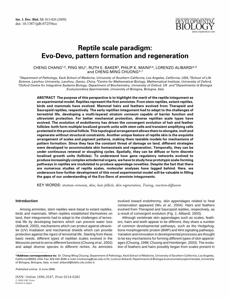
evolved toward endothermy, skin appendages related to heatconservation appeared (Wu et al., 2004). Hairs and feathersevolved from Therapsid and Sauropsid reptiles, respectively, asa result of convergent evolution (Fig. 1; Alibardi, 2003).
Although vertebrate skin appendages such as scales, feath-ers, hairs and teeth appear to be different, they share a numberof common developmental pathways, such as the Hedgehog,bone morphogenetic protein (BMP) and Wnt signaling pathways.Variation and innovation in developmental processes are thoughtto be key mechanisms for forming different types of skin append-ages (Chuong, 1998; Chuong and Homberger, 2003). The evolu-tion of feathers and hairs possibly began from scales present in
814 C. Chang et al.
early reptiles in which the modulation and re-organization of generegulatory networks led to new forms of skin appendages (Chuong,1998; Wagner. 2007; Table 1). To thoroughly understand theorigin and evolution of amniote skin appendages, we must includereptile scales in these analyses since the prototype reptile scalesrepresent the basal state (see section on "Evo-Devo"). Reptilesmay have obtained a periodic arrangement of scales early inevolution (see section on "Pattern Formation"), because thishelps partition the unit of renewal and regeneration in an effectivetopo-biological configuration (see section on "Regeneration").Subsequently, essential, novel evolutionary mechanisms to trans-form scales into feathers and hairs include the formation offollicular structures and the ability for proximal-distal elongation offilaments (Fig. 1; Chuong et al., 2000, 2003, 2007; Alibardi, 2003,2004a; Prum, 1999; 2005; Sawyer and Knapp, 2003).
In this review, our objective is to provide a perspective thathighlights the value of the reptile scale as an experimental modelfor furthering our understanding of the Evo-Devo of amnioteinteguments, biological pattern formation, and the different strat-egies of regeneration in amniotes. It is not intended to provide adetailed account of reptilian skin biology. Those interested in thistopic can be referred to the more specific references on reptileinteguments (Maderson, 1985; Landmann, 1986; Alibardi, 2003).
Reptile scale typesHere we will introduce the basic type of reptilian scales. Over
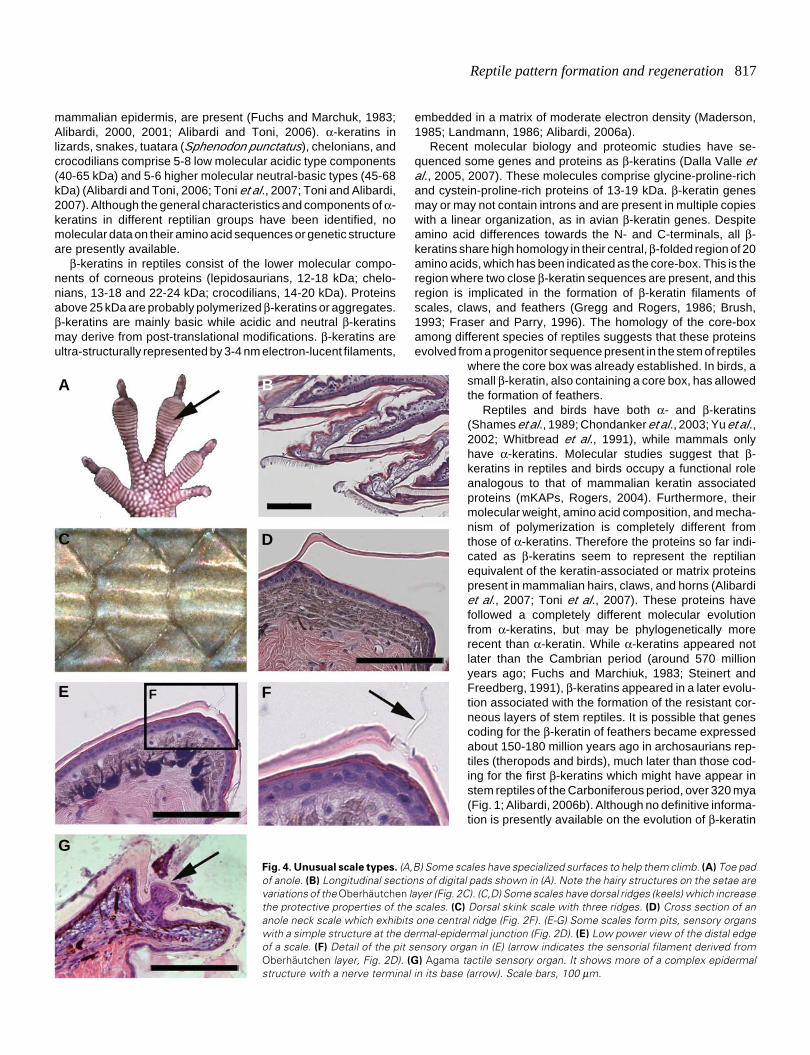
time, diverse scale types evolved in the Mesozoic period (al-though some of the lineages involved are now extinct) and inextant reptiles. Some types with rather unusual characteristicsare shown schematically in Fig. 2. An exemplary green iguana(Iguana iguana) with different types and arrangement of scales isshown in Fig. 3. Some unusual scales are shown in Fig. 4.
Extant reptiles are represented by four orders: Crocodilia(alligators and crocodiles), Chelonia (tortoises and turtles),Squamata (lizards and snakes), and Rhynchocephalia (the tuat-ara or Sphenodon punctatus) (Alexander, 1970; Maderson, 1985;Landmann, 1986). Crocodilian scales show relatively few varia-tions in gross morphology, are generally only a little overlappedand show a large surface composed mainly of hard β-keratinized,stratified epidermis (Alibardi and Thompson, 2000, 2001, 2002;Alibardi, 2003, 2006a, 2006b; Alibardi and Toni, 2006). Thenarrow hinge regions among scales have a thinner epidermiscontaining both α- and β-keratin. Chelonians have a soft, foldedskin in the limbs, tail, neck (most aquatic turtles), and a scaled skinin the shell (Maderson, 1985; Alibardi, 2003). In limbs and tail ofterrestrial turtles, hard scales containing β-keratin are also present,but β-keratin is decreased or absent in the hinge regions. A fewlayers of keratinocytes are covered by a thick, multi-layeredcorneus layer in the carapace and plastron.
In squamates, there are non-overlapping scales which do notappear to exhibit anterior-posterior polarity (Fig. 2A). They areseen in the scales on the head of snakes and lizards, and theround scales (tuberculate scales, Fig. 3F) on the sides of the bodyof the green iguana. However, the most frequently occurring is theoverlapping scale, which has distinct outer and inner surfaces(Fig. 2B, 3 C-E). This is the most common scale type on the bodyof lizards and snakes. The overlapping scale is asymmetric, withthe hinge region assigned to the posterior end. The outer surfaceconsists of a strongly cornified epidermis, which provides stiff-ness for the scale. During embryonic development, the morpho-genesis of overlapping scales passes through the flat two-layeredepidermis stage, the symmetric scale anlagen stage, the asym-metric scale anlagen stage, and the β-keratinizing asymmetricscale stage (Maderson, 1985; Alibardi, 1996, 1998).
In different species, some overgrowths and / or skin protru-sions are formed in different body regions. For example, micro-ornamentation, pits, sensory receptors, spiny, horny, crest, scutes,carapace, plastron, leather-like, and mosaic head scales and soon (Pianka et al., 2003, Landmann, 1986; Wu et al., 2004). Somelizards have elongated scales in a specific region of the body,such as the green iguana crest (frill) on the back (Fig. 2, 3). Insome elongated frills, we observe a follicle-like structure at thebase (Fig. 3B).
The pits are sensory organs in reptilian scales located in theinner surface, or hinge area, of some scales. They display follicle-like structures and can vary in their extension and depth. How-
Fig. 1. Potential relationship among
amniote skin appendages and key
evolutionary novel events. The reptil-ian integument shows some basal char-acteristics in comparison to mamma-lian and avian integuments. Mammalsevolved about 225 million years agofrom Therapsid-type reptiles, while birdsevolved about 175 million years agofrom archosaurian Saurospids-type rep-tiles. Key events that led to evolution-ary novelty are shown in blue italiccharacters.
Reptile pattern formation and regeneration 815
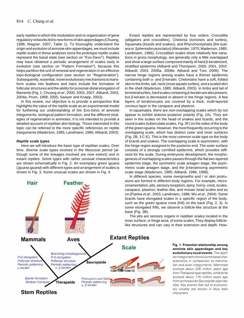
Fig. 2. Schematic drawings showing different types of reptile scales. Scales (resting phase) are shown in multiple layers with names labeled inpanel B. (A) Non-overlapping tuberculate type scales. (B) Overlapping scales commonly seen in squamates. (C) Variations of microstructures fromthe Oberhäutchen layer illustrating short spines in a, b and long setaes in c (such as those in the adhesive pad lamellae in geckos, Fig. 4B). (D) Pitson the scales of anole, gecko and iguana (mainly epidermal sensory organs; Fig. 4 E,F). (E) Tactile sensory organ on the hinge side of a scale in Agama.Some follicle-like structures have clustered dermal cells associated to their base; Fig. 4G). (F) Scales with ridges are seen on the back of skink or theneck of anole. (G) Frills, or very elongated scales, are seen on the back of iguana (Fig. 3B). (H) The horn on the head of chameleon contains a bonyelement core (osteoderm). (I) Scales on the limb of crocodilians show only minor overlapping. (J) Keeled scales with a central, elevated corneous ridgeare seen on the dorsal body of crocodilians and some armored agamid lizards (e.g. Australian spiny desert lizard or molok). Legends: a, fine ‘hair’ onscales of anoles; b, Micro-ornamentation on scales of snakes; c, Toe pad of anole or gecko;*, dermal cells clustered at the base of sensory organsin Agama; AK, α keratin; BK, β keratin; BP, bone element.
Non-overlapping scales Overlapping scales Special scale types Hairs Feathers Keratin Alpha, beta Alpha, beta Alpha, beta Alpha Alpha, beta
Elongation No Yes, but very limited longer Can be very long Can be very long Follicular structure No No Primitive? Yes Yes
Stem cells No ? ? Hair bulge Follicular bulge Proliferating cells Random Diffuse in general with some cells
preferentially distributed ? Localized in the proximal follicle Localized in the proximal follicle
Dermal papilla No No Some proto-dermal condensation? Yes Yes Structure of appendages
Epithelial layer covering a dermal core
Epithelial layer covering a dermal core Depending on different examples Epithelial cord Epithelial layer with mesenchymal core lost when mature
Regeneration Sloughing renewal cycle Sloughing renewal cycle ? Episodic molting and regeneration Episodic molting and regeneration
TABLE 1
COMPARISON OF REPTILE SCALES, AVIAN FEATHERS AND MAMMALIAN HAIRS
ever, they are different from the truefollicles in hairs and feathers becausethe structures are essentially only epi-dermal without dermal participation(Fig. 2, 4, Maderson, 1972; Von Dur-ing and Miller, 1979). For example, insome gecko-like lizards, they merelyrepresent the elongation of a singleOberhäutchen cell, with the onion-liketermination of sensory nerves under-neath the specific region where thehair-like organ is located (Maderson,1985).
In other cases, terminal tactile senseorgans contain a dermal componentconsisting of fibrocyte layers. Thereare also Merkel cells for mechano-sensory function. Pits that containsense receptor organs on the scalesof Agama form in a special site of thescale tip, and consist of a unique epi-dermal outgrowth (Fig. 4G). Thus,while overlapping scales are morewidespread, there are some unusualscale shapes and structures (Fig. 2,4). Some may show features whichare the preludes of proximal-distalelongation or follicle invagination (Fig.1), but they do not cumulate in realappendage follicles as seen in hairs orfeathers.
Evo-Devo
Initial adaptation to terrestrial lifeThe integument forms a critical in-
terface between the organism and itsenvironment. To live in the terrestrialenvironment successfully, reptilian an-
GB
C
D
E
F
H
I
J
A
816 C. Chang et al.
cestors had to deal with the huge differences between land andthe aquatic environment (Alibardi, 2003). The major challengewas the production of a barrier in the skin that limited the loss ofwater by transpiration or evaporation. There was also the need todeal with the intense UV irradiation of the land environment andto provide mechanical protection on the rough terrestrial environ-ment, much harsher than the aquatic environment of the amphib-ian ancestors of reptiles. The skin of reptiles responded to thesechallenges in the following ways.
1. Development of a barrier to prevent water loss to theexternal environment. The mesos- and α-layers of the extantsquamate epidermis played key roles in the development of thisbarrier (Bentley et al., 1966; Davis et al., 1980; Dunson andMazzotti, 1988; Landmann, 1979, 1986; Landmann et al., 1981;Lillywhite and Maderson, 1982; 2006; Menon et. al., 1986, 1996;Lillywhite, 2006). Formation of a corneum barrier involves thesynthesis of cornified proteins such as loricrin-like proteins andcomplex lipids and waxes. 2. Development of a radiation resistantmechanism to prevent the skin from being damaged by UVirradiation. The latter process in Squamates is obtained by bio-chemical, morphological and physical mechanisms (Chang, 2003;Ringvold, 2003). 3. Development of a tenacious skin shield, bothin the epidermis and in the dermis, to prevent mechanical damagefrom the harsh terrestrial environment. Hard proteins (β-keratinsor sKAPs) (KAP: keratin associated proteins) have been thesolution to this problem in reptiles and birds (Landmann, 1986;Maderson and Alibardi, 2000; Maderson, 2003; Alibardi et al.,2007; Toni et al., 2007). 4. Development of an effective heatinsulation mechanism to live in an environment in which the rangeof temperatures experienced daily or seasonally can be huge. Inthis regard, reptiles have not been very successful and their dailyand annual activities are thus limited (Pianka and Vitt, 2003). 5.Development of different adaptive structures related to specialenvironments. For example, various sensory functions havedeveloped on the skin with a pit-like morphology (Von During andMiller, 1979; Cooper and Greenberg, 1992; Pianka and Vitt, 2003;Landmann, 1986).
Molecular evolution of ααααα- and βββββ-keratinThere are two main types of intermediate filament proteins in
vertebrate keratinocytes: α- and β-keratins. Reptiles have both α-and β-keratins. α-keratogenic tissue provides a barrier to waterloss while an overlying β-keratogenic layer provides mechanicalstiffness to the skin (Alexander, 1970; Baden et al., 1970, 1974;Fraser and Parry, 1996; Gregg and Rogers, 1986; Landmann,1986; Maderson, 1985; Presland et al., 1989a, 1989b; Carver andSawyer, 1987; Sawyer et al., 2000; Steinert and Freedberg, 1991;Alibardi et al., 2002, 2005; Alibardi, 2000, 2001, 2002a, 2002b;Alibardi and Toni, 2006; Wyld and Brush, 1979, 1983).
α-, or "soft" keratin, is present in all vertebrate species whereasβ-, or "hard" keratin, is present only in birds and reptiles. Overlap-ping scales and the production of β-keratins provide strongprotection (hard scales, ramphoteca, claws) but still allow somemechanical plasticity through the inter-scale regions which aremade of α-keratins. α-keratins are divided into type I (acidic, 40-55 kDa) and type II (basic to neutral, 56-70 kDa), and they formthe 8-10 nm intermediate filament cytoskeletal network of epithe-lial cells. Previous studies have shown that in lizard epidermis,acidic and scarse basic cytokeratins, similar to those of the
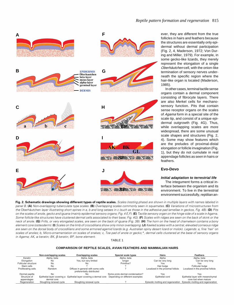
Fig. 3. Arrangement and different types of scales in iguana. (A) Anadult iguana showing different scale types in different regions. (B-F) Leftcolumn: scales from regions designated in (A). Right column: H&Estaining of their histological sections on the right. (B) Frills from themidline of the neck. Note the elongated scales compared with those in(C-F). (C) Scales from the dorsal trunk. (D) Scales from the ventral trunk.(E) Scales from the tail. (F) Tuberculate scales from the lateral neckregion. Scale bars, 500 μm.
B
C
D
E
F
A
Reptile pattern formation and regeneration 817
G
B
C D
E F
A
F
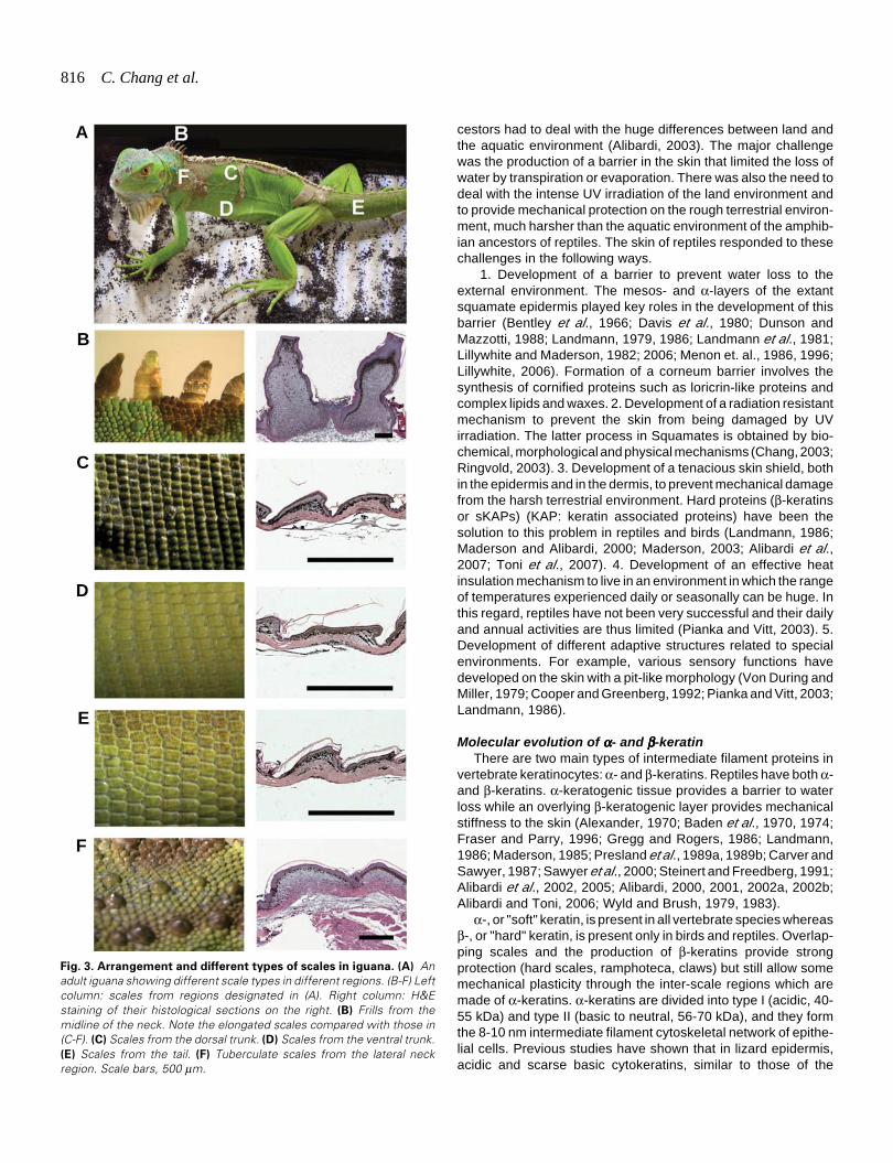
Fig. 4. Unusual scale types. (A,B) Some scales have specialized surfaces to help them climb. (A) Toe padof anole. (B) Longitudinal sections of digital pads shown in (A). Note the hairy structures on the setae arevariations of the Oberhäutchen layer (Fig. 2C). (C,D) Some scales have dorsal ridges (keels) which increasethe protective properties of the scales. (C) Dorsal skink scale with three ridges. (D) Cross section of ananole neck scale which exhibits one central ridge (Fig. 2F). (E-G) Some scales form pits, sensory organswith a simple structure at the dermal-epidermal junction (Fig. 2D). (E) Low power view of the distal edgeof a scale. (F) Detail of the pit sensory organ in (E) (arrow indicates the sensorial filament derived fromOberhäutchen layer, Fig. 2D). (G) Agama tactile sensory organ. It shows more of a complex epidermalstructure with a nerve terminal in its base (arrow). Scale bars, 100 μm.
where the core box was already established. In birds, asmall β-keratin, also containing a core box, has allowedthe formation of feathers.
Reptiles and birds have both α- and β-keratins(Shames et al., 1989; Chondanker et al., 2003; Yu et al.,2002; Whitbread et al., 1991), while mammals onlyhave α-keratins. Molecular studies suggest that β-keratins in reptiles and birds occupy a functional roleanalogous to that of mammalian keratin associatedproteins (mKAPs, Rogers, 2004). Furthermore, theirmolecular weight, amino acid composition, and mecha-nism of polymerization is completely different fromthose of α-keratins. Therefore the proteins so far indi-cated as β-keratins seem to represent the reptilianequivalent of the keratin-associated or matrix proteinspresent in mammalian hairs, claws, and horns (Alibardiet al., 2007; Toni et al., 2007). These proteins havefollowed a completely different molecular evolutionfrom α-keratins, but may be phylogenetically morerecent than α-keratin. While α-keratins appeared notlater than the Cambrian period (around 570 millionyears ago; Fuchs and Marchiuk, 1983; Steinert andFreedberg, 1991), β-keratins appeared in a later evolu-tion associated with the formation of the resistant cor-neous layers of stem reptiles. It is possible that genescoding for the β-keratin of feathers became expressedabout 150-180 million years ago in archosaurians rep-tiles (theropods and birds), much later than those cod-ing for the first β-keratins which might have appear instem reptiles of the Carboniferous period, over 320 mya(Fig. 1; Alibardi, 2006b). Although no definitive informa-tion is presently available on the evolution of β-keratin
mammalian epidermis, are present (Fuchs and Marchuk, 1983;Alibardi, 2000, 2001; Alibardi and Toni, 2006). α-keratins inlizards, snakes, tuatara (Sphenodon punctatus), chelonians, andcrocodilians comprise 5-8 low molecular acidic type components(40-65 kDa) and 5-6 higher molecular neutral-basic types (45-68kDa) (Alibardi and Toni, 2006; Toni et al., 2007; Toni and Alibardi,2007). Although the general characteristics and components of α-keratins in different reptilian groups have been identified, nomolecular data on their amino acid sequences or genetic structureare presently available.
β-keratins in reptiles consist of the lower molecular compo-nents of corneous proteins (lepidosaurians, 12-18 kDa; chelo-nians, 13-18 and 22-24 kDa; crocodilians, 14-20 kDa). Proteinsabove 25 kDa are probably polymerized β-keratins or aggregates.β-keratins are mainly basic while acidic and neutral β-keratinsmay derive from post-translational modifications. β-keratins areultra-structurally represented by 3-4 nm electron-lucent filaments,
embedded in a matrix of moderate electron density (Maderson,1985; Landmann, 1986; Alibardi, 2006a).
Recent molecular biology and proteomic studies have se-quenced some genes and proteins as β-keratins (Dalla Valle etal., 2005, 2007). These molecules comprise glycine-proline-richand cystein-proline-rich proteins of 13-19 kDa. β-keratin genesmay or may not contain introns and are present in multiple copieswith a linear organization, as in avian β-keratin genes. Despiteamino acid differences towards the N- and C-terminals, all β-keratins share high homology in their central, β-folded region of 20amino acids, which has been indicated as the core-box. This is theregion where two close β-keratin sequences are present, and thisregion is implicated in the formation of β-keratin filaments ofscales, claws, and feathers (Gregg and Rogers, 1986; Brush,1993; Fraser and Parry, 1996). The homology of the core-boxamong different species of reptiles suggests that these proteinsevolved from a progenitor sequence present in the stem of reptiles
818 C. Chang et al.
from the likely more primitive α-keratin (Sawyer and Knapp,2003), recent molecular studies have increased our understand-ing. These studies have indicated that basic amniotes possesseda common ancestral protein rich in glycine and proline sequencesthat diversified in the evolutive lineage leading to reptiles andbirds versus that leading to mammals (Alibardi et al., 2007; Toniet al., 2007). In reptiles and birds, β-keratins were formed while inmammals high-glycine and high-sulfur proteins developed for thispurpose. Both are small proteins that associate with α-keratins tomake a very hard corneous material, utilized for hard scales,claws, hairs, horns etc. With more genomes sequenced, furtherstudies on the molecular evolution of α- and β-keratins, theirassociated proteins and other hard keratins will provide clues onamniote evolution.
Scale as a basal state for feather and hair follicle evolutionScales, feathers and hairs are all skin appendages and, as
such, they share some signaling pathway networks. One insight-ful experiment using reciprocal epithelial-mesenchymal recombi-nation among developing lizard, mouse and chicken skins dem-onstrates this relationship (Dhouailly, 1975). The results showedthat the epidermis of these animals can understand the messagefrom dermis to "make an appendage". However, due to the lackof information in their genomes, when lizard epidermis is encoun-tered with chicken mesenchyme, scales will form but arranged infeather arrays.
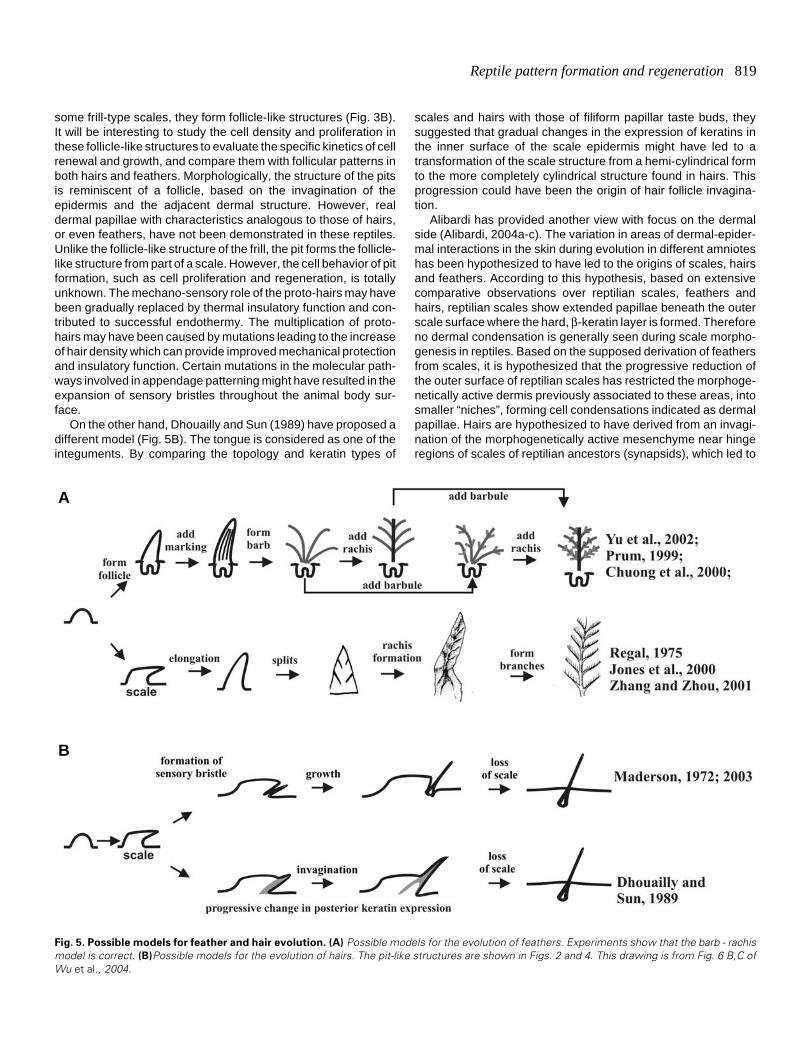
The origin and early evolutionary history of feather and hair isnot clear, but there are several hypothetical models (Fig. 5; Brush,1993; Prum, 1999; Chuong et al., 2000, 2003. Prum and Brush,2002; Alibardi, 2004a,b,c). One key feature shared by the hair andthe feather follicle is that both have adopted a proximal - distalgrowth mode. Another key shared feature is the invagination ofthe epidermis to form follicles, although produced via differentdevelopmental mechanisms. Both steps are important evolution-ary novelties.
From reptile skin to avian feathersReptilian scales and avian feathers have been considered as
homologous structures (Maderson, 1972). The epidermal cellswhich make feathers and avian scales arise from the sameprogenitors, suggesting that feathers evolved through modifica-tion of the embryonic epidermis (Sawyer and Knapp, 2003;Sawyer et al., 2005). However, there are some fundamentaldifferences in the development of scales and feathers. Normalscales do not form follicular structures. The mature scales aremade of an epithelial shell and a mesenchymal core (Wu et al.,2004). The outside of the epithelial shell is the suprabasal cor-neous (horny) layer.
Compared with reptilian scales, avian feathers have a muchmore complex topological organization (Yu et al., 2004; Lucasand Stettenheim, 1972). After the feather bud protrudes andelongates, the localized proliferation zone gradually shifts throughthe shaft and localizes proximally to the base of the feather(Chondankar et al., 2003). In the meantime, the epidermis sur-rounding the feather starts to invaginate into the dermis to formthe follicle. The dermal papilla is situated at the base of the follicle,and induces the epithelial collar above to continue proliferation(Yue et al., 2005) and branch (Yu et al., 2002; Yue et al., 2006).
How were reptilian scales transformed into feathers? In a
previous study, Regal (1975) suggested that reptilian scales firstunderwent elongation, later through etching of the elongatedscales to produce the branched feather vanes, and finally theinter-woven pennaceous feather barbs became plumulaceous.The Triassic archosaur fossil, Longisquama, which exhibitsbranches in its elongated scale, appears to support this hypoth-esis (Jones et al., 2000). Yet, developmental and molecularevidence and fossilized feathers suggest that feathers evolvedfrom conical hair-like outgrowths, with a cylindrical follicle (Prum,1999; Prum and Brush, 2002; Chuong et al., 2000; Yu et al., 2002;Alibardi, 2006b).
Since the 1990’s, some fantastic fossils have been discoveredin Jehol Biota (120 million years ago) in northern China (Zhou etal., 2003). They are unique because the integument and append-ages are well preserved due to the geology of that time (shallowlake with frequent volcanic eruptions in the vicinity). Some ofthese fossils are bona fide dinosaurs as judged by the skeleton,but there are feather-like appendages on their integument (Chenet al., 1998; Xu et al., 1999, 2001, 2003, Prum and Brush, 2002).Are they real feathers? Some are, and while some do not fulfill allthe definitions of a true feather (Chuong et al., 2003), they dorepresent precursor appendages of feathers and can be namedproto-feathers.
Laboratory experiments showed retinoic acid can producefeather growth on a scale (Dhouailly et al., 1980). Ectopic expres-sion of β-catenin in the scale can cause a small region of the scaleepidermis to become feather follicles (Widelitz et al., 2000).Suppression of BMP receptor activity and over-expression ofdelta can produce similar results (reviewed in Widelitz et al.,2003). However, the experimental observations have alwaysbeen that a small proportion of scale epidermis is activated tobecome feather primordia. It invaginates to form a feather follicle,and together they become "feathery scales". Conversion of awhole scale into one feather, as predicted by Jones et al., 2000,is never observed. Rather, it appears that a small group of stemcells in the scale is activated by molecular perturbation and thesecells go on to form a feather.
The levels of BMPs and Shh were shown to regulate the patternof branching morphogenesis (relative amounts of rachis andramus, Yu et al., 2002; Harris et al., 2002). When the anterior -posterior Wnt 3a gradient in bilaterally symmetric flight feathers isflattened experimentally, the feathers are converted into radiallysymmetric downy-like feathers (Yue et al., 2006). Together, theseresults shed light on how feathers were built stepwise in develop-ment and evolution through multiple evolutionary novelties overapproximately 50 million years (Fig. 5A; Chuong et al., 2000,2001, 2003; Prum, 1999, 2005; Prum and Brush, 2002).
From reptile skin to mammalian hairUnlike the fantastic discovery of feathered dinosaurs and
protofeathers in fossils (reviewed in Prum and Brush,; Chuong etal., 2003), so far, there have been no paleontological records toprovide clues which can help elucidate the presumed evolution-ary steps taken from reptilian scales to mammalian hairs(Spearman, 1964; Maderson, 1972; Alibardi, 2004a,b,c). Severalhypotheses have been proposed as to the origin of hair (Fig. 5B).Maderson (1970, 1972; 2003) hypothesized that hairs arose fromreptilian sensory appendages of the mechanoreceptor type (pit-organ) that were located in the hinge region of ancient reptiles. In
Reptile pattern formation and regeneration 819
some frill-type scales, they form follicle-like structures (Fig. 3B).It will be interesting to study the cell density and proliferation inthese follicle-like structures to evaluate the specific kinetics of cellrenewal and growth, and compare them with follicular patterns inboth hairs and feathers. Morphologically, the structure of the pitsis reminiscent of a follicle, based on the invagination of theepidermis and the adjacent dermal structure. However, realdermal papillae with characteristics analogous to those of hairs,or even feathers, have not been demonstrated in these reptiles.Unlike the follicle-like structure of the frill, the pit forms the follicle-like structure from part of a scale. However, the cell behavior of pitformation, such as cell proliferation and regeneration, is totallyunknown. The mechano-sensory role of the proto-hairs may havebeen gradually replaced by thermal insulatory function and con-tributed to successful endothermy. The multiplication of proto-hairs may have been caused by mutations leading to the increaseof hair density which can provide improved mechanical protectionand insulatory function. Certain mutations in the molecular path-ways involved in appendage patterning might have resulted in theexpansion of sensory bristles throughout the animal body sur-face.
On the other hand, Dhouailly and Sun (1989) have proposed adifferent model (Fig. 5B). The tongue is considered as one of theinteguments. By comparing the topology and keratin types of
scales and hairs with those of filiform papillar taste buds, theysuggested that gradual changes in the expression of keratins inthe inner surface of the scale epidermis might have led to atransformation of the scale structure from a hemi-cylindrical formto the more completely cylindrical structure found in hairs. Thisprogression could have been the origin of hair follicle invagina-tion.
Alibardi has provided another view with focus on the dermalside (Alibardi, 2004a-c). The variation in areas of dermal-epider-mal interactions in the skin during evolution in different amnioteshas been hypothesized to have led to the origins of scales, hairsand feathers. According to this hypothesis, based on extensivecomparative observations over reptilian scales, feathers andhairs, reptilian scales show extended papillae beneath the outerscale surface where the hard, β-keratin layer is formed. Thereforeno dermal condensation is generally seen during scale morpho-genesis in reptiles. Based on the supposed derivation of feathersfrom scales, it is hypothesized that the progressive reduction ofthe outer surface of reptilian scales has restricted the morphoge-netically active dermis previously associated to these areas, intosmaller “niches”, forming cell condensations indicated as dermalpapillae. Hairs are hypothesized to have derived from an invagi-nation of the morphogenetically active mesenchyme near hingeregions of scales of reptilian ancestors (synapsids), which led to
Fig. 5. Possible models for feather and hair evolution. (A) Possible models for the evolution of feathers. Experiments show that the barb - rachismodel is correct. (B)Possible models for the evolution of hairs. The pit-like structures are shown in Figs. 2 and 4. This drawing is from Fig. 6 B,C ofWu et al., 2004.
B
A
820 C. Chang et al.
the formation of the dermal papilla associated with hair.With regards to keratin evolution, early theropsid amniotes,
including mammalian ancestors, never possessed and later lostβ-keratogenic tissue. Their α-keratogenic epidermis was tough-ened by the evolution of mammalian-type HRP (histidine-richprotein, mainly filaggrins) (Maderson, 2003) and their hard deriva-tives, such as hairs, claws and horns, evolved specialized typesof KAPs, rich in glycine-tyrosine or in cysteine (Rogers, 2004;Alibardi et al., 2007).
Pattern Formation
Scale pattern: size, number, arrangementThe arrangements of reptile scales and pigment patterns in
different species are so exquisite, specific and stable, they areusually used as characteristics for the classification of species invertebrates (Landmann, 1986; Pianka et al., 2003). Interestingly,scales also show regional specificity with different sizes andarrangements in different parts of the body surface (for example,the green iguana in Fig. 3). Because most scales do not elongatetoo much in length, the patterns are usually exquisite and obvious,without being obscured as in hair or feather filaments, makingthem a good model for the study of biological pattern formation.In contrast to other types of patterns observed in the skin, such asthose of leopard and other vertebrate species, lizard or snake skinis relatively accessible experimentally and thereby providing uswith paradigm systems for furthering our understanding in thisarea.
ModelsOne of the first mechanisms suggested for self-organized
biological pattern formation was that of diffusion-driven instability
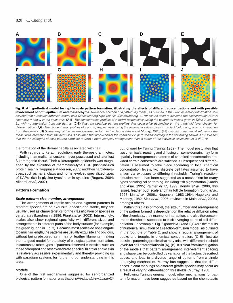
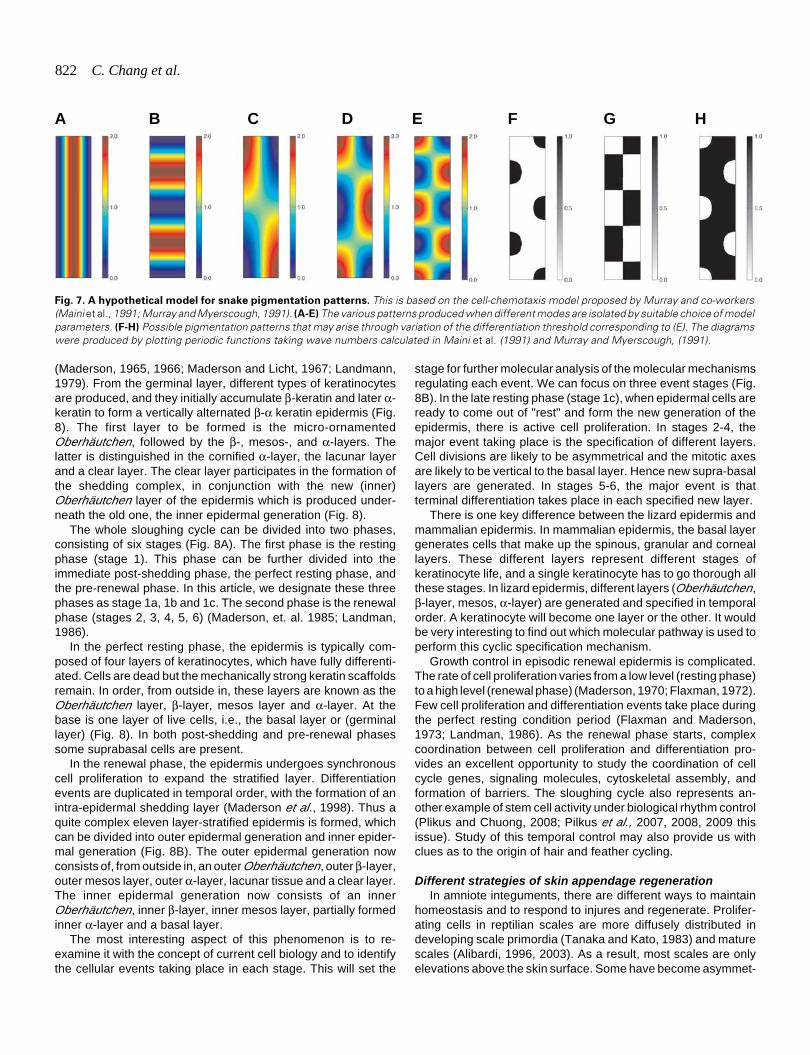
Fig. 6. A hypothetical model for reptile scale pattern formation, illustrating the effects of different concentrations and with possible
involvement of both epithelium and mesenchyme. Numerical solution of a patterning model, as outlined in the Supplementary Information. Weassume that a reaction-diffusion model with Schnakenberg-type kinetics (Schnakenberg, 1979) can be used to describe the concentration of twochemicals v and w in the epidermis. (A,B) The concentration profiles of v and w respectively, using the parameter values given in Table 2 (column3), with no interaction from the dermis. (C-E) illustrate possible pattern profiles that could arise depending on the threshold level chosen fordifferentiation. (F,G) The concentration profiles of v and w, respectively, using the parameter values given in Table 2 (column 4), with no interactionfrom the dermis. (H) Spatial map of the pattern assumed to form in the dermis (Shaw and Murray, 1990). (I,J) Results of numerical solution of themodel with interaction from the dermis: it is assumed that production of the chemical v is perturbed according to the patterning shown in (C). We seethat the wavelengths of each pattern combine to form a more complex arrangement than in either of the individual cases shown in (F,G,H).
put forward by Turing (Turing, 1952). The model postulates thattwo chemicals, reacting and diffusing on some domain, may formspatially heterogeneous patterns of chemical concentration pro-vided certain constraints are satisfied. Subsequent cell differen-tiation is assumed to take place according to local chemicalconcentration levels, with discrete cell fates assumed to havearisen via exposure to differing thresholds. Turing’s reaction-diffusion model has been suggested as a mechanism for manytypes of biological patterning, including fish pigmentation (Kondoand Asai, 1995; Painter et al., 1999; Kondo et al., 2009, thisissue), feather bud, scale and hair follicle formation (Jung et al.,1998; Lin et al., 2006,; Nagorcka, 1983-1984; Nagorcka andMooney, 1982; Sick et al., 2006; reviewed in Maini et al., 2006),amongst others.
Within this class of model, the size, number and arrangementof the pattern formed is dependent on the relative diffusion ratesof the chemicals, their manner of interaction, and also the concen-tration thresholds supposed to elicit diverging paths of cell differ-entiation. For example, Fig. 6 (panels A,B and F,G) are the resultsof numerical simulation of a reaction-diffusion model, as outlinedin the footnote of Table 2, and show a regular arrangement ofpeaks and troughs in chemical concentration. (C-E) illustratepossible patterning profiles that may arise with different thresholdlevels for cell differentiation in (A), (B). It is clear from investigationof the models that pattern arrangement, inter-element spacingand shape can be controlled by variation of the factors describedabove, and lead to a diverse range of patterns from a singleunderlying mechanism. Murray has suggested that the differ-ences in coat markings on different giraffe species may occur asa result of varying differentiation thresholds (Murray, 1988).
Following Turing’s original model, other mechanisms for pat-tern formation have been suggested based on the chemotactic
F G H I J
A B C D E
Reptile pattern formation and regeneration 821
ability of cells to respond to their environment. The most well-known models for cell-chemotaxis were proposed by Patlak(Patlak, 1963) and Keller and Segel (Keller and Segel, 1970). Theunderlying mechanism involves cells moving up gradients inchemical concentration (chemotaxis) and amplification of thesegradients as cells secrete the chemical. As with the reaction-diffusion model, spatially heterogeneous patterns of chemicalconcentration may arise, and these are mirrored by patterns in celldensity.
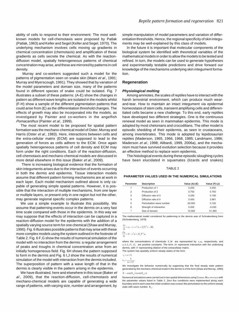
Murray and co-workers suggested such a model for thepatterns of pigmentation seen on snake skin (Maini et al., 1991;Murray and Myerscough, 1991). They showed that by variation ofthe model parameters and domain size, many of the patternsfound in different species of snake could be isolated. Fig. 7illustrates a subset of these patterns: (A-E) show the changes inpattern as different wave lengths are isolated in the models whilst(F-H) show a sample of the different pigmentation patterns thatcould arise from (E) as the differentiation threshold changes. Theeffects of growth may also be incorporated into the models, asinvestigated by Painter and co-workers in the angelfishPomacanthus (Painter et al., 1999).
The most recent model to be proposed for spatial patternformation was the mechano-chemical model of Oster, Murray andHarris (Oster et al., 1983). Here, interactions between cells andthe extra-cellular matrix (ECM) are supposed to lead to thegeneration of forces as cells adhere to the ECM. Once againspatially heterogeneous patterns of cell density and ECM mayform under the right conditions. Each of the reaction-diffusion,cell-chemotaxis and mechano-chemical models are discussed inmore detail elsewhere in this issue (Baker et al., 2009).
There is increasing biological evidence that the formation ofskin integuments arises due to the interaction of events occurringin both the dermis and epidermis. Tissue interaction modelsassume that different pattern forming mechanisms are at work ineach layer. Each model mechanism outlined above is only ca-pable of generating simple spatial patterns. However, it is pos-sible that the interaction of multiple mechanisms, from one layeror multiple layers, or present only in one region but not the other,may generate regional specific complex patterns.
We use a simple example to illustrate this possibility. Weassume that patterning events occur in the dermis on a very fasttime scale compared with those in the epidermis. In this way wemay suppose that the effects of interaction can be captured in areaction-diffusion model for the epidermis with the addition of aspatially varying source term for one chemical (Shaw and Murray,1990). Fig. 6 illustrates possible patterns that may arise with thesemore complex models using the system outlined in the footnote ofTable 2. Fig. 6 F,G show the results of numerical simulation of themodel with no interaction from the dermis: a regular arrangementof peaks and troughs in chemical concentration arise from aninitially homogeneous field. Fig. 6H shows the pattern supposedto form in the dermis and Fig. 6 I,J show the results of numericalsimulation of the model with interaction from the dermis included.The superposition of pattern with a wave length of that in thedermis is clearly visible in the pattern arising in the epidermis.
We have illustrated, here and elsewhere in this issue (Baker etal., 2009), that the reaction-diffusion, cell-chemotaxis andmechano-chemical models are capable of generating a widerange of patterns, with varying size, number and arrangement, by
simple manipulation of model parameters and variation of differ-entiation thresholds. Hence, the regional specificity of skin integu-ments may be well-explained by this class of models.
In the future it is important that molecular components of thebiological system be identified with theoretical variables of themathematical models in order to allow the models to be tested andrefined. In turn, the models can be used to generate hypothesesand experimentally testable predictions and drive forward ourknowledge of the mechanisms underlying skin integument forma-tion.
Regeneration
Physiological moltingAmong amniotes, the scales of reptiles have to interact with the
harsh terrestrial environment, which can produce much wear-and-tear. How to maintain an intact integument via epidermalhomeostasis of stem cells, transient amplifying cells and differen-tiated cells became a new challenge. To this end, early reptileshave developed two different strategies. One is the continuousrenewal model as seen in mammalian epidermis. This mode isadopted by most chelonians and crocodilians. The other model isepisodic shedding of their epidermis, as seen in crustaceans,among invertebrates. This mode is adopted by lepidosaurianreptiles (Maderson, 1965, 1966, 1968, 1985; Landmann, 1986;Maderson et al., 1998; Alibardi, 1999, 2006a), and the mecha-nism must have survived evolution selection because it providesan effective method to replace worn epidermis.
The histological events during these episodic sloughing cycleshave been elucidated in squamates (lizards and snakes)
Parameter Description Value (A)-(E) Value (F)-(J)
1 Production of 1 0.250 0.250
2 Production of 2 0.750 0.750
3 Diffusion rate of 3 0.230 0.195
4 Diffusion rate of 4 5.000 3.961
5 Perturbation wave number 20.000 0.122
6 Strength of interaction 0.000 -0.030
7 Size of domain 10.000 51.363
TABLE 2
PARAMETER VALUES USED IN THE NUMERICAL SIMULATIONS
The mathematical model considered for patterning in the dermis was of Schnakenberg form(Schnakenberg, 1979)
∂∂= − + + ∇ −
∂∂
= − + ∇
vta v v w D v
wtb v w D w
v
w
2 2
2 2
δθ ,
,
where the concentrations of chemicals V W, are represented by v w, , respectively, anda b D Dv w, , , ,δ are positive constants. The term δθ represents interaction with the underlyingdermis, with θ representing dilation of the extracellular matrix.The system has spatially uniform steady states of the form
v a b
w ba b
0
0 2
= +
=+
,
( ).
We investigate the behavior numerically by supposing that the final steady state patterngenerated by the mechano-chemical model in the dermis is of the form (Shaw and Murray, 1990)
θ ∼ cos( )cos( ).K x K ym m
Numerical simulations were carried out in two spatial dimensions using COMSOL MULTIPHYSICS withthe parameter values listed in Table 2. Zero flux conditions were implemented along eachboundary and in each case the initial condition was a wave-like perturbation to the uniform steadystate with wave number Km .
822 C. Chang et al.
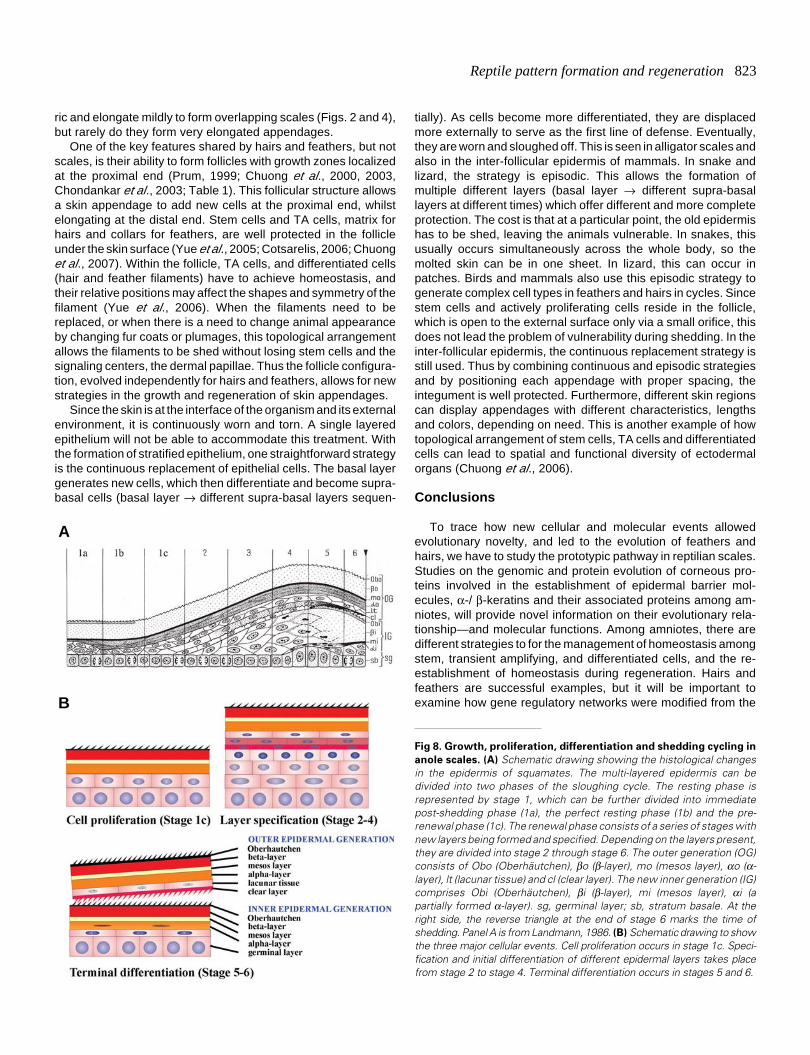
(Maderson, 1965, 1966; Maderson and Licht, 1967; Landmann,1979). From the germinal layer, different types of keratinocytesare produced, and they initially accumulate β-keratin and later α-keratin to form a vertically alternated β-α keratin epidermis (Fig.8). The first layer to be formed is the micro-ornamentedOberhäutchen, followed by the β-, mesos-, and α-layers. Thelatter is distinguished in the cornified α-layer, the lacunar layerand a clear layer. The clear layer participates in the formation ofthe shedding complex, in conjunction with the new (inner)Oberhäutchen layer of the epidermis which is produced under-neath the old one, the inner epidermal generation (Fig. 8).
The whole sloughing cycle can be divided into two phases,consisting of six stages (Fig. 8A). The first phase is the restingphase (stage 1). This phase can be further divided into theimmediate post-shedding phase, the perfect resting phase, andthe pre-renewal phase. In this article, we designate these threephases as stage 1a, 1b and 1c. The second phase is the renewalphase (stages 2, 3, 4, 5, 6) (Maderson, et. al. 1985; Landman,1986).
In the perfect resting phase, the epidermis is typically com-posed of four layers of keratinocytes, which have fully differenti-ated. Cells are dead but the mechanically strong keratin scaffoldsremain. In order, from outside in, these layers are known as theOberhäutchen layer, β-layer, mesos layer and α-layer. At thebase is one layer of live cells, i.e., the basal layer or (germinallayer) (Fig. 8). In both post-shedding and pre-renewal phasessome suprabasal cells are present.
In the renewal phase, the epidermis undergoes synchronouscell proliferation to expand the stratified layer. Differentiationevents are duplicated in temporal order, with the formation of anintra-epidermal shedding layer (Maderson et al., 1998). Thus aquite complex eleven layer-stratified epidermis is formed, whichcan be divided into outer epidermal generation and inner epider-mal generation (Fig. 8B). The outer epidermal generation nowconsists of, from outside in, an outer Oberhäutchen, outer β-layer,outer mesos layer, outer α-layer, lacunar tissue and a clear layer.The inner epidermal generation now consists of an innerOberhäutchen, inner β-layer, inner mesos layer, partially formedinner α-layer and a basal layer.
The most interesting aspect of this phenomenon is to re-examine it with the concept of current cell biology and to identifythe cellular events taking place in each stage. This will set the
stage for further molecular analysis of the molecular mechanismsregulating each event. We can focus on three event stages (Fig.8B). In the late resting phase (stage 1c), when epidermal cells areready to come out of "rest" and form the new generation of theepidermis, there is active cell proliferation. In stages 2-4, themajor event taking place is the specification of different layers.Cell divisions are likely to be asymmetrical and the mitotic axesare likely to be vertical to the basal layer. Hence new supra-basallayers are generated. In stages 5-6, the major event is thatterminal differentiation takes place in each specified new layer.
There is one key difference between the lizard epidermis andmammalian epidermis. In mammalian epidermis, the basal layergenerates cells that make up the spinous, granular and corneallayers. These different layers represent different stages ofkeratinocyte life, and a single keratinocyte has to go thorough allthese stages. In lizard epidermis, different layers (Oberhäutchen,β-layer, mesos, α-layer) are generated and specified in temporalorder. A keratinocyte will become one layer or the other. It wouldbe very interesting to find out which molecular pathway is used toperform this cyclic specification mechanism.
Growth control in episodic renewal epidermis is complicated.The rate of cell proliferation varies from a low level (resting phase)to a high level (renewal phase) (Maderson, 1970; Flaxman, 1972).Few cell proliferation and differentiation events take place duringthe perfect resting condition period (Flaxman and Maderson,1973; Landman, 1986). As the renewal phase starts, complexcoordination between cell proliferation and differentiation pro-vides an excellent opportunity to study the coordination of cellcycle genes, signaling molecules, cytoskeletal assembly, andformation of barriers. The sloughing cycle also represents an-other example of stem cell activity under biological rhythm control(Plikus and Chuong, 2008; Pilkus et al., 2007, 2008, 2009 thisissue). Study of this temporal control may also provide us withclues as to the origin of hair and feather cycling.
Different strategies of skin appendage regenerationIn amniote integuments, there are different ways to maintain
homeostasis and to respond to injures and regenerate. Prolifer-ating cells in reptilian scales are more diffusely distributed indeveloping scale primordia (Tanaka and Kato, 1983) and maturescales (Alibardi, 1996, 2003). As a result, most scales are onlyelevations above the skin surface. Some have become asymmet-
Fig. 7. A hypothetical model for snake pigmentation patterns. This is based on the cell-chemotaxis model proposed by Murray and co-workers(Maini et al., 1991; Murray and Myerscough, 1991). (A-E) The various patterns produced when different modes are isolated by suitable choice of modelparameters. (F-H) Possible pigmentation patterns that may arise through variation of the differentiation threshold corresponding to (E). The diagramswere produced by plotting periodic functions taking wave numbers calculated in Maini et al. (1991) and Murray and Myerscough, (1991).
A B C D E F G H
Reptile pattern formation and regeneration 823
ric and elongate mildly to form overlapping scales (Figs. 2 and 4),but rarely do they form very elongated appendages.
One of the key features shared by hairs and feathers, but notscales, is their ability to form follicles with growth zones localizedat the proximal end (Prum, 1999; Chuong et al., 2000, 2003,Chondankar et al., 2003; Table 1). This follicular structure allowsa skin appendage to add new cells at the proximal end, whilstelongating at the distal end. Stem cells and TA cells, matrix forhairs and collars for feathers, are well protected in the follicleunder the skin surface (Yue et al., 2005; Cotsarelis, 2006; Chuonget al., 2007). Within the follicle, TA cells, and differentiated cells(hair and feather filaments) have to achieve homeostasis, andtheir relative positions may affect the shapes and symmetry of thefilament (Yue et al., 2006). When the filaments need to bereplaced, or when there is a need to change animal appearanceby changing fur coats or plumages, this topological arrangementallows the filaments to be shed without losing stem cells and thesignaling centers, the dermal papillae. Thus the follicle configura-tion, evolved independently for hairs and feathers, allows for newstrategies in the growth and regeneration of skin appendages.
Since the skin is at the interface of the organism and its externalenvironment, it is continuously worn and torn. A single layeredepithelium will not be able to accommodate this treatment. Withthe formation of stratified epithelium, one straightforward strategyis the continuous replacement of epithelial cells. The basal layergenerates new cells, which then differentiate and become supra-basal cells (basal layer → different supra-basal layers sequen-
tially). As cells become more differentiated, they are displacedmore externally to serve as the first line of defense. Eventually,they are worn and sloughed off. This is seen in alligator scales andalso in the inter-follicular epidermis of mammals. In snake andlizard, the strategy is episodic. This allows the formation ofmultiple different layers (basal layer → different supra-basallayers at different times) which offer different and more completeprotection. The cost is that at a particular point, the old epidermishas to be shed, leaving the animals vulnerable. In snakes, thisusually occurs simultaneously across the whole body, so themolted skin can be in one sheet. In lizard, this can occur inpatches. Birds and mammals also use this episodic strategy togenerate complex cell types in feathers and hairs in cycles. Sincestem cells and actively proliferating cells reside in the follicle,which is open to the external surface only via a small orifice, thisdoes not lead the problem of vulnerability during shedding. In theinter-follicular epidermis, the continuous replacement strategy isstill used. Thus by combining continuous and episodic strategiesand by positioning each appendage with proper spacing, theintegument is well protected. Furthermore, different skin regionscan display appendages with different characteristics, lengthsand colors, depending on need. This is another example of howtopological arrangement of stem cells, TA cells and differentiatedcells can lead to spatial and functional diversity of ectodermalorgans (Chuong et al., 2006).
Conclusions
To trace how new cellular and molecular events allowedevolutionary novelty, and led to the evolution of feathers andhairs, we have to study the prototypic pathway in reptilian scales.Studies on the genomic and protein evolution of corneous pro-teins involved in the establishment of epidermal barrier mol-ecules, α-/ β-keratins and their associated proteins among am-niotes, will provide novel information on their evolutionary rela-tionship—and molecular functions. Among amniotes, there aredifferent strategies to for the management of homeostasis amongstem, transient amplifying, and differentiated cells, and the re-establishment of homeostasis during regeneration. Hairs andfeathers are successful examples, but it will be important toexamine how gene regulatory networks were modified from the
Fig 8. Growth, proliferation, differentiation and shedding cycling in
anole scales. (A) Schematic drawing showing the histological changesin the epidermis of squamates. The multi-layered epidermis can bedivided into two phases of the sloughing cycle. The resting phase isrepresented by stage 1, which can be further divided into immediatepost-shedding phase (1a), the perfect resting phase (1b) and the pre-renewal phase (1c). The renewal phase consists of a series of stages withnew layers being formed and specified. Depending on the layers present,they are divided into stage 2 through stage 6. The outer generation (OG)consists of Obo (Oberhäutchen), βo (β-layer), mo (mesos layer), αo (α-layer), lt (lacunar tissue) and cl (clear layer). The new inner generation (IG)comprises Obi (Oberhäutchen), βi (β-layer), mi (mesos layer), αi (apartially formed α-layer). sg, germinal layer; sb, stratum basale. At theright side, the reverse triangle at the end of stage 6 marks the time ofshedding. Panel A is from Landmann, 1986. (B) Schematic drawing to showthe three major cellular events. Cell proliferation occurs in stage 1c. Speci-fication and initial differentiation of different epidermal layers takes placefrom stage 2 to stage 4. Terminal differentiation occurs in stages 5 and 6.
B
A
824 C. Chang et al.
basal network for scales in a succession of evolutionary novelcellular events. We have identified the formation of localizedgrowth zones and the invagination of follicles as a key step. Thespatial arrangement of these localized growth units is not onlyimportant for integument function but also for understandingbiological pattern formation. Since most scales are short in height,their arrangement, size and shapes are clearly revealed, makingthem an exquisite model for experimental analyses on howbiological patterns form. While research on reptile scales hasbeen carried out, the analyses of molecules involved in scalemorphogenesis have lagged behind. Here we underscore howthe development of this novel experimental model will be valuablein filling in the gaps of our understanding of the biology of amnioteinteguments.
AcknowledgementThis work is supported by grants from NIA and NIAMS (CMC). Dr.
Alibardi is supported by a 60% University of Bologna Grant. Dr. ChengChang is supported by grants from China NSFC 30770240 and LanzhouUniversity. REB is supported by an RCUK Fellowship, a MicrosoftEuropean Research Fellowship and a Junior Research Fellowship fromSt Hugh’s College, Oxford. PKM is partially supported by a Royal Society-Wolfson Merit Award.
References
ALEXANDER, N. J. (1970). Comparison of α an β keratin in reptiles. Z. Zellforsch.110: 153-165.
ALIBARDI, L. (1996) Scale morphogenesis during embryonic development in thelizard Anolis lineatopus. J. Anat. 188:713-725.
ALIBARDI, L. (1998). Differentiation of the epidermis during scale formation inembryos of lizard. J. Anat. 192: 173-186.
ALIBARDI, L. (1999). Keratohyalin-like granules and differentiation of the sheddingcomplex in embryonic and regenerating epidermis of lizards and Sphenodon(Reptilia, Lepidosauria). Amphibia-Reptilia. 20: 11-23.
ALIBARDI, L. (2000). Ultrastructural localization of alpha-keratins in the regenerat-ing epidermis of the lizard Podarcis muralis during formation of the sheddinglayer. Tissue Cell. 32: 153-162.
ALIBARDI, L. (2001). Keratohyalin-like granules in lizard epidermis: evidence fromcytochemical, autoradiographic, and microanalytical studies. J. Morphol. 248:64-79.
ALIBARDI, L. (2002a). Histidine uptake in the epidermis of lizards and snakes inrelation to the formation of the shedding complex. J. Exp. Zool. 292: 331-344.
ALIBARDI, L. (2002b). Loricrine-like immunoreactivity during keratinization in lizardepidermis. J. Morphol. 254: 132-138.
ALIBARDI, L. (2003). Aaptation to the land: The skin of reptiles in comparison to thatof amphibians and endotherm amniotes. J. Exp. Zoolog. B. Mol. Dev. Evol.298(1): 12-41.
ALIBARDI, L. (2004a). Dermo-epidermal interactions in reptilian scales: specula-tions on the evolution of scales, feathers, and hairs. J. Exp. Zoolog. B. Mol. Dev.Evol. 302(4): 365-383.
ALIBARDI, L. (2004b). Fine structure and immunocytochemistry of monotremehairs with emphasis on the inner root sheath and trichohyaline-based cornifica-tion during hair evolution. J. Morphol. 261: 345-363.
ALIBARDI, L. (2004c). Comparative aspects of the inner root sheath in adult anddeveloping hairs of mammals in relation to the evolution of hairs. J. Anat. 205:179-200.
ALIBARDI, L. (2006a). Structural and immunocytochemical characterization ofkeratinization in vertebrate epidermis and epidermal derivatives. Int. Rev. Cytol.253: 177–259.
ALIBARDI, L. (2006b). Cells of embryonic and regenerating germinal layers withinbarb ridges: implication for the development, evolution and diversification offeathers. J. Subm. Cytol. Pathol. 38: 51-76.
ALIBARDI, L., SAWYER, R. H. (2002). Immunocytochemical analysis of β-keratinsin the epidermis of chelonians, lepidosaurians, and archosaurians. J. Exp. Zool.293: 27–38.
ALIBARDI, L., THOMPSON, M. B. (2000). Scale morphogenesis and ultrastructureof dermis during embryo development in the alligator (Alligator mississippiensis,Crocodilia, Reptilia). Acta Zool-Stockolm. 81: 325-338.
ALIBARDI, L., THOMPSON, M. B. (2001). Fine structure of the developing epider-mis in the embryo of the american alligator (Alligator mississippiensis, Crocodlilia,Reptilia). J. Anat. 198: 265-282.
ALIBARDI, L., THOMPSON, M. B. (2002). Keratinization and ultrastructure of theepidermis of late embryonic stages in the alligator (Alligator mississippiensis).J. Anat. 201: 71-84.
ALIBARDI, L., TONI, M. (2005). Immunolocalization and characterization of corni-fication proteins in snake epidermis. Anat. Rec. A: Discov. Mol. Cell Evol. Biol.282: 138–146.
ALIBARDI, L., TONI, M. (2006). Cytochemical, biochemical and molecular aspectsof the process of keratinization in the epidermis of reptilian scales. Prog.Histochem. Cytochem. 40: 173–134.
ALIBARDI, L., TONI, M., DALLLA, V. L. (2007). Review. Hard keratins in reptilianepidermis in comparison to those of birds and mammals. Exp. Dermatol. 16:961-976.
BADEN, H. P., MADERSON, P. F. A. (1970). Morphological and biophysicalidentification of fibrous proteins in the amniote epidermis. J. Exp. Zool. 174: 225-232.
BADEN, H. P., SVIOKLA. S., ROTH, I. (1974). The structural protein of reptilianscales. J. Exp. Zool. 187: 287-294.
BAKER, R. E., SCHNELL, S., PHILIP, PK, (2009). Waves and patterning indevelopmental biology: vertebrate segmentation and feather bud formation ascase studies. Int. J. Dev. Biol. 53: 783-794. (doi: 10.1387/ijdb.072493rb)
BENTLEY, P. J., SCHMIDT-NIELSEN, K. (1966). Cutaneous water-loss in reptiles.Science. 151: 1547-1549.
BRUSH, A. H. (1993). The origin of feather: a novel approach. In: Farner, D., Kling,J., Parker, K. editors. Avian biology. New York: Academic Press. pp. 121–162.
CARVER, W. E., SAWYER, R. H. (1987). Development and keratinization of theepidermis in the common lizard, Anolis carolinensis. J. Exp. Zool. 243: 435-443.
CHANG, C., ZHENG, R. L. (2003). Effects of ultraviolet B on epidermal morphology,shedding, lipid peroxide, and antioxidant enzymes in Cope’s rat snake (Elaphetaeniura). J. Photochem. Photobiol. B. Biol. 72: 79-85.
CHEN, P., DONG, Z., ZHEN, S. (1998). An exceptionally well preserved theropoddinosaur from the Yixian Formation of China. Nature. 391: 147–52.
CHODANKAR, R., CHANG, C.-H., YUE, Z., SUKSAWEANG, S., BURRUS, L.,CHUONG, C.-M., AND WIDELITZ, R.B. 2003. Shift of Localized Growth Zones(LoGZ) Contributes to the Morphogenesis of Skin Appendages: Associationwith Wnt/B-catenin activities. J. Invest. Dermatol. 120: 19-26.
CHUONG, C.-M. (1998). Molecular Basis of Epithelial Appendage Morphogenesis.Landes Bioscience, Austin, TX: Landes Bioscience.
CHUONG, C.-M., CHODANKAR, R., WIDELITZ, R.B., JIANG, T. X. (2000). Evo-Devo of Feathers and Scales: Building complex epithelial appendages. Curr.Opin. in Dev. and Gen. 10: 449-456.
CHUONG, C.-M., COTSARELIS, G., STENN, K. (2007). Defining hair follicles in theage of stem cell bio-engineering. J. Invest. Dermatol. 127: 2098-2100.
CHUONG, C.-M., HOMBERGER, D. G. (2003). Development and Evolution of theAmniote Integument: Current Landscape and Future Horizon. J. Exp. Zool.298B: 1-11.
CHUONG, C.-M., HOU, L. H., CHEN, P. J., WU, P., PATEL, N., CHEN, Y. P. (2001).Dinosaur’s feather and Chicken’s tooth? Tissue engineering of the integument.Eur. J. Dermatol. 11: 286-292.
CHUONG C.-M., NICKLOFF, B.J., ELIAS P.M., GOLDSMITH, L.A., MACHER, E.,MADERSON, P.A., SUNDBERG, J.P., TAGAMI, H., PLONGKA, P.M.,THESTRUP-PEDERSEN, K., BERNARD, B.A., SCRODER, J.M., DOTTO, P.,CHANG C,-H., WILLIAMS, M.L., FEINGOLD, K.R., KING, L.E., KLIGMAN,A.M., REES, J.L., CHRISTOPHERS, E. (2002). What is the ‘true’ function ofskin. Exp. Dermatol. 11: 159-187.
CHUONG, C-M., WU, P., ZHANG, F.-C., XU, X., YU, M., WIDELITZ, R. B., JIANG,T. -X., HOU, L. (2003). Adaptation to the Sky: Defining the Feather with
Reptile pattern formation and regeneration 825
Integument fossils from Mesozoic China and experimental evidence frommolecular laboratories. J. Exp. Zool. 298B: 42-56.
CHUONG, C-M., WU, P., PLIKUS MV, JIANG, TX, WIDELITZ, RB. (2006). Engi-neering Stem cells into organs: Topobiological transformations demonstratedby beak, feather and other ectodermal organ morphogenesis. Curr.Top. Dev.Biol. 72: 237-274.
COOPER, W. E., GREENBERG, N. (1992). Reptilian coloration and behaviour. InC Gans, F Billett, MADERSON, P. F. A., eds. Biology of the reptilia. Vol. 18.Physiology E. Chicago-London: Chicago University Press, pp. 298-423.
COTSARELIS G. (2006). Epithelial stem cells: a folliculocentric view. J InvestDermatol. 126: 1459-1468.
DALLA VALLE, L., NARDI, A., TOFFOLO, V., NIERO, C., TONI, M., ALIBARDI, L.(2007). Cloning and characterization of scale β-keratins in the differentiatingepidermis of geckoes show they are glycine-proline-serine-rich proteins with acentral motif homologous to avian beta-keratins. Dev. Dyn. 236: 374–388.
DALLA VALLE, L., TOFFOLO, V., BELVEDERE, P., ALIBARDI, L. (2005). Isolationof a mRNA encoding a glycine-proline-rich beta-keratin expressed in theregenerating epidermis of lizard. Dev. Dyn. 234: 934–947.
DAVIS, J. E., SPOTILA, J. R., SCHEFLER, W. C. (1980). Evaporative water lossfrom the american alligator, Alligator mississippiensis: the relative importanceof respiratory and cutaneous components and the regulatory role of the skin.Comp. Biochem. Phys. 67A: 439-446.
DHOUAILLY, D. (1975). Formation of cutaceous appendages in dermo-epidermalrecombinaitons between reptiles, birds and mammals. Wilhelm Roux’ Arch.Entwicklungsmech Org. 177: 323-40.
DHOUAILLY, D., HARDY, M. H., SENGEL, P. (1980). Formation of feathers onchick foot scales: a stage-dependent morphogenetic response to retinoic acid.J. Embryol. Exp. Morphol. 58: 63-78.
DHOUAILLY, D., SUN, T. T. (1989). The mammalian tongue filiform papillae: atheoretical model for primitive hairs. In Trends in human hair growth andAlopecia research Eds. D. van Neste, LaCapelle, J. M., Antoine, J. L. KluwerAcad. Pub. Boston, pp. 29-34.
DUNSON, W. A., MAZZOTTI, F. J. (1988). Some aspects of water and sodiumexchange of freshwater crocodilians in fresh water and sea water: role of theintegument. Comp. Biochem. Phys. 90A: 391-396.
FLAXMAN, B. A. (1972). Cell differentiation and its control in the vertebrateepidermis. Am. Zool. 12: 13-25.
FLAXMAN, B. A., MADERSON, P. F. A. (1973). Relationship between pattern of cellmigration from the germinal layer and changing patterns of differentiation in thelizard epidermis. J. Exp. Zool. 183, 209-216.
FRASER, R. D., PARRY, D. A. (1996). The molecular structure of reptilian keratin.Int. J. Biol. Macromol. 19: 207–211.
FUCHS, E., MARCHUK, D. (1983). Type I and type II keratins have evolved fromlower eukaryotes to form the epidermal intermediate filaments in mammalianskin. Proc. Natl. Acad. Sci. USA. 80: 5857–5861.
GREGG, K., ROGERS, G. (1986). Feather keratins: composition, structure andbiogenesis. In: Bereither-Hahn J, Matoltsy G, Sylvia- Richards K, editors.Biology of the integument: vertebrates. New York: Springer-Verlag. pp. 666-694.
HARRIS, M. P., FALLON, J. F., PRUM, R. O. (2002). Shh-Bmp2 signaling moduleand the evolutionary origin and diversification of feathers. J. Exp. Zool. 294(2):160-176.
JONES, T. D., RUBEN, J. A., MARTIN, L. D., KUROCHKIN, E. N., FEDUCCIA, A.,MADERSON, P. F., HILLENIUS, W. J., GEIST, N. R., ALIFANOV, V. (2000).Nonavian feathers in a late Triassic archosaur. Science. 288: 2202-2205.
JUNG, H.-S., FRANCIS-WEST, F., WIDELITZ, R.B, JIANG, T.-X., TING, S.,TICKLE, C., WOLPERT, L. AND CHUONG, C.-M., (1998). Local inhibitoryaction of BMPs and their relationships with activators in feather formation:Implications for periodic patterning. Dev Biol 196: 11-23.
KELLER, E. F., SEGEL, L. A., (1970). Initiation of slime mold aggregation viewedas an instability. J. Theor. Biol. 26: 399-415.
KONDO, S., ASAI, R. (1995). A reaction-diffusion wave on the skin of the marineangelfish Pomacanthus. Nature. 376: 765-768.
KONDO, S, IWASHITA, M. AND YAMAGUCHI, M. (2009). How animals get theirskin patterns: fish pigment pattern as a live Turing wave. Int. J. Dev. Biol. 53:
851-856. (doi: 10.1387/ijdb.072502sk).
LANDMANN, L. (1979). Keratin formation and barrier mechanisms in the epidermisof Natrix natrix (Reptilia: Serpentes): an ultrastructural study. J. Morphol. 162:93-126.
LANDMANN, L. (1986). The skin of Reptiles: epidermis and dermis. In: Bereither-Hahn J, Matoltsy G, Sylvia-Richards K, editors. Biology of the integument:vertebrate. New York: Springer Verlag. pp. 150-187.
LANDMANN, L., STOLINSKI, C., MARTIN, B. (1981). The permeability barrier inthe epidermis of the grass snake during the resting stage of the sloughing cycle.Cell Tissue Res. 215: 369-382.
LILLYWHITE, H. B. (2006). Water relations of tetrapod integument. J. Exp. Biol.209: 202-226.
LILLYWHITE, H. B., MADERSON, P. F. A. (1982). Skin structure and permeability.In Gans, C., Pough, F. H. eds. Biology of the Reptilia; Physiological Ecology.Vol. 12. New York, London: Academic Press, pp. 397-442.
LIN, C.-M., JIANG, T. X., WIDELITZ, R. B., CHUONG, C.-M. (2006). Molecularsignaling in feather morphogenesis. Curr. Opin. Cell Biol. 18: 730-741.
LUCAS, A. M., STTETENHEIM, P. R. (1972). Avian Anatomy Integument. In:Agriculture Handbook 362. Agricultural Research Services. Washington, DC:US Department of Agriculture.
MADERSON, P. F. A. (1965). Histological changes in the epidermis of snakesduring the sloughing cycle. J. Zool. (London) 146: 98-113.
MADERSON, P. F. A. (1966). Histological changes in the epidermis of the Tokay(Gekko gecko) during the sloughing cycle. J. Morphol. 119: 39-50.
MADERSON, P. F. A. (1968). Observations on the epidermis of the Tuatara(Sphenodon punctatus). J. Anat. 103: 311-320.
MADERSON, P. F. A. (1970). Lizard hands and lizard glands: Models for evolution-ary study. Forma. Et. Functio. 3: 179-204.
MADERSON, P. F. A. (1972). When? Why? and How? Some speculations on theevolution of the vertebrate integument. Am. Zool. 12: 159-171.
MADERSON, P. F. A. (1985). Some developmental problems of the reptilianintegument. In: Gans C, Billett F, Maderson PF, editors. Biology of reptilia. NewYork: John Wiley & Sons. pp. 525–598.
MADERSON, P.F.A. (2003). Mammalian skin evolution: a reevaluation. Exp.Dermatol. 12: 233-236.
MADERSON, P. F. A., ALIBARDI, L. (2000). The development of the sauropsidintegument: a contribution to the problem of the origin and evolution of feathers.Am. Zool. 40: 513-529.
MADERSON, P. F. A., LICHT, P. (1967). The epidermal morphology and sloughingfrequency in normal and prolactin injected Anolis caroli;zensis (Iguanidae,Lacertilia). J. Morphol. 123: 157-172.
MADERSON, P. F. A. (1985). Some developmental problems of the reptilianintegument; MADERSON, P. F. A., GANS, C., BILLETT, F. eds. Biology of theReptilia. Vol. 14. New York: John Wiley & Sons; pp. 525–598.
MADERSON, P. F. A., RABINOWITZ, T., TANDLER, B., ALIBARDI, L. (1998).Ultrastructural contributions to an understanding of the cellular mechanismsinvolved in lizard skin shedding with comments on the function and evolution ofa unique Lepidosaurian phenomenon. J. Morphol. 236: 1–24.
MAINI, P. K., MYERSCOUGH, M. R., MURRAY, J. D., WINTERS, K. H. (1991).Bifurcating spatially heterogeneous solutions in a chemotaxis model for biologi-cal pattern formation. Bull. Math. Biol. 53: 701-719.
MAINI, PK, BAKER, RE, CHUONG, CM., (2006). The Turing model comes ofmolecular age. Science. 314: 1397-1398.
MENON, G. K., BROWN, B. E., ELIAS, P. M. (1986). Avian epidermal differentia-tion: role of lipids in permeability barrier function. Tissue Cell. 18: 71-82.
MENON, G. K., MADERSON, P. F. A., DREWES, R. C., BAPTISTA, L. F., PRICE,L. F., ELIAS, P. M. (1996). Ultrastructural organization of avian stratum corneumlipids as the basis for facultative cutaneous waterproofing. J. Morphol. 227: 1-13.
MURRAY, J. D. (1988). How the leopard gets its spots. Sci. Amer. 258: 80-87.
MURRAY, J. D., MYERSCOUGH, M. R. (1991). Pigmentation pattern formation onsnakes. J. Theor. Biol. 149: 361-375.
NAGORCKA, B. N., (1983-1984). Evidence for a reaction-diffusion system as amechanism controlling mammalian hair growth. Biosystems. 16: 323-332.
826 C. Chang et al.
NAGORCKA, B. N., MOONEY, J. R. (1982). The role of a reaction-diffusion systemin the formation of hair fibres. J. Theor. Biol. 98: 575-607.
OSTER, G., MURRAY, J. D., HARRIS, A. K. (1983). Mechanical aspects ofmesenchymal morphogenesis. J. Embryol. Exp. Morphol. 78: 83-125.
PAINTER, K. J., MAINI, P. K., OTHMER, H. G. (1999). Stripe formation in juvenilePomacanthus explained by a generalized Turing mechanism with chemotaxis.Proc. Natl. Acad. Sci USA. 96: 5549-5554.
PATLAK, C. A. (1963). Random walk with persistence and external bias. Bull. Math.Biophys. 15: 311-338.
PIANKA, E. R., VITT, L. J. (2003). Lizards: Windows to the Evolution of Diversity.University of California Press; 1st ed. pp. 1-346
PLIKUS, M. V., SUNDBERG, J. P., CHUONG, C. -M. (2007). Mouse Skin Ectoder-mal Organs. In The Mouse in Biomedical Research, Academic Press. NY. pp.691-730.
PLIKUS M.V. and CHUONG C.M. (2008). Complex hair cycle domain patterns andregenerative hair waves in living rodents. J. Invest Dermatol. 128: 1071-1080.
PLIKUS M. V., MAYER J. A., DE LA CRUZ D., BAKER R. E., MAINI P. K., MAXSONR. AND CHUONG C.M. (2008). Cyclic dermal BMP signaling regulates stem cellactivation during hair regeneration. Nature. 451: 340-344.
PLIKUS, M.V. WIODELITZ, R.B., MAXSON R. and CHUONG, C.M. (2009).Analyses of regenerative wave patterns in adult hair follicle populations revealmacro-environmental regulation of stem cell activity. Int. J. Dev. Biol. 53: 857-868. doi: (10.1387/ijdb.072564mp)
PRESLAND, R. B., GREGG, K., MOLLOY, P. L., MORRIS, C. P., CROCKER, L. A.,ROGERS, G. E. (1989a). Avian keratin genes. I. A molecular analysis of thestructure and expression of a group of feather keratin genes. J. Mol. Biol. 209:549-59.
PRESLAND, R. B., WHITBREAD, L. A., ROGERS, G. E. (1989b). Avian keratingenes. II. Chromosomal arrangement and close linkage of three gene families.J. Mol. Biol. 209: 561-76.
PRUM, R. O. (1999). Development and evolutionary origin of feathers. J. Exp. Zool.285: 291-306.
PRUM, R. O. (2005). Evolution of the morphological innovations of feathers. J. Exp.Zoolog. B. Mol. Dev. Evol. 304(6):570-9.
PRUM, R. O., BRUSH, A. H. (2002). The evolutionary origin and diversification offeathers. Q. Rev. Biol. 77: 261-95.
REGAL, P. J. (1975). The evolutionary origin of feathers. Q. Rev. Biol. 50: 35-66.
ROGERS, G. (2004). Hair follicle differentiation and regulation. Int. J. Dev. Biol. 48:163–170.
RINGVOLD, A., ANDERSSEN, E., JELLUM, E., BJERKÅS, E., SONERUD, G. A.,HAALAND, P. J., DEVOR, T. P., KJØNNIKSEN, I. (2003). UV-AbsorbingCompounds in the Aqueous Humor from Aquatic Mammals and Various Non-Mammalian Vertebrates. Ophthalmic Res. 35: 208–216
SAWYER, R. H., GLENN, T. C., FRENCH, J. O., MAYS, B., SHAMES, R. B.,BARNES, G. L., RHODES, W., ISHIKAWA, Y. (2000). The expression of beta-keratins in the epidermal appendages of reptiles and birds. Am. Zool. 40: 530–539.
SAWYER, R. H., KNAPP, L. W. (2003). Avian skin development and the evolution-ary origin of feathers. J. Exp. Zool. Part. B Mol. Dev. Evol. 298: 57-72.
SAWYER R. H., ROGERS L., WASHINGTON L., GLENN T.C., KNAPP L.W.(2005). Evolutionary origin of the feather epidermis. Dev Dyn 232: 256-67.
SCHNAKENBERG, J. (1979). Simple chemical reaction systems with limit cyclebehaviour. J. Theor. Biol. 81, 389-400.
SHAMES, R. B., KNAPP, L. W., CARVER, W. E., WASHINGTON, L. D., SAWYER,R. H. (1989). Keratinization of the outer surface of the avian scuatate scale:interrelationship of alpha and beta keratin filaments in a cornifying tissue. CellTissue Res. 257: 85-92.
SHAW, L. J., MURRAY, J. D. (1990). Analysis of a model for complex skin patterns.
SIAM J. Appl. Math. 50: 628-648.
SICK, S. et al. (2006). WNT and DKK determine hair follicle spacing through areaction-diffusion mechanism. Science. 314: 1447-1450.
SPEARMAN, R.I.C. (1964). The evolution of mammalian keratinized structures. In:Ebling, F. J. editor. The mammalian epidermis and its derivatives. London:Zoological Society. pp. 67-81.
STEINERT, P. M., FREEDBERG, I. M. (1991). Molecular and cellular biology ofkeratins. In: Goldsmith LA, editor. Physiology, biochemistry, and molecularbiology of the skin. New York: Oxford University Press. pp. 113-147.
TANAKA, S., KATO, Y. (1983). Epigenesis in developing avian scales. II. Cellproliferation in relation to morphogenesis and differentiation in the epidermis. J.Exp. Zool. 225: 271-83.
TONI, M., ALIBARDI, L. (2007). Alpha- and beta-keratins of the snake epidermis.Zoology. 110: 41–47
TONI, M., VALLE, L. D., ALIBARDI, L. (2007). Hard (Beta-)keratins in the epidermisof reptiles: composition, sequence, and molecular organization. J. Proteome.Res. 6:3377-92.
TURING, A. M. (1952). The chemical basis of morphogenesis. Roy. Soc. Lond. Phil.Trans. B. 237: 37-72.
VON DURING, M., MILLER, M. L. (1979). Sensory nerve endings of the skin anddeeper structures. In: GANS, C. eds. Biology of the Reptilia, Neurology AAcademic Press, London, New York and San Francisco. 9: 407-441.
WAGNER, G. P. (2007). The developmental genetics of homology. Nat. Rev.Genet. 8:473-9.
WHITBREAD, L. A., GREGG, K., ROGERS, G. E. (1991). The structure andexpression of a gene encoding chick claw keratin. Gene. 101: 223–229.
WIDELITZ, R. B., JIANG, T. -X., LU, J. -F., CHUONG, C. -M. (2000). Beta cateninin epithelial morphogenesis: Conversion of part of avian foot scales into featherbuds with a mutated beta catenin. Dev. Biol. 219: 98-114.
WIDELITZ, R. B., JIANG, T. -X., YU, M., WU, P., YUE, Z., CHUONG, C. -M. (2003).Molecular biology of feather morphogenesis: A testable model of Evo-Devoresearch. J. Exp. Zool. 298B: 109-222.
WU, P., HOU, L., PLIKUS, M., HUGHES, M., SCEHNET, J., SUKSAWEANG, S.,WIDELTZ, R. W., CHUONG, C. -M. (2004). Evo-Devo of amniote integumentsand appendages. Int. J. Dev. Biol. 48: 249-270.
WYLD, J. A., BRUSH, A. H. (1979). The molecular heterogeneity and diversity ofreptilian keratins. J. Mol. Evol. 12: 331–347.
WYLD, J. A., BRUSH, A. H. (1983). Keratin diversity in the reptilian epidermis. J.Exp. Zool. 225: 387–396.
XU, X., WANG, X. L., WU, X. C. (1999b). A dromaeosaurid dinosaur with afilamentous integument from the Yixian Formation of China. Nature. 401: 262–266.
XU, X., ZHOU, Z., PRUM, R. O. (2001). Branched integumental structures inSinornithosaurus and the origin of feathers. Nature. 410: 200-204.
XU, X., ZHOU, Z., WANG, X., KUANG, X., ZHANG, F., DU, X. (2003). Four-wingeddinosaurs from China. Nature. 421: 335-340.
YU, M., WU, P., WIDLITZ, R. B., CHUONG, C. -M. (2002). The Morphogenesis offeathers. Nature 420: 308-312.
YU, M., YUE, Z., WU, P., WU, D.-Y., MAYER, J. A., MEDINA, M., WIDELITZ, R. B.,JIANG, T. -X., CHUONG, C. -M. (2004). The developmental biology of featherfollicles. Int. J. Dev. Biol. 48:181-191.
YUE, Z., JIANG, T. -X., WIDELITZ. R. B., AND CHUONG, C. -M. (2005). Mappingstem cell activities in the feather follicle. Nature 438: 1026-1029.
YUE, Z., JIANG, T. X., WIDELITZ, R. B., CHUONG, C. -M. (2006) Wnt3a gradientconverts radial to bilateral feather symmetry via topological arrangement ofepithelia. Proc. Natl. Acad. Sci USA. 103: 951-955.
ZHOU, Z., BARRETT, P.M., HILTON, J. (2003). An exceptionally preserved LowerCretaceous ecosystem. Nature 421: 807-14.
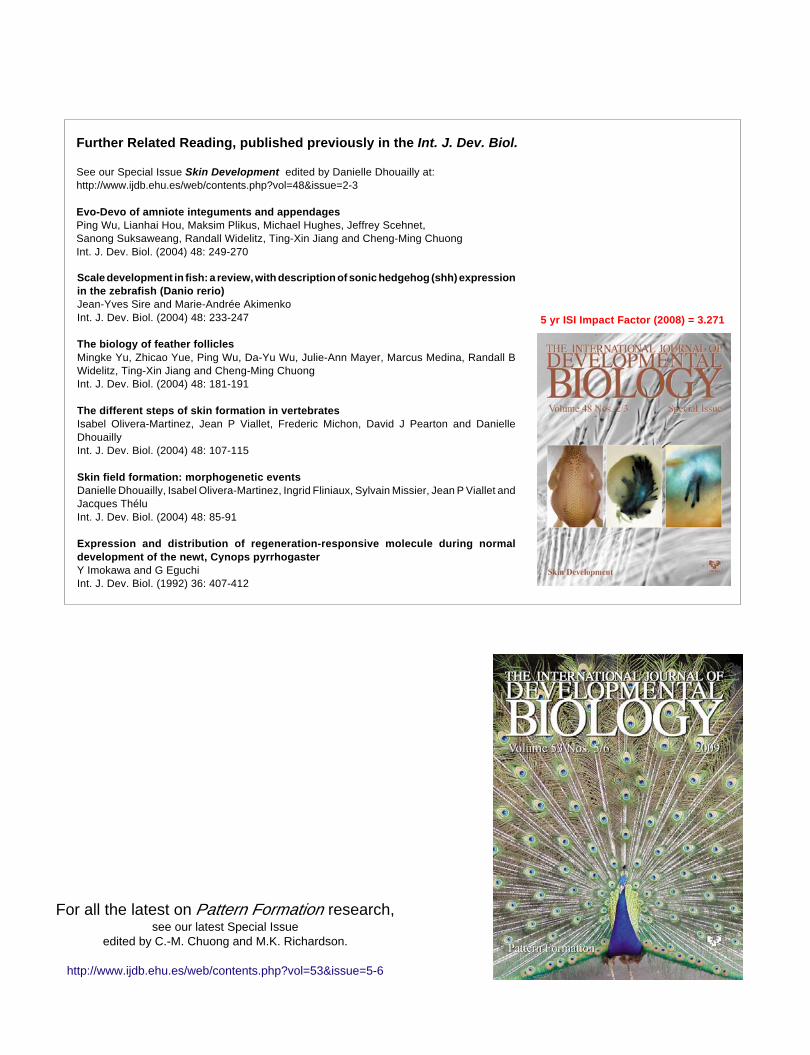
Reptile pattern formation and regeneration 827
Further Related Reading, published previously in the Int. J. Dev. Biol.
See our Special Issue Skin Development edited by Danielle Dhouailly at:http://www.ijdb.ehu.es/web/contents.php?vol=48&issue=2-3
Evo-Devo of amniote integuments and appendagesPing Wu, Lianhai Hou, Maksim Plikus, Michael Hughes, Jeffrey Scehnet,Sanong Suksaweang, Randall Widelitz, Ting-Xin Jiang and Cheng-Ming ChuongInt. J. Dev. Biol. (2004) 48: 249-270
Scale development in fish: a review, with description of sonic hedgehog (shh) expressionin the zebrafish (Danio rerio)Jean-Yves Sire and Marie-Andrée AkimenkoInt. J. Dev. Biol. (2004) 48: 233-247
The biology of feather folliclesMingke Yu, Zhicao Yue, Ping Wu, Da-Yu Wu, Julie-Ann Mayer, Marcus Medina, Randall BWidelitz, Ting-Xin Jiang and Cheng-Ming ChuongInt. J. Dev. Biol. (2004) 48: 181-191
The different steps of skin formation in vertebratesIsabel Olivera-Martinez, Jean P Viallet, Frederic Michon, David J Pearton and DanielleDhouaillyInt. J. Dev. Biol. (2004) 48: 107-115
Skin field formation: morphogenetic eventsDanielle Dhouailly, Isabel Olivera-Martinez, Ingrid Fliniaux, Sylvain Missier, Jean P Viallet andJacques ThéluInt. J. Dev. Biol. (2004) 48: 85-91
Expression and distribution of regeneration-responsive molecule during normaldevelopment of the newt, Cynops pyrrhogasterY Imokawa and G EguchiInt. J. Dev. Biol. (1992) 36: 407-412
For all the latest on Pattern Formation research,see our latest Special Issue
edited by C.-M. Chuong and M.K. Richardson.
http://www.ijdb.ehu.es/web/contents.php?vol=53&issue=5-6
5 yr ISI Impact Factor (2008) = 3.271