requirement for myd88 signaling in b cells and dendritic ... · cells for germinal center...
TRANSCRIPT

of July 13, 2018.This information is current as
Lyn-Deficient MiceAnti-Nuclear Antibody Production inand Dendritic Cells for Germinal Center Requirement for MyD88 Signaling in B Cells
Clifford A. Lowell, Baidong Hou and Anthony L. DeFrancoRamos-Hernández, Patrizia Scapini, Ming Ji, Haitao Shao, Zhaolin Hua, Andrew J. Gross, Chrystelle Lamagna, Natalia
http://www.jimmunol.org/content/192/3/875doi: 10.4049/jimmunol.1300683December 2013;
2014; 192:875-885; Prepublished online 30J Immunol
MaterialSupplementary
3.DCSupplementalhttp://www.jimmunol.org/content/suppl/2013/12/30/jimmunol.130068
Referenceshttp://www.jimmunol.org/content/192/3/875.full#ref-list-1
, 24 of which you can access for free at: cites 56 articlesThis article
average*
4 weeks from acceptance to publicationFast Publication! •
Every submission reviewed by practicing scientistsNo Triage! •
from submission to initial decisionRapid Reviews! 30 days* •
Submit online. ?The JIWhy
Subscriptionhttp://jimmunol.org/subscription
is online at: The Journal of ImmunologyInformation about subscribing to
Permissionshttp://www.aai.org/About/Publications/JI/copyright.htmlSubmit copyright permission requests at:
Email Alertshttp://jimmunol.org/alertsReceive free email-alerts when new articles cite this article. Sign up at:
Print ISSN: 0022-1767 Online ISSN: 1550-6606. Immunologists, Inc. All rights reserved.Copyright © 2014 by The American Association of1451 Rockville Pike, Suite 650, Rockville, MD 20852The American Association of Immunologists, Inc.,
is published twice each month byThe Journal of Immunology
by guest on July 13, 2018http://w
ww
.jimm
unol.org/D
ownloaded from
by guest on July 13, 2018
http://ww
w.jim
munol.org/
Dow
nloaded from

The Journal of Immunology
Requirement for MyD88 Signaling in B Cells and DendriticCells for Germinal Center Anti-Nuclear Antibody Productionin Lyn-Deficient Mice
Zhaolin Hua,* Andrew J. Gross,†,‡ Chrystelle Lamagna,x Natalia Ramos-Hernandez,†
Patrizia Scapini,x,{ Ming Ji,† Haitao Shao,* Clifford A. Lowell,x Baidong Hou,*,† and
Anthony L. DeFranco†
The intracellular tyrosine kinase Lyn mediates inhibitory receptor function in B cells and myeloid cells, and Lyn2/2 mice
spontaneously develop an autoimmune and inflammatory disease that closely resembles human systemic lupus erythematosus.
TLR-signaling pathways have been implicated in the production of anti-nuclear Abs in systemic lupus erythematosus and mouse
models of it. We used a conditional allele of Myd88 to determine whether the autoimmunity of Lyn2/2 mice is dependent on TLR/
MyD88 signaling in B cells and/or in dendritic cells (DCs). The production of IgG anti-nuclear Abs, as well as the deposition of
these Abs in the glomeruli of the kidneys, leading to glomerulonephritis in Lyn2/2 mice, were completely abolished by selective
deletion ofMyd88 in B cells, and autoantibody production and glomerulonephritis were delayed or decreased by deletion ofMyd88
in DCs. The reduced autoantibody production in mice lacking MyD88 in B cells or DCs was accompanied by a dramatic decrease
in the spontaneous germinal center (GC) response, suggesting that autoantibodies in Lyn2/2 mice may depend on GC responses.
Consistent with this view, IgG anti-nuclear Abs were absent if T cells were deleted (TCRb2/2 TCRd2/2 mice) or if T cells were
unable to contribute to GC responses as the result of mutation of the adaptor molecule SAP. Thus, the autoimmunity of Lyn2/2
mice was dependent on T cells and on TLR/MyD88 signaling in B cells and in DCs, supporting a model in which DC hyperactivity
combines with defects in tolerance in B cells to lead to a T cell–dependent systemic autoimmunity in Lyn2/2 mice. The Journal of
Immunology, 2014, 192: 875–885.
The human autoimmune disease systemic lupus erythe-matosus (SLE) is characterized by production of auto-antibodies against multiple self-Ags, of which nuclear
autoantigens, such as dsDNA and ribonucleoproteins (RNPs), arepredominant (1). A similar spontaneously developing autoimmu-nity characterized by anti-nuclear Ab production is seen in a va-riety of genetically determined mouse models, some of which are
multigenic and others of which result from spontaneous or tar-geted mutations of known genes (2). One of the better studied ofthe latter category is the Lyn2/2 mouse, which develops a highlypenetrant autoimmune and inflammatory disease characterized byanti-dsDNA IgG Abs and glomerulonephritis (3–5). Lyn is a Srcfamily protein tyrosine kinase that is required for the function ofa number of inhibitory receptors on B cells and myeloid cells. InB cells, the functions of both the sialic acid–binding Ig super-family member CD22 and of the inhibitory FcgRIIB depend onthe ability of Lyn to phosphorylate tyrosines in their cytoplasmictails, catalyzing the recruitment to the membrane of the inhibitoryphosphatases SHP-1 and SHIP-1 (4, 6, 7). Autoimmunity of Lyn-deficient mice likely involves a combination of compromisedtolerance of B cells, which is due to the loss of these inhibitorypathways, and hyperactivity of myeloid cells, which drive acti-vation of T cells and inflammatory disease (8–11).Like most human autoimmune diseases, lupus has a strong genetic
susceptibility component that is multigenic in the great majority ofpatients (1, 12). Among the genes that contribute to lupus suscep-tibility in humans are those encoding components of Lyn-inhibitorypathways. For example, some individuals of European descent havea single nucleotide polymorphism in the 59 untranslated region ofthe LYN gene that is mildly protective for the development of lupus(odds ratio, 0.80) (12). More impressively, loss-of-function allelesof SIAE, which encodes a sialic acid acetyl esterase that is neces-sary to create the ligand for CD22, contributes a large increase in thesusceptibility to lupus and several other autoimmune diseases (oddsratio, ∼8) in a small, but significant, fraction of individuals (13).Given that Lyn+/2 mice exhibit a mild lupus phenotype (14), itis possible that less frequent alleles of Lyn, other than those examinedin genome-wide association analysis, and/or alleles of genes
*Key Laboratory of Infection and Immunity, Institute of Biophysics, Chinese Acad-emy of Sciences, Beijing 100101, China; †Department of Microbiology and Immu-nology, University of California, San Francisco, San Francisco, CA 94143;‡Department of Medicine, University of California, San Francisco, San Francisco,CA 94143; xDepartment of Laboratory Medicine, University of California, San Fran-cisco, San Francisco, CA 94143; and {Division of Pathology, Department of Pathologyand Diagnostics, University of Verona, Verona 37134, Italy
Received for publication March 12, 2013. Accepted for publication November 18,2013.
This work was supported by research grants from the National Institutes of Health(R01AI072058 and P01AI078869 to A.L.D. and P01AI078869 and R01AI068150 toC.A.L.), as well as by the National Natural Science Foundation of China (Grants31170848 to B.H. and 31200669 to Z.H.). B.H. is supported by an award from theNational Thousand Talent Plan of China (2011).
Address correspondence and reprint requests to Dr. Anthony L. DeFranco or Dr.Baidong Hou, Department of Microbiology and Immunology, University of Califor-nia, San Francisco, 513 Parnassus Avenue, Box 0414, HSE-1001F, San Francisco,CA 94143 (A.L.D.) or Key Laboratory of Infection and Immunity, Institute of Bio-physics, Chinese Academy of Sciences, 15 Datun Road, Chaoyang District, Beijing100101, China (B.H.). E-mail addresses: [email protected] (A.L.D.) [email protected] (B.H.)
The online version of this article contains supplemental material.
Abbreviations used in this article: ANA, anti-nuclear Ab; C3, complement 3; DC,dendritic cell; GC, germinal center; RNP, ribonucleoprotein; SLE, systemic lupuserythematosus; SmRNP, Smith Ag ribonucleoprotein; Tfh, follicular helper T.
Copyright� 2014 by The American Association of Immunologists, Inc. 0022-1767/14/$16.00
www.jimmunol.org/cgi/doi/10.4049/jimmunol.1300683
by guest on July 13, 2018http://w
ww
.jimm
unol.org/D
ownloaded from

encoding the other components of Lyn-dependent inhibitory path-ways contribute significantly to lupus susceptibility in humans.Recent studies (15) in several mouse models of lupus implicated
TLR9 and TLR7 in the spontaneous production of anti-dsDNA andanti-RNP IgG, respectively. For example, MRL/lpr mice are pro-tected from the development of glomerulonephritis when combinedwith loss-of-function mutation of TLR7, either alone or in combi-nation with mutation of TLR9 (16). Similarly, deletion of the TLR-signaling component MyD88 prevents spontaneous lupus-like diseasein Lyn-deficient mice (17). Conversely, the Yaa autoimmune accel-erator locus of mice turns out to be a duplication onto the Y chro-mosome of a small region of the X chromosome that includes TLR7,resulting in increased expression of TLR7 (18–20). The possiblerelevance of TLR7 and TLR9 to lupus-like autoimmunity was ini-tially suggested by in vitro studies of Marshak-Rothstein andcoworkers (15), demonstrating a marked synergy for B cell activationin vitro between BCR engagement and TLR9 or TLR7 engagement.This synergy was shown to operate in vivo as well (21). Althoughthose studies strongly suggest that the contribution of TLR7 andTLR9 to lupus-like autoantibody production is by their action innucleic acid–recognizing B cells, TLR7 and TLR9 are also potentactivators of dendritic cells (DCs) and, moreover, induce type 1 IFNproduction by plasmacytoid DCs (22). A number of studies impli-cated type 1 IFNs in the pathogenesis of human lupus (23), so it isalso possible that the nucleic acid–recognizing TLRs play key roles inDCs for the development or propagation of lupus-like autoimmunity.Recently, we used cell type–specific deletion of the key TLR-
signaling component MyD88 to dissect the cellular basis for TLR9stimulation of Ab responses to immunization. We found that in vivoIgG responses to soluble protein–CpG oligonucleotide conjugateswere dependent primarily on TLR9/MyD88 signaling in DCs,whereas IgG responses to virus-like particles or inactivated in-fluenza virus particles were primarily dependent on TLR/MyD88signaling in B cells (24). In this study, we addressed the role ofMyD88 signaling in B cells and in DCs for the spontaneous de-velopment of lupus-like autoimmunity in Lyn2/2 mice. We foundthat anti-dsDNA and anti-RNP IgG in this model were likely pro-duced by germinal center (GC) responses that were highly depen-dent on TLR/MyD88 signaling in B cells. TLR/MyD88 signaling inDCs also played a major, although less essential, role in autoanti-body production. In contrast, TLR/MyD88 signaling in DCs wasessential for accumulation of activated T cells and splenomegaly,reinforcing the notion that Lyn-deficient autoimmune disease hasboth autoantibody and inflammatory components and that TLR/MyD88 signaling contributes to both disease manifestations.
Materials and MethodsMice
B6 (000664; C57BL/6J) mice were from The Jackson Laboratory. Lyn2/2
Myd88fl mice (B6.129P2-Myd88tm1Defr), Myd88fl/fl Cd11c-Cre mice, andMyd88fl/fl Cd79a-Cre mice were described previously (25–27) and werebackcrossed onto the C57BL/6 background for $10 generations. Thesemice were bred to generate Lyn2/2 Myd88fl/fl, Lyn2/2 Myd88fl/fl Cd79a-Cre, or Lyn2/2 Myd88fl/fl Cd11c-Cre mice used in this study. Sex-matchedlittermates were used in all experiments, and data were pooled and analyzedaccording to the age group. Sap2/2 mice (28) were obtained from Dr. PamelaSchwartzberg (National Institutes of Health, Bethesda, MD). tcrb2/2 tcrd2/2
mice (29, 30) were from The Jackson Laboratory. Animals were housed inspecific pathogen–free animal facilities at the University of California, SanFrancisco or the Institute of Biophysics under conditions that met the re-spective institutional and national guidelines. Animal use in each facility wasapproved by the respective Institutional Animal Care and Use Committee.
Anti-nuclear Ab immunofluorescence
Serum was diluted at 1:40 or 1:160 in PBS containing 1% FBS and appliedto fixed and permeabilized HEp-2 anti-nuclear Ab (ANA) slides (Bio-Rad).
After overnight incubation at 4˚C, ANAs were detected by Alexa Fluor488–conjugated goat anti-mouse IgG (Fcg fragment specific; JacksonImmunoResearch). DAPI was included in the last wash of slides. Slideswere visualized with a regular fluorescence microscope under a 403 ob-jective lens and imaged with an Olympus digital camera. Nuclear regionswere defined with DAPI staining, and the average Alexa 488 fluorescenceintensity of nuclear regions was quantified with ImageJ (National Institutesof Health).
Serum Ig and autoantibody measurement
Levels of serum IgM and IgG were measured using ELISA-based quantifi-cation kits (Bethyl Laboratories), according to the manufacturer’s instructions.For anti-dsDNA Ig ELISA, 96-well flat-bottom plates (BD Falcon) werecoated with 20 ng/well linearized pUC19 plasmid in 100 mM Tris-HCl (pH7.3). For anti-Smith Ag ribonucleoprotein (SmRNP) Ig ELISA, plates werecoated with 2 U/ml Smith Ag RNP complex Ag (ImmunoVision) in car-bonate buffer (pH 9.6). After overnight incubation at 4˚C, plates wereblocked with PBS containing 1% BSA and 0.05% Tween 20 for 1 h. Seriallydiluted sera were added to the plates and incubated for 2 h at room tem-perature. After washing with PBS containing 0.05% Tween 20, autoanti-bodies were detected by HRP-conjugated goat anti-mouse IgM Ab (m chainspecific) or anti-mouse total IgG (Fcg fragment specific; both from BethylLaboratories). The assays were developed by adding HRP substrate 3,39,5,59-tetramethylbenzidine (Vector Laboratories) and stopped by the addition of2 N sulfuric acid. The absorbance at 450 nm was measured with a microplatereader (Spectra Max Plus; MDS Analytical Technologies). For total IgM orIgG, the concentration was calculated according to standard samples suppliedby Bethyl Laboratories. For anti-dsDNA or anti-smRNP Ig, the relativeamount was represented as absorbance values at the same dilutions (1:40for IgG and 1:160 for IgM).
Histology and immunofluorescence analysis
For kidney histological analysis, both kidneys from a mousewere cut in halflaterally, fixed in 10% formalin, embedded in paraffin, and stained withH&E at the Institute of Biophysics Pathology Core. The presence andseverity of nephritis were evaluated in a blinded fashion, by one of theauthors, with stained sections that contained glomeruli away from themedulla. Scoring of glomerulonephritis and interstitial nephritis was doneusing a 0 to 3 scale (0 = absent, 1 = mild, 2 = moderate, 3 = severe):scoring for glomeruli was based on glomerular size, glomerular hyper-cellularity, and presence of glomerular sclerosis, whereas for interstitialdisease, it was based on the degree of inflammatory infiltrate and alterationin tissue architecture.
For histological analysis of the GC response, spleen samples from 5–7-mo-old mice were placed in Tissue-Tek O.C.T. compound (Sakura) andimmediately frozen with a mixture of ethanol and dry ice. Cryostat sec-tions (7 mm in thickness) were fixed in ice-cold acetone and stained withbiotin-conjugated rat anti-mouse IgD (eBioscience) and FITC-conjugatedanti-mouse GL-7 (BD Bioscience). HRP-conjugated extravidin (Sigma)and alkaline phosphatase–conjugated anti-FITC (Jackson Immuno-Research) were used as secondary Abs. Enzyme reactions were developedwith conventional substrates for peroxidases (diaminobenzidine/H2O2) andalkaline phosphatase (Fast Red; both from Sigma). Sections were counter-stained with hematoxylin (Fisher).
To detect complement 3 (C3) deposition, kidneys were placed in O.C.T.compound and snap-frozen in methyl-butane chilled in liquid nitrogen.Cryostat sections (15 mm in thickness) were fixed in ice-cold acetone andstained with FITC-conjugated anti-mouse C3 (Cappel Laboratories) for 1 hat room temperature. The average intensity of FITC in the glomerularregions was quantified by ImageJ.
Flow cytometry
Splenocytes were obtained by treating the organs with digestion medium(RPMI 1640, 25 mM HEPES, 50 mg/ml Liberase [Roche] and 100 mg/mlDNase I [Worthington]). Bone marrow cells were obtained by flushing tibiaand femur with medium. Kidney cells were prepared as previously described(8). Briefly, kidneys were pressed through a 70-mm cell strainer (BD Falcon).After washing, the cells were resuspended in 33% Percoll solution andcentrifuged at 2000 rpm for 20 min at room temperature. Kidney-infiltratingcells were obtained from cell pellets. For staining for flow cytometry, single-cell suspensions were blocked with anti-CD16/CD32 Ab and then stainedwith fluorescent Abs in ice-cold flow cytometry buffer (PBS supplementedwith 2 mM EDTA, 1% heat-inactivated FBS, and 0.02% sodium azide). TheAbs included FITC-labeled anti-Ly6G (1A8), anti-Ly6C (HK1.4), anti-CD44(IM7), anti–GL-7, and anti-CD43 (S7) Abs; PE-labeled anti-CD86 (GL1),anti–PD-1 (RMP1-30), anti-TCRb (H57-597), and anti-CD23 (B3B4) Abs;
876 MyD88 SIGNALING IN Lyn2/2 AUTOIMMUNITY
by guest on July 13, 2018http://w
ww
.jimm
unol.org/D
ownloaded from

PerCP-Cy5.5–labeled anti-F4/80 (BM8), anti-ICOS (7E.17G9), and anti-IgM(RMM-1) Abs; PE-Cy7–labeled anti-CD11b (M1/70), anti-CD95 (Jo2), andanti-CD62L (MEL-14) Abs; allophycocyanin- or Alexa Fluor 647–labeledanti-CD11c (HL3), anti-CD8 (53-6.7), anti-CD138 (281-2), and anti-CD62L(MEL-14) Abs; Alexa Fluor 700–labeled anti–I-Ab (M5/114.15.2), anti-CD11b (M1/70), and anti-CD45.2 (104) Abs; allophycocyanin-Cy7–labeledanti-B220 (RA3-6B2), anti-CD19 (6D5), and Pacific Blue–labeled anti-IgD(11-26c.2a). All fluorochrome-conjugated mAbs were purchased from BDPharmingen, eBioscience, or BioLegend. Biotinylated anti-CD86 (GL1) oranti-CD93 (AA4.1) Abs were detected with streptavidin-conjugated PacificOrange (Invitrogen). After the final wash, the cells were resuspended in flowcytometry buffer containing 0.4 mM DAPI (Invitrogen). All data were col-lected on an LSR II flow cytometer (Becton Dickinson) and analyzed withFlowJo software (TreeStar).
Immature, transitional, and mature B cell populations were identified in thespleen and bone marrow of mice using fluorescently conjugated Abs againstsurface B220, CD23 (BD Biosciences), CD93 (clone AA4.1; eBioscience),and IgM F(ab)9 monomer (Jackson ImmunoResearch). In the spleen, surfaceCD93 was used to distinguish immature (B220+, CD93+) and mature (B220+,CD932) B cells. Within the immature B cell population, surface CD23 andIgM levels were used to identify immature-transitional T1 (IgMhi, CD23lo-neg),T2 (IgMhi, CD23hi-int), and T3 (IgMlo, CD23hi-int) subsets. Within the matureB cell population, surface expression of CD23, CD21, IgM, and CD43 wereused to distinguish follicular B cells (CD23+, CD21lo, IgMlo-int), marginalzone B cells (CD23lo-neg, CD21hi, IgMhi, CD432), and B1 B cells (CD23lo-neg,CD21neg-lo, IgMlo, CD43+). These markers were also used to distinguishimmature and pro/pre-B cells (B220+, CD93+) and mature recirculatingB cells (B220+ CD932) in the bone marrow. Surface IgM and CD23 levelswithin the CD93+ population distinguish newly formed immature B cells(IgM+, CD23lo-neg), BM-T1 cells (IgMhi, CD23lo-neg), BM-T2 cells (IgMhi,CD23int), and pro/pre-B cells (IgM2, CD232). Absolute cell numbers withineach population were back-calculated from total splenocytes or total cells/femur and tibia.
RNA extraction and quantitative RT-PCR
For quantifying cytokine expression, mouse spleens were harvested andsnap-frozen in liquid nitrogen. Total RNAwas extracted with the RNeasy kit(QIAGEN) with on-column DNase digestion. cDNA was transcribed fromtotal RNAwith the iScript cDNA Synthesis Kit (Bio-Rad). The primer pairsfor IL-12p40, IL-6, and BAFF were described previously (8). Transcriptswere quantified by PCR with iTaq SYBR Green SuperMix with ROX (Bio-Rad), and the levels of cytokine transcripts were normalized to the levels ofHPRT mRNA.
Statistical analysis
Statistical significance for data with multiple groups was calculated witha one-way ANOVA; if a significant difference was observed betweengroups, the Bonferroni posttest was used to assess the statistical differencebetween specific groups and either the wild-type (WT) group or the Lyn2/2
group. In cases in which there was a large difference between the WTgroup and the three groups with Lyn deficiency (B cell number data, Fig.3E, Fig. 6B, Supplemental Fig. 1B), this analysis was inadequate to detectdifferences between the Lyn-deficiency groups, because all were greatlydifferent from the WT group. In these cases, a secondary statistical anal-ysis was performed with just the Lyn-deficiency groups. In the figures, thissecondary statistical analysis is denoted by different symbols. When therewere three such groups, this secondary analysis again used one-way ANOVA;however, in cases in which only two groups remained, the Mann–Whitney Utest was used. All p values # 0.05 were considered significant and are in-dicated in the figures.
ResultsAblation of MyD88 signaling in either B cells or DCsattenuates autoimmunity in Lyn2/2 mice
To examine the contribution of TLR signaling in B cells and DCsto the spontaneous autoimmunity caused by Lyn deficiency, micewith a conditional allele of Myd88 (Myd88fl) were crossed toLyn2/2 mice, and transgenic Cre alleles that express selectivelyin B cells (Cd79a-Cre) or in DCs (CD11c-Cre) were also intro-duced. Progenies of genotypes Lyn2/2 Myd88fl/fl, Lyn2/2 Myd88fl/fl
Cd79a-Cre, or Lyn2/2 Myd88fl/fl Cd11c-Cre (hereafter referred toas Lyn2/2, Lyn2/2 B-Myd882/2, and Lyn2/2 DC-Myd882/2 mice,respectively) were generated, housed under specific pathogen–free
conditions, and examined for autoimmune phenotypes at dif-ferent ages.The development of autoantibodies by these mice was initially
screened by ANA immunofluorescence, with mouse sera collectedat 5–7 mo of age. As previously described (4, 5), most Lyn2/2
mice exhibited elevated titers of ANAs by this age (Fig. 1A, 1B).The development of IgG ANAs was almost completely abolishedin Lyn2/2 B-Myd882/2 mice, and it was substantially attenuatedin mice lacking MyD88 selectively in DCs (Fig. 1A, 1B).Next, we used ELISA to examine the development of Abs to
dsDNA and smRNP, two major types of ANAs seen in SLEpatients (1), as well as in animal models of SLE. All Lyn2/2 micedeveloped IgM autoantibodies against both of these nuclear Ags,typically by 4 mo of age, and a substantial majority also de-veloped IgG anti-dsDNA and IgG anti-smRNP. Deletion ofMyD88 in DCs delayed production of these autoantibodies, but8–10-mo-old mice still had substantial titers of IgG and IgManti-dsDNA and IgM anti-smRNP. In contrast, deletion ofMyd88 in B cells prevented the production of IgG autoanti-bodies in Lyn2/2 B-Myd882/2 mice at all time points testedand attenuated the production of IgM autoantibodies (Fig. 1C–F). Total serum IgM and IgG levels were somewhat reduced inthese mice compared with Lyn2/2 mice (Fig. 1G, 1H), but thedifferences were generally #2-fold, indicating that there wasa selective defect in the production of autoantibodies.Anti-dsDNA IgG Abs are often deposited in the glomeruli of the
kidneys and can lead to glomerulonephritis, as observed in Lyn-deficient mice (8, 31). Indeed, immunofluorescent staining clearlyrevealed a large amount of C3 deposition in the glomeruli of mostof the 5–7-mo-old Lyn2/2 mice (Fig. 2A, 2B). In agreement withthe almost complete abolishment of IgG autoantibodies in Lyn2/2
B-Myd882/2 mice, C3 deposition was not evident in the glomeruliof these mice at a corresponding age. Moreover, the nephritis typ-ically seen in old Lyn2/2 mice by H&E staining of kidney tissuesections was substantially decreased, but not completely absent, inLyn-deficient mice deleted for MyD88 in either B cells or DCs (Fig.2C, 2D); the numbers of infiltrating inflammatory cells (CD45+),mainly CD11b+ neutrophils and macrophages, were correspond-ingly reduced (Fig. 2E). The splenomegaly and myeloprolifera-tion that are characteristic of aged Lyn2/2 mice (.6 mo old)were corrected by deletion of Myd88 in DCs but apparently notby deletion of Myd88 in B cells (Fig. 3A, 3B, Supplemental Fig.1A). Together, these results are consistent with the observationthat ablating MyD88 signaling in B cells or DCs reduces IgGautoantibody production in Lyn2/2 mice but also indicates thatsome of the inflammatory phenotypes are independent of auto-antibody production.
Autoantibody production in Lyn2/2 mice requires SAPexpression and T cell help
Abs against dsDNA obtained from MRL/lpr mice exhibit somaticmutations that increase reactivity to DNA (32), which is consistentwith a GC origin for these autoantibody responses. However, Ig-transgenic B cells with a rheumatoid factor specificity in thesemice make an extrafollicular Ab response with substantial num-bers of somatic mutations, even in the absence of Ag-specificT cell help (21, 33). Moreover, mice overexpressing BAFF alsospontaneously produce ANAs in a T cell–independent fashion(34). Thus, there are precedents for extrafollicular production ofANAs in some mouse models.To address this issue, we first examined Lyn2/2 mice for the
frequency of GC phenotype B cells. The frequency of CD95+GL-7+
GC B cells relative to the number of splenic B cells already wassignificantly increased in Lyn2/2 mice compared with WT mice
The Journal of Immunology 877
by guest on July 13, 2018http://w
ww
.jimm
unol.org/D
ownloaded from

by 2–4 mo, and this increase became more dramatic at 5–7 mo(Fig. 3C, 3D), correlating with the time course of the developmentof IgG ANAs. However, it should be noted that Lyn2/2 mice havesubstantially fewer splenic B cells than do WT mice (Supple-mental Fig. 1B), so the absolute numbers of GC B cells weresimilar in Lyn2/2 mice and WT mice (Fig. 3E). In agreement withflow cytometry analysis, staining of spleen sections also clearlyrevealed the existence of IgDlowGL-7+ GC structures in multiplefollicles of Lyn2/2 mice (Fig. 3F) at 5–7 mo of age. In contrast,GCs were not common in WT mice housed in the same mouseroom (data not shown). Interestingly, the spontaneous GC re-sponse seen in Lyn2/2 mice was greatly reduced upon deletion ofMyd88 in B cells (Fig. 3D, 3E). In contrast, the mesenteric lymphnodes of Lyn2/2 and Lyn2/2 B-Myd882/2 mice contained similarnumbers of GC B cells (data not shown), suggesting that the lattermice did not have a general defect in mounting GC reactions.To examine whether Th cells and the GC reaction contribute to
the generation of IgG ANAs, we crossed Lyn2/2 mice to mice
deleted for TCR genes (tcrb2/2 tcrd2/2) or to mice lacking theSAP signaling adaptor protein, which is critical for follicularhelper T (Tfh) cells to interact with GC B cells and promote GCAb responses (35, 36). Lyn-deficient mice lacking T cells orlacking the SAP adaptor molecule made greatly attenuated levelsof anti-dsDNA IgG Abs, as measured by ELISA (Fig. 3G). Thesegenetic results strongly support the hypothesis that there is a GC-origin of lupus-like autoantibodies in Lyn-deficient mice.
Activation and expansion of self-reactive T cells in Lyn2/2
mice require MyD88 signaling in DCs and B cells
Lyn2/2 mice exhibit a dramatic expansion of activated T cells asautoimmune disease progresses (8, 37). Therefore, we next in-vestigated the role of MyD88 in DCs and B cells in the hyper-reactivity of T cells in Lyn2/2 mice. As previously reported (8),Lyn2/2 mice accumulate increasing numbers of CD4+ and CD8+
T cells with an activated or effector memory phenotype (CD44+
CD62Llow) as they age. As Lyn-deficient mice lacking MyD88 in
FIGURE 1. Roles of MyD88 signaling in B
cells and in DCs for development of autoanti-
bodies in Lyn2/2 mice. (A and B) Anti-nuclear IgG
Abs of individual mice were screened using sera of
5–7-mo-old mice. Representative ANA fluores-
cence images (original magnification 340) of 1:40
diluted sera of the indicated genotype (A), as well
as summarized relative fluorescence intensity of
sera from individual mice (s) and means of each
mouse group (bars) (B). Serum IgM and IgG Abs
to dsDNA (C, D) and to anti-smRNP (E, F) of in-
dividual mice of the four genotypes indicated and
of different age groups were measured by ELISA.
The relative amount of autoantibody in each
mouse (s) and the mean of each mouse group
(bars) are shown. (G and H) Total serum IgM and
IgG levels of mice of the different genotypes and
age groups were quantified by ELISA. Data are
presented as mean 6 SE of WT (n = 6, 6, 6 mice,
respectively, in each age group), Lyn2/2 (n = 6, 13,
11), Lyn2/2 B-MyD882/2 (n = 6, 10, 7), and Lyn2/2
DC-MyD882/2 (n = 7, 8, 7) mice. Differences be-
tween WT or Lyn2/2 and other groups are indicated
by brackets. *p , 0.05, **p , 0.01.
878 MyD88 SIGNALING IN Lyn2/2 AUTOIMMUNITY
by guest on July 13, 2018http://w
ww
.jimm
unol.org/D
ownloaded from

B cells or in DCs aged, these populations did not increase at all(CD4+ T cells) or nearly as much (CD8+ T cells) (Fig. 4A–C,Supplemental Fig. 1C, 1D). Thus, increased spontaneous activa-tion of T cells in Lyn2/2 mice was largely dependent on MyD88signaling in both DCs and B cells.Many of the activated CD4+ T cells also expressed ICOS and
PD-1, two markers that have been used to identify Tfh cells. Thepercentage and absolute number of those cells already wereslightly elevated in Lyn2/2 mice at 2–4 mo of age compared withWT mice, and they especially increased in older mice (Fig. 4E,4F). In another recent study (38), the number of Tfh cells at 4–5mo of age was roughly normal in Lyn2/2 mice. Importantly, de-letion of Myd88 in either DCs or B cells substantially reduced theexpansion of these cells in Lyn2/2 mice (Fig. 4E, 4F).
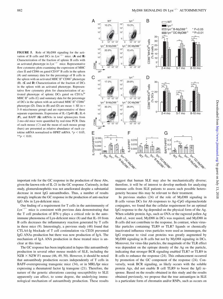
Splenic B cells and DCs in Lyn2/2 mice have elevatedexpression of MHC class II and CD86 that is not dependent onMyD88 signaling
TLR agonists have well-established adjuvant properties for pro-moting T cell responses (39). To investigate the possibility thatTLRs of DCs or B cells promote their ability to present self-Ags toT cells, we examined the expression of MHC class II and of thecostimulatory molecule CD86 on splenic B cells and on DCs ofLyn2/2 mice with or without DC or B cell MyD88. In Lyn2/2
mice, an increased proportion of B cells exhibited elevated levelsof MHC class II and CD86 by 2–4 mo of age (Fig. 5A, 5B). Thenumber of such B cells was not dependent on the expression of
MyD88 in B cells (Fig. 5A, 5B), suggesting that the loss of Lyn-based attenuation of BCR signaling in B cells may be sufficient forthis phenotypic alteration. Similarly, DCs in young Lyn2/2 micehad spontaneously elevated MHC class II expression and CD86expression. Deletion of Myd88 in DCs had little effect on theirexpression of MHC class II and CD86 (Fig. 5C, 5D). Together,these data indicate that the requirement for MyD88 in B cells andin DCs for development of autoimmunity in Lyn2/2 mice was notcaused by changes in the expression of MHC class II and CD86.Next, we examined the role of MyD88 signaling in DCs and
B cells in Lyn2/2 mice for the induction of cytokines that havebeen implicated in the pathogenesis of autoimmunity and in-flammation in this model (8, 40). In total splenocytes from Lyn2/2
mice at 2 mo of age, increased mRNA expression of the cytokinesIL-12p40 and BAFF was readily detected (Fig. 5E–G), demon-strating that enhanced inflammatory responses arise even beforethe development of overt autoimmunity in these mice. Interest-ingly, deletion of Myd88 in DCs substantially reduced the ex-pression of IL-12p40 and IL-6 in the spleens of young Lyn2/2
mice (Fig. 5E, 5F), suggesting that Lyn-deficient DCs are sensi-tized to have enhanced responses to endogenous or ubiquitousTLR ligands and are a major producer of proinflammatory cyto-kines in these mice. This is in agreement with other recent reports(10, 11) describing a proinflammatory role for Lyn-deficient DCs.As mentioned above, the incidence of splenomegaly was greatlyreduced in aged (8–10 mo) Lyn2/2 DC-MyD882/2 mice com-pared with Lyn2/2 mice (Fig. 3A, 3B). In comparison, deletion of
FIGURE 2. Effect of ablation of MyD88 signaling
in B cells and in DCs for complement deposition and
inflammation in the kidneys of Lyn2/2 mice. De-
position of C3 was detected by immunofluorescent
staining of frozen kidney sections of 5–7-mo-old
mice of the indicated genotype; representative images
(original magnification 320) (A) and summarized
data (B) are shown. (C) Kidney sections of the dif-
ferent genotypes at 8–10 mo of age were stained with
H&E to assess pathological changes associated with
glomerulonephritis (original magnification 340). (D)
Sections from individual mice were graded, in a
blinded fashion, on a scale of 0 to 3 (0 = absent, 1 =
mild, 2 = moderate, 3 = severe) for the degree of
histological abnormality in the glomeruli and for the
degree of inflammation in the interstitial regions. (E)
Numbers of inflammatory cells in the kidneys were
measured by flow cytometry in six mice of each
genotype at 8–10 mo of age. Data are mean 6 SE
and are representative of two separate experiments.
*p , 0.05, **p , 0.01.
The Journal of Immunology 879
by guest on July 13, 2018http://w
ww
.jimm
unol.org/D
ownloaded from

B cell Myd88 had little or no effect on the expression of IL-12p40or BAFF (Fig. 5E–G).
Effect of cell-intrinsic MyD88 signaling on peripheral numbersof B cells in Lyn2/2 mice
Lyn deficiency has substantial B cell–intrinsic effects on the numbersof B cells in the spleen (8), decreasing their overall numbers (Sup-plemental Fig. 1B), apparently as a result of enhanced cell death atthe T1 or T2 immature stages in the spleen and decreased maturationto the follicular mature state and/or decreased survival of these cells
(41–43). Interestingly, although deletion of Myd88 in Lyn2/2 B cellsdid not affect bone marrow populations of B cell precursors or im-mature B cells compared with Lyn2/2 mice (Fig. 6A), the decreasednumber of B cells in the spleens of young Lyn2/2 mice was furtherexacerbated by deletion ofMyd88 in B cells (Fig. 6B). The reductionin B cells in Lyn2/2B-Myd882/2 mice began at the T1 stage in thespleen and persisted in subsequent developmental stages until mat-uration (Fig. 6B, 6C). Despite these decreases in mature B cellnumber, the numbers of plasma cells in the spleen were elevatedin Lyn2/2 and Lyn2/2B-Myd882/2 mice compared with WT mice
FIGURE 3. Role of GC response in autoantibody production by Lyn2/2 mice and effect of ablation of MyD88 on GCs. Effect of deletion of Myd88 in B
cells or DCs for the splenic hypercellularity that accumulates over time in Lyn-deficient mice; spleen weights of individual mice at 8–10 mo of age for the
indicated genotypes (A) and numbers of total splenocytes (mean6 SE) for mice of different ages (B). Analysis of GC B cells; representative flow cytometry
profiles (C) and summarized data from mice with different genotypes and at different ages, represented either as percentage of total splenic B cells that have
a GC B cell phenotype (D) or as absolute number of GC B cells/spleen in the indicated mice (E). Data in (D) and (E) are mean 6 SE of five to eight mice/
group and are representative of three separate experiments. (F) Representative immunohistological staining of GCs in the spleens of 5–7-mo-old Lyn2/2
and Lyn2/2 B-MyD882/2 mice. IgD+ cells (brown) and GL-7+ cells (red) are shown with hematoxylin counterstaining (original magnification 34 or 320,
as indicated). (G) Effect of genetic deletion of T cells or the adaptor molecule SAP on ANA production of Lyn2/2 mice at 5–6 mo of age. Shown are the
relative amounts of anti-dsDNA IgG measured by ELISA in individual mice (s) and means of each mouse group (bars). *p , 0.05, **p , 0.01, ANOVA,
as described in Materials and Methods. A secondary statistical analysis in which the WT group was excluded was also performed on the data in (E). #p ,0.05, ##p , 0.01.
880 MyD88 SIGNALING IN Lyn2/2 AUTOIMMUNITY
by guest on July 13, 2018http://w
ww
.jimm
unol.org/D
ownloaded from

(Supplemental Fig. 1E). These results suggest that MyD88 signalingin B cells promotes their survival at the immature T1 stage in thespleen, but it does not greatly affect maturation or survival at sub-sequent stages of development in the spleen and is not required fordifferentiation into plasma cells, in agreement with the circulatinglevels of total IgM and IgG in these mice (Fig. 1G, 1H). We observeda modest increase in the percentage of CD93+CD23+IgMlow T3B cells and a small decrease in the percentage of follicular B cells inthe spleens of Lyn2/2B-Myd882/2 mice, such that the ratio of T3/follicular B cells was slightly, but significantly, increased by the lossof MyD88 in B cells (Fig. 6D). The T3 B cell population previouslywas shown to include many anergic autoreactive B cells (44). Ourdata suggest that intrinsic MyD88 signaling may keep some self-reactive B cells from becoming anergic, although other explanationsare possible.
DiscussionLyn is an intracellular protein tyrosine kinase that is critical forinhibitory receptor function in B cells and in DCs, and Lyn2/2micespontaneously develop a lupus-like autoimmune and inflammatorydisease (4, 5). To address the mechanism by which nucleic acid–recognizing TLRs contribute to spontaneous production of ANAsin the Lyn2/2 mouse model of SLE, we used cell type–specificdeletion of the gene encoding MyD88, a key adaptor moleculerequired for intracellular signaling by most TLRs, including TLR7and TLR9. Deletion of Myd88 selectively in B cells completelyblocked production of the anti-dsDNA and anti-RNP IgG Abs,which are among the most characteristic autoantibodies seen in
human SLE and are rarely produced in other human diseases(1), and ameliorated glomerulonephritis. In addition, deletion ofMyd88 selectively in DCs delayed production of IgG anti-dsDNA,blocked production of IgG anti-smRNP, largely abrogated accu-mulation of activated phenotype T cells in the spleen, and ame-liorated glomerulonephritis. These results demonstrate that TLR/MyD88 signaling is required in both B cells and DCs for thedevelopment of autoimmune disease in these mice.The demonstration that MyD88/TLR signaling in B cells is re-
quired for anti-nuclear IgG production in Lyn2/2 mice adds to theaccumulating evidence indicating that dual stimulation of DNA-and RNP-specific B cells by the BCR and by TLR9 or TLR7, re-spectively, is a key underlying mechanism in the breakdown oftolerance to nuclear self-Ags in mouse models of lupus (15, 45, 46).Although TLR signaling can boost Ab responses in multiple ways(47), we recently showed that it can boost GC responses dramati-cally in response to virus-like particles and inactivated virions (24).It is likely that an analogous mechanism, perhaps in response tofragments from apoptotic cells, participates in the production ofanti-dsDNA and anti-RNP IgG Abs in Lyn2/2 mice, because it waslargely dependent on MyD88 expression in B cells, on the presenceof T cells, and on the expression of SAP, which is required for GCresponses (Fig. 3G). SAP is an adaptor for SLAM family adhesionmolecules, and its function in T cells is important at early stages ofthe GC response by acting to stabilize interactions between acti-vated B cells and cognate Th cells (36). Moreover, a recent reportfrom another group (38) found that Lyn2/2 IL-212/2 mice failed toproduce anti-dsDNA IgG Abs, which is also consistent with an
FIGURE 4. Roles of MyD88 signaling in DCs and B cells for the activation and expansion of T cells in Lyn2/2 mice. (A–C) Age-dependent accu-
mulation of activated phenotype T cells in Lyn2/2 mice and effect of ablation ofMyd88 in B cells or DCs. Representative flow cytometry plots of activated
CD4+ T cells (CD44hi CD62Llow) in spleens of 5–7-mo-old mice of the indicated genotype (A) and summarized data for CD4+ T cells (B) and CD8+ T cells
(C) from mice of different ages. (D–F) Age-dependent accumulation of Tfh phenotype cells in Lyn2/2 mice. Representative flow cytometry plots of Tfh
cells (PD-1+ ICOS+) for gated CD4+ T cells in the spleen (D) and summarized data (E, F). Data in (B), (C), (E), and (F) are mean6 SE (n = 5–8 mice/group)
and are representative of three separate experiments. *p , 0.05, **p , 0.01.
The Journal of Immunology 881
by guest on July 13, 2018http://w
ww
.jimm
unol.org/D
ownloaded from
important role for the GC response in the production of these Abs,given the known role of IL-21 in the GC response. Curiously, in thatstudy, glomerulonephritis was not ameliorated despite a substantialdecrease in most IgG autoantibodies. Thus, a number of resultsstrongly implicate the GC response in the production of anti-nuclearIgG Abs in Lyn-deficient mice.Our finding of a requirement for T cells in the autoimmunity of
Lyn2/2 mice is consistent with previous data demonstrating thatthe T cell production of IFN-g plays a critical role in the auto-immune phenomena of Lyn-deficient mice (8) and that IL-10 fromB cells decreases the inflammatory reaction generated by T cellsin these mice (9). Interestingly, a previous study (48) found thatCTLA4-Ig blockade of T cell costimulation via CD28 preventedIgG ANAs production but there was now production of IgA. Themechanism of IgA ANA production in these treated mice is un-clear at this time.The GC response has been implicated in lupus-like autoantibody
production in several other mouse models of SLE, including theNZB 3 NZW F1 mouse (46, 49, 50). However, it should be notedthat autoantibody production occurs independently of T cells inBAFF-overexpressing transgenic mice (34), or in MRL/lpr miceexpressing a rheumatoid factor Ig transgene (21). Therefore, thenature of the genetic alterations causing susceptibility to SLEapparently can affect, to some degree, the underlying immu-nological mechanism of autoantibody production. These results
suggest that human SLE may also be mechanistically diverse;therefore, it will be of interest to develop methods for analyzingimmune cells from SLE patients to assess such possible hetero-geneity because this may be relevant to their treatment.In previous studies (24) of the role of MyD88 signaling in
B cells versus DCs for Ab responses to Ag–CpG oligonucleotideconjugates, we found that the cellular requirement for an optimalIgG response to the Ag depended on the physical form of the Ag.When soluble protein Ags, such as OVA or the ragweed pollen AgAmb a1, were used, MyD88 in DCs was required, and MyD88 inB cells did not contribute to the response. In contrast, when virus-like particles containing TLR9 or TLR7 ligands or chemicallyinactivated influenza virus particles were used as immunogen, theIgG response to viral coat proteins was greatly augmented byMyD88 signaling in B cells but not by MyD88 signaling in DCs.Moreover, for virus-like particles, the magnitude of the TLR effectwas dependent on the epitope density of the Ag on the particle,indicating that stronger BCR signaling enabled TLR7 or TLR9 inB cells to enhance the response (24). This enhancement occurredby promotion of the GC component of the response (24). Con-versely, weak BCR signaling, as likely occurs with the solubleprotein Ags, did not enable B cell TLR9 to boost the IgG re-sponse. Based on the results obtained in this study and the resultsobtained previously, we propose that the true autoantigen in SLEis a particulate form of chromatin and/or RNPs, such as occurs on
FIGURE 5. Role of MyD88 signaling for the acti-
vation of B cells and DCs in Lyn2/2 mice. (A and B)
Characterization of the fraction of splenic B cells with
an activated phenotype in Lyn2/2 mice. Representative
flow cytometry plots examining the expression of MHC
class II and CD86 on gated CD19+ B cells in the spleen
(A) and summary data for the percentage of B cells in
the spleen with an activated MHC II+ CD86+ phenotype
(B). (C and D) Characterization of the fraction of DCs
in the spleen with an activated phenotype. Represen-
tative flow cytometry plots for characterization of ac-
tivated phenotype of splenic DCs gated on CD11chi
MHC II+ cells (C) and summary data for the percentage
of DCs in the spleen with an activated MHC II+ CD86+
phenotype (D). Data in (B) and (D) are mean 6 SE (n =
5–8 mice/mouse group) and are representative of three
separate experiments. Expression of IL-12p40 (E), IL-6
(F), and BAFF (G) mRNAs in total splenocytes from
2-mo-old mice were quantified by real-time PCR. Data
of each mouse (s) and the mean of each mouse group
(bars) are presented as relative abundance of each cy-
tokine mRNA normalized to HPRT mRNA. *p , 0.05,
**p , 0.01.
882 MyD88 SIGNALING IN Lyn2/2 AUTOIMMUNITY
by guest on July 13, 2018http://w
ww
.jimm
unol.org/D
ownloaded from

apoptotic fragments released from dying cells. This proposal isconsistent with the fact that many autoantibodies obtained frommouse SLE-prone strains bind to apoptotic blebs (51).The observation mentioned above—that strong BCR signaling is
required to enable TLR7 or TLR9 signaling in B cells to enhancetheir GC response—suggests why Lyn deficiency of B cells cre-ates a strong susceptibility for the development of anti-dsDNA andanti-RNP IgG Abs. Low-affinity DNA- or RNP-reactive mature oranergic follicular B cells in WT mice presumably have weak BCRsignaling that is attenuated by the Lyn/CD22/SHP-1 feedback-inhibitory pathway; therefore, even if they acutely encounter ap-optotic blebs, their low level of BCR signaling does not synergizewith TLR7 or TLR9 signaling to promote a GC response. Incontrast, Lyn-deficient B cells of the same specificity have exag-gerated BCR signaling; therefore, we hypothesize that they canenter into and participate in GC responses, leading to the pro-duction of class-switched and affinity-matured pathogenic auto-antibodies.Several recent studies (10, 11) implicated dysregulation of DCs
by loss of Lyn as also being an important contributor to the au-toimmune phenomena in Lyn2/2 mice. Consistent with this viewis our demonstration that MyD88 signaling in DCs contributesimportantly to the autoimmune phenotypes of Lyn2/2 mice. Thecharacteristic expansion of activated phenotype T cells that is seenin Lyn2/2 mice as they get older was abrogated by deletion ofMyd88 in DCs (Fig. 4), indicating that TLR/MyD88 signaling inDCs provides a necessary activation signal that synergizes withthe effects of loss of Lyn-dependent inhibitory signaling in thesecells. This interpretation is supported by the recent report (11) thatmice in which Lyn is deleted only in DCs also develop a severelupus-like autoimmunity. Thus, we propose that Lyn2/2 micedevelop a severe lupus-like autoimmunity and inflammatory dis-ease as a result of the combination of at least two defects: the
deficiency of Lyn in B cells compromises their cell-intrinsic tol-erance mechanisms by allowing TLR7 and TLR9 to promote ac-tivation of self DNA- and RNP-reactive B cells, and the dysregulationof Lyn-deficient DCs leads to excessive activation of T cells, which,in turn, can promote increased affinity IgG responses of the DNA-and RNP-specific B cells. In addition, Lyn-deficient DCs and theT cells activated by them provide a self-reinforcing inflammatoryresponse that can also contribute to inflammatory disease (8, 11). Thislatter component of the disease of Lyn-deficient mice is especiallyevident if Lyn is selectively deleted in DCs (11) or if the B cells areunable to produce IL-10 to inhibit it (9).Studies in mouse models indicate that the presence of genetic
susceptibility loci is required for spontaneous breakdown of tol-erance to nuclear autoantigens. Although most natural suscepti-bility loci in human and mouse remain poorly understood, a subsetof identified loci alters regulation of BCR signaling, includingablation of Lyn (4, 31), B cell–specific deletion of SHP-1 (52),deletion of CD22 (53), and a point mutation of CD45 thatincreases the activity of some Src family tyrosine kinases whiledecreasing the activity of Lyn (54, 55). Strikingly, Lyn, CD22, andSHP-1 work together in a feedback-inhibitory pathway to limitBCR signaling, especially in mature B cells (4, 56). Geneticanalysis in human SLE patients suggests that this pathway islikely compromised in some SLE patients (12, 13). Thus, theLyn2/2 mouse model of lupus is highly relevant to a subset ofhuman SLE patients. Our results indicate that TLR/MyD88signaling is likely to be necessary for ANA production in thissubset of patients.
AcknowledgmentsWe thank Derek Rookhuizen for helpful discussions related to analysis of
Tfh cells by flow cytometry and Yongmei Hu for assistance with animal
husbandry.
FIGURE 6. Effect of cell-intrinsic MyD88 signaling on B cell development in Lyn2/2 mice. Effect of ablation of Myd88 in B cells for the numbers of
B cell precursors, immature B cells, and mature B cells in the bone marrow (A) or spleen (B, C) of 2–4-mo-old Lyn2/2 mice. Also shown for com-
parison purposes are B cell populations from WT mice. (A) Percentage of bone marrow cells that were CD19+CD93+IgM2 pro- and pre-B (Pro/Pre),
CD19+CD93+IgM+CD232 newly formed immature B cells, CD19+CD93+IgM+CD23+ T2 B (T2), and CD19+CD932IgM+CD23+ recirculating B cells.
Also shown are the absolute numbers (B) and percentage (C) of CD19+CD93+IgM+CD232 T1 B (T1), CD19+CD93+IgMhiCD23+ T2 B (T2), CD19+
CD93+IgMlowCD23+ T3 B (T3), CD19+CD932IgM+CD23+ follicular B (Fo), CD19+CD23lo-negCD21hiIgMhiCD432 marginal zone (MZ), and CD19+
CD932CD232 CD21lo-negCD43+B1 B cells (B1) in the spleens. (D) Ratio of T3 B cells/follicular B cells in the spleens of Lyn2/2 and Lyn2/2
B-Myd882/2 mice. Data are mean6 SE (n = 4–6 mice/group) and are representative of three separate experiments. *p, 0.05, **p, 0.01, ANOVA. As
described in Materials and Methods, a secondary statistical analysis in which the WT group was excluded was also performed on the data in (B): #p ,0.05, ##p , 0.01.
The Journal of Immunology 883
by guest on July 13, 2018http://w
ww
.jimm
unol.org/D
ownloaded from

DisclosuresThe authors have no financial conflicts of interest.
References1. Rahman, A., and D. A. Isenberg. 2008. Systemic lupus erythematosus. N. Engl.
J. Med. 358: 929–939.2. Pathak, S., and C. Mohan. 2011. Cellular and molecular pathogenesis of sys-
temic lupus erythematosus: lessons from animal models. Arthritis Res. Ther. 13:241.
3. Hibbs, M. L., D. M. Tarlinton, J. Armes, D. Grail, G. Hodgson, R. Maglitto,S. A. Stacker, and A. R. Dunn. 1995. Multiple defects in the immune system ofLyn-deficient mice, culminating in autoimmune disease. Cell 83: 301–311.
4. Xu, Y., K. W. Harder, N. D. Huntington, M. L. Hibbs, and D. M. Tarlinton. 2005.Lyn tyrosine kinase: accentuating the positive and the negative. Immunity 22: 9–18.
5. Scapini, P., S. Pereira, H. Zhang, and C. A. Lowell. 2009. Multiple roles of Lynkinase in myeloid cell signaling and function. Immunol. Rev. 228: 23–40.
6. Smith, K. G., D. M. Tarlinton, G. M. Doody, M. L. Hibbs, and D. T. Fearon.1998. Inhibition of the B cell by CD22: a requirement for Lyn. J. Exp. Med. 187:807–811.
7. Chan, V. W., C. A. Lowell, and A. L. DeFranco. 1998. Defective negative reg-ulation of antigen receptor signaling in Lyn-deficient B lymphocytes. Curr. Biol.8: 545–553.
8. Scapini, P., Y. Hu, C. L. Chu, T. S. Migone, A. L. Defranco, M. A. Cassatella,and C. A. Lowell. 2010. Myeloid cells, BAFF, and IFN-gamma establish aninflammatory loop that exacerbates autoimmunity in Lyn-deficient mice. J. Exp.Med. 207: 1757–1773.
9. Scapini, P., C. Lamagna, Y. Hu, K. Lee, Q. Tang, A. L. DeFranco, andC. A. Lowell. 2011. B cell-derived IL-10 suppresses inflammatory disease inLyn-deficient mice. Proc. Natl. Acad. Sci. USA 108: E823–E832.
10. Krebs, D. L., M. K. Chehal, A. Sio, N. D. Huntington, M. L. Da, P. Ziltener,M. Inglese, N. Kountouri, J. J. Priatel, J. Jones, et al. 2012. Lyn-dependentsignaling regulates the innate immune response by controlling dendritic cellactivation of NK cells. J. Immunol. 188: 5094–5105.
11. Lamagna, C., P. Scapini, J. A. van Ziffle, A. L. DeFranco, and C. A. Lowell.2013. Hyperactivated MyD88 signaling in dendritic cells, through specific de-letion of Lyn kinase, causes severe autoimmunity and inflammation. Proc. Natl.Acad. Sci. USA 110: E3311–E3320.
12. Lu, R., G. S. Vidal, J. A. Kelly, A. M. Delgado-Vega, X. K. HowardS. R. Macwana, N. Dominguez, W. Klein, C. Burrell, I. T. Harley, et al;BIOLUPUS and GENLES Multicenter Collaborations. 2009. Genetic associa-tions of LYN with systemic lupus erythematosus. Genes Immun. 10: 397–403.
13. Surolia, I., S. P. Pirnie, V. Chellappa, K. N. Taylor, A. Cariappa, J. Moya, H. Liu,D. W. Bell, D. R. Driscoll, S. Diederichs, et al. 2010. Functionally defectivegermline variants of sialic acid acetylesterase in autoimmunity. Nature 466: 243–247.
14. Tsantikos, E., M. J. Maxwell, N. Kountouri, K. W. Harder, D. M. Tarlinton, andM. L. Hibbs. 2012. Genetic interdependence of Lyn and negative regulators ofB cell receptor signaling in autoimmune disease development. J. Immunol. 189:1726–1736.
15. Green, N. M., and A. Marshak-Rothstein. 2011. Toll-like receptor driven B cellactivation in the induction of systemic autoimmunity. Semin. Immunol. 23: 106–112.
16. Nickerson, K. M., S. R. Christensen, J. Shupe, M. Kashgarian, D. Kim, K. Elkon,and M. J. Shlomchik. 2010. TLR9 regulates TLR7- and MyD88-dependent au-toantibody production and disease in a murine model of lupus. J. Immunol. 184:1840–1848.
17. Silver, K. L., T. L. Crockford, T. Bouriez-Jones, S. Milling, T. Lambe, andR. J. Cornall. 2007. MyD88-dependent autoimmune disease in Lyn-deficientmice. Eur. J. Immunol. 37: 2734–2743.
18. Pisitkun, P., J. A. Deane, M. J. Difilippantonio, T. Tarasenko, A. B. Satterthwaite,and S. Bolland. 2006. Autoreactive B cell responses to RNA-related antigens dueto TLR7 gene duplication. Science 312: 1669–1672.
19. Subramanian, S., K. Tus, Q.-Z. Li, A. Wang, X.-H. Tian, J. Zhou, C. Liang,G. Bartov, L. D. McDaniel, X. J. Zhou, et al. 2006. A Tlr7 translocationaccelerates systemic autoimmunity in murine lupus. Proc. Natl. Acad. Sci. USA103: 9970–9975.
20. Ehlers, M., H. Fukuyama, T. L. McGaha, A. Aderem, and J. V. Ravetch. 2006.TLR9/MyD88 signaling is required for class switching to pathogenic IgG2a and2b autoantibodies in SLE. J. Exp. Med. 203: 553–561.
21. Herlands, R. A., S. R. Christensen, R. A. Sweet, U. Hershberg, andM. J. Shlomchik. 2008. T cell-independent and toll-like receptor-dependentantigen-driven activation of autoreactive B cells. Immunity 29: 249–260.
22. Gilliet, M., W. Cao, and Y. J. Liu. 2008. Plasmacytoid dendritic cells: sensingnucleic acids in viral infection and autoimmune diseases. Nat. Rev. Immunol. 8:594–606.
23. Banchereau, J., and V. Pascual. 2006. Type I interferon in systemic lupuserythematosus and other autoimmune diseases. Immunity 25: 383–392.
24. Hou, B., P. Saudan, G. Ott, M. L. Wheeler, M. Ji, L. Kuzmich, L. M. Lee,R. L. Coffman, M. F. Bachmann, and A. L. DeFranco. 2011. Selective utilizationof Toll-like receptor and MyD88 signaling in B cells for enhancement of theantiviral germinal center response. Immunity 34: 375–384.
25. Hou, B., B. Reizis, and A. L. DeFranco. 2008. Toll-like receptors activate innateand adaptive immunity by using dendritic cell-intrinsic and -extrinsic mecha-nisms. Immunity 29: 272–282.
26. Caton, M. L., M. R. Smith-Raska, and B. Reizis. 2007. Notch-RBP-J signalingcontrols the homeostasis of CD82 dendritic cells in the spleen. J. Exp. Med.204: 1653–1664.
27. Hobeika, E., S. Thiemann, B. Storch, H. Jumaa, P. J. Nielsen, R. Pelanda, andM. Reth. 2006. Testing gene function early in the B cell lineage in mb1-cre mice.Proc. Natl. Acad. Sci. USA 103: 13789–13794.
28. Czar, M. J., E. N. Kersh, L. A. Mijares, G. Lanier, J. Lewis, G. Yap, A. Chen,A. Sher, C. S. Duckett, R. Ahmed, and P. L. Schwartzberg. 2001. Alteredlymphocyte responses and cytokine production in mice deficient in the X-linkedlymphoproliferative disease gene SH2D1A/DSHP/SAP. Proc. Natl. Acad. Sci. USA98: 7449–7454.
29. Itohara, S., P. Mombaerts, J. Lafaille, J. Iacomini, A. Nelson, A. R. Clarke,M. L. Hooper, A. Farr, and S. Tonegawa. 1993. T cell receptor delta gene mutantmice: independent generation of alpha beta T cells and programmed rear-rangements of gamma delta TCR genes. Cell 72: 337–348.
30. Mombaerts, P., A. R. Clarke, M. L. Hooper, and S. Tonegawa. 1991. Creation ofa large genomic deletion at the T-cell antigen receptor beta-subunit locus inmouse embryonic stem cells by gene targeting. Proc. Natl. Acad. Sci. USA 88:3084–3087.
31. Yu, C. C., T. S. Yen, C. A. Lowell, and A. L. DeFranco. 2001. Lupus-like kidneydisease in mice deficient in the Src family tyrosine kinases Lyn and Fyn. Curr.Biol. 11: 34–38.
32. Radic, M. Z., and M. Weigert. 1994. Genetic and structural evidence for antigenselection of anti-DNA antibodies. Annu. Rev. Immunol. 12: 487–520.
33. William, J., C. Euler, S. R. Christensen, and M. J. Shlomchik. 2002. Evolution ofautoantibody responses via somatic hypermutation outside of germinal centers.Science 297: 2066–2070.
34. Groom, J. R., C. A. Fletcher, S. N. Walters, S. T. Grey, S. V. Watt, M. J. Sweet,M. J. Smyth, C. R. Mackay, and F. Mackay. 2007. BAFF and MyD88 signalspromote a lupuslike disease independent of T cells. J. Exp. Med. 204: 1959–1971.
35. Crotty, S., E. N. Kersh, J. Cannons, P. L. Schwartzberg, and R. Ahmed. 2003.SAP is required for generating long-term humoral immunity. Nature 421: 282–287.
36. Cannons, J. L., S. G. Tangye, and P. L. Schwartzberg. 2011. SLAM familyreceptors and SAP adaptors in immunity. Annu. Rev. Immunol. 29: 665–705.
37. Tsantikos, E., C. Quilici, K. W. Harder, B. Wang, H. J. Zhu, G. P. Anderson,D. M. Tarlinton, and M. L. Hibbs. 2009. Perturbation of the CD4 T cell com-partment and expansion of regulatory T cells in autoimmune-prone Lyn-deficientmice. J. Immunol. 183: 2484–2494.
38. Gutierrez, T., J. M. Mayeux, S. B. Ortega, N. J. Karandikar, Q. Z. Li, D. Rakheja,X. J. Zhou, and A. B. Satterthwaite. 2013. IL-21 promotes the production of anti-DNA IgG but is dispensable for kidney damage in lyn-/- mice. Eur. J. Immunol.43: 382–393.
39. Coffman, R. L., A. Sher, and R. A. Seder. 2010. Vaccine adjuvants: puttinginnate immunity to work. Immunity 33: 492–503.
40. Tsantikos, E., S. A. Oracki, C. Quilici, G. P. Anderson, D. M. Tarlinton, andM. L. Hibbs. 2010. Autoimmune disease in Lyn-deficient mice is dependent onan inflammatory environment established by IL-6. J. Immunol. 184: 1348–1360.
41. Gross, A. J., I. Proekt, and A. L. DeFranco. 2011. Elevated BCR signaling anddecreased survival of Lyn-deficient transitional and follicular B cells. Eur. J.Immunol. 41: 3645–3655.
42. Shahaf, G., A. J. Gross, M. Sternberg-Simon, D. Kaplan, A. L. DeFranco, andR. Mehr. 2012. Lyn deficiency affects B-cell maturation as well as survival. Eur.J. Immunol. 42: 511–521.
43. Meade, J., C. Fernandez, and M. Turner. 2002. The tyrosine kinase Lyn is re-quired for B cell development beyond the T1 stage in the spleen: rescue by over-expression of Bcl-2. Eur. J. Immunol. 32: 1029–1034.
44. Merrell, K. T., R. J. Benschop, S. B. Gauld, K. Aviszus, D. Decote-Ricardo,L. J. Wysocki, and J. C. Cambier. 2006. Identification of anergic B cells withina wild-type repertoire. Immunity 25: 953–962.
45. Hwang, S. H., H. Lee, M. Yamamoto, L. A. Jones, J. Dayalan, R. Hopkins,X. J. Zhou, F. Yarovinsky, J. E. Connolly, M. A. Curotto de Lafaille, et al. 2012.B cell TLR7 expression drives anti-RNA autoantibody production and exacer-bates disease in systemic lupus erythematosus-prone mice. J. Immunol. 189:5786–5796.
46. Walsh, E. R., P. Pisitkun, E. Voynova, J. A. Deane, B. L. Scott, R. R. Caspi, andS. Bolland. 2012. Dual signaling by innate and adaptive immune receptors isrequired for TLR7-induced B-cell-mediated autoimmunity. Proc. Natl. Acad.Sci. USA 109: 16276–16281.
47. Rawlings, D. J., M. A. Schwartz, S. W. Jackson, and A. Meyer-Bahlburg. 2012.Integration of B cell responses through Toll-like receptors and antigen receptors.Nat. Rev. Immunol. 12: 282–294.
48. Oracki, S. A., E. Tsantikos, C. Quilici, A. Light, T. Schmidt, A. M. Lew,J. E. Martin, K. G. Smith, M. L. Hibbs, and D. M. Tarlinton. 2010. CTLA4Igalters the course of autoimmune disease development in Lyn-/- mice. J. Immunol.184: 757–763.
49. Wofsy, D. 1993. Treatment of murine lupus with anti-CD4 monoclonal anti-bodies. Immunol. Ser. 59: 221–236.
50. Shlomchik, M. J. 2009. Activating systemic autoimmunity: B’s, T’s, and tolls.Curr. Opin. Immunol. 21: 626–633.
51. Cline, A. M., and M. Z. Radic. 2004. Murine lupus autoantibodies identifydistinct subsets of apoptotic bodies. Autoimmunity 37: 85–93.
52. Pao, L. I., K. P. Lam, J. M. Henderson, J. L. Kutok, M. Alimzhanov, L. Nitschke,M. L. Thomas, B. G. Neel, and K. Rajewsky. 2007. B cell-specific deletion ofprotein-tyrosine phosphatase Shp1 promotes B-1a cell development and causessystemic autoimmunity. Immunity 27: 35–48.
884 MyD88 SIGNALING IN Lyn2/2 AUTOIMMUNITY
by guest on July 13, 2018http://w
ww
.jimm
unol.org/D
ownloaded from

53. Jellusova, J., U. Wellmann, K. Amann, T. H. Winkler, and L. Nitschke. 2010.CD22 x Siglec-G double-deficient mice have massively increased B1 cellnumbers and develop systemic autoimmunity. J. Immunol. 184: 3618–3627.
54. Hermiston, M. L., A. L. Tan, V. A. Gupta, R. Majeti, and A. Weiss. 2005. Thejuxtamembrane wedge negatively regulates CD45 function in B cells. Immunity23: 635–647.
55. Zikherman, J., R. Parameswaran, M. Hermiston, and A. Weiss. 2013. The structuralwedge domain of the receptor-like tyrosine phosphatase CD45 enforces B celltolerance by regulating substrate specificity. J. Immunol. 190: 2527–2535.
56. Gross, A. J., J. R. Lyandres, A. K. Panigrahi, E. T. Prak, and A. L. DeFranco.2009. Developmental acquisition of the Lyn-CD22-SHP-1 inhibitory pathwaypromotes B cell tolerance. J. Immunol. 182: 5382–5392.
The Journal of Immunology 885
by guest on July 13, 2018http://w
ww
.jimm
unol.org/D
ownloaded from