research article adipose tissue-derived mesenchymal...
TRANSCRIPT

Research ArticleAdipose Tissue-Derived Mesenchymal Stem Cells ExertIn Vitro Immunomodulatory and Beta Cell Protective Functionsin Streptozotocin-Induced Diabetic Mice Model
Hossein Rahavi,1 Seyed Mahmoud Hashemi,2,3 Masoud Soleimani,4
Jamal Mohammadi,1,5 and Nader Tajik1
1Division of Transplant Immunology and Immunogenetics, Immunology Research Center (IRC),Iran University of Medical Sciences, Tehran, Iran2Department of Immunology, School of Medicine, Shahid Beheshti University of Medical Sciences, Tehran, Iran3Department of Stem Cell Biology, Stem Cell Technology Research Center, Tehran, Iran4Department of Hematology, School of Medical Sciences, Tarbiat Modares University, Tehran, Iran5Department of Immunology, School of Medicine, Tehran University of Medical Sciences, Tehran, Iran
Correspondence should be addressed to Nader Tajik; [email protected]
Received 20 December 2014; Revised 14 March 2015; Accepted 15 March 2015
Academic Editor: Hiroshi Okamoto
Copyright © 2015 Hossein Rahavi et al.This is an open access article distributed under the Creative Commons Attribution License,which permits unrestricted use, distribution, and reproduction in any medium, provided the original work is properly cited.
Regenerative and immunomodulatory properties of mesenchymal stem cells (MSCs) might be applied for type 1 diabetes mellitus(T1DM) treatment. Thus, we proposed in vitro assessment of adipose tissue-derived MSCs (AT-MSCs) immunomodulation onautoimmune response along with beta cell protection in streptozotocin- (STZ-) induced diabetic C57BL/6 mice model. MSCs wereextracted from abdominal adipose tissue of normalmice and cultured to proliferate. Diabeticmice were prepared by administrationof multiple low-doses of streptozotocin. Pancreatic islets were isolated from normal mice and splenocytes prepared from normaland diabetic mice. Proliferation, cytokine production, and insulin secretion assays were performed in coculture experiments. AT-MSCs inhibited splenocytes proliferative response to specific (islet lysate) and nonspecific (PHA) triggers in a dose-dependentmanner (𝑃 < 0.05). Decreased production of proinflammatory cytokines, such as IFN-𝛾, IL-2, and IL-17, and increased secretion ofregulatory cytokines such as TGF-𝛽, IL-4, IL-10, and IL-13 by stimulated splenocytes were also shown in response to islet lysate orPHA stimulants (𝑃 < 0.05). Finally, we demonstrated that AT-MSCs could effectively sustain viability as well as insulin secretionpotential of pancreatic islets in the presence of reactive splenocytes (𝑃 < 0.05). In conclusion, it seems that MSCs may provide anew horizon for T1DM cell therapy and islet transplantation in the future.
1. Introduction
Type 1 diabetes mellitus (T1DM) is identified by the progres-sive autoimmune destruction of pancreatic beta cells, whichresults in a dramatic decrease of insulin production and con-sequent metabolic complications. Transplantation of humancadaveric pancreas or allogeneic islet cells could be consid-ered therapeutic in this condition. However, the scarcity ofcadaveric pancreas donors necessitates search for alternativecell sources [1]. In addition, replacement of the beta cell deficitalong with regulation of autoimmune response to cells thatexpress insulin is crucial for a T1DM definitive cure. Thus, in
recent years, the usage of cell sources that modulate immunesystem along with islet cell replacement has received muchattention [2].
Mesenchymal stem cells (MSCs) represent a rare hetero-geneous subset of multipotent stromal cells localized inmanydifferent adult and fetal tissues. They have self-renewal andmultidifferentiation capacity that can give rise to diverse line-ages of mesenchymal origin, including osteoblasts, adipo-cytes, and chondrocytes, and have also shown their potentialfor differentiating into nonmesodermal origin cells [3]. Dueto these properties, MSCs might be useful in tissue regen-eration and cell-based therapies [4]. Although multipotent
Hindawi Publishing CorporationJournal of Diabetes ResearchVolume 2015, Article ID 878535, 10 pageshttp://dx.doi.org/10.1155/2015/878535

2 Journal of Diabetes Research
MSCs are usually isolated from bone marrow (BM), morerecently, adipose tissue-derived MSCs (AT-MSCs) due tomore quantities, simple accessibility, and also the betterimmunomodulatory properties were represented as anotheralternative source for MSCs [5, 6]. Numerous recent stud-ies indicated that MSCs possess immunomodulatory orimmunosuppressive effects both in vitro and in vivo onseveral immune cells, not only T lymphocytes but also on Blymphocytes, dendritic cells (DCs), and NK cells [7]. In vitrostudies have identified that the immunomodulatory functionof MSCs can be addressed by both cell-cell contact [8] andsoluble factors [9, 10].
MSCs can inhibit immune cells proliferation, reduceinflammatory cytokines secretion, and alter immune celltypes to regulatory clones. They exert immune regula-tion by the secretion of anti-inflammatory factors, suchas interleukin-10 (IL-10) [11], transforming growth factor-𝛽 (TGF-𝛽), indoleamine 2,3-dioxygenase (IDO) [12, 13],nitric oxide [9], prostaglandin-E2 (PGE-2) [14], and humanleukocyte antigen-G (HLA-G) [15]. In addition,MSCs inducecell cycle arrest and apoptosis of T lymphocytes [11, 16] andcause lymphocytes to secrete regulatory cytokines, especiallyIL-10 [11].
Mesenchymal stem cells due to their low immunogenicityand immunomodulatory properties as well as high degree ofdifferentiation and proliferation potential might be useful ininhibiting the autoimmunity and regenerating the insulin-secreting cells [17]. Furthermore, many studies declared thatthe regenerative role ofMSCs could bemediated by protectiveeffects on functional islet cells and also differentiation potencyto insulin-producing cells in vivo and in vitro [18]. The idealuse of MSCs in autoimmune diabetes regenerative therapycan only be obtained after we learn their immunomodulatorycharacteristics in detail. The aim of this research was toevaluate the immunomodulatory effects of adipose-derivedMSCs on autoimmune response besides islet protective func-tion in streptozotocin- (STZ-) induced diabetic mice model.This study may provide some basic information regardingapplication of human MSCs in treating type 1 diabetes.
2. Materials and Methods
2.1. Mice and Induction of Experimental Type 1 Diabetes.Female inbred C57BL/6 mice (6–8 weeks old) were pur-chased from the Pasteur Institute, Tehran, Iran. They weregiven sterilized water and autoclaved standard mouse chowthroughout the study. Diabetes was induced in the mice byintraperitoneal injection of multiple low-doses (50mg/kg,4 consecutive days) of streptozotocin (STZ) (Sigma, USA).STZ was solubilized in the sodium citrate buffer, pH 4.5, andinjected within 10min of preparation.
2.2. Isolation and Expansion of Adipose Tissue-Derived Mes-enchymal Stem Cell (AT-MSCs). Abdominal adipose tissuewas taken from C57Bl/6 mice after sacrificing, washed 3times with phosphate-buffered saline (PBS), and minced.Extracellular matrix was digested with 0.075% type I colla-genase (37∘C and 5% CO
2for 30min) and centrifuged at
500 g for 5min; then the pellet was cultured in high glu-cose Dulbecco’s modified Eagle’s medium (DMEM, GIBCO)containing 10% fetal bovine serum (FBS, GIBCO), 2mM L-glutamine, penicillin, and streptomycin (all from Invitrogen)as MSCs culture media and incubated at 37∘C in 5% CO
2.
After 48 h, nonadherent cells were removed and fresh mediawere added. When adherent cells were confluent, they weretrypsinized, harvested, and expanded. All the experimentswere performed using AT-MSCs at passage 3.
2.3. Immunophenotype Analyses. The cell surface markers onAT-MSCs were assessed usingmonoclonal antibodies againstmouse CD73, CD105, CD29, CD90, CD31, CD11b, CD45, andCD34 (all from eBioscience).The AT-MSCs at passage 3 weredetached with 0.25% trypsin/EDTA and resuspended to 5 ×10⁵ cells in PBS. The cells were incubated with the specificor isotype control antibodies (mouse IgG1-FITC and mouseIgG1-PE, eBioscience) in 100𝜇L of 3% bovine serum albumin(BSA, Sigma) in PBS for 1 hour at 4∘C. The cells were thenfixed with 1% paraformaldehyde (Sigma) and analyzed usinga FACS Calibur flow cytometer (BD Biosciences, San Diego,CA) and Cyflogic software (CyFlo Ltd.).
2.4. Multilineage Differentiation. The AT-MSCs at passage 3were analyzed for their ability to differentiate into osteoblast,adipocyte, and chondrocyte. For osteogenic differentiation,cells were cultured in medium containing 10mM beta-glyce-rophosphate (Merck), 50mg/mL ascorbic acid biphosphate(Sigma), and 100 nM dexamethasone (Sigma). After 3-weekinduction, cells were stainedwith alizarin red to assessminer-alization. For adipogenic differentiation, cells were cultured inthe presence of 250 nM dexamethasone (Sigma), 0.5mM 3-isobutyl-1-methylxanthine (Sigma), 5mM insulin (Sigma),and 100mM indomethacin (Sigma). After 3 weeks oil redstaining was used to determine the accumulation of oildroplets in the cytoplasm. For differentiation to chondro-cytes, 1 × 10⁴ cells were centrifuged to form a pelletedmicromass and then treated with TGF-beta (10 ng/mL;Merck), ascorbic acid biphosphate (50 𝜇g/Ml), dexam-ethasone (10𝜇M), and insulin transferrin selenium (ITS)(50 𝜇g/Ml; Sigma) for 3 weeks. Chondrocyte differentiationwas assessed by alcian blue staining on sections obtainedfrom micromasses.
2.5. Isolation of Pancreatic Islets. Pancreatic islets were iso-lated by amodified collagenase digestionmethod [19]. Briefly,pancreas was excised from C57BL/6 mice under sterile con-ditions, inflated by collagenase type XI (1mg/mL; Sigma) forfew moments, and minced into small pieces. The enzymaticdigestion of the pancreatic tissue was fulfilled by collagenasetype XI in 37∘C water bath for 20min with interim agitation.Then, digested contents were filtered through 500 and 100 𝜇mcell strainers, respectively, to capture the islets and allowthe small exocrine cells to pass. The enriched islets werehand picked using a sampler under a stereomicroscope. Thepurified islets were counted and characterized by dithizone(DTZ) staining. To evaluate islet cell function, islets (𝑛 =10) were stimulated in RPMI-1640 culture solution with lowglucose (5.6mmol/L) and incubated for 4 hours at 37∘C for

Journal of Diabetes Research 3
Table 1: Test and control groups in coculture experiments.
Control groups Test groupsNormal mice Diabetic mice Normal mice Diabetic miceSP N SP D SP N + lysate SP D + lysateSP N + PHA SP D + PHA SP N + lysate + MSCs SP D + lysate + MSCsSP N + MSCs SP D + MSCsSP N + PHA +MSCs SP D + PHA +MSCs
AT-MSCsSP N: normal splenocytes; SP D: diabetic splenocytes; PHA: phytohemagglutinin; lysate: pancreatic islet cells lysate; AT-MCSs: adipose tissue-derivedmesenchymal stem cells. SP and AT-MSCs monocultures were used as blank in experiments.
detection of the total levels of insulin in the culture solution.The RPMI-1640 culture solution was then switched to highglucose (16.7mmol/L) and culture performed under the samecondition (37∘C, 4 hours) for insulin determination. Isletcells lysate was prepared by freezing and thawing 10 islets in0.5mL of RPMI-1640 medium supplemented with 10% FBS(assuming one islet contains 1000 single cells) [20].
2.6. Splenocytes Proliferation Assay. The spleen was removedfrom the normal and diabetic mice and placed in cold RPMI-1640 media. Splenocytes were extracted using a 5mL syringewith a 23G needle. RBC was lysed with ammonium chloridesolution and cells were washed twice. Cell suspensions werewashed in cold RPMI-1640 media and counted and viabilitywas assessed by 0.2% trypan blue. RPMI-1640 supplementedwith 10% heat inactivated FBS, 100mg/mL streptomycin, 100units/mL penicillin, 2mM L-glutamine, and 10mM HEPESwas used as splenocyte culture medium. In proliferationassay, normal and diabetic splenocytes were cocultured withAT-MSCs in the MSCs culture mediummixed 1 : 1 with freshsplenocyte culture medium (mixed culture medium). Priorto final plating, optimized concentration of splenocyteswith or without phytohemagglutinin (PHA, GIBCO) wasdetermined at dilutions of 1, 2, 3, 4, and 5 × 10⁵ cells in 96-wellplate byMTT assay. Final density of splenocytes was adjustedto 2.5 × 10⁵ cells per well for coculture with AT-MSCs.AT-MSCs at passage 3 were harvested and adjusted to 2 ×10²/mL, 1 × 10³/mL, and 5 × 10³/mL inMSCs culturemediumcontaining 10% FBS. A 100 𝜇L suspension of AT-MSCs wasplated into 96-well plates and incubated for 24 h at 37∘C.After the AT-MSCs reached 70% confluence, the mediumwas removed, and 100 𝜇L of fresh medium containing 5 𝜇Lof mitomycin-C (1 𝜇g/𝜇L; Sigma) was added for 1 h at 37∘Cto mitotically inactivate the AT-MSCs. After that mediumwas removed and inactivated AT-MSCs were washed twicewith PBS. AT-MSCs were resuspended in 100𝜇L of mixedculture medium, cocultured with 2.5 × 10⁵ splenocytes, andstimulated by 5 𝜇g/mL PHA (10 𝜇L) or 10 𝜇L of islet cellslysate in control and test groups, respectively, for 48 h at 37∘C.The test and control groups in proliferation experimentswere designed as in Table 1 representing the content of eachwell in culture plate. Three ratios of AT-MSCs to splenocyteswere used: 1 : 50, 1 : 250, and 1 : 1250. The suppressive effectof AT-MSCs on splenocytes proliferation in the absenceand presence of PHA (as nonspecific stimulator) and isletcells lysate (as specific stimulator) was determined by MTT
[3-(4,5-dimethylthiazol-2-yl)-2,5-diphenyl tetrazolium bro-mide] assay. The cells, cultured in a 96-well plate, were incu-bated for 4 h in the presence of MTT (5mg/mL; Sigma)followed by addition of 0.1mL dimethyl sulfoxide (DMSO).The formazan crystals were dissolved and the absorbancewas read at 570 nm by ELISA reader. The proliferation indexwas calculated using the following formula:Proliferation index
=OD (MSCs + Splenocytes ± stimulator) −OD (MSCs)
OD (Splenocytes).
(1)In order to examine the proliferation of splenocytes (in thepresence of AT-MSCs), AT-MSCs monoculture was usedas blank to subtract background absorbance of AT-MSCs.To provide the optimum condition, the MTT assay wasrepeated several times with different ratios of AT-MSCsto splenocytes, various concentrations of PHA, and alsodifferent periods of coculture incubation.
2.7. Cytokine Production Assay. After being trypsinized, AT-MSCs were adjusted to 2.5 × 10⁴/mL and plated (1mL) in 24-well plates. After theAT-MSCs reached 70%confluence, 10 𝜇Lof mitomycin-C (1 𝜇g/𝜇L) was added into each well. After1 h at 37∘C and 5% CO
2, the medium was removed and cells
were washed twice with PBS. Then 12.5 × 10⁵ splenocytes in1mL of mixed culture medium were added and coculturedin the presence of 50𝜇L of PHA or 50𝜇L islet lysate for72 h at 37∘C. The groups were similar to those described forproliferation. The supernatants of each group were collectedand evaluated by Multi-Analyte ELISArray Kit (MEM-003A,SABiosciences) for TGF-𝛽, IL-10, IL-4, and IL-13 as regula-tory cytokines and IFN-𝛾, IL-2, and IL-17A as inflammatorycytokines. In order to examine cytokine production bysplenocytes, AT-MSCs were used as blank to subtract back-ground absorbance of the cytokines produced by AT-MSCs.
2.8. Insulin Secretion Assay. AT-MSCs at passage 3 wereseeded in 96-well plates at density of 5 × 10³ cells/well. Afterreaching the ideal confluency, the medium was removed andcells were washed twice with PBS. Then 2.5 × 10⁵ normaland diabetic splenocytes were added and cocultured with 10freshly isolated islets (assuming one islet contains 1000 singlecells) in 100𝜇L ofmixed culturemedium (low glucose RPMI-1640 and high glucose DMEM) supplemented with 10% FBS

4 Journal of Diabetes Research
for 24 h at 37∘C. After incubation period, the medium wasremoved slowly; then the islets were challenged with 100 𝜇Lstimulatorymedium containing high glucoseDMEM supple-mented with 5mmol/L theophylline for a 10min period. Thesupernatants of each group were collected and evaluated bymouse insulin ELISA kit (EZRMI-13k,Millipore).The groupswere designed as follows: islet cells (positive control), isletcells + normal splenocytes, islet cells + diabetic splenocytes,islet cells + normal splenocytes + MSCs, islet cells + diabeticsplenocytes + MSCs, and splenocytes (negative control).
2.9. Statistical Analysis. All data are expressed as the mean ±SD. Statistical analysis was done using the one-way analysisof variance (ANOVA) to compare results. Values of 𝑃 < 0.05were considered to be statistically significant.
3. Results
3.1. Induction of ExperimentalDiabetes. In this study, diabeticmicemodelwas developed by administration ofmultiple low-doses of STZ. The blood glucose levels of ≥300mg/dL weremonitored within 1 week of STZ treatment. In addition, theinsulin levels of 4.95± 0.52 ng/dL in normalmice decreased to<0.5 ng/dL in diabetic mice and pancreatic islets destructionwas confirmed by histopathological examination.
3.2. Characterization of AT-MSCs. MSCs seeded to the cul-ture flasks sparsely and the cells displayed a fibroblast-likemorphology during the early days of incubation. After 6–8 days, the cells gradually grew to form small colonies thatwere termed colony-forming units. As growth continued,colonies gradually expanded in size and the adjacent onesinterconnected with each other. These primary cells reachedmonolayer confluence after plating for 10–12 days in theirfirst passages. In later passages, MSCs appeared to adopt auniform fibroblast-like morphology.
AT-MSCs at passage 3 were evaluated for the expressionof specific cell surfacemarkers.The cells lackedCD11b, CD34,CD45, and CD31 whereas they were all positive for CD73,CD90, CD105, and CD29 expression. Moreover, AT-MSCswere able to differentiate toward osteogenic, adipogenic, andchondrogenic lineages. After 21 days, osteogenesis of AT-MSCs was demonstrated by mineralization of the extracellu-lar matrix with alizarin red staining. Additionally, lipid drop-lets were detectable by oil red O staining after three weeksof adipocytic induction. After 21 days of induction, chondro-genic differentiation of AT-MSCs was achieved. More than80% of all cells stained positively with alcian blue showed theglucose amino glycan (GAG) biosynthesis in the cell pellets(a figure illustrating AT-MSCs characterization has beenpresented as supplementary data in Supplementary Materialavailable online at http://dx.doi.org/10.1155/2015/878535).
3.3. Isolation of Pancreatic Islets. The perfusion of pancreasthrough common bile duct is a complicated manipulationand needs professional experience. Moreover, purification ofislets by Ficoll gradient method could have toxic side effectson islet cells. In this study, we modified the procedure byexcluding the in situ pancreas perfusion and Ficoll gradient
steps. The islet count showed that the efficiency of isolationvaried depending on animal age and strain. The yield ofisolation was between 30 and 50 islets for each pancreas.However, the duration of whole procedure decreased to 30–40min for each mouse. The purified islets showed intactmorphology with smooth surface and were free of exocrinecells as confirmed with DTZ staining. The viability of isletswas greater than 95% as determined by trypan blue.
3.4. AT-MSCs Reduce In Vitro Proliferation of Mitogen-Stimulated Splenocytes. This experiment was designed toinvestigate whether AT-MSCs could inhibit proliferation ofsplenocytes that were triggered by PHA as a nonspecificstimulator. Firstly, to assess which concentration of spleno-cytes exerts optimized proliferation, we cultured resting cellsat dilutions of 1, 2, 3, 4, and 5 × 10⁵ in the absence orpresence of PHA. After drawing standard curve, the finaldensity of normal splenocytes was adjusted to 2.5 × 10⁵ cellsfor coculture with AT-MSCs.
To evaluate antiproliferative effect of AT-MSCs, they wereadded to PHA-stimulated splenocytes cultures with a ratioof AT-MSCs to splenocytes of 1 : 50, 1 : 250, and 1 : 1250 andcell proliferation was determined. As expected, the restingsplenocytes showed a strong proliferation response in thepresence of PHA (Figure 1; 𝑃 < 0.05), whereas addition ofAT-MSC to PHA-stimulated splenocytes resulted in a sig-nificant reduction of splenocytes proliferation in a number-dependent manner (Figure 1; 𝑃 < 0.05). Higher inhibitionof splenocytes proliferation was shown at the lowest dilutiontested (1 : 50). The results also indicated that AT-MSCs couldinduce cell death of some splenocytes in the absence ofmitogen (Figure 1; 𝑃 < 0.05). The greater cell death ofsplenocytes seemed to occur at the highest density of AT-MSCs (1 : 50). In mitogen-induced proliferation experiment,no significant difference was detected between normal anddiabetic splenocytes in the pattern of reduced proliferationand response to mitogen.
3.5. AT-MSCs Exert Antiproliferative Effect on SplenocytesStimulated with Islet Cells Lysate. After confirming thatAT-MSCs could inhibit proliferation of mitogen-stimulatedsplenocytes, we then investigated whether AT-MSCs couldinhibit proliferation of splenocytes triggered by specific stim-ulator. AT-MSCs were cocultured with islet lysate-stimulatedsplenocytes as previously described and cell proliferationwas assessed. The normal splenocytes did not proliferateupon addition of islet lysate. In contrast, diabetic cellsshowed a significant proliferation (Figure 2; 𝑃 < 0.05).Again, the dose-dependent effect of AT-MSCs on diabeticsplenocytes proliferation inhibitionwas observed in presenceof islet lysate (Figure 2; 𝑃 < 0.05). At the lowest dilutiontested (1 : 50), more significant inhibition of splenocytesproliferation was observed. The diabetic splenocytes did notsignificantly proliferate in the absence of islet lysate. However,the results demonstrated that AT-MSCs could induce celldeath in some of these splenocytes (Figure 2; 𝑃 < 0.05).
3.6. Effect of AT-MSCs on Cytokines Produced by Splenocytes.To determine whether AT-MSCs modulate cytokine secre-tion by splenocytes, we prepared a coculture design with

Journal of Diabetes Research 5
0
0.5
1
1.5
2
2.5
Normal splenocytes proliferation against PHAPr
olife
ratio
n in
dex
−PHA +PHA
∗
∗
AT-MSCs + SP normalMSCs/SP ratio1 : 501 : 250
1 : 1250−MSCs
(a)
MSCs/SP ratio
0
0.5
1
1.5
2
2.5
Diabetic splenocytes proliferation against PHA
Prol
ifera
tion
inde
x
1 : 501 : 250
1 : 1250−MSCs
−PHA +PHA
∗
∗
AT-MSCs + SP diabetic
(b)
Figure 1: Proliferation index of splenocytes from normal (a) and diabetic (b) mice cocultured with AT-MSCs in presence of PHA asnonspecific stimulator. The resting splenocytes showed a sharp proliferation response in the presence of PHA. AT-MSCs caused cell death insome resting splenocytes in the absence of mitogen (proliferation index < 1.0). AT-MSCs could suppress normal and diabetic splenocytesproliferation in response to PHA in a dose-dependent manner, where greater concentration of AT-MSCs induced cell death in somesplenocytes (proliferation index < 1.0). Each bar represents the average of five independent experiments. ∗ indicates significant differencebetween groups (𝑃 < 0.05). PHA: phytohemagglutinin; SP: splenocytes.
00.20.40.60.8
11.2
Normal splenocytes proliferation against islet cells lysate
Prol
ifera
tion
inde
x
−lysate +lysate
∗ ∗
AT-MSCs + SP normalMSCs/SP ratio1 : 501 : 250
1 : 1250−MSCs
(a)
0
0.5
1
1.5
2
2.5
Prol
ifera
tion
inde
x
Diabetic splenocytes proliferation against islet cells lysate
∗
∗
−lysate +lysateAT-MSCs + SP diabetic
MSCs/SP ratio1 : 501 : 250
1 : 1250−MSCs
(b)
Figure 2: Proliferation index of splenocytes from normal (a) and diabetic (b) mice cocultured with AT-MSCs in presence of islet cells lysateas specific stimulator. Diabetic splenocytes showed a sharp proliferation in the presence of lysate, in which normal splenocytes remainednonproliferated. AT-MSCs caused cell death of some resting splenocytes in the absence of lysate (proliferation index < 1.0). AT-MSCs couldsuppress diabetic splenocytes proliferation in response to lysate in a dose-dependent manner, where higher concentration of AT-MSCsinduced cell death in some splenocytes (proliferation index< 1.0). Each bar represents the average of five independent experiments.∗ indicatessignificant difference between groups (𝑃 < 0.05). Lysate: pancreatic islet cells lysate; SP: splenocytes.
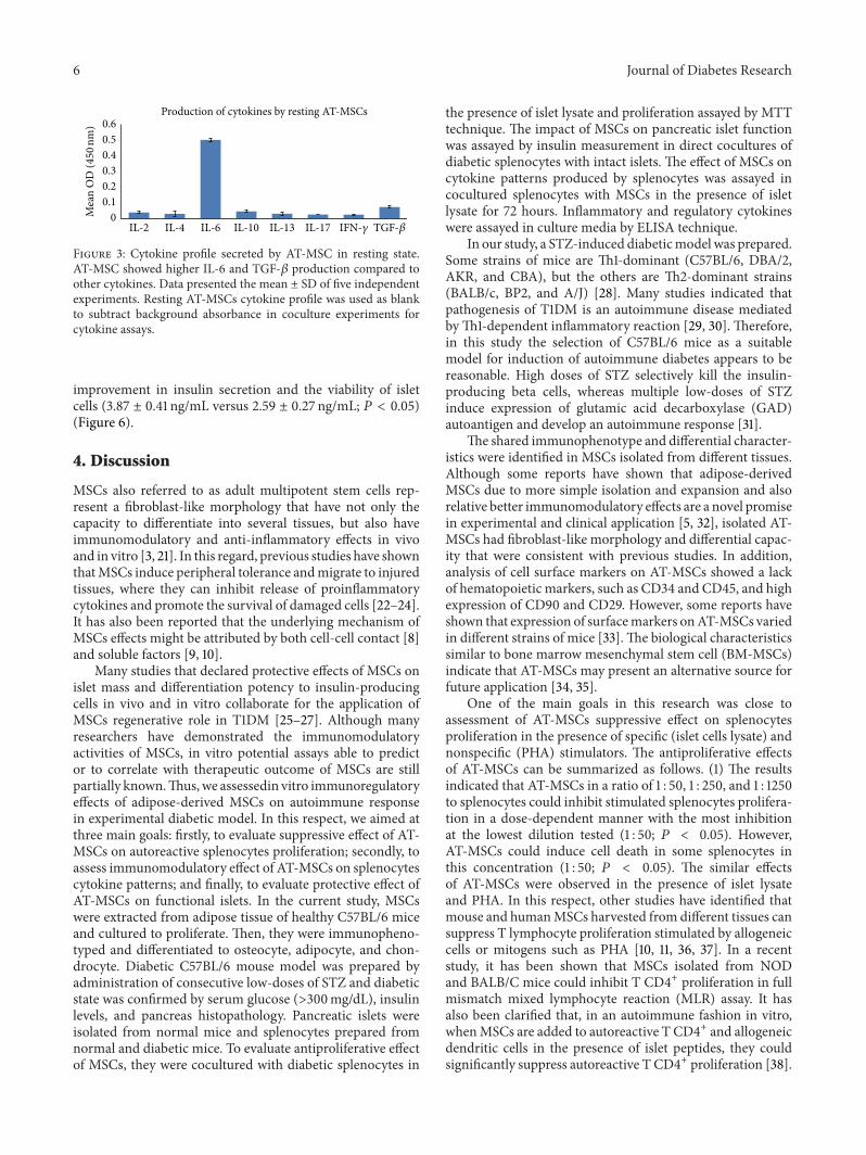
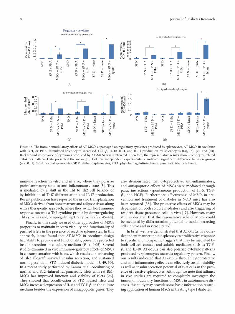
splenocytes supplemented by PHA or islet cells lysate. Anal-ysis of cytokines production by resting AT-MSCs showedthat they consistently secreted TGF-𝛽 and IL-6 in higherlevels compared to other cytokines (Figure 3; 𝑃 < 0.05).As previously described, AT-MSCs were used as blank tosubtract background absorbance of their cytokines produc-tion. The results demonstrated that addition of AT-MSCs tolysate-stimulated diabetic splenocytes significantly decreasedIFN-𝛾, IL-2, and IL-17 production by splenocytes (Figure 4;𝑃 < 0.05). The similar results were obtained when AT-MSCswere added to PHA-triggered splenocytes. In addition, thisexperiment indicated that AT-MSCs significantly increasedTGF-𝛽, IL-10, IL-4, and IL-13 production by splenocytes(Figure 5; 𝑃 < 0.05). Although the increased production
of these cytokines was also observed in the presence ofPHA, increasing levels were significantly prominent in AT-MSCs coculturedwith lysate-stimulated diabetic splenocytes,particularly for IL-13 (Figure 5).
3.7. Protective Effect of AT-MSCs on Insulin Secretion of Islets.To determine protective ability of AT-MSCs on islet cellsfunction, islets were cocultured with reactive splenocytesand insulin content assay was performed. This experimentindicated that the crude islets alone secreted significantinsulin levels when inducedwith stimulatorymedium (6.45 ±0.69 ng/mL). However, diabetic splenocytes significantlydecreased insulin secretion by islets (2.59 ± 0.27 ng/mL; 𝑃 <0.05). In contrast, coculturewithAT-MSCs led to a significant
6 Journal of Diabetes Research
00.10.20.30.40.50.6
IL-2 IL-4 IL-6 IL-10 IL-13 IL-17
Production of cytokines by resting AT-MSCs
IFN-𝛾 TGF-𝛽
Mea
n O
D (450
nm)
Figure 3: Cytokine profile secreted by AT-MSC in resting state.AT-MSC showed higher IL-6 and TGF-𝛽 production compared toother cytokines. Data presented the mean ± SD of five independentexperiments. Resting AT-MSCs cytokine profile was used as blankto subtract background absorbance in coculture experiments forcytokine assays.
improvement in insulin secretion and the viability of isletcells (3.87 ± 0.41 ng/mL versus 2.59 ± 0.27 ng/mL; 𝑃 < 0.05)(Figure 6).
4. Discussion
MSCs also referred to as adult multipotent stem cells rep-resent a fibroblast-like morphology that have not only thecapacity to differentiate into several tissues, but also haveimmunomodulatory and anti-inflammatory effects in vivoand in vitro [3, 21]. In this regard, previous studies have shownthatMSCs induce peripheral tolerance andmigrate to injuredtissues, where they can inhibit release of proinflammatorycytokines and promote the survival of damaged cells [22–24].It has also been reported that the underlying mechanism ofMSCs effects might be attributed by both cell-cell contact [8]and soluble factors [9, 10].
Many studies that declared protective effects of MSCs onislet mass and differentiation potency to insulin-producingcells in vivo and in vitro collaborate for the application ofMSCs regenerative role in T1DM [25–27]. Although manyresearchers have demonstrated the immunomodulatoryactivities of MSCs, in vitro potential assays able to predictor to correlate with therapeutic outcome of MSCs are stillpartially known.Thus,we assessedin vitro immunoregulatoryeffects of adipose-derived MSCs on autoimmune responsein experimental diabetic model. In this respect, we aimed atthree main goals: firstly, to evaluate suppressive effect of AT-MSCs on autoreactive splenocytes proliferation; secondly, toassess immunomodulatory effect of AT-MSCs on splenocytescytokine patterns; and finally, to evaluate protective effect ofAT-MSCs on functional islets. In the current study, MSCswere extracted from adipose tissue of healthy C57BL/6 miceand cultured to proliferate. Then, they were immunopheno-typed and differentiated to osteocyte, adipocyte, and chon-drocyte. Diabetic C57BL/6 mouse model was prepared byadministration of consecutive low-doses of STZ and diabeticstate was confirmed by serum glucose (>300mg/dL), insulinlevels, and pancreas histopathology. Pancreatic islets wereisolated from normal mice and splenocytes prepared fromnormal and diabetic mice. To evaluate antiproliferative effectof MSCs, they were cocultured with diabetic splenocytes in
the presence of islet lysate and proliferation assayed by MTTtechnique. The impact of MSCs on pancreatic islet functionwas assayed by insulin measurement in direct cocultures ofdiabetic splenocytes with intact islets. The effect of MSCs oncytokine patterns produced by splenocytes was assayed incocultured splenocytes with MSCs in the presence of isletlysate for 72 hours. Inflammatory and regulatory cytokineswere assayed in culture media by ELISA technique.
In our study, a STZ-induced diabeticmodel was prepared.Some strains of mice are Th1-dominant (C57BL/6, DBA/2,AKR, and CBA), but the others are Th2-dominant strains(BALB/c, BP2, and A/J) [28]. Many studies indicated thatpathogenesis of T1DM is an autoimmune disease mediatedbyTh1-dependent inflammatory reaction [29, 30]. Therefore,in this study the selection of C57BL/6 mice as a suitablemodel for induction of autoimmune diabetes appears to bereasonable. High doses of STZ selectively kill the insulin-producing beta cells, whereas multiple low-doses of STZinduce expression of glutamic acid decarboxylase (GAD)autoantigen and develop an autoimmune response [31].
The shared immunophenotype and differential character-istics were identified in MSCs isolated from different tissues.Although some reports have shown that adipose-derivedMSCs due to more simple isolation and expansion and alsorelative better immunomodulatory effects are a novel promisein experimental and clinical application [5, 32], isolated AT-MSCs had fibroblast-like morphology and differential capac-ity that were consistent with previous studies. In addition,analysis of cell surface markers on AT-MSCs showed a lackof hematopoietic markers, such as CD34 and CD45, and highexpression of CD90 and CD29. However, some reports haveshown that expression of surfacemarkers onAT-MSCs variedin different strains of mice [33].The biological characteristicssimilar to bone marrow mesenchymal stem cell (BM-MSCs)indicate that AT-MSCs may present an alternative source forfuture application [34, 35].
One of the main goals in this research was close toassessment of AT-MSCs suppressive effect on splenocytesproliferation in the presence of specific (islet cells lysate) andnonspecific (PHA) stimulators. The antiproliferative effectsof AT-MSCs can be summarized as follows. (1) The resultsindicated that AT-MSCs in a ratio of 1 : 50, 1 : 250, and 1 : 1250to splenocytes could inhibit stimulated splenocytes prolifera-tion in a dose-dependent manner with the most inhibitionat the lowest dilution tested (1 : 50; 𝑃 < 0.05). However,AT-MSCs could induce cell death in some splenocytes inthis concentration (1 : 50; 𝑃 < 0.05). The similar effectsof AT-MSCs were observed in the presence of islet lysateand PHA. In this respect, other studies have identified thatmouse and humanMSCs harvested from different tissues cansuppress T lymphocyte proliferation stimulated by allogeneiccells or mitogens such as PHA [10, 11, 36, 37]. In a recentstudy, it has been shown that MSCs isolated from NODand BALB/C mice could inhibit T CD4+ proliferation in fullmismatch mixed lymphocyte reaction (MLR) assay. It hasalso been clarified that, in an autoimmune fashion in vitro,whenMSCs are added to autoreactive T CD4+ and allogeneicdendritic cells in the presence of islet peptides, they couldsignificantly suppress autoreactive T CD4+ proliferation [38].

Journal of Diabetes Research 7
00.040.080.120.16
Inflammatory cytokines
∗
∗ ∗
IFN-𝛾 production by splenocytes
SP N
SPN+
PHA
SP D
SPD+
PHA
SPD+
lysa
te
SPN+
lysa
te
Mea
n re
sidua
lO
D (450
nm)
MSC
s+SP
N+
PHA
MSC
s+SP
D+
PHA
MSC
s+SP
D+
lysa
te
(a)
00.05
0.10.15
0.20.25
IL-17 production by splenocytes
∗ ∗∗
SP N
SPN+
PHA
SP D
SPD+
PHA
SPD+
lysa
te
SPN+
lysa
te
Mea
n re
sidua
lO
D (450
nm)
MSC
s+SP
N+
PHA
MSC
s+SP
D+
PHA
MSC
s+SP
D+
lysa
te
(b)
00.05
0.10.15
0.20.25
0.3
IL-2 production by splenocytes
∗
∗
∗
SP N
SPN+
PHA
SP D
SPD+
PHA
SPD+
lysa
te
SPN+
lysa
te
Mea
n re
sidua
lO
D (450
nm)
MSC
s+SP
N+
PHA
MSC
s+SP
D+
PHA
MSC
s+SP
D+
lysa
te(c)
Figure 4: The immunomodulatory effects of AT-MSCs at passage 3 on inflammatory cytokines produced by splenocytes. Addition of AT-MSCs to islet, or PHA, stimulated splenocytes decreased IFN-𝛾, IL-2, and IL-17 production by splenocytes ((a), (b), and (c)). Backgroundabsorbance of cytokines produced by AT-MSCs was subtracted. Thus, the representative results show splenocytes related cytokines pattern.Data presented the mean ± SD of five independent experiments. ∗ indicates significant difference between groups (𝑃 < 0.05). SP N: normalsplenocytes; SP D: diabetic splenocytes; PHA: phytohemagglutinin; lysate: pancreatic islet cells lysate.
We found that suppressive effects of MSCs were dependenton cell number. Several reports have demonstrated thatimmunosuppressive effects of MSCs occur in a dose-relatedmanner in which it is considered independent of MHCinteraction [39–41]. Although target cell-MSC interactionsmay be an important factor, the immunosuppressive effectof MSCs can also be mediated through the secretion ofsoluble molecules such as IL-10, TGF-𝛽, IDO, NO, PGE-2, and HLA-G that are triggered following cross talk withtarget cells [4]. (2) AT-MSCs caused some cell death in restingsplenocytes without each type of stimulators (𝑃 < 0.05). Thismay suggest that MSCs can induce cellular death throughcell-cell contact as well as secretion of NO and IL-10 [9,11]; also, they can drive splenocytes to produce IL-10 [11].Although, in accordance with previous studies, we suggestthat MSCs may induce cell death of splenocytes rather thancell cycle arrest [11], complementary studies are required toidentify molecular mechanisms in detail. (3) In the absenceof AT-MSCs, normal and diabetic splenocytes in responseto nonspecific stimulator (PHA) showed similar fashion. Incontrast, in response to islet lysate normal splenocytes werenot responsive, whereas a sharp proliferation was obtained indiabetic splenocytes (𝑃 < 0.05).
In the next stage, we aimed at evaluating the effect ofAT-MSCs on cytokine patterns produced by splenocytes.Analysis of cytokines production by AT-MSCs alone showedthat they consistently secreted TGF-𝛽 and IL-6 in higherlevels compared to other cytokines (𝑃 < 0.05). Severalpublications have confirmed spontaneous secretion ofTGF-𝛽1, IL-6, hepatocyte growth factor (HGF), HLA-G, andPGE-2 that associated with immunomodulatory propertiesof MSCs [42, 43]. Our in vitro experiment showed thatAT-MSCs decreased production of IFN-𝛾, IL-2, and IL-17and increased production of TGF-𝛽, IL-4, IL-10, and IL-13by stimulated splenocytes in response to islet lysate or PHA(𝑃 < 0.05). However, the increases in regulatory cytokineswere significantly prominent in AT-MSCs cocultured withlysate-stimulated diabetic splenocytes, particularly for IL-13.Pathogenesis of many autoimmune diseases was mediatedby several types of helper T lymphocytes (Th) such as Th1,Th2, and Th17 identified by secretion of distinct cytokineprofiles [44]. Th1 cells produced IFN-𝛾, IL-2, and TNF-𝛼and Th2 cells secreted IL-4, IL-5, IL-6, IL-10, and IL-13.Regulatory T cells and Th3 cells produce TGF-𝛽 and IL10and another T helper subtype (Th17) produces IL-17. It hasbeen demonstrated that MSCs have capacity to modulate
8 Journal of Diabetes Research
00.10.20.30.40.50.6
Regulatory cytokines TGF-𝛽 production by splenocytes
∗∗
∗
Mea
n re
sidua
lO
D (450
nm)
SP N
SPN+
PHA
SP D
SPD+
PHA
SPD+
lysa
te
SPN+
lysa
te
MSC
s+SP
N+
PHA
MSC
s+SP
D+
PHA
MSC
s+SP
D+
lysa
te
(a)
00.10.20.30.40.50.6
IL-10 production by splenocytes
∗ ∗
∗
Mea
n re
sidua
lO
D (450
nm)
SP N
SPN+
PHA
SP D
SPD+
PHA
SPD+
lysa
te
SPN+
lysa
te
MSC
s+SP
N+
PHA
MSC
s+SP
D+
PHA
MSC
s+SP
D+
lysa
te
(b)
00.05
0.10.15
0.20.25
IL-4 production by splenocytes
∗∗
∗
Mea
n re
sidua
lO
D (450
nm)
SP N
SPN+
PHA
SP D
SPD+
PHA
SPD+
lysa
te
SPN+
lysa
te
MSC
s+SP
N+
PHA
MSC
s+SP
D+
PHA
MSC
s+SP
D+
lysa
te
(c)
00.10.20.30.4
IL-13 production by splenocytes∗
Mea
n re
sidua
lO
D (450
nm)
SP N
SPN+
PHA
SP D
SPD+
PHA
SPD+
lysa
te
SPN+
lysa
te
MSC
s+SP
N+
PHA
MSC
s+SP
D+
PHA
MSC
s+SP
D+
lysa
te
(d)
Figure 5:The immunomodulatory effects of AT-MSCs at passage 3 on regulatory cytokines produced by splenocytes. AT-MSCs in coculturewith islet, or PHA, stimulated splenocytes increased TGF-𝛽, IL-10, IL-4, and IL-13 production by splenocytes ((a), (b), (c), and (d)).Background absorbance of cytokines produced by AT-MCSs was subtracted. Therefore, the representative results show splenocytes relatedcytokines pattern. Data presented the mean ± SD of five independent experiments. ∗ indicates significant difference between groups(𝑃 < 0.05). SP N: normal splenocytes; SP D: diabetic splenocytes; PHA: phytohemagglutinin; lysate: pancreatic islet cells lysate.
immune reaction in vitro and in vivo, where they polarizeproinflammatory state to anti-inflammatory state [3]. Thisis mediated by a shift in the Th1 to Th2 cell balance orby inhibition of Th17 differentiation and IL-17 production.Recent publications have reported the in vivo transplantationof MSCs derived from bonemarrow and adipose tissue alongwith a therapeutic approach, where they switch host immuneresponse towards a Th2 cytokine profile by downregulatingTh1 cytokines and/or upregulatingTh2 cytokines [22, 45–48].
Finally, in this study we used other approaches of MSCsproperties to maintain in vitro viability and functionality ofpurified islets in the presence of reactive splenocytes. In thisapproach, it was found that AT-MSCs in coculture designhad ability to provide islet functionality, proven by protectedinsulin secretion in coculture medium (𝑃 < 0.05). Severalstudies examined in vivo immunoregulatory effects of MSCsin cotransplantation with islets, which resulted in enhancingof islet allograft survival, insulin secretion, and sustainednormoglycemia in STZ-induced diabetic model [43, 49, 50].In a recent study performed by Karaoz et al. coculturing ofnormal and STZ-injured rat pancreatic islets with rat BM-MSCs has improved function and viability of islets [26].They showed that cocultivation of STZ-injured islets andMSCs increased expression of IL-6 and TGF-𝛽1 in the culturemedium besides the expression of antiapoptotic genes. They
also demonstrated that cytoprotective, anti-inflammatory,and antiapoptotic effects of MSCs were mediated throughparacrine actions (spontaneous production of IL-6, TGF-𝛽1, and HGF). Furthermore, effectiveness of MSCs in pre-vention and treatment of diabetes in NOD mice has alsobeen reported [38]. The protective effects of MSCs may bedependent on both soluble mediators and also triggering ofresident tissue precursor cells in vivo [17]. However, manystudies declared that the regenerative role of MSCs couldbe mediated by differentiation potential to insulin-secretingcells in vivo and in vitro [18, 25].
In brief, we have demonstrated that AT-MSCs in a dose-dependent manner inhibit splenocytes proliferative responseto specific and nonspecific triggers that may be mediated byboth cell-cell contact and soluble mediators such as TGF-𝛽1 and IL-10. AT-MSCs can also polarize cytokine patternsproduced by splenocytes toward a regulatory pattern. Finally,our results indicated that AT-MSCs through cytoprotectiveand anti-inflammatory effects can effectively sustain viabilityas well as insulin secretion potential of islet cells in the pres-ence of reactive splenocytes. Although we note that adjunctin vivo studies are required to completely investigate theimmunomodulatory functions of MSCs in autoimmune dis-eases, this study may provide some basic information regard-ing application of human MSCs in treating type 1 diabetes.

Journal of Diabetes Research 9
0
1
2
3
4
5
6
7Is
let
Insulin release assayIn
sulin
cont
ent (
ng/m
L)
∗ ∗
SPN+
islet
SPD+
islet
MSC
s+SP
N+
islet
MSC
s+SP
D+
islet
Figure 6: Pancreatic islet insulin release assay. Intact islets alonesecreted distinct amount of insulin in response to stimulatorymedium (6.45 ± 0.69 ng/mL). However, the insulin secretion lev-els decreased after coculture with diabetic splenocytes (2.59 ±0.27 ng/mL). Interestingly, addition of AT-MSCs improved insulinsecretion by injured islets (3.87 ± 0.41 ng/mL). Data presented themean ± SD of five independent experiments. ∗ indicates significantdifference between groups (𝑃 < 0.05). SP N: normal splenocytes; SPD: diabetic splenocytes; islet: mouse pancreatic islet.
Conflict of Interests
The authors indicate no potential conflict of interests.
Acknowledgments
The authors acknowledge funding of this study providedby Iran University of Medical Sciences and the Stem CellTechnology Research Center.
References
[1] C. Aguayo-Mazzucato and S. Bonner-Weir, “Stem cell therapyfor type 1 diabetes mellitus,”Nature Reviews Endocrinology, vol.6, no. 3, pp. 139–148, 2010.
[2] P. K. Mishra, S. R. Singh, I. G. Joshua, and S. C. Tyagi, “Stemcells as a therapeutic target for diabetes,” Frontiers in Bioscience,vol. 15, no. 2, pp. 461–477, 2010.
[3] A. Uccelli, L. Moretta, and V. Pistoia, “Mesenchymal stem cellsin health and disease,” Nature Reviews Immunology, vol. 8, no.9, pp. 726–736, 2008.
[4] R. Meisel, A. Zibert, M. Laryea, U. Gobel, W. Daubener, and D.Dilloo, “Human bone marrow stromal cells inhibit allogeneicT-cell responses by indoleamine 2,3-dioxygenase-mediatedtryptophan degradation,” Blood, vol. 103, no. 12, pp. 4619–4621,2004.
[5] B. Puissant, C. Barreau, P. Bourin et al., “Immunomodulatoryeffect of human adipose tissue-derived adult stem cells: com-parison with bonemarrowmesenchymal stem cells,”TheBritishJournal of Haematology, vol. 129, no. 1, pp. 118–129, 2005.
[6] E. Ivanova-Todorova, I. Bochev, M. Mourdjeva et al., “Adiposetissue-derived mesenchymal stem cells are more potent sup-pressors of dendritic cells differentiation compared to bonemarrow-derived mesenchymal stem cells,” Immunology Letters,vol. 126, no. 1-2, pp. 37–42, 2009.
[7] H. K. Salem and C.Thiemermann, “Mesenchymal stromal cells:current understanding and clinical status,” Stem Cells, vol. 28,no. 3, pp. 585–596, 2010.
[8] H. Sheng, Y. Wang, Y. Jin et al., “A critical role of IFN𝛾in priming MSC-mediated suppression of T cell proliferationthrough up-regulation of B7-H1,” Cell Research, vol. 18, no. 8,pp. 846–857, 2008.
[9] K. Sato, K. Ozaki, I. Oh et al., “Nitric oxide plays a critical role insuppression of T-cell proliferation by mesenchymal stem cells,”Blood, vol. 109, no. 1, pp. 228–234, 2007.
[10] X. Lu, T. Liu, L. Gu et al., “Immunomodulatory effects ofmesenchymal stem cells involved in favoring type 2 T cellsubsets,” Transplant Immunology, vol. 22, no. 1-2, pp. 55–61,2009.
[11] S.-H. Yang, M.-J. Park, I.-H. Yoon et al., “Soluble mediatorsfrom mesenchymal stem cells suppress T cell proliferation byinducing IL-10,” Experimental and Molecular Medicine, vol. 41,no. 5, pp. 315–324, 2009.
[12] K. Nemeth, A. Keane-Myers, J. M. Brown et al., “Bone marrowstromal cells use TGF-beta to suppress allergic responses in amouse model of ragweed-induced asthma,” Proceedings of theNational Academy of Sciences of the United States of America,vol. 107, no. 12, pp. 5652–5657, 2010.
[13] O. Delarosa, E. Lombardo, A. Beraza et al., “Requirement ofIFN-𝛾-mediated indoleamine 2,3-dioxygenase expression inthe modulation of lymphocyte proliferation by human adipose-derived stem cells,” Tissue Engineering—Part A, vol. 15, no. 10,pp. 2795–2806, 2009.
[14] R. Yanez, A. Oviedo, M. Aldea, J. A. Bueren, andM. L. Lamana,“Prostaglandin E2 plays a key role in the immunosuppressiveproperties of adipose and bone marrow tissue-derived mes-enchymal stromal cells,”Experimental Cell Research, vol. 316, no.19, pp. 3109–3123, 2010.
[15] Z. Selmani, A.Naji, E.Gaiffe et al., “HLA-G is a crucial immuno-suppressive molecule secreted by adult human mesenchymalstem cells,” Transplantation, vol. 87, no. 9, pp. S62–S66, 2009.
[16] L. Jarvinen, L. Badri, S. Wettlaufer et al., “Lung residentmesenchymal stem cells isolated from human lung allograftsinhibit T cell proliferation via a soluble mediator,” Journal ofImmunology, vol. 181, no. 6, pp. 4389–4396, 2008.
[17] M. J. Hoogduijn, F. Popp, R. Verbeek et al., “The immunomod-ulatory properties of mesenchymal stem cells and their use forimmunotherapy,” International Immunopharmacology, vol. 10,no. 12, pp. 1496–1500, 2010.
[18] V. Chandra, G. Swetha, S. Phadnis, P. D. Nair, and R. R. Bhonde,“Generation of pancreatic hormone-expressing islet-like cellaggregates frommurine adipose tissue-derived stem cells,” StemCells, vol. 27, no. 8, pp. 1941–1953, 2009.
[19] P. E. Lacy and M. Kostianovsky, “Method for the isolation ofintact islets of Langerhans from the rat pancreas.,”Diabetes, vol.16, no. 1, pp. 35–39, 1967.
[20] D. Chen, N. Zhang, S. Fu et al., “CD4+CD25+ regulatory T-cells inhibit the islet innate immune response and promote isletengraftment,” Diabetes, vol. 55, no. 4, pp. 1011–1021, 2006.
[21] R. Abdi, P. Fiorina, C. N. Adra, M. Atkinson, andM. H. Sayegh,“Immunomodulation by mesenchymal stem cells: a potential
10 Journal of Diabetes Research
therapeutic strategy for type 1 diabetes,” Diabetes, vol. 57, no. 7,pp. 1759–1767, 2008.
[22] G.Constantin, S.Marconi, B. Rossi et al., “Adipose-derivedmes-enchymal stem cells ameliorate chronic experimental autoim-mune encephalomyelitis,” Stem Cells, vol. 27, no. 10, pp. 2624–2635, 2009.
[23] A. Uccelli, A. Laroni, and M. S. Freedman, “Mesenchymalstem cells for the treatment of multiple sclerosis and otherneurological diseases,” The Lancet Neurology, vol. 10, no. 7, pp.649–656, 2011.
[24] N. L. Payne, G. Sun, C.Mcdonald et al., “Distinct immunomod-ulatory and migratory mechanisms underpin the therapeuticpotential of human mesenchymal stem cells in autoimmunedemyelination,” Cell Transplantation, vol. 22, no. 8, pp. 1409–1425, 2013.
[25] D.-Q. Tang, L.-Z. Cao, B. R. Burkhardt et al., “In vivo and invitro characterization of insulin-producing cells obtained frommurine bone marrow,” Diabetes, vol. 53, no. 7, pp. 1721–1732,2004.
[26] E. Karaoz, Z. S. Genc, P. C. Demircan, A. Aksoy, and G.Duruksu, “Protection of rat pancreatic islet function and via-bility by coculture with rat bone marrow-derived mesenchymalstem cells,” Cell Death and Disease, vol. 1, article e36, 2010.
[27] A. Scuteri, E. Donzelli, V. Rodriguez-Menendez et al., “A doublemechanism for the mesenchymal stem cells’ positive effect onpancreatic islets,” PLoS ONE, vol. 9, no. 1, Article ID e84309,2014.
[28] V. De Vooght, J. A. J. Vanoirbeek, K. Luyts, S. Haenen, B.Nemery, and P. H. M. Hoet, “Choice of mouse strain influencesthe outcome in a mouse model of chemical-induced asthma,”PloS ONE, vol. 5, no. 9, Article ID e12581, 2010.
[29] K. M. Gillespie, “Type 1 diabetes: pathogenesis and prevention,”Canadian Medical Association Journal, vol. 175, no. 2, pp. 165–170, 2006.
[30] T. L. van Belle, K. T. Coppieters, and M. G. von Herrath, “Type1 diabetes: etiology, immunology, and therapeutic strategies,”Physiological Reviews, vol. 91, no. 1, pp. 79–118, 2011.
[31] T. L. VanBelle, P. Taylor, andM.G. vonHerrath, “Mousemodelsfor type 1 diabetes,” Drug Discovery Today: Disease Models, vol.6, no. 2, pp. 41–45, 2009.
[32] Y. Zhu, T. Liu, K. Song, X. Fan, X. Ma, and Z. Cui, “Adipose-derived stem cell: a better stem cell than BMSC,” Cell Biochem-istry and Function, vol. 26, no. 6, pp. 664–675, 2008.
[33] S. M. Hashemi, Z. M. Hassan, A. A. Pourfathollah, S. Soudi, A.Shafiee, and M. Soleimani, “Comparative immunomodulatoryproperties of adipose-derived mesenchymal stem cells condi-tioned media from BALB/c, C57BL/6, and DBAmouse strains,”Journal of Cellular Biochemistry, vol. 114, no. 4, pp. 955–965,2013.
[34] A. Schaffler and C. Buchler, “Concise review: adipose tissue-derived stromal cells—basic and clinical implications for novelcell-based therapies,” StemCells, vol. 25, no. 4, pp. 818–827, 2007.
[35] I. Bochev, G. Elmadjian, D. Kyurkchiev et al., “Mesenchymalstem cells from human bone marrow or adipose tissue dif-ferently modulate mitogen-stimulated B-cell immunoglobulinproduction in vitro,” Cell Biology International, vol. 32, no. 4,pp. 384–393, 2008.
[36] M. Sioud, A. Mobergslien, A. Boudabous, and Y. Fløisand,“Mesenchymal stem cell-mediated T cell suppression occursthrough secreted galectins,” International Journal of Oncology,vol. 38, no. 2, pp. 385–390, 2011.
[37] E. Svobodova,M. Krulova, A. Zajicova et al., “The role ofmousemesenchymal stem cells in differentiation of naive T-cells intoanti-inflammatory regulatory T-cell or proinflammatory helperT-cell 17 population,” Stem Cells and Development, vol. 21, no. 6,pp. 901–910, 2012.
[38] P. Fiorina, M. Jurewicz, A. Augello et al., “Immunomodulatoryfunction of bone marrow-derived mesenchymal stem cellsin experimental autoimmune type 1 diabetes,” The Journal ofImmunology, vol. 183, no. 2, pp. 993–1004, 2009.
[39] C. Bocelli-Tyndall, L. Bracci, G. Spagnoli et al., “Bone marrowmesenchymal stromal cells (BM-MSCs) from healthy donorsand auto-immune disease patients reduce the proliferation ofautologous- and allogeneic-stimulated lymphocytes in vitro,”Rheumatology, vol. 46, no. 3, pp. 403–408, 2007.
[40] M. L. Weiss, C. Anderson, S. Medicetty et al., “Immuneproperties of human umbilical cord Wharton’s jelly-derivedcells,” Stem Cells, vol. 26, no. 11, pp. 2865–2874, 2008.
[41] M. Wang, Y. Yang, D. Yang et al., “The immunomodulatoryactivity of human umbilical cord blood-derived mesenchymalstem cells in vitro,” Immunology, vol. 126, no. 2, pp. 220–232,2009.
[42] A. E. Aksu, E. Horibe, J. Sacks et al., “Co-infusion of donor bonemarrow with host mesenchymal stem cells treats GVHD andpromotes vascularized skin allograft survival in rats,” ClinicalImmunology, vol. 127, no. 3, pp. 348–358, 2008.
[43] I. Boumaza, S. Srinivasan, W. T. Witt et al., “Autologous bonemarrow-derived rat mesenchymal stem cells promote PDX-1and insulin expression in the islets, alter T cell cytokine patternand preserve regulatory T cells in the periphery and inducesustained normoglycemia,” Journal of Autoimmunity, vol. 32,no. 1, pp. 33–42, 2009.
[44] L. Steinman, “A brief history of T(H)17, the first major revisionin the T(H)1/T(H)2 hypothesis of T cell-mediated tissue dam-age,” Nature Medicine, vol. 13, no. 2, pp. 139–145, 2007.
[45] P. Batten, P. Sarathchandra, J. W. Antoniw et al., “Humanmesenchymal stem cells induce T cell anergy and downregulateT cell allo-responses via the TH2 pathway: relevance to tissueengineering human heart valves,”Tissue Engineering, vol. 12, no.8, pp. 2263–2273, 2006.
[46] C. Bouffi, C. Bony, G. Courties, C. Jorgensen, and D. Noel,“IL-6-dependent PGE2 secretion by mesenchymal stem cellsinhibits local inflammation in experimental arthritis,” PLoSONE, vol. 5, no. 12, Article ID e14247, 2010.
[47] P. J. Darlington, M.-N. Boivin, C. Renoux et al., “ReciprocalTh1andTh17 regulation bymesenchymal stem cells: implication formultiple sclerosis,” Annals of Neurology, vol. 68, no. 4, pp. 540–545, 2010.
[48] E. W. Choi, I. S. Shin, H. W. Lee et al., “Transplantation ofCTLA4Ig gene-transduced adipose tissue-derived mesenchy-mal stem cells reduces inflammatory immune response andimproves Th1/Th2 balance in experimental autoimmune thy-roiditis,” The Journal of Gene Medicine, vol. 13, no. 1, pp. 3–16,2011.
[49] N. Sakata, M. Goto, G. Yoshimatsu, S. Egawa, and M. Unno,“Utility of co-transplanting mesenchymal stem cells in islettransplantation,” World Journal of Gastroenterology, vol. 17, no.47, pp. 5150–5155, 2011.
[50] M. G. Solari, S. Srinivasan, I. Boumaza et al., “Marginalmass islet transplantation with autologous mesenchymal stemcells promotes long-term islet allograft survival and sustainednormoglycemia,” Journal of Autoimmunity, vol. 32, no. 2, pp.116–124, 2009.

Submit your manuscripts athttp://www.hindawi.com
Stem CellsInternational
Hindawi Publishing Corporationhttp://www.hindawi.com Volume 2014
Hindawi Publishing Corporationhttp://www.hindawi.com Volume 2014
MEDIATORSINFLAMMATION
of
Hindawi Publishing Corporationhttp://www.hindawi.com Volume 2014
Behavioural Neurology
EndocrinologyInternational Journal of
Hindawi Publishing Corporationhttp://www.hindawi.com Volume 2014
Hindawi Publishing Corporationhttp://www.hindawi.com Volume 2014
Disease Markers
Hindawi Publishing Corporationhttp://www.hindawi.com Volume 2014
BioMed Research International
OncologyJournal of
Hindawi Publishing Corporationhttp://www.hindawi.com Volume 2014
Hindawi Publishing Corporationhttp://www.hindawi.com Volume 2014
Oxidative Medicine and Cellular Longevity
Hindawi Publishing Corporationhttp://www.hindawi.com Volume 2014
PPAR Research
The Scientific World JournalHindawi Publishing Corporation http://www.hindawi.com Volume 2014
Immunology ResearchHindawi Publishing Corporationhttp://www.hindawi.com Volume 2014
Journal of
ObesityJournal of
Hindawi Publishing Corporationhttp://www.hindawi.com Volume 2014
Hindawi Publishing Corporationhttp://www.hindawi.com Volume 2014
Computational and Mathematical Methods in Medicine
OphthalmologyJournal of
Hindawi Publishing Corporationhttp://www.hindawi.com Volume 2014
Diabetes ResearchJournal of
Hindawi Publishing Corporationhttp://www.hindawi.com Volume 2014
Hindawi Publishing Corporationhttp://www.hindawi.com Volume 2014
Research and TreatmentAIDS
Hindawi Publishing Corporationhttp://www.hindawi.com Volume 2014
Gastroenterology Research and Practice
Hindawi Publishing Corporationhttp://www.hindawi.com Volume 2014
Parkinson’s Disease
Evidence-Based Complementary and Alternative Medicine
Volume 2014Hindawi Publishing Corporationhttp://www.hindawi.com