research article clinical profiles of dengue infection...
TRANSCRIPT
Research ArticleClinical Profiles of Dengue Infection during an Outbreakin Northern India
Anish Laul,1 Poonam Laul,2 Vamsi Merugumala,2 Ravi Pathak,2
Urvashi Miglani,2 and Pinkee Saxena2
1Maulana Azad Medical College, No. 2, Bahadur Shah Zafar Marg, New Delhi, Delhi 110002, India2Deen Dayal Upadhyay Hospital, Hari Nagar, Clock Tower, New Delhi, Delhi 110064, India
Correspondence should be addressed to Poonam Laul; poonam [email protected]
Received 2 August 2016; Revised 25 September 2016; Accepted 8 November 2016
Academic Editor: Shyam Sundar
Copyright © 2016 Anish Laul et al. This is an open access article distributed under the Creative Commons Attribution License,which permits unrestricted use, distribution, and reproduction in any medium, provided the original work is properly cited.
Introduction.Dengue fever is an arboviral disease, which is transmitted bymosquito vector and presents as varied clinical spectrumof dengue fever (DF), dengue hemorrhagic fever (DHF), dengue shock syndrome (DSS), and expanded dengue syndrome (EDS)with atypical presentations, thus posing a diagnostic dilemma. Unless we are aware of these presentations, diagnosis as well asearly initiation of treatment becomes difficult.We studied the various clinical presentations of dengue infection during an outbreakof disease in 2015. Materials and Methods. A total of 115 confirmed cases of dengue infection from Department of Medicine ofDeen Dayal Upadhyay Hospital, New Delhi, were enrolled in this observational study. Results.The common signs and symptomsof dengue infection were fever, headache, body ache, backache, retro-orbital pain, bleeding manifestations, and rash in 100%,87%, 86%, 58%, 41%, 21%, and 21%, respectively. Nonspecific or warning signs and symptoms included vomiting, weakness,abdominal pain, breathlessness, vertigo, sweating, and syncope. Other possible signs and symptoms of coinfections, comorbidities,or complications included diarrhea, sore throat, and neurological manifestations. There were seven patients with coinfections andfour with comorbidities.The final diagnosis of these patientswasDF (73%), DHF (16.5%), DSS (1.7%), and EDS (4.3%). Among EDSpatients, the atypical presentations included encephalopathy, lateral rectus nerve palsy, acalculous cholecystitis, and myocarditis.Four patients required ICU care and there was no death in this study. Conclusion. Knowledge of atypical presentations is a must forearly diagnosis and timely intervention to prevent life-threatening complications.
1. Introduction
Dengue is a viral disease of tropical regions. The causativeagent is one of serotypes of a single stranded RNAvirus calledDENV 1, 2, 3, and 4 [1]. It is a tropical disease, which istransmitted by theAedes aegyptimosquito. Incubation periodis 4 to 7 days (range 3–14 days) [2]. Once the symptomsstart, person can remain infectious for next six to sevendays. In addition to mosquito bite, dengue has been reportedto be transmitted by transfusion of blood from an infecteddonor, injuries by infected sharps to health care workers,transplantation of organs and tissues from infected donors,and from infected pregnant mother to her fetus by verticaltransmission [3–5]. This viral infection has a wide clinicalspectrum ranging from asymptomatic disease to undiffer-entiated fever (or viral syndromes), classical dengue fever
(DF), dengue hemorrhagic fever (DHF), or dengue shocksyndrome (DSS) and expanded dengue syndrome (EDS) [2].After incubation period ends, the disease is followed by threephases: febrile, critical, and recovery phase [2]. In the recentyears, dengue has crossed geographic borders and has spreadto many new countries. Currently, it is endemic to southeastAsia and western pacific regions [2]. So dengue fever hasbecome a major international public concern particularly intropical and subtropical regions affecting urban as well asrural areas.Though several measures are taken to prevent andcontrol it, recurrent outbreaks have been reported in India.The first outbreak of dengue fever in India was in 1812 [6].
Severalmajor outbreaks took place after this in years 1836,1906, 1911, 1972, 2005, 2010, and 2015. An increasing numberof cases of dengue are being reported with atypical presen-tations as frequent epidemics are occurring. As awareness of
Hindawi Publishing CorporationJournal of Tropical MedicineVolume 2016, Article ID 5917934, 7 pageshttp://dx.doi.org/10.1155/2016/5917934
2 Journal of Tropical Medicine
Table 1
DF/DHF Grade Signs and symptoms Laboratory
DF With or without haemorrhagicmanifestations
Fever with two of the following:(i) Headache.
(ii) Retro-orbital pain.(iii) Myalgia.
(iv) Arthtralgia/bone pain.(v) Rash.
(vi) Haemorrhagic manifestations.(vii) No evidence of plasma leakage.
Leucopenia (wbc ≤5000 cells/mm3).(i) Thrombocytopenia (platelet count
<150 000 cells/mm3).(ii) Rising haematocrit (5%–10%).(iii) No evidence of plasma loss.
DHF I
Fever and haemorrhagicmanifestation (positive tourniquet
test) and evidence of plasmaleakage.
Thrombocytopenia<100 000 cells/mm3; HCT rise ≥20%.
DHF II As in Grade I plus spontaneousbleeding.
Thrombocytopenia<100 000 cells/mm3; HCT rise ≥20%.
DHF III
As in Grade I or II plus circulatoryfailure (weak pulse, narrow pulse
pressure (≤20 mmHg),hypotension, restlessness).
Thrombocytopenia<100 000 cells/mm3; HCT rise ≥20%.
DHF IV As in Grade III plus profound shockwith undetectable BP and pulse.
Thrombocytopenia <100 000 cells/mm3; HCT rise ≥20%.
this disease is increasing, rare manifestations are also beingreported. During an outbreak in 2015 in Northern India, thepresent study was conducted in tertiary care hospital in Delhihighlighting the varied clinical presentations of dengue fever.
2. Materials and Methods
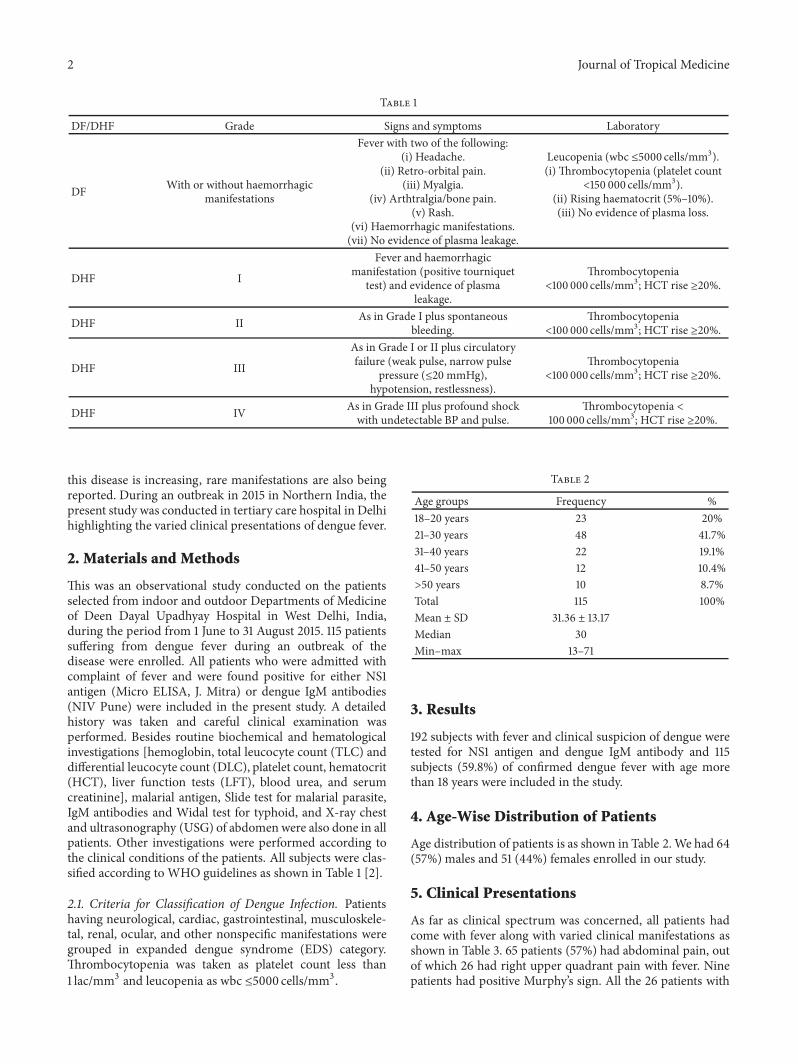
This was an observational study conducted on the patientsselected from indoor and outdoor Departments of Medicineof Deen Dayal Upadhyay Hospital in West Delhi, India,during the period from 1 June to 31 August 2015. 115 patientssuffering from dengue fever during an outbreak of thedisease were enrolled. All patients who were admitted withcomplaint of fever and were found positive for either NS1antigen (Micro ELISA, J. Mitra) or dengue IgM antibodies(NIV Pune) were included in the present study. A detailedhistory was taken and careful clinical examination wasperformed. Besides routine biochemical and hematologicalinvestigations [hemoglobin, total leucocyte count (TLC) anddifferential leucocyte count (DLC), platelet count, hematocrit(HCT), liver function tests (LFT), blood urea, and serumcreatinine], malarial antigen, Slide test for malarial parasite,IgM antibodies and Widal test for typhoid, and X-ray chestand ultrasonography (USG) of abdomen were also done in allpatients. Other investigations were performed according tothe clinical conditions of the patients. All subjects were clas-sified according to WHO guidelines as shown in Table 1 [2].
2.1. Criteria for Classification of Dengue Infection. Patientshaving neurological, cardiac, gastrointestinal, musculoskele-tal, renal, ocular, and other nonspecific manifestations weregrouped in expanded dengue syndrome (EDS) category.Thrombocytopenia was taken as platelet count less than1 lac/mm3 and leucopenia as wbc ≤5000 cells/mm3 .
Table 2
Age groups Frequency %18–20 years 23 20%21–30 years 48 41.7%31–40 years 22 19.1%41–50 years 12 10.4%>50 years 10 8.7%Total 115 100%Mean ± SD 31.36 ± 13.17Median 30Min–max 13–71
3. Results
192 subjects with fever and clinical suspicion of dengue weretested for NS1 antigen and dengue IgM antibody and 115subjects (59.8%) of confirmed dengue fever with age morethan 18 years were included in the study.
4. Age-Wise Distribution of Patients
Age distribution of patients is as shown in Table 2.We had 64(57%) males and 51 (44%) females enrolled in our study.
5. Clinical Presentations
As far as clinical spectrum was concerned, all patients hadcome with fever along with varied clinical manifestations asshown in Table 3. 65 patients (57%) had abdominal pain, outof which 26 had right upper quadrant pain with fever. Ninepatients had positive Murphy’s sign. All the 26 patients with

Journal of Tropical Medicine 3
Table 3
Symptoms Number of patients Percentage%
Fever 115 100
Headache 100 87
Body ache 99 86
Backache 67 58
Retro-orbital pain 47 41
Bleed (anyhemorrhagicmanifestation)
24 21
Rashes 25 21
Vomiting 78 68
Weakness 79 68
Pain abdomen 65 57
Breathlessness 22 19
Vertigo 16 14
Sweating 15 13
Syncope 14 12
Diarrhoea 31 27
Sore throat 29 25
Neurologicalmanifestations
3 2
Itching 22 19
Others 9 8
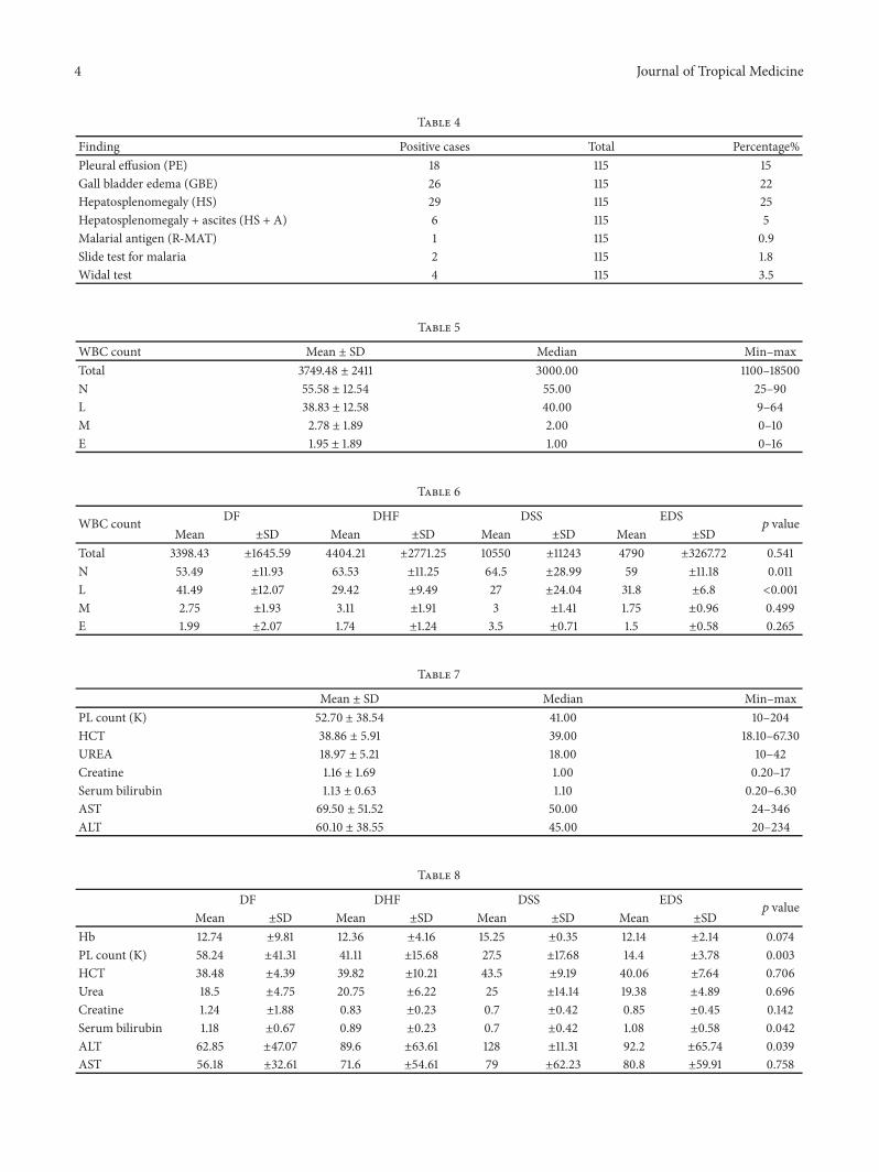
pain had edematous gall bladder without gallstones. Diag-nosis of acalculous cholecystitis was made and all patientsresponded well to supportive therapy. 29 patients (25%) hadassociated hepatomegaly and six patients (5%) had ascitesassociated with hepatosplenomegaly. All 65 patients (57%)of pain abdomen had elevated AST and 49% had elevatedALT as shown in Table 7. There was significant difference inALT among the four groups with elevated levels indicatingthe severest and atypical forms of infections in form of DHF,DSS, andEDS (𝑝 value 0.039) as shown inTable 8. All patientswith raised AST and ALT were discharged once they showedfalling trend and no patient reported to have any hepaticsequelae.
One patientwith jaundice and enzymes in range of 200 IUalso recovered completely within two weeks. All other causesof jaundice were ruled out in this. Only one patient inour study had blood urea of 42mg% and serum creatinine1.6mg%; however he also recovered with supportive treat-mentwithout any complication.Therewas nohepatic or renalfailure reported in our study.
22 patients (19%) had breathlessness, out of which 18patients (16%) had pleural effusion on chest X-ray as shownin Table 4. Two patients were known asthmatic, one hadcome in respiratory failure, and another had dyspnoea dueto congestive heart failure.
6. Investigations (X-Ray, UltrasoundAbdomen, Test for Coinfections)
For more details see Table 4.
7. Blood Investigations
90 patients (78%) had thrombocytopenia with platelet countless than one lac. There was significant difference in theplatelet count among the four groups, with DHF and DSShaving significantly low values (𝑝 value 0.003). 80 patients(70%) had leucopenia with relatively decreased lymphocytecount among the four groups, themean value being the lowestin DSS and DHF group, respectively (𝑝 value < 0.001) (Tables5 and 6).
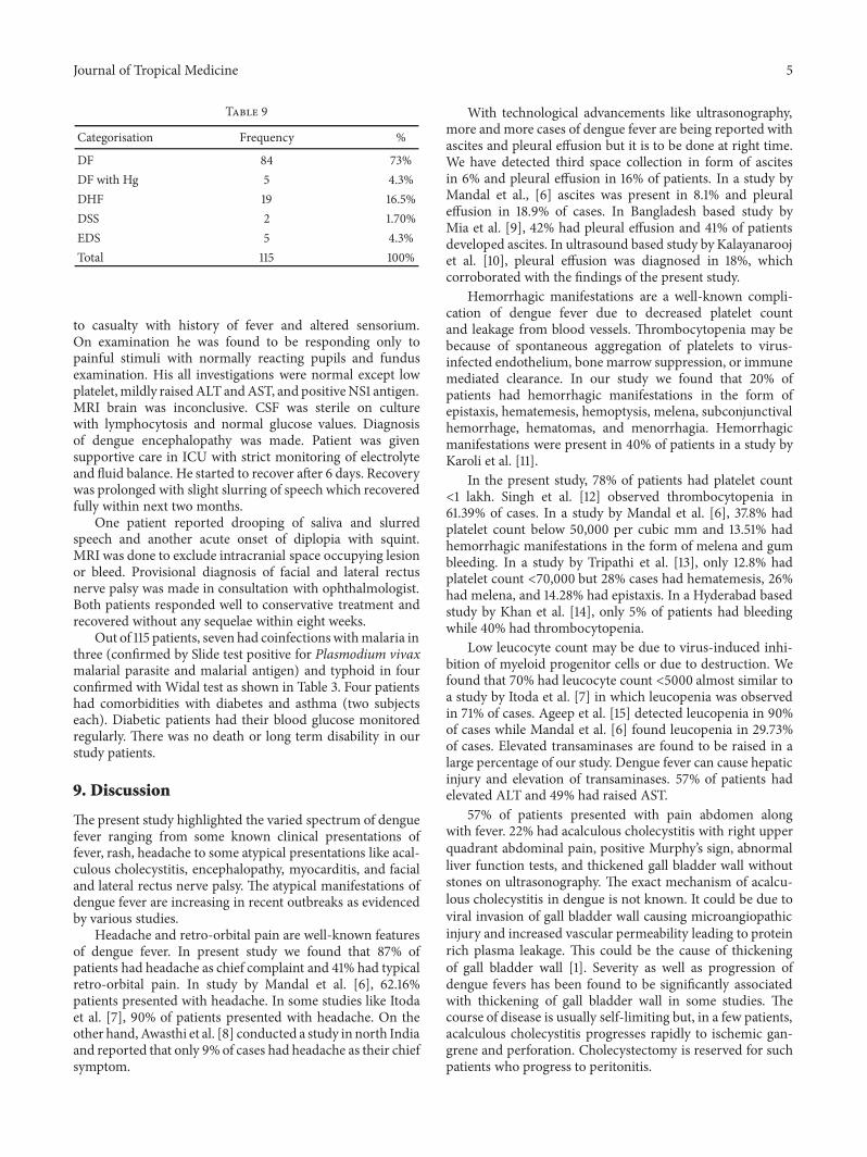
Clinical categorisation of subjects was done as shown inTable 9. Nineteen patients were diagnosed with DHF andtwo with DSS. 24 patients (20%) had hemorrhagic man-ifestations in form of epistaxis, hematemesis, hemoptysis,melena, subconjunctival hemorrhage, and menorrhagia. Tenpatients required both blood and platelet transfusion and fiverequired only platelet transfusion. A total of thirty units ofplatelets were transfused. Intravenous fluid therapy was givenaccording to WHO guidelines.
8. Clinical Categorisation
Four patients (3%) required ICU care because of DSS,myocarditis, and encephalopathy. One patient of DSS hadcome with acute respiratory distress in shock with saturationof 79% and needed intubation in casualty and was transferredto ICU and put on ventilator. He had pleural effusion as wellas ascites and very low platelet count. He was monitoredwith central line, transfused 6 units of platelets, and requiredICU care for five days. Total hospital stay was eight days andpatient recovered well without any residual disease.
Another patient of DSS with BP of 60/40 and hae-matemesis required ICU care with platelet as well as bloodtransfusion.His hospital staywas of 6 days. Patient respondedwell to platelet, blood, and fluid therapy. Another patient ofencephalopathy also required ICU care.
The fourth patient requiring ICU care was an eighteen-year-old girl. She had come with fever and breathlessnesswhich on physical examination revealed congestive heart fail-ure. Patient was hypoxemic and hypotensive. She had throm-bocytopenia, EKG revealed sinus tachycardia, X-ray chestrevealed bilateral pleural effusion, and echocardiographyshowed pericardial effusion, global hypokinesia, and reducedleft ventricular systolic function with ejection fraction of40%. Clinical diagnosis of myocarditis was made. She wastreated in ICU with standard decongestive therapy, positiveinotropic support, and platelet infusion. Patient respondedwell to treatment and had ejection fraction of 56% at the timeof discharge with no long term sequelae on follow-up.
Five patients (4.3%) had expanded dengue syndrome(EDS) affecting nervous system (encephalopathy, facial, andlateral rectus nerve palsy) in three, cardiovascular system(myocarditis) in one, and gastrointestinal system in one(jaundice). The patient with encephalopathy was brought
4 Journal of Tropical Medicine
Table 4
Finding Positive cases Total Percentage%Pleural effusion (PE) 18 115 15Gall bladder edema (GBE) 26 115 22Hepatosplenomegaly (HS) 29 115 25Hepatosplenomegaly + ascites (HS + A) 6 115 5Malarial antigen (R-MAT) 1 115 0.9Slide test for malaria 2 115 1.8Widal test 4 115 3.5
Table 5
WBC count Mean ± SD Median Min–maxTotal 3749.48 ± 2411 3000.00 1100–18500N 55.58 ± 12.54 55.00 25–90L 38.83 ± 12.58 40.00 9–64M 2.78 ± 1.89 2.00 0–10E 1.95 ± 1.89 1.00 0–16
Table 6
WBC count DF DHF DSS EDS𝑝 value
Mean ±SD Mean ±SD Mean ±SD Mean ±SDTotal 3398.43 ±1645.59 4404.21 ±2771.25 10550 ±11243 4790 ±3267.72 0.541N 53.49 ±11.93 63.53 ±11.25 64.5 ±28.99 59 ±11.18 0.011L 41.49 ±12.07 29.42 ±9.49 27 ±24.04 31.8 ±6.8 <0.001M 2.75 ±1.93 3.11 ±1.91 3 ±1.41 1.75 ±0.96 0.499E 1.99 ±2.07 1.74 ±1.24 3.5 ±0.71 1.5 ±0.58 0.265
Table 7
Mean ± SD Median Min–maxPL count (K) 52.70 ± 38.54 41.00 10–204HCT 38.86 ± 5.91 39.00 18.10–67.30UREA 18.97 ± 5.21 18.00 10–42Creatine 1.16 ± 1.69 1.00 0.20–17Serum bilirubin 1.13 ± 0.63 1.10 0.20–6.30AST 69.50 ± 51.52 50.00 24–346ALT 60.10 ± 38.55 45.00 20–234
Table 8
DF DHF DSS EDS𝑝 value
Mean ±SD Mean ±SD Mean ±SD Mean ±SDHb 12.74 ±9.81 12.36 ±4.16 15.25 ±0.35 12.14 ±2.14 0.074PL count (K) 58.24 ±41.31 41.11 ±15.68 27.5 ±17.68 14.4 ±3.78 0.003HCT 38.48 ±4.39 39.82 ±10.21 43.5 ±9.19 40.06 ±7.64 0.706Urea 18.5 ±4.75 20.75 ±6.22 25 ±14.14 19.38 ±4.89 0.696Creatine 1.24 ±1.88 0.83 ±0.23 0.7 ±0.42 0.85 ±0.45 0.142Serum bilirubin 1.18 ±0.67 0.89 ±0.23 0.7 ±0.42 1.08 ±0.58 0.042ALT 62.85 ±47.07 89.6 ±63.61 128 ±11.31 92.2 ±65.74 0.039AST 56.18 ±32.61 71.6 ±54.61 79 ±62.23 80.8 ±59.91 0.758
Journal of Tropical Medicine 5
Table 9
Categorisation Frequency %
DF 84 73%DF with Hg 5 4.3%DHF 19 16.5%DSS 2 1.70%EDS 5 4.3%Total 115 100%
to casualty with history of fever and altered sensorium.On examination he was found to be responding only topainful stimuli with normally reacting pupils and fundusexamination. His all investigations were normal except lowplatelet,mildly raisedALT andAST, and positiveNS1 antigen.MRI brain was inconclusive. CSF was sterile on culturewith lymphocytosis and normal glucose values. Diagnosisof dengue encephalopathy was made. Patient was givensupportive care in ICU with strict monitoring of electrolyteand fluid balance. He started to recover after 6 days. Recoverywas prolonged with slight slurring of speech which recoveredfully within next two months.
One patient reported drooping of saliva and slurredspeech and another acute onset of diplopia with squint.MRI was done to exclude intracranial space occupying lesionor bleed. Provisional diagnosis of facial and lateral rectusnerve palsy was made in consultation with ophthalmologist.Both patients responded well to conservative treatment andrecovered without any sequelae within eight weeks.
Out of 115 patients, seven had coinfectionswithmalaria inthree (confirmed by Slide test positive for Plasmodium vivaxmalarial parasite and malarial antigen) and typhoid in fourconfirmed with Widal test as shown in Table 3. Four patientshad comorbidities with diabetes and asthma (two subjectseach). Diabetic patients had their blood glucose monitoredregularly. There was no death or long term disability in ourstudy patients.
9. Discussion
The present study highlighted the varied spectrum of denguefever ranging from some known clinical presentations offever, rash, headache to some atypical presentations like acal-culous cholecystitis, encephalopathy, myocarditis, and facialand lateral rectus nerve palsy. The atypical manifestations ofdengue fever are increasing in recent outbreaks as evidencedby various studies.
Headache and retro-orbital pain are well-known featuresof dengue fever. In present study we found that 87% ofpatients had headache as chief complaint and 41% had typicalretro-orbital pain. In study by Mandal et al. [6], 62.16%patients presented with headache. In some studies like Itodaet al. [7], 90% of patients presented with headache. On theother hand,Awasthi et al. [8] conducted a study in north Indiaand reported that only 9%of cases had headache as their chiefsymptom.
With technological advancements like ultrasonography,more and more cases of dengue fever are being reported withascites and pleural effusion but it is to be done at right time.We have detected third space collection in form of ascitesin 6% and pleural effusion in 16% of patients. In a study byMandal et al., [6] ascites was present in 8.1% and pleuraleffusion in 18.9% of cases. In Bangladesh based study byMia et al. [9], 42% had pleural effusion and 41% of patientsdeveloped ascites. In ultrasound based study by Kalayanaroojet al. [10], pleural effusion was diagnosed in 18%, whichcorroborated with the findings of the present study.
Hemorrhagic manifestations are a well-known compli-cation of dengue fever due to decreased platelet countand leakage from blood vessels. Thrombocytopenia may bebecause of spontaneous aggregation of platelets to virus-infected endothelium, bone marrow suppression, or immunemediated clearance. In our study we found that 20% ofpatients had hemorrhagic manifestations in the form ofepistaxis, hematemesis, hemoptysis, melena, subconjunctivalhemorrhage, hematomas, and menorrhagia. Hemorrhagicmanifestations were present in 40% of patients in a study byKaroli et al. [11].
In the present study, 78% of patients had platelet count<1 lakh. Singh et al. [12] observed thrombocytopenia in61.39% of cases. In a study by Mandal et al. [6], 37.8% hadplatelet count below 50,000 per cubic mm and 13.51% hadhemorrhagic manifestations in the form of melena and gumbleeding. In a study by Tripathi et al. [13], only 12.8% hadplatelet count <70,000 but 28% cases had hematemesis, 26%had melena, and 14.28% had epistaxis. In a Hyderabad basedstudy by Khan et al. [14], only 5% of patients had bleedingwhile 40% had thrombocytopenia.
Low leucocyte count may be due to virus-induced inhi-bition of myeloid progenitor cells or due to destruction. Wefound that 70% had leucocyte count <5000 almost similar toa study by Itoda et al. [7] in which leucopenia was observedin 71% of cases. Ageep et al. [15] detected leucopenia in 90%of cases while Mandal et al. [6] found leucopenia in 29.73%of cases. Elevated transaminases are found to be raised in alarge percentage of our study. Dengue fever can cause hepaticinjury and elevation of transaminases. 57% of patients hadelevated ALT and 49% had raised AST.
57% of patients presented with pain abdomen alongwith fever. 22% had acalculous cholecystitis with right upperquadrant abdominal pain, positive Murphy’s sign, abnormalliver function tests, and thickened gall bladder wall withoutstones on ultrasonography. The exact mechanism of acalcu-lous cholecystitis in dengue is not known. It could be due toviral invasion of gall bladder wall causing microangiopathicinjury and increased vascular permeability leading to proteinrich plasma leakage. This could be the cause of thickeningof gall bladder wall [1]. Severity as well as progression ofdengue fevers has been found to be significantly associatedwith thickening of gall bladder wall in some studies. Thecourse of disease is usually self-limiting but, in a few patients,acalculous cholecystitis progresses rapidly to ischemic gan-grene and perforation. Cholecystectomy is reserved for suchpatients who progress to peritonitis.
6 Journal of Tropical Medicine
We had five cases of EDS with neurological, cardiovas-cular, and gastrointestinal involvement. The unusual atypicalmanifestations, in this study, were of neurological involve-ment in 3 (2.6%) patients. Neurological involvement indengue may occur because of neurotropism of the virus,immunological mechanisms, cerebral anoxia, intracranialhemorrhage, hyponatremia, cerebral edema, hepatic failure,renal failure, or release of toxic products. Peter et al. [16]reported isolated facial nerve palsy in dengue fever and Shiv-anthan et al. [17] have also reported isolated 6th nerve palsy.Donnio et al. [18] reported dengue with a rare presentation ofoculomotor nerve palsy. Sanjay et al. [19] reported a case ofoptic neuropathy associated with dengue fever.
One patient was categorised as EDS due to myocardi-tis. This patient had benign though prolonged self-limitingcourse. Bhasin et al. [20] also reported a case of DHF withmyocarditis mimicking DSS with hypotension and pul-monary edema.
One patient reported acute respiratory distress syndromeand needed ICU care. ARDS in dengue is due to increasedpermeability of alveolar capillary membrane, which resultsin alveolar edema leading to pulmonary dysfunction [21].Anam et al. [22] had an unusual EDS case presenting aspancreatitis and haemothorax with respiratory distress. Thiscomplication requires early recognition and promptmanage-ment for favorable outcome.
10. Conclusion
Dengue is a challenging disease with multisystemic, var-ied, atypical, and sometimes life-threatening presentations.Awareness of these rare manifestations goes a long way inearly recognition, correct diagnosis, prompt intervention,and appropriate treatment. Every aspect of dengue viral infec-tion remains a clinical challenge. A continuous seroepidemi-ological surveillance, timely interventions, vaccines research,and vector control measures are needed to identify thecases so that outbreaks, complications, and mortality can beminimized.
Competing Interests
The authors declare that there is no conflict of interestsregarding publication of this paper.
References
[1] S. Gulati and A. Maheshwari, “Atypical manifestations ofdengue,” Tropical Medicine and International Health, vol. 12, no.9, pp. 1087–1095, 2007.
[2] WHO, “Comprehensive guidelines for prevention and con-trol of dengue and dengue hemorrhagic fever,” World HealthOrganization, Regional Office for South-East Asia, http://apps.searo.who.int/pds docs/B4751.pdf?ua=1.
[3] B. Pozzetto, M. Memmi, and O. Garraud, “Is transfusion-transmitted dengue fever a potential public health threat?”World Journal of Virology, vol. 4, no. 2, pp. 113–123, 2015.
[4] F. L. Tan, D. L. Loh, K. Prabhakaran, P. A. Tambyah, and H.K. Yap, “Dengue haemorrhagic fever after living donor renal
transplantation,” Nephrology Dialysis Transplantation, vol. 20,no. 2, pp. 447–448, 2005.
[5] S. H. Pouliot, X. Xiong, E. Harville et al., “Maternal dengueand pregnancy outcomes: a systematic review,” Obstetrical andGynecological Survey, vol. 65, no. 2, pp. 107–118, 2010.
[6] S. K.Mandal, J. Ganguly, K. Sil et al., “Clinical profiles of denguefever in a teaching hospital of eastern India,”National Journal ofMedical Research, vol. 3, no. 2, pp. 173–176, 2013.
[7] I. Itoda, G. Masuda, A. Suganuma et al., “Clinical features of 62imported cases of dengue fever in Japan,” American Journal ofTropical Medicine andHygiene, vol. 75, no. 3, pp. 470–474, 2006.
[8] S. Awasthi, V. K. Singh, S. Kumar, A. Kumar, and S. Dutta,“The changing clinical spectrum of Dengue fever in the 2009epidemic in north India: a tertiary teaching hospital basedstudy,” Journal of Clinical and Diagnostic Research, vol. 6, no. 6,pp. 999–1002, 2012.
[9] M. W. Mia, A. M. Nurullah, A. Hossain, and M. M. Haque,“Clinical and sonographic evaluation of dengue fever in Bang-ladesh: a study of 100 cases,” Dinajpur Medical College Journal,vol. 3, pp. 29–34, 2010.
[10] S. Kalayanarooj, D. W. Vaughn, S. Nimmannitya et al., “Earlyclinical and laboratory indicators of acute dengue illness,” TheJournal of Infectious Diseases, vol. 176, no. 2, pp. 313–321, 1997.
[11] R. Karoli, J. Fatima, Z. Siddiqi, K. I. Kazmi, and A. R. Sultania,“Clinical profile of dengue infection at a teaching hospital inNorth India,” Journal of Infection in Developing Countries, vol.6, no. 7, pp. 551–554, 2012.
[12] N. P. Singh, R. Jhamb, S. K. Agarwal et al., “The 2003 outbreak ofdengue fever inDelhi, India,” Southeast Asian Journal of TropicalMedicine and Public Health, vol. 36, no. 5, pp. 1174–1178, 2005.
[13] B. K. Tripathi, B. Gupta, R. S. K. Sinha, S. Prasad, and D. K.Sharma, “Experience in adult population in dengue outbreakin Delhi,” Journal of Association of Physicians of India, vol. 46,no. 3, pp. 273–276, 1998.
[14] A. H. Khan, A. S. Hayat, N. Masood, N. M. Solangi, and T. Z.Shaikh, “Frequency and clinical presentation of dengue feverat tertiary care hospital of Hyderabad/Jamshoro,” Journal of theLiaquat University of Medical and Health Sciences, vol. 9, no. 2,pp. 88–94, 2010.
[15] A. K. Ageep, A. A. Malik, and M. S. Elkarsani, “Clinicalpresentations and laboratory findings in suspected cases ofdengue virus,” Saudi Medical Journal, vol. 27, no. 11, pp. 1711–1713, 2006.
[16] S. Peter, N. Malhotra, P. Peter, and R. Sood, “Isolated Bell’spalsy—an unusual presentation of dengue infection,” AsianPacific Journal of Tropical Medicine, vol. 6, no. 1, pp. 82–84, 2013.
[17] M. C. Shivanthan, E. C. Ratnayake, B. C.Wijesiriwardena, K. C.Somaratna, and L. K. G. K. Gamagedara, “Paralytic squint dueto abducens nerve palsy: a rare consequence of dengue fever,”BMC Infectious Diseases, vol. 12, article 156, 2012.
[18] A.Donnio, L. Beral, S. Olindo, A. Cabie, andH.Merle, “Dengue,a new etiology of oculomotor paralysis,” Canadian Journal ofOphthalmology, vol. 45, no. 2, pp. 183–184, 2010.
[19] S. Sanjay, A. M. Wagle, and K. G. Au Eong, “Optic neuropathyassociated with dengue fever,” Eye, vol. 22, no. 5, pp. 722–724,2008.
[20] A. Bhasin, R. Kumar, K. Chandra, and R. K. Singal, “Denguefever with myocarditis,” Journal, Indian Academy of ClinicalMedicine, vol. 14, no. 2, pp. 187–189, 2013.
[21] L. C. S. Lum,M.K.Thong, Y. K. Cheah, and S. K. Lam, “Dengue-associated adult respiratory distress syndrome,” Annals of Trop-ical Paediatrics, vol. 15, no. 4, pp. 335–339, 1995.

Journal of Tropical Medicine 7
[22] A. M. Anam, R. Rabbani, F. Shumy, and M. M. I. Polash,“Subsequent pancreatitis and haemothorax in a patient of expa-nded dengue syndrome,” Tropical Doctor, vol. 46, no. 1, pp. 40–42, 2016.

Submit your manuscripts athttp://www.hindawi.com
Stem CellsInternational
Hindawi Publishing Corporationhttp://www.hindawi.com Volume 2014
Hindawi Publishing Corporationhttp://www.hindawi.com Volume 2014
MEDIATORSINFLAMMATION
of
Hindawi Publishing Corporationhttp://www.hindawi.com Volume 2014
Behavioural Neurology
EndocrinologyInternational Journal of
Hindawi Publishing Corporationhttp://www.hindawi.com Volume 2014
Hindawi Publishing Corporationhttp://www.hindawi.com Volume 2014
Disease Markers
Hindawi Publishing Corporationhttp://www.hindawi.com Volume 2014
BioMed Research International
OncologyJournal of
Hindawi Publishing Corporationhttp://www.hindawi.com Volume 2014
Hindawi Publishing Corporationhttp://www.hindawi.com Volume 2014
Oxidative Medicine and Cellular Longevity
Hindawi Publishing Corporationhttp://www.hindawi.com Volume 2014
PPAR Research
The Scientific World JournalHindawi Publishing Corporation http://www.hindawi.com Volume 2014
Immunology ResearchHindawi Publishing Corporationhttp://www.hindawi.com Volume 2014
Journal of
ObesityJournal of
Hindawi Publishing Corporationhttp://www.hindawi.com Volume 2014
Hindawi Publishing Corporationhttp://www.hindawi.com Volume 2014
Computational and Mathematical Methods in Medicine
OphthalmologyJournal of
Hindawi Publishing Corporationhttp://www.hindawi.com Volume 2014
Diabetes ResearchJournal of
Hindawi Publishing Corporationhttp://www.hindawi.com Volume 2014
Hindawi Publishing Corporationhttp://www.hindawi.com Volume 2014
Research and TreatmentAIDS
Hindawi Publishing Corporationhttp://www.hindawi.com Volume 2014
Gastroenterology Research and Practice
Hindawi Publishing Corporationhttp://www.hindawi.com Volume 2014
Parkinson’s Disease
Evidence-Based Complementary and Alternative Medicine
Volume 2014Hindawi Publishing Corporationhttp://www.hindawi.com