research article - jcim (journal16... · traditional chinese medicine wrist pulse-taking is...
TRANSCRIPT

March 2016, Vol.14, No.2 100 Journal of Integrative Medicine
Journal homepage: www.jcimjournal.com/jimwww.elsevier.com/locate/issn/20954964
Available also online at www.sciencedirect.com. Copyright © 2016, Journal of Integrative Medicine Editorial Office. E-edition published by Elsevier (Singapore) Pte Ltd. All rights reserved.
http://dx.doi.org/10.1016/S2095-4964(16)60233-9Received June 12, 2015; accepted July 22, 2015.Correspondence: Arthur de Sá Ferreira, D.Sc.; E-mail: [email protected], [email protected]
● Research ArticleTraditional Chinese medicine wrist pulse-taking is associated with pulse waveform analysis and hemodynamics in hypertensionNathalia Gomes Ribeiro de Moura1, Ivan Cordovil2, Arthur de Sá Ferreira1,3
1. Postgraduate Program of Rehabilitation Science, Augusto Motta University Center, 21041-010, Rio de Janeiro, RJ, Brazil
2. Division of Arterial Hypertension, National Institute of Cardiology, 22240-002, Rio de Janeiro, RJ, Brazil3. Department of Physical Therapy, Salgado de Oliveira University, 24030-060, Niterói, RJ, Brazil
ABSTRACT
BACKGROUND: Pulse wave analysis (PWA) quantifies the phenomenon of pulse waveform propagation in patients with cardiovascular diseases, whereas pulse image analysis (PIA) is a subjective examination in traditional Chinese medicine.
OBJECTIVE: This study evaluated the association of PIA with PWA and hemodynamics in patients with hypertension.
DESIGN, SETTING, PARTICIPANTS AND INTERVENTIONS: This observational, cross-sectional study enrolled 45 patients (26 men, (55.2 ± 10.3) years, systolic blood pressure (155 ± 28) mmHg, diastolic blood pressure (93 ± 17) mmHg) for assessment of clinical and laboratorial data.
MAIN OUTCOME MEASURES: Primary outcomes comprised: pattern differentiation based on an automated method; PIA at the radial artery using the ‘simultaneous pressing’ method for identification of factors such as strength (strong/weak), depth (superficial/deep), and speed (fast/moderate/slow); and PWA at the same artery using a noninvasive system.
RESULTS: Significant multivariate main effects were observed for depth (l=0.648, F5,29 =3.149, P=0.022, h2 =0.352), strength (l=0.608, F5,29 =3.736, P=0.010, h2 =0.392), and speed (l=0.535, F5,29 =5.302, P=0.002, h2 =0.465). General effects comprised high values of PWA and blood pressure for superficial, strong, and fast pulse images. A strong pulse was found for pulse pressure ≥ 62.5 mmHg and systolic blood pressure ≥ 149.5 mmHg, whereas a superficial pulse was found for heart rate ≥ 58.25 beats/min; a fast pulse was found for heart rate ≥ 69.6 beats/min and pulse wave velocity ≥ 9.185 m/s.
CONCLUSION: Associations were explained by LaPlace’s law, arterial remodeling in hypertension, alongside the traditional criterion for classifying speed in pulse images. PIA is associated with PWA and hemodynamics in patients with hypertension. Systolic and pulse pressures, heart rate, and pulse wave velocity are quantitative variables that have information to describe the qualitative pulse images such as strength, depth and speed.
Keywords: medicine, Chinese traditional; hypertension; syndrome differentiation; rehabilitation
Citation: Moura NGR, Cordovil I, Ferreira AS. Traditional Chinese medicine wrist pulse-taking is associated with pulse waveform analysis and hemodynamics in hypertension. J Integr Med. 2016; 14(2): 100–113.
March 2016, Vol.14, No.2101Journal of Integrative Medicine
www.jcimjournal.com/jim
1 Introdution
Hypertension stands out as a public health problem worldwide being the cardiovascular disease with the highest prevalence in developed countries[1]. Advancing age, physical inactivity, excessive body mass, and other factors contribute to the clustering of total cardiovascular risk[2–7]. High blood pressure is responsible for arterial remodeling[8−10], a maladaptive process that requires early diagnosis and immediate treatment[6,11]. Hypertension is allegedly a ‘silent disease’ albeit some studies[12−14] reported signs or symptoms mostly secondary to hypertensive crisis or target-organ damage[6]. Therefore, it is essential to monitor the disease progression and the effects of therapeutic interventions from blood pressure measures to the functional capacity level[6,15]. In this sense, pulse wave analysis (PWA) is a non-invasive and reliable method for assessment of the cardiovascular system. PWA allows quantifying changes in vascular impedance due to arterial stiffness and/or endothelial dysfunction in patients with cardiovascular diseases[16]. Surrogate measures of arterial stiffness and peripheral arterial resistance[17,18] are amongst the variables related to the prognosis of cardiovascular diseases that can be determined from PWA[19]. Evidence shows that proximal arteries convey similar information from PWA to those obtained from peripheral arteries such as the aorta and radial arteries, respectively[20−22].
Traditional Chinese medicine (TCM) is a health practice with systematic and philosophic reasoning based on the relationships between humanity and nature. Under the contemporary paradigm of ‘pattern/disease’[23], TCM literature relates patterns or Zheng to diseases such as hypertension[24−29]. Diagnosis in TCM is achieved by pattern differentiation using solely clinical examinations—inspection, auscultation-olfaction, inquiry, and palpation[24−29]. Particularly, pulse image analysis (PIA) is an important palpatory procedure[30]; it consists of the palpation of the radial artery pulse bilaterally at three different positions and depths from which qualitative ‘pulse images’ are perceived[31−33]. Previous studies investigated PIA using PWA[34−41], albeit they presented important limitations either from the traditional or conventional medicine perspectives. For instance, theories developed in animal models[34,36] were not yet experimented in humans, and presented no in-deep theoretical correspondence with TCM. Studies[37,39−41] with healthy participants in theoretical correspondence with TCM cannot be extrapolated to patients with hypertension without proper evidence. Incomplete descriptions of diagnostic criteria for hypertension and traditional descriptions of PIA were also found[38,39]. Finally, limited analysis comparing normotensive with hypertensive
participants—and not among patients with hypertension —was reported[38].
Whilst important aspects related to the pulse image remain neglected[42], PWA is highlighted as a possible link between pulse palpation in TCM and cardiology in conventional medicine[19]. Therefore, this study aimed to investigate the association of PIA with PWA and hemodynamics in patients with hypertension. Considering that (1) hypertension presents different phenotypes due to a combination of genetic and environmental factors, (2) anatomic-functional changes observed in the course of hypertension impact primarily in the arteries thus changing the waveform morphology, and (3) the radial artery can be used for both PWA and PIA, we hypothesized that quantitative variables derived from pulse waveform propagation and hemodynamics convey clinical information related to TCM’s pulse images.
2 Materials and methods
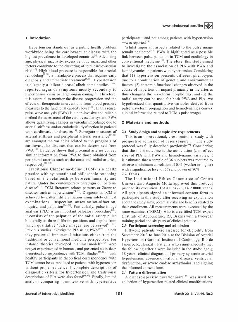
2.1 Study design and sample size requirementsThis is an observational, cross-sectional study with
prospective admission of cases (Figure 1). The study protocol was fully described previously[43]. Considering that the main outcome is the association (i.e., effect size) of PIA with PWA and hemodynamic variables, it is estimated that a sample of 36 subjects was required to observe a minimum correlation of 0.41 (weaker or higher) with a significance level of 5% and power of 80%.2.2 Ethics
The Insti tut ional Ethics Committee of Centro Universitário Augusto Motta approved this protocol prior to its execution (CAAE 34723714.2.0000.5235). All participants signed an informed consent form to participate in this study after receiving an explanation about the study aims, potential risks and benefits related to their enrollment. All measurements were executed by the same examiner (NGRM), who is a certified TCM expert (Institute of Acupuncture, RJ, Brazil) with a two-year training period and six years of clinical practice.2.3 Participant screening and admission
Fifty-one patients were assessed for eligibility from September 2013 to June 2014 at the Division of Arterial Hypertension (National Institute of Cardiology, Rio de Janeiro, RJ, Brazil). Patients who simultaneously met the following criteria were included in the study: age ≥ 18 years; clinical diagnosis of primary systemic arterial hypertension; absence of valvular disease, ventricular dysfunction, or severe cardiac arrhythmias; and signing the informed consent form.2.4 Pattern differentiation
A disease-specific questionnaire [44] was used for collection of hypertension-related clinical manifestations.

www.jcimjournal.com/jim
March 2016, Vol.14, No.2 102 Journal of Integrative Medicine
Data from each participant were typed into a software interface for automated diagnosis using the Pattern Differentiation Algorithm (PDA) model[45,46]. Briefly, the model applied two quantitative criteria to identify the underlying pattern: the explained information (F%, the ratio of the number of manifestations describing a pattern in dataset to the total number of manifestations reported by the patient) and the available information (N%, the ratio of the number of manifestations describing a pattern in dataset to the total number of manifestations for that pattern in the dataset). PDA suggested a list of candidate patterns ranked simultaneously by increasing F% and decreasing N%. Only a single candidate pattern was identifiable as the primary diagnosis with simultaneous highest F% and lowest N% values, otherwise no pattern was identified at all. Accuracy of PDA for pattern differentiation among 73 Zangfu patterns was estimated in 94.7% (sensitivity=89.8%; specificity=99.5%)[46].2.5 PIA and PWA
Measurements were performed between 08:00 and 11:00 AM as recommended for both PWA[16] and PIA[31,32]. PIA measurements were first obtained after 10-minute rest period in the sitting position. Wrist pulse-taking was performed bilaterally using the ‘simultaneous pressing’ technique from the deepest level to the surface in both
wrists[31−33]. In sequence, PWA measurements were obtained after 10-minute rest period in supine position in the right upper extremity. Two noninvasive piezoelectric transducers (model PT-102, iWorx Systems Inc., Dover, NH, USA) were attached to the brachial and radial arteries with Velcro® straps and were connected to a preamplifier running into a 14-bit acquisition board (USB 6009, National Instruments, Dallas, TX, USA) before connecting to a computer. At the radial artery the transducer was positioned above the styloid process, which corresponds to the Guan position of the TCM pulse. Pressure pulse waveform signals were acquired simultaneously from these two arterial sites at a sampling rate of 1.0 kHz/channel during 60 s using the Arterial Function Analysis (AFA) system[47−49] developed in LabVIEW (National Instruments, Dallas, TX, USA). Pulse waveforms were visually inspected on the computer screen before data acquisition to increase the overall quality of the blood pressure signals.2.6 Data analyses of control and outcome variables
Control variables include[6]: body weight and height as measured by an analog scale anda stadiometer; the body mass index (weight/height2); systolic and diastolic blood pressures (SBP and DBP, respectively) and heart rate (HR), as measured with an automated oscillometric
Figure 1 Study flowchartSBP: systolic blood pressure; DBP: diastolic blood pressure; PP: pulse pressure; MBP: mean arterial pressure; CVD: cardiovascular disease; LDL-C: low-density lipoprotein cholesterol; HDL-C: high-density lipoprotein cholesterol; PWA: pulse ware analysis; PIA: pulse image analysis.

March 2016, Vol.14, No.2103Journal of Integrative Medicine
www.jcimjournal.com/jim
device (BP3AF1-3 model, G-TECH, Shenzhen, China); calculated pulse pressure (PP=SBP-DBP) and mean arterial pressure (MBP=DBP+PP/3]); self-reported duration of hypertension; medication classes in use; left ventricular hypertrophy as assessed from the electrocardiogram; and blood chemistry analysis. Control variables were used for classification of hypertension grade[6] and obesity grade[50]. Additionally, the following risk factors for cardiovascular diseases were assessed: cigarette smoking (self-report of current or previous use); smoking history (self-report of number of packs per week, multiplied by the number of years of smoking); obesity (Quetelet index ≥ 30 kg/m2); physical inactivity (self-report of no regular exercise practice); dyslipidemia (low-density lipoprotein cholesterol (LDL-C)>70 mg/dL, high-density lipoprotein cholesterol (HDL-C)<50 mg/dL and triglycerides >150 mg/dL)[50]; diabetes mellitus (fasting blood glucose >126 mg/dL)[51]; and history of premature cardiovascular disease (<55 years in men, <65 years in women)[6].
The following outcome variables were obtained: the hypertension-related TCM pattern, PIA, PWA, and hemodynamics. Five hypertension-related patterns were available for identification[44]: Liver-fire blazing upwards; Kidney-yin deficiency and Liver-yang rising; obstruction of phlegm and dampness of Heart/Liver/Gallbladder; Qi and blood deficiency leading to Liver-yang rising; or Kidney-yin/yang deficiency. Pulse images were annotated for PIA cohich comprised[31−33]: strength (levels: strong, weak), depth (levels: superficial, deep), speed (levels: fast, moderate, slow) and rhythm (levels: regular, intermittent). The AFA software averaged the pulse wave velocity (PWV, 1st derivative method[47]) and the reflection index (IR1,2: first-to-second systolic peak ratio[49]) on a sequence of at 10 heart beats.2.7 Statistical analysis
Statistical analysis was performed using SPSS 22 (IBM Corp., USA). Descriptive analysis was displayed as mean±standard deviation, median [minimum; maximum], or frequency (%) accordingly to the variable type. Significance was set to P < 0.05 (two-tailed).
Multivariate analysis-of-variance was used to test for main and interaction effects of factors such as pulse image strength (strong=1, weak=0), depth (superficial=1, deep=0), and speed (fast=2, moderate=1, slow=0) on variables obtained from PWA and hemodynamics. Gender (male=1), age, and body mass index were inputted into the general linear model as covariates because they comprise important confounders of PWA[16] and PIA[31,32]. Wilks’ λ, F-test results and respective P-value from null hypothesis testing were reported alongside partial η2 as effect size (correlation)[52]. Discrimination analysis between levels on each factor was performed with receiver-operating characteristic (ROC) curves. The optimal cut-off value for detection of different levels on each factor
(following Youden’s method), the area under the ROC curve with 95% confidence interval (95% CI), sensitivity and specificity were reported solely for variables with significant univariate effects. Cramer’s V coefficient was used to analyze the effect size (correlation) of antihypertensive medication on PIA. Correlation values were qualitatively described as no association (0.00), negligible (0.01 to 0.20), weak (0.21 to 0.40), moderate (0.41 to 0.70), strong (0.71 to 0.99), or perfect association (1.00)[53].
Intrarater test-retest reliability for hemodynamic variables was assessed by the two-way random-effects intra-class correlation coefficient for single measurements (ICC2,1) using an absolute agreement definition and its respective 95% CI. Reliability for each PIA factor was assessed using Cohen’s k (kappa)[54] and its respective 95% CI. Reliability was qualitatively described as excellent or acceptable (> 0.75), fair to good (0.40 to 0.75), or poor (< 0.40); minimum reference value of reliability was set to at least fair to good (H0: k=0.40).
3 Results
Six patients were excluded because they lacked of complete data related to outcome variables. The remaining 45 patients (Table 1) enrolled in the study underwent clinical history, physical and laboratory examination for the confirmation of their clinical status following international guidelines[6]. Another sample of 15 patients (7 men) had their PIA and blood pressure measurements performed twice 30-minute apart to assess intrarater, test-retest reliability.3.1 Occurrence of patterns and clinical manifestations in hypertension
The most frequent pattern found (Table 2) was Kidney-yin deficiency and Liver-yang rising (46.7%), followed by Liver-fire blazing upwards and obstruction of phlegm and dampness of Heart/Liver/Gallbladder (17.8% each). The least frequent pattern was Kidney-yin/yang deficiency (15.6%). One participant (2.2%) could not have its primary pattern clearly identified by PDA.
The frequency of manifestations varied according to the identified pattern. No pathognomonic manifestation was found and no patients reported convulsions either. Frequent nocturnal urination was the manifestation most commonly reported (77.8%) by the patients. Four manifestations were reported by the majority of patients, with occurrences ranging from 66.7% (pale tongue) to 51.1% (insomnia). Sixteen manifestations were seldom observed, occurring between 46.7% (mental fatigue; headache) to 26.7% (dizziness). Finally, other 16 rarely observed manifestations occurred in range 24.4% (weak legs; tinnitus; slippery pulse) to 6.7% (aphasia).

www.jcimjournal.com/jim
March 2016, Vol.14, No.2 104 Journal of Integrative Medicine
Table 1 Descriptive analysis of the studied sample
Variable ValuesGender (n, %) 45 (100%)
Men 26 (58%)Women 19 (42%)
Age (mean ± standard deviation, years) 55.2 ± 10.3Body mass (mean ± standard deviation, kg) 83.8 ± 17.7Body height (mean ± standard deviation, m) 1.67 ± 0.10BMI (mean ± standard deviation, kg/m2) 30.0 ± 4.6BMI classification (n, %)
Normal range 8 (17.8%)Overweight 17 (37.8%)Obesity I 14 (31.1%)Obesity II 4 (8.9%)Obesity III 2 (4.4%)
Duration of hypertension (mean ± standard deviation, months) 156 ± 146Heart rate (mean ± standard deviation, beats/min) 63.3 ± 9.9Blood pressure (mean ± standard deviation, mmHg)
Systolic 155 ± 28Diastolic 93 ± 17Mean 114 ± 20Pulse 62 ± 16
Blood pressure classification (n, %)Optimal 1 (2.2%)Normal 14 (31.1%)Normal high 3 (6.7%)Grade-I 13 (28.9%)Grade-II 8 (17.8%)Grade-III 6 (13.3%)
Smoking (mean ± standard deviation, packs) 30 ± 61Risk factor classification (n, %)
Premature CVD 40 (88.9%)Dyslipidemia 37 (82.2%)Obesity 20 (44.4%)Ventricular hypertrophy 16 (35.6%)Sedentary 15 (33.3%)Smoking 11 (24.4%)Diabetes 10 (22.2%)
Medication type (n, %) B-blocker 32 (71.1%)AT2 antagonist 32 (71.1%)Diuretic 29 (64.4%)ACE inhibitors 6 (13.3%)Vasodilator 5 (11.1%)Sympatholytic 1 (2.2%)
Serum chemistry (mean ± standard deviation, mg/dL) Total cholesterol 206 ± 50Triglycerides 158 ± 88LDL-C 131 ± 41HDL-C 44 ± 13Creatinine 0.99 ± 0.33Glucose 117 ± 41Uric acid 6.2 ± 1.8
BMI: body mass index; CVD: cardiovascular disease; AT2: angiotensin Ⅱ ; ACE: angiotensin converting enzyme; LDL-C: low-density lipoprotein cholesterol; HDL-C: high-density lipoprotein cholesterol.

March 2016, Vol.14, No.2105Journal of Integrative Medicine
www.jcimjournal.com/jim
Table 2 Descriptive analysis of clinical manifestations in patients with hypertension (n=45)
Clinical manifestation
Liver-fire blazing upwards
(n=8, 17.8%)
Kidney-yin deficiency and
Liver-yang rising (n=21, 46.7%)
Obstruction of phlegm and
dampness of Heart/Liver/Gallbladder
(n=8, 17.8%)
Kidney-yin/yang deficiency (n=7, 15.6%)
Total(n=45, 100%)
Frequent nocturnal urination 7 (87.5%) 16 (76.2%) 6 (75.0%) 5 (71.4%) 35 (77.8%)Pale tongue 5 (62.5%) 13 (61.9%) 6 (75.5%) 5 (71.4%) 30 (66.7%)Numbness in feet and hands 4 (50.0%) 14 (66.7%) 4 (50.0%) 2 (28.6%) 25 (55.6%)Blurred vision 4(50.0%) 16 (76.2%) 2 (25.0%) 0 (0.0%) 23 (51.1%)Insomnia 4(50.0%) 13 (61.9%) 3 (37.5%) 2 (28.6%) 23 (51.1%)Mental fatigue 4 (50.0%) 8 (38.1%) 4 (50.0%) 4 (57.1%) 21 (46.7%)Headache 4 (50.0%) 10 (47.6%) 5 (62.5%) 1 (14.3%) 21 (46.7%)Deep pulse 0 (0.0%) 11 (52.4%) 1 (12.5%) 7 (100.0%) 20 (44.4%)Numbness in the limbs 0 (0.0%) 16 (76.2%) 3 (37.5%) 0 (0.0%) 20 (44.4%)Thin pulse 0 (0.0%) 12 (57.1%) 0 (0.0%) 7 (100.0%) 20 (44.4%)Strong pulse 7 (87.5%) 6 (28.6%) 7 (87.5%) 0 (0.0%) 20 (44.4%)Peeled tongue 5 (62.5%) 9 (42.9%) 4 (50.0%) 1 (14.3%) 19 (42.2%)Irritability 5 (62.5%) 12 (57.1%) 1 (12.5%) 0 (0.0%) 18 (40.0%)Heavy limb sensation 3 (37.5%) 12 (57.1%) 2 (25.0%) 0 (0.0%) 18 (40.0%)Wiry pulse 4 (50.0%) 8 (38.1%) 5 (62.5%) 1 (14.3%) 18 (40.0%)Shortness of breath 3 (37.5%) 10 (47.3%) 2 (25.0%) 1 (14.3%) 17 (37.8%)Palpitation 2 (25.0%) 10 (47.6%) 3 (37.5%) 1 (14.3%) 17 (37.8%)Greasy and thick tongue coating 2 (25.0%) 9 (42.9%) 4 (50.0%) 1 (14.3%) 16 (35.6%)Nausea 3 (37.5%) 7 (33.3%) 2 (25.0%) 0 (0.0%) 13 (28.9%)Excessive dreaming 2 (25.0%) 8 (38.1%) 2 (25.0%) 1 (14.3%) 13 (28.9%)Dizziness 2 (25.0%) 8 (38.1%) 2 (25.0%) 0 (0.0%) 12 (26.7%)Weak legs 2 (25.0%) 5 (23.8%) 3 (37.5%) 0 (0.0%) 11 (24.4%)Tinnitus 1 (12.5%) 9 (42.9%) 0 (0.0%) 1 (14.3%) 11 (24.4%)Slippery pulse 3 (37.5%) 3 (14.3%) 5 (62.5%) 0 (0.0%) 11 (24.4%)Yellow tongue coating 5 (62.5%) 1 (4.8%) 1 (12.5%) 2 (28.6%) 10 (22.2%)Constipation 3 (37.5%) 3 (14.3%) 0 (0.0%) 2 (28.6%) 9 (20.0%)Sexual impotence 1 (12.5%) 6 (28.6%) 0 (0.0%) 1 (14.3%) 8 (17.8%)Severe dizziness 1 (12.5%) 4 (19.0%) 1 (12.5%) 1 (14.3%) 8 (17.8%)Red eyes 3 (37.5%) 3 (14.3%) 0 (0.0%) 1 (14.3%) 8 (17.8%)Red face 2 (25.0%) 4 (19.0%) 0 (0.0%) 1 (14.3%) 8 (17.8%)Fast pulse 3 (37.5%) 5 (23.8%) 0 (0.0%) 0 (0.0%) 8 (17.8%)Red tongue 1 (12.5%) 3 (14.3%) 1 (12.5%) 2 (28.6%) 7 (15.6%)Stroke 0 (0.0%) 5 (23.8%) 0 (0.0%) 0 (0.0%) 5 (11.1%)Congested feeling in the chest 4 (50.0%) 1 (4.8%) 0 (0.0%) 0 (0.0%) 5 (11.1%)Vomiting 1 (12.5%) 3 (14.3%) 0 (0.0%) 0 (0.0%) 5 (11.1%)Fainting 1 (12.5%) 2 (9.5%) 0 (0.0%) 0 (0.0%) 4 (8.9%)Aphasia 0 (0.0%) 3 (14.3%) 0 (0.0%) 0 (0.0%) 3 (6.7%)Convulsions 0 (0.0%) 0 (0.0%) 0 (0.0%) 0 (0.0%) 0 (0%)
No patient was identified with pattern “Qi and blood deficiency leading to Liver-yang rising”. One patient (2.2%) was not possible to identify the primary pattern using the automated method, but the corresponding manifestation was summed up in the total column.

www.jcimjournal.com/jim
March 2016, Vol.14, No.2 106 Journal of Integrative Medicine
3.2 Effects of pulse image analysis on PWA and hemodynamics
Descriptive analysis (Table 3) showed that the majority of participants had a strong pulse (53%), a superficial pulse (56%), or a moderate pulse image (84%). All participants exhibited a regular rhythm and thus this pulse image was not assessed for effect size or reliability because it was a constant value within the studied sample.
Significant multivariate interaction effects were not observed among all factors strength*depth*speed (λ= 1.000), neither for the pairwise analysis of strength*speed (λ=0.887, P=0.602, η2 =0.113), depth*speed (λ=0.878, P=0.557, η2 =0.122), nor strength*depth (λ=0.876, P=0.545, η2 =0.124). Significant multivariate main effects were observed for depth (λ=0.648, F5,29 =3.149, P=0.022, η2 =0.352), strength (λ=0.608, F5,29 =3.736, P=0.010, η2=0.392), and speed (λ=0.535, F5,29 =5.302, P=0.002, η2 =0.465). General effects for depth comprised higher values for superficial than deep pulse images, with significant univariate main effects observed for PWV (F1,33 =6.179, P=0.018), HR (F1,33 =5.605, P=0.024), and PP (F1,33 =4.926, P=0.033). General effects for strength comprised higher values for strong than weak pulse images, with significant univariate main effects observed for PP (F1,33 =17.124, P<0.001) and SBP (F1,33 =7.670, P=0.009). Finally, general effects for speed comprised higher values for fast than moderate pulse images, with significant univariate main effects observed for PWV (F1,33 =7.930, P=0.008) and HR (F1,33 =6.952, P=0.013).
No significant multivariate main effects were observed for neither of the covariates gender (λ=0.831, P=0.344, η2 =0.169), age (λ=0.951, P=0.910, η2 =0.049), nor body mass index (λ=0.733, P=0.093, η2 =0.267).
Figure 2 presents representative pressure pulse waveforms of all patients with hypertension. It can be noticed differences among patients in waveform morphology and baseline changes due to respiratory rate.
ROC curve analysis (Figure 3) showed a significant discriminative power as assessed by the area under the curve regarding strength for both PP (0.742, 95% CI=[0.598; 0.886], P=0.006) and SBP (0.729, 95% CI=[0.578; 0.881], P=0.009). Pulse images were detected as strong separately with PP≥62.5 mmHg (sensitivity =58%, specificity=81%) or SBP≥149.5 mmHg (sensitivity=75%, specificity=67%). Within factor depth significant discriminative power was observed for HR (0.684, 95% CI=[0.521; 0.874], P=0.040) but neither for PP (0.594, 95% CI=[0.420; 0.769], P=0.293) nor PWV (0.357, 95% CI=[0.186; 0.529], P=0.112). Pulse images were detected as superficial with HR≥58.25 beats/min (sensitivity=83%, specificity=53%). Finally, both HR (0.762, 95% CI=[0.533; 0.991], P=0.030) and PWV (0.746, 95% CI=[0.589; 0.904], P=0.041) showed significant discriminative power within factor speed. Pulse images were detected as fast separately with HR≥69.6 beats/min (sensitivity=71%, specificity=83%) or PWV≥9.185 m/s (sensitivity=100%, specificity=53%).
Table 3 Association of pulse image analysis with pulse waveform analysis and hemodynamic variables
Index
Strength Depth Speed Group-average (n=45, 100%)
Strong (n=24, 53%)
Weak (n=21, 47%)
P
Superficial (n=25, 56%)
Deep (n=20, 44%)
P
Fast (n=7, 16%)
Moderate (n=38, 84%)
P
PWV (m/s) 9.8±2.4 10.1±2.5 0.077 9.4±2.2 10.7±2.6 0.018* 11.7±1.6 9.6±2.4 0.008* 9.9±2.4
IR1,2 (%) 97.3±22.2 93.0±15.6 0.761 97.3±21.9 92.8±15.8 0.322 90.7±17.5 96.2±19.8 0.396 95.3±19.4
SBP (mmHg) 165±29 144±22 0.009* 159±28 150±27 0.534 162±40 154±25 0.832 155±28
DBP (mmHg) 96±19 89±15 0.438 96±19 90±14 0.366 106±24 91±15 0.139 93±17
MBP (mmHg) 119±21 108±17 0.096 117±22 110±17 0.806 125±29 112±17 0.335 114±20
PP (mmHg) 68±17 55±13 <0.001* 63±15 60±18 0.033* 56±19 63±16 0.176 62±16
HR (beats/min) 64±10 62±10 0.103 66±10 59±9 0.024* 72±12 62±9 0.013* 63±10
PWV: pulse wave velocity; IR1,2: reflection index; SBP: systolic blood pressure; DBP: diastolic blood pressure; MBP: mean blood pressure; PP: pulse pressure; HR: heart rate. *Statistical significant difference at P<0.05 after controlling for confounders (gender, age, and body mass index). Data are presented as mean ± standard deviation.

March 2016, Vol.14, No.2107Journal of Integrative Medicine
www.jcimjournal.com/jim
Figure 2 Pressure pulse waveforms with four to five heartbeats of all patients with systemic arterial hypertensionPatients are in sequence from top to bottom, left to right panels.
3.3 Association of PIA with antihypertensive medication and reliability for PIA and hemodynamics
Significant though weak association was observed between diuretics usage and speed (V=0.319, P=0.032),
but neither with strength (V=0.136, P=0.360) nor depth (V=0.104, P=0.486). No other significant associations of PIA with any antihypertensive drug classes were observed, including β-blocker (strength: V=0.007, P=0.965;

www.jcimjournal.com/jim
March 2016, Vol.14, No.2 108 Journal of Integrative Medicine
depth: V=0.077, P=0.607; speed: V=0.138, P=0.354), angiotensin converting enzyme inhibitors (strength: V=0.026, P=0.860; depth: V=0.044, P=0.769; speed: V=0.012, P=0.936), sympatholitycs (strength: V=0.141, P=0.344; depth: V=0.135, P=0.366; speed: V=0.065, P=0.664), vasodilators (strength: V=0.189, P=0.205; depth: V=0.174, P=0.243; speed: V=0.043, P=0.771), or angiotensinⅡ antagonists (strength: V=0.190, P=0.202; depth: V=0.219, P=0.141; speed: V=0.003, P=0.984).
Excellent reliability was found for all hemodynamic variables, including PP (ICC2,1 =0.945, 95% CI=[0.848; 0.981], P<0.001), SBP (ICC2,1=0.944, 95% CI=[0.827; 0.981], P<0.001), DPB (ICC2,1=0.928, 95% CI=[0.798; 0.975], P < 0.001), and MBP (ICC2,1=0.887, 95% CI=[0.637; 0.963], P<0.001). Fair to good reliability was found for PIA regarding depth (k=0.615, 95% CI= [0.256; 0.975], P=0.028). Poor reliability was found for PIA regarding both speed (k=0.375, 95% CI=[−0.078; 0.828], P<0.001) and strength (k=0.196, 95% CI=[−0.300; 0.693], P=0.619).
4 Discussion
The major outcomes of this study comprised the association of PIA with PWA and hemodynamics in patients with hypertension. Such association confirms our hypothesis that quantitative variables derived from pulse waveform propagation and hemodynamics convey clinical information to describe qualitative pulse images as reported in TCM. The Chinese art of palpating the radial artery for pulse diagnosis is essentially a subjective analysis, and the last two decades have witnessed the growth of importance of research related to quantification of pulse diagnosis[55]. Therefore, our findings regarding PWA and hemodynamics filled an important gap to link pulse palpation in TCM and conventional cardiology[19].4.1 Hypertension presents with different manifestation profiles
The most frequent (Kidney-yin deficiency and Liver-yang rising) and the least frequent (Qi and blood deficiency leading to Liver-yang rising) patterns were expected outcomes and are in agreement with previous reporting in 43 patients with hypertension[44]. Conversely, the pattern obstruction of phlegm and dampness of Heart/Liver/Gallbladder was not reported in that study[44] but was identified in our sample, reinforcing the likelihood of its occurrence in this population[24−29]. Overall, these results suggest that our sample is representative of the most frequent TCM patterns related to hypertension. Nonetheless, the automated diagnostic model did not uniquely diagnose one patient; although PDA’s accuracy is high (up to 94.7%)[46], its objective criteria might fail
Figure 3 Receiver-operating characteristic curves for discrimination of pulse images strength (upper panel), depth (center panel), and speed (lower panel) on quantitative variables significantly associated with those pulse images

March 2016, Vol.14, No.2109Journal of Integrative Medicine
www.jcimjournal.com/jim
due to co-occurrence of manifestations[56] as is the case in hypertension. PDA was already used in clinical studies related to hypertension[44] and is being integrated into a large project on computerization of Chinese medicine[57], which strengths our confidence in these findings and facilitates the reproducibility of the methods adopted in this study in other samples.
The occurrence of manifestations showed that patients reported the same most common manifestation regardless of the identified pattern, namely frequent nocturnal urination. As a matter of fact, nocturia is a prevalent symptom of hypertension with an estimated prevalence in range 18% to 51%[58], which may help explain its high occurrence in all patterns. Some interesting findings are worth noticing for an integrative medicine approach. Patients with all patterns affecting the Liver-gan reported blurred vision, reinforcing the role attributed to the Liver-gan as related to the eyes in TCM[24−29] and to target-organ damage[6]. Deep or thin pulses were frequently palpated among patterns with deficiency of vital substances; most importantly, they were observed in all patients with the most severe deficiency pattern, namely Kidney-yin/yang deficiency. Wiry pulse was palpated in patients with any pattern, which helps explain why it is considered as the most representative pulse in the literature related to hypertension in TCM[24−29]. Fast pulse was palpated in patterns where the yang aspect was uprising, supporting the pair yang-function in TCM theory. Conversely, stroke and aphasia were manifestations only expressed by patients who had Kidney-yin deficiency or Liver-yang rising pattern, with a lower frequency of the latter manifestation (23.8% versus 14.3%, respectively)—stressing the pair yin-structure in TCM theory. Altogether, these data not only further support the existence of different hypertension-related patterns but also provide some insights on the TCM’s interpretation of hypertension itself.4.2 Anatomic-functional changes in hypertension impact in the arteries and change the pulse waveform
Absence of significant multivariate interaction effects among PIA factors on PWA and hemodynamics variables suggess that each pulse image was not dependent on any level of the other pulse images. Conversely, the observed significant main effects for each pulse image suggest that PWA and hemodynamic variables were different between levels of the same factor.
High values of PP and SBP were found for the strong pulse image rather than the weak pulse image, with up to 39% of the total variance explained by this factor. It is worth noticing that cut-off value for PP is much higher than the value of PP corresponding to a borderline optimal blood pressure (120−80 = 40 mmHg), whilst the cut-off value for SBP is nearly centered in grade-I hypertension
range of values. This association can be explain by the LaPlace’s law applied to the circulatory system[59]: pressure causes tangential tension, which dictates that the variable being palpated at the radial artery is thus wall tension rather than transmural pressure. According to LaPlace’s law, large arterial wall tension might result from an interaction among proportionally large SBP, large inner radius, and/or small wall thickness. As a matter of fact, arteries adapt to sustained high blood pressure by remodeling their wall constituents and/or changing their inner radius[8−10]. This means that for a given stage of arterial remodeling in hypertension (i.e., inner-radius to wall-thickness ratio), the larger the SBP is the larger the wall tension will be. Moreover, in conditions of increased pulsatile flow (i.e., large PP) wall motion is further increased in the radial direction[60], which combined with the large SBP explains the ‘strong’ feeling as perceived upon pulse-taking.
High values for PWV and HR were found for the fast pulse image rather than the moderate pulse image, also with up to 47% of the total variance explained by this factor. Pulse image speed is objectively classified in TCM[31−33] using the quantity of the patient’s heartbeats during an examiner’s complete respiratory cycle, i.e., heartbeats:cycle ratio, as slow (<4:1), moderate (4:1 or 5:1), or fast (>5:1) pulse image. Healthy adults[61] present HR in the range from 60 to 100 heartbeats/min and a respiratory rate of roughly 16 cycles/min, yielding 3.8:1 to 6.3:1 ratios. Assuming the above-cited respiratory rate, the cut-off for a HR to be classified as ‘moderate’ yields a 4.4:1 ratio, which is consistent with the traditional criterion. The association of speed with PWV is straightforward explained by the elastic, viscous and inertial properties of the arterial wall[62]: an increase in HR shortens the time for an artery to recoil, thus yielding a stiffer vessel and increasing the PWV.
Higher values of PP, HR, and PWV were found for the superficial pulse image rather than the deep pulse image, with up to 35% of the total variance explained by this factor. The external compression of the radial artery through its palpation at the skin surface changes its cross-sectional area from circular to nearly elliptical one because of the rigid base provided by the styloid process of the radius bone. Because the deformation of an elliptical vessel requires less energy than a circular one, the expansion in the direction of the compression is largely increased[60]. Additionally, the radial artery becomes closer to the skin and subcutaneous tissue under compression. These facts, combined with the increased wall motion in the radial direction due to a large PP[60], may explain the ‘superficial’ feeling as perceived upon pulse-taking. In contrast, the explanation for the association of a superficial pulse image with large HR or PWV was unexpected and is unclear. Although the

www.jcimjournal.com/jim
March 2016, Vol.14, No.2 110 Journal of Integrative Medicine
reasoning about the relationship between HR and PWV also holds here, it does not provide additional insights on their association with depth. We may argue that a large PP and HR—and thus PWV—reflects an increased sympathetic tone, but how these information contributes directly or indirectly to a perception of a more superficial pulse image deserves attention in future studies.
IR1,2 was not significantly associated with any factors of PIA, although values of IR1,2 above the group-average were observed for strong, superficial, and moderate pulse images separately. A large value of IR1,2 indicates increased total vascular impedance because it occurs due to a high amplitude and/or speed of the wave reflection. A study[36] found that the radial augmentation index (AIx) was significantly higher in patients with hypertension than in healthy subjects in all three positions bilaterally, but was not significantly different among these positions in patients with hypertension. Other two studies solely on healthy subjects found controversial results of no differences[37] or significant differences[41] of ‘radial AIx’ among the three positions for pulse palpation. Therefore, there is a need to further explore the contributions of pulse wave reflection for the perceived pulse image at the wrist according to TCM.4.3 Effects of antihypertensive drugs on the association of PIA with PWA and hemodynamics
All patients were under antihypertensive pharmacological treatment and washout was not conducted due to ethical constraints as in previous studies[34,44]. It is plausible that medications might have influenced findings such as the high occurrence of a moderate pulse image or frequent nocturnal urination, which does not allow a clear distinction between the natural history of hypertension and the adverse effects of antihypertensive therapy in this study’s outcomes. Nonetheless, the lack of strong and consistent association of PIA with antihypertensive therapy does not support this confounding effect; most importantly, it reflects the everyday clinical practice in patients using both conventional and TCM interventions. Future studies are warranted to verify the association of PIA with PWA and hemodynamics in untreated patients with hypertension.4.4 Study’s limitations and strengths
Developing a study for unveiling the relationship between PIA and PWA in patients with hypertension is particularly challenging; to combine traditional theories with contemporary instruments and procedures without mutual compromise is virtually impossible. Therefore, an appraisal of this study’s limitations is warranted. First, this study largely enrolled middle-aged patients with primary hypertension and thus the results should not be extrapolated to younger/older groups or other types of hypertension (e.g., secondary hypertension).
Second, PWA was performed only at position guan as in previous studies on PWA[36] whereas PIA was performed simultaneously at all three positions. However, a software simulation study[63] using an electric-analogue model of the forearm arterial system showed no differences among the three positions in harmonics of the pulse waveform regardless of the palpation depth. Finally, intrarater reliability varied from fair to good to poor depending on the analyzed factor; because reliability for hemodynamics was excellent, these results suggest that intrarater subjectivity played a role for PIA. Nonetheless, these data are consistent with a review[64] reporting inter-rater reliability for pulse palpation ranging from poor to excellent agreement.
Our study presents major improvements over previous studies in this subject. To the best of our knowledge, this is the first study that systematically investigated the association of PIA with PWA and hemodynamics in patients with hypertension. Major novelties of this study rely on the simultaneous and comprehensive application of traditional and contemporary methods for (i) pattern differentiation using software-implemented traditional guidelines[24−29] and diagnosis using international guidelines for hypertension[6], (ii) pulse-taking using traditional examination for PIA[31,32] and advanced biomedical instrumentation for PWA[47−49], and (iii) controlling for major confounders of PIA and PWA. Moreover, both AFA and PDA comprise robust automated systems that may contribute for analyzing the relationship of PIA with PWA in other cardiovascular diseases such as coronary artery disease of congestive heart failure. The potential benefits for the population with hypertension or related disorders include a better understanding of the relationship between cardiology in TCM and conventional medicine.
5 Conclusions
Hemodynamic and PWA variables have information to describe the qualitative pulse images such as strength, depth and speed as reported in TCM. Strong pulse images are associated to both pulse pressure and systolic pressure, superficial pulse images are associated to heart rate, and fast pulse images are associated to both pulse wave velocity and heart rate. Antihypertensive medication presented weak, no significant association with pulse images. Prevalence of TCM patterns and clinical manifestations in patients with hypertension highlights the need for an early assessment and intervention to prevent target-organ damage.
6 Author’s contribution
NGRM designed the study protocol, performed data

March 2016, Vol.14, No.2111Journal of Integrative Medicine
www.jcimjournal.com/jim
acquisition, and drafted the manuscript. IC assisted data acquisition and drafted the manuscript. ASF designed the study protocol, developed the computational methods, and drafted the manuscript. All authors revised and approved the final version of the manuscript.
7 Funding
This study is supported by the grant from the Fundação Carlos Chagas Filho de Amparo à Pesquisa do Estado do Rio de Janeiro (FAPERJ).
8 Competing interests
None declared.
REFERENCES
1 World Health Organization. A global brief on hypertension. Silent killer, global public health crisis. Geneva: World Health Organization. 2013.
2 Knuiman MW, Divitini ML, Welborn TA, Bartholomew HC. Familial correlations, cohabitation effects, and heritability for cardiovascular risk factors. Ann Epidemiol. 1996; 6(3): 188–194.
3 World Health Organization. Obesity and overweight. Fact sheet Nº311. (2015-01) [2015-05-03]. http://www.who.int/mediacentre/factsheets/fs311/en/.
4 Stranges S, Wu T, Dorn JM, Freudenheim JL, Muti P, Farinaro E, Russel M, Nochajski TH, Trevisan M. Relationship of alcohol drinking pattern to risk of hypertension: a population-based study. Hypertension. 2004; 44(6): 813–819.
5 Fagard RH. Physical activity, physical fitness and the incidence of hypertension. J Hypertens. 2005; 23(2): 265–267.
6 Mancia G, Fagard R, Narkiewicz K, Rédon J, Zanchetti A, Bohm M, Christiaens T, Cifkova R, De Backer G, Dominiczak A, Galderisi M, Grobbe D, Jaarsma T, Kirchhof P, Kjeldsen S, Laurent S, Manolis A, Nilsson P, Ruilope L, Schmieder R, Sirnes P, Sleight P, Viigimaa M, Waeber B, Zannad F. The task force for the management of arterial hypertension of the European Society of Hypertension (ESH) and of the European Society of Cardiology (ESC). J Hypertens. 2013; 31(7): 1281–1357.
7 World Health Organization. Global health observatory (GOD) data. Geneva: World Health Organization. 2013.
8 Mourad JJ, Girerd X, Boutourie P, Safar M, Laurent S. Opposite effects of remodeling and hypertrophy on arterial compliance in hypertension. Hypertension. 1998; 31(1 Part 2): 529–533.
9 Ward MR, Pasterkamp G, Yeung AC, Borst C. Arterial remodeling mechanisms and clinical implications. Circulation. 2000; 102(10): 1186–1191.
10 Arribas SM, Hinek A, González MC. Elastic fibres and vascular structure in hypertension. Pharmacol Ther. 2006; 111(3): 771–791.
11 Ferreira Ade S. Integrative medicine for hypertension: the earlier the better for treating who and what are not yet ill. Hypertens Res. 2013; 36(7): 583–585.
12 Bulpitt CJ, Dollery CT, Carne S. Change in symptoms of hypertensive patients after referral to hospital clinic. Br Heart J. 1976; 38(2): 121–128.
13 Zizi F, Jean-Louis G, Magai C, Greenidge KC, Wolintz AH, Heath-Phillip O. Sleep complaints and visual impairment among older Americans: a community-based study. J Gerontol A Biol Sci Med Sci. 2002; 57(10): M691–M694.
14 Suka M, Yoshida K, Sugimori H. Persistent insomnia is a predictor of hypertension in Japanese male workers. J Occup Health. 2003; 45(6): 344–350.
15 Hajjar I, Lackland DT, Cupples LA, Lipsitz LA. Association between concurrent and remote blood pressure and disability in older adults. Hypertension. 2007; 50(6): 1026–1032.
16 Bortel LMV, Duprez D, Starmans-Kool MJ, Safar ME, Giannattasio C, Cockcroft J, Kaiser DR, Thuillez C. Clinical applications of arterial stiffness, Task-force III: Recommendations for the user procedures. Am J Hypertens. 2002; 15(5): 445–452.
17 O’Rourke MF, Pauca A, Jiang XJ. Pulse wave analysis. Br J Clin Pharmacol. 2001; 51(6): 507–522.
18 Ghasemzadeh N, Maziar Zafari A. A brief journey into the history of the arterial pulse. Cardiol Res Practice. 2011; 2011: Article ID 164832.
19 Ferreira AS, Lopes AJ. Pulse waveform analysis as a bridge between pulse examination in Chinese medicine and cardiology. Chin J Integr Med. 2013; 19(4): 307–314.
20 Wilkinson IB, Fuchs SA, Jansen IM, Spratt JC, Murray GD, Cockcroft JR, Webb DJ. Reproducibility of pulse wave velocity and augmentation index measured by pulse wave analysis. J Hypertens. 1998; 16(12 Part 2): 2079–2084.
21 Millasseau SC, Patel SJ, Redwood SR, Ritter JM, Chowienczyk PJ. Pressure wave reflection assessed from peripheral pulse: is a transfer function necessary? Hypertension. 2003; 41(5): 1016–1020.
22 Munir S, Guilcher A, Kamalesh T, Clapp B, Redwood S, Marber M, Chowienczyk. Peripheral augmentation index defines the relationship between central and peripheral pulse pressure. Hypertension. 2008; 51(1): 112–118.
23 Shin BC, Kim S, Cho YH. Syndrome pattern and its application in parallel randomized controlled trials. Chin J Integr Med. 2013; 19(3): 163–171.
24 O’Connor J, Bensky D. Acupuncture: a comprehensive text. Seattle: Eastland Press. 1987.
25 Auteroche B, Navailh P. O diagnóstico na medicina chinesa. São Paulo: Editora Andrei. 1986. Portuguese.
26 Maciocia G. Os Fundamentos da Medicina Chinesa: Um

www.jcimjournal.com/jim
March 2016, Vol.14, No.2 112 Journal of Integrative Medicine
Texto Abrangente para Acupunturistas e Fitoterapeutas. São Paulo: Roca. 1996. Portuguese.
27 Yamamoto C. Pulsologia: Arte e Ciencia do Diagnóstico na Medicina Oriental. 2nd ed. São Paulo: Ground. 1998. Portuguese.
28 Ross J. Combinacao Dos Pontos de Acupuntura: a Chave para o Exito Clinico. São Paulo: Roca. 2003. Portuguese.
29 Maciocia G. Diagnóstico na Medicina Chinesa: Um Guia Geral. São Paulo: Roca. 2006. Portuguese.
30 Ferreira AS, Lopes AJ. Chinese medicine pattern differentiation and its implications for clinical practice. Chin J Integr Med. 2011; 17(11): 818–823.
31 Yang SZ. The pulse classic: a translation of the Mai Jing. Boulder: Blue Poppy Enterprise, Inc. 1997.
32 Flaws B. The Lakeside Master’s study of the pulse: a translation of the Bin Hu Mai Xue Bai Shuo Jie. Boulder: Blue Poppy Enterprises, Inc. 1999.
33 World Health Organization. International standard terminologies on traditional medicine in the Western Pacific Region. Geneva: World Health Organization. 2007.
34 Lu WA. Pulse spectrum analysis in primary hypertension patients. Taipei City Med J. 2006; 3(9): 23–32.
35 Liu SH, Tyan CC. Quantitative analysis of sensor for pressure waveform measurement. Biomed Eng Online. 2010; 9: 6.
36 Huang CM, Chang HC, Kao ST, Li TC, Wei CC, Chen CC, Chen FJ, Tsou SS. Radial pressure pulse and heart rate variability in normotensive and hypertensive subjects. J Altern Complement Med. 2011; 17(10): 945–952.
37 Jeon YJ, Kim JU, Lee HJ, Lee J, Ryu HH, Lee YJ, Kim JY. A clinical study of the pulse wave characteristics as the three pulse diagnosis positions of Chon, Gwan and Cheok. Evid-Based Complement Altern Med. 2011; 2011: 904056.
38 Tang ACY, Chung JWY, Wong TKS. Validation of a novel tradicional Chinese medicine pulse diagnostic model using an artificial neural network. Evid-Based Complement Altern Med. 2012; 2012: 685094.
39 Luo CH, Chung YF, Hu CS, Yeh CC, Si XC, Feng DH, Lee YC, Huang SI, Yeh SM, Liang CH. Possibility of quantifying TCM finger-reading sensations: I. Bi-Sensing Pulse Diagnosis Instrument. Eur J Integr Med. 2012; 4: e255–e262.
40 O’Brien KA, Birch S, Abbas E, Movsessian P, Hook M, Komesaroff PA. Traditional East Asian Medical Pulse Diagnosis: A preliminary physiologic investigation. J Altern Complement Med. 2013; 19(10): 793–798.
41 Yim YK, Lee C, Lee HJ, Park KS. Gender and Measuring-position differences in the radial pulse of healthy individuals. J Acupunct Meridian Stud. 2014; 7(6): 324–330.
42 Ferreira AS, Moura NGR. Asserted and neglected issues linking evidence-based and Chinese medicines for cardiac rehabilitation. World J Cardiol. 2014; 6(5): 295–303.
43 Moura NGR, Ferreira AS. Pulse waveform analysis of Chinese pulse images and its association with disability in hypertension. J Acupunct Meridian Stud. 2015; Epub ahead of print.
44 Luiz AB, Cordovil I, Barbosa Filho J, Ferreira AS. Zangfu zheng (patterns) are associated with clinical manifestations of zang shang ( target -organ damage) in ar ter ia l hypertension. Chin Med. 2011; 6: 23.
45 Ferreira AS. Statistical validation of strategies for Zang-Fu single pattern differentiation. J Chin Integr Med. 2008; 6(11): 1109–1116.
46 Ferreira AS. Diagnostic accuracy of pattern differentiation algorithm based on traditional Chinese medicine theory: a stochastic simulation study. Chin Med. 2009; 4: 24.
47 Ferreira AS, Santos MAR, Barbosa Filho J, Cordovil I, Souza MN. Determination of radial artery compliance can increase the diagnostic power of pulse wave velocity measurement. Physiol Measur. 2004; 25(1): 37–50.
48 Ferreira AS, Barbosa Filho J, Cordovil I, Souza MN. Three-section transmission-line arterial model for noninvasive assessment of vascular remodeling in primary hypertension. Biomed Signal Process Control. 2009; 4(1): 2–6.
49 Ferreira AS, Filho JB, Cordovil I, Souza MN. Noninvasive pressure pulse waveform analysis of flow-mediated vasodilatation evoked by post-occlusive reactive hyperemia maneuver. Biomed Signal Process Control. 2012; 7(6): 616–621.
50 World Health Organization Western Pacific Region, International Association for the Study of Obesity, International Obesity Task Force. Redefining obesity and its treatment. Geneva: World Health Organization. 2000.
51 American Diabetes Association. Standards of medical care in diabetes – 2014. Diabetes Care. 2014; 37(Suppl.1): S14–S80.
52 Richardson JTE. Eta squared and partial eta squared as measures of effect size in educational research. Educ Res Rev. 2011; 6(2): 135–147.
53 World Health Organization. Health research methodology: a guide for training in research methods. 2nd ed. Manila, Philippines: World Health Organization Regional Office for the Western Pacific Region. 2001.
54 Cohen J. A coefficient of agreement for nominal scales. Educ Psychol Measurem. 1960; 20(1): 37–46.
55 Xu LS, Wang KQ, Zhang D. Modern researches on traditional Chinese pulse diagnosis. Eur J Oriental Med. 2004; 4(6): 46–54.
56 Sá Ferreira A. Misdiagnosis and undiagnosis due to pattern similarity in Chinese medicine: a stochastic simulation study using pattern differentiation algorithm. Chin Med. 2011; 6: 13.
57 Ferre i ra AS. Promoting in tegrat ive medicine by computerization of traditional Chinese medicine for scientific research and clinical practice: The SuiteTCM project. J Integr Med. 2013; 11(2): 135–139.

March 2016, Vol.14, No.2113Journal of Integrative Medicine
www.jcimjournal.com/jim
58 Feldstein CA. Nocturia in arterial hypertension: a prevalent, underreported, and sometimes underestimated association. J Am Soc Hypertens. 2013; 7(1): 75–84.
59 Let íc M. Fee l ing wal l tens ion in an in te rac t ive demonstration of Laplace’s law. Adv Physiol Educ. 2012; 36(2): 176.
60 Rutherford RB. Vascular surgery. 4th ed. Philadelphia: W.B. Saunders Company. 1995.
61 Silbernagl S, Despopoulos A. Fisiologia: texto e atlas. 5ª edição. Porto Alegre: Artmed. 2003. Portuguese.
62 Lantelme P, Mestre C, Lievre M, Gressard A, Milon H. Heart rate: an important confounder of pulse wave velocity assessment. Hypertension. 2002; 39(6): 1083–1087.
63 Ferreira AS. Resonance phenomenon during wrist pulse-taking: a stochastic simulation, model-based study of the ‘pressing with one finger’ technique. Biomed Signal Process Control. 2013; 8(3): 229–236.
64 O’Brien KA, Birch S. A review of the reliability of traditional East Asian medicine diagnosis. J Altern Complement Med. 2009; 15(4): 353–366.
Submission Guide
Journal of Integrative Medicine (JIM) is an international, peer-reviewed, PubMed-indexed journal, publishing papers on all aspects of integrative medicine, such as acupuncture and traditional Chinese medicine, Ayurvedic medicine, herbal medicine, homeopathy, nutrition, chiropractic, mind-body medicine, Taichi, Qigong, meditation, and any other modalities of complementary and alternative medicine (CAM). Article
types include reviews, systematic reviews and meta-analyses, randomized controlled and pragmatic trials, translational and patient-centered effectiveness outcome studies, case series and reports, clinical trial protocols, preclinical and basic science studies, papers on methodology and CAM history or education, editorials, global views, commentaries, short communications, book reviews, conference proceedings, and letters to the editor.
● No submission and page charges ● Quick decision and online first publication
For information on manuscript preparation and submission, please visit JIM website. Send your postal address by e-mail to [email protected], we will send you a complimentary print issue upon receipt.