research article mutational screening of pkd2 gene in ... · transmembrane domains, exon1 and exon2...
TRANSCRIPT

RESEARCH ARTICLE
Mutational screening of PKD2 gene in north Indian polycystic kidney
disease patients revealed 28 genetic variations
SONAM RAJ, RANA GOPAL SINGH, PARIMAL DAS*
Short running title: Raj et al. PKD2 novel genetic variants in North Indian PKD patients
Key words: Polycystic Kidney Disease; Autosomal Dominant Polycystic Kidney Disease;
End Stage Renal Disease; PKD2; PC2
Abstract
Background: Polycystic kidney disease (PKD) is a systemic disorder which adds majority of
renal patients to end stage renal disease. Autosomal dominant polycystic kidney disease
(ADPKD) is more prevalent and leading cause of dialysis and kidney transplant. Linkage
analysis revealed some closely linked loci two of which are identified as PKD1, PKD2 and an
unidentified locus to ADPKD. Methods: This study was performed using PCR and
automated DNA sequencing in 84 cases and 80 controls to test potential candidature of PKD2
as underlying cause of PKD by In-silico and statistical analysis. Results: Two associated
symptoms, hypertension (19%) and liver cyst (31%) have major contribution to PKD. Gender
based analysis reveled that familial female patients (27%) and familial male patients (33%)
are more hypertensive. Liver cyst, the second major contributing symptom presented by large
percentage of sporadic males (46%). Genetic screening of all 15 exons of PKD2 revealed 8
pathogenic (c.854_854delG, c.915C>A, c.973C>T, c.1050_1050delC, c.1604_1604delT,
c.1790T>C, c.2182_2183delAG, c.2224C>T) and 8 likely pathogenic (g.11732A>G,
c.646T>C, c.1354A>G, g.39212G>C, c.1789C>A, c.1849C>A, c.2164G>T, c.2494A>G)
DNA sequence variants. Conclusion: In our study 27.38% (23/84) cases shown
pathogenic/likely pathogenic variants in PKD2 gene. Some regions of PKD2 prone for

genetic variation suggest to be linked with disease pathogenesis. This apparent hot spot
regions holding higher frequency (50%) of pathogenic/likely pathogenic genetic variants
constituting single nucleotide variants than large deletion and insertion actually represents
only 41.08% of coding sequence of PKD2. Statistically significant association for IVS3-
22AA genotype was observed with PKD while association of IVS4+62C>T was found
insignificant.
Introduction
Polycystic kidney is the most frequently found renal abnormality associated with several
genetic and non genetic disorders. Among all types of polycystic kidney diseases (PKD),
worldwide prevalence of Autosomal Dominant Polycystic Kidney Disease (ADPKD) is
1:400-1000 individuals while Autosomal Recessive Polycystic Kidney Disease (ARPKD) is
1:20,000 (1). Among all types of cystic kidney disorders, ADPKD contributes 10% of End
Stage Renal Disease (ESRD) in human (2). Cardinal symptoms of ADPKD are adult onset of
bilateral multiple renal cysts of variable sizes originated from 1% of all nephrons.
Dedifferentiation of nephronic epithelial cells to primitive state along with thickening,
splitting, and disorganization of basement membrane are initial steps of cyst generation. It is
prime cause of destruction of structural integrity of kidneys and hence creates hindrance in
normal functioning of nephron which ultimately leads to complete renal failure (3). Due to
expansion of cyst affected individuals show enlarged kidneys and increased abdominal mass
(4). Patients have complications of flank pain, haematuria, frequent urinary tract infection,
cyst infection and cyst haemorrhage. Sometimes renal stone, extra renal cyst such as liver
cyst, prostatic cyst, cyst in testis and ovary are also present in ADPKD patients. Non cystic
manifestations like cardiovascular defects, intracranial aneurysm and hypertension are also
common in these patients (5). Diagnosis can further be confirmed by radiology (Ultrasound,
CT scan and MRI) and genetic procedures (6). ADPKD shows genetic heterogeneity because

more than one gene is responsible for ADPKD which are PKD1 (localized to the 16p13.1)
(7), PKD2 (localized to the 4p21) (8) and unidentified locus/loci (9). Mutations in PKD1
gene accounts for 85% ADPKD cases while remaining 15% occurs due to mutation in PKD2
gene (10). The second gene PKD2 contains 15 exons acquires a 68Kb genomic region
transcribes 5.3Kb mRNA of 2904 bp long coding sequence which encodes 968 amino acid
long 110 KDa integral membrane protein polycystin2 (PC2). PKD2 gene encodes two major
segments transmembrane region and intracellular region having both N and C terminals.
Exon1 is 660 bp long and is very GC- rich. Exon2, exon6, exon7, exon8 and exon9 code for
transmembrane domains, exon1 and exon2 code for N- terminal region, exon11, exon12,
exon13, exon14 and exon15 form C-terminal region and exon3, exon4 and exon5 code for
extracellular loop (11). PC2 shares structural feature with transient receptor potential (TRP)
channel as well as voltage activated calcium and sodium channel. It can form homo and
hetero-multimer with the help of its C-terminal tail containing two different sites for PC2 and
PC1 protein-protein interaction respectively (12). This protein is expressed in all segments of
nephron except glomeruli. Sub-cellular localization of PC2 is confined to Golgi
compartment, endoplasmic reticulum, and plasma membrane. PC2 expressed in kidney, heart,
ovary, testis, vascular smooth muscles and small intestine (13). Molecular studies performed
in animal models and human kidneys reveals that several proteins are involved in
cystogenesis and among all these two cilliary proteins PC1 and PC2 found prominently (14).
Genetic screening of two candidate genes has been performed in several populations using
conventional and high throughput sequencing techniques (15). A report from Germany
published that 30 sporadic cases were reported 24/30 PKD1 and 6/30 PKD2 mutations (16).
Twenty two genetic variants were reported from screening of PKD2 gene in 115 Czech PKD
patients (17). In 56 Czech PKD patients 2 likely pathogenic variants were reported in PKD2
gene (18). Large number (643) of Italian PKD patients presented 452 novel genetic variants

(19). Clinical presentation of ADPKD2 (Autosomal dominant polycystic kidney disease 2) is
milder than ADPKD1 (Autosomal dominant polycystic kidney disease 1) affected
individuals. Individuals having mutation in both genes have severe clinical presentation.
Polycystic kidney disease in severe cases not only affects the overall health of the individual,
it leads the patient to regular dialysis and eventually leads to death due to complete renal
failure. The treatment of patient affected by the PKD is complex, expensive and lifelong. The
study was planned to test the potential candidature of PKD2 gene as underlying cause of
polycystic kidney disease as well as work towards identification and characterization of novel
genetic variants responsible for Polycystic Kidney resulting chronic renal failure in adult.
Identification of the novel and reported DNA sequence variants and SNPs will help us
developing molecular genetic tests for diagnosis of different developmental anomalies of
cystic kidney prevalent in human, defining genetic predisposition for those diseases, finding
genetically compatible kidney donor in future and the risk assessment studies. The spectrum
of genetic variants identified in this study includes one/two nucleotide deletion responsible
for truncated protein formation and single nucleotide substitution causing missense and silent
variation at protein level. From screening of PKD2 gene we can draw the correlation between
the genotype and clinical phenotype of cystic patients.
Materials and methods
Based on clinical investigation like family history, symptoms and ultasonography, 84
polycystic patients were enrolled after informed consent from SSL Hospital, BHU (Institution
Ethical Review board approval number: F.Sc./Ethics Committee/2015-16/6). Peripheral
blood was collected from 84 patients and 80 healthy controls in heparinised syringe or in
EDTA vials with ethical consideration. DNA was extracted using phenol-chloroform method
for mutational screening of complete PKD2 gene from polycystic patients and controls. The
quantity and quality of extracted DNA was assayed by spectrophotometer (Nanodrop 2000)

as well as 0.8% agarose gel electrophoresis. Patients were further categorized on the basis of
gender, age, and clinical symptoms to find out the specific association of PKD2 sequence
variants with disease pathophysiology. Genetic screening of candidate gene PKD2 was
performed using various molecular techniques (PCR and automated DNA sequencing)
followed by In-silico and statistical analysis.
Polymerase chain reaction (PCR):16 sets of primers were used to amplify 15 exons and its
flanking region (12, 17). Some new primers were designed using Primer3 software (primer
sequence available on request). The PCR of reaction volume 25 ul (1.5mM MgCl2 and 30ng
of genomic DNA) was performed using ABI Veriti 96 well thermal cycler and the cycling
parameters were as follows: initial denaturation of 95⁰C for 5min, followed by 30 cycles of
95⁰C for 1min, annealing 53-55⁰C for 1min, 72⁰C for 1min and final extension of 72⁰C for
10min with 4⁰C hold. PCR product was run on 2% agarose gel to check large deletion and
insertion in amplified region.
DNA sequencing and analysis: PCR products were cleaned by using ExonucleaseI and
rShrimp Alkaline Phosphatase enzyme (USB Affimetrix, USA) and then cycle sequencing
reaction (ABI Big Dye Terminator V3.1) was set in ABI Veriti with cycling parameter as
follows: initial denaturation of 95⁰C for 1min, followed by 25cycles of 95⁰C for 1min, 55⁰C
for 5sec, 60⁰C for 4min and final 4⁰C hold. Cycle sequencing products were cleaned using
EDTA-alcohol method and run in automated sequencer ABI 3130 Genetic Analyzer.
Sequencing data was analyzed using sequencing analysis software V5.2 (ABI, USA).
Sequences were compared with reference genomic sequence available on NCBI using basic
local alignment search tool (BLAST). In-silico analysis was performed using Polyphen-2,
PROVEAN, SIFT and Mutation Taster software’s to predict the effect of genetic changes.
Genetic variants harbored by Indian patients were crosschecked with ExAC genome browser
and ADPKD mutation database V3.1 (http://pkdb.mayo.edu). The allelic frequencies of the

SNPs examined were tested for Hardy & Weinberg equilibrium using Chi-square test (P-
values <0.05). The genetic risk among case and control groups was compared and tested by
Fisher’s exact test for calculating odds ratio with a 95% class interval. Two tailed p value of
<0.05 was interpreted as statistical significant (using Graphpad software).
Results
Clinical symptoms screening: We have been identifying familial and sporadic cases of PKD
for understanding the pathophysiology of PKD in patient cohort from northern INDIA.
Eighty four northern Indian individuals, clinically diagnosed with Polycystic Kidney Disease
at SS Hospital of Banaras Hindu University were enrolled for the present study. There is
equal percentage of females to males (48.8:51.2) and 30.95% of those have positive family
history (familial: FAM) while rest 69.05% have negative family history (sporadic: SPO).
Four sub-categories were made two are female group: females with positive family history
(FFAM: 13.1%) and with negative family history (FSPO: 35.7%) and rest two are from male
group: male with positive family history (MFAM: 17.86%) and negative family history
(MSPO: 33.33%). Among renal and extra-renal associated clinical symptoms presented by
PKD patients: extra renal cyst, hypertension, CKD (chronic kidney disease) and stone in
different organs is major contributor (fig1a). Two other main associated symptoms like
hypertension (HTN) and presence of liver cysts were found in 19% and 31% of polycystic
patients respectively. Sub-categorization according to gender and family history reflects
maximum frequency of HTN patients were contributed by females having positive family
history (FFAM: 27%) and males having positive family history (MFAM: 33%) (fig 1b).
Presence of liver cyst in males having negative family history (MSPO: 46%) contributed
more to another main associated symptom of PKD (fig 1c). Co-occurrence of liver cyst and
hypertension in PKD patients was found in case of males having positive family history
(MFAM: 20%) and negative family history (MSPO: 14%) (fig 1d).

Mutation screening: All 15 exons and its flanking region of PKD2 gene were screened in 84
north-Indian polycystic kidney disease patients. As a result of the PCR-DNA sequencing
analysis of polycystic patients, we identified total 28 DNA sequence variants in the PKD2
gene. Sequencing results were compared with reference genomic sequence NC_000004.12,
mRNA sequence NM_000297.3 and protein sequence NP_000288.1 downloaded from NCBI.
All variants were analyzed In-silico using Polyphen-2, PROVEAN, SIFT and Mutation
Taster software’s. In-silico results were compared with ExAC browser and ADPKD
mutation database (PKDB) for marking them as novel or known (table 1A and 1B). Twenty
eight genomic changes were presented by PKD patients. Eighteen were spread in exonic
region (six truncating, ten missense and two silent) and ten were in intronic region (two
pathogenic and eight polymorphism). Ten novel (3 protein truncating: c.854_854delG,
c.1050_1050delC, c.1604_1604delT; 5 missense: c.646T>C, c.915C>A, c.1789C>A,
c.1790T>C, c.1849C>A; 2 intronic: g.11732A>G, g.28713C>A) and eighteen reported (3
protein truncating: c.973C>T, c.2182_2183delAG, c.2224C>T; 5 missense: c.83G>C,
c.1354A>G, c.2164G>T, c.2398A>C, c.2494A>G; 2 silent: c.1359A>G, c.2460C>T; 8
intronic: g.11761T>C, g.11775C>T, g.28743T>C, g.30562G>A, g.30896C>T, g.39212G>C,
g.48386C>T, g.67319G>A) variants were spread in the entire PKD2 gene covering exon1,
exon2, exon4, exon6, exon7 exon8, exon11, exon13, IVS1, IVS3, IVS4, IVS6, IVS7 and
IVS14. Out of sixteen pathogenic/likely pathogenic variants: seven were reported and nine
were novel variants presented by Indian PKD patients (fig 2a, 2b, 2c, 2d, 2e, 2f, 2g, 2h, 2i).
In-silico analysis predicted that fifty percent of pathogenic/likely pathogenic variants (14/28)
were harbored by exonic region only. Total 12 polymorphism/likely neutral (one novel and
eleven reported) variants were found in which two were missense and two were silent
variants present in exonic region, and rest eight polymorphisms were present in IVS1, IVS3,
IVS4, IVS7 and IVS14. Genetic screening for novel/known variants those present in exon4,

exon7, exon8 and their flanking regions were absent in 80 age related (30y-70y) healthy
control individuals. One reported SNP g.48386C>T (rs372552957) was present in
heterozygous state in two control individuals. Occurrence and allele frequency of DNA
sequence variants (following Hardy-Weinberg equilibrium) presented by patients suggest
them to be rare, unique therefore would have high impact on pathogenesis of PKD (table 2).
Two intronic variants harbored by flanking regions of exon4 were found in both cases and
controls. Reported polymorphism IVS3-22G>A were found more frequent in controls (minor
allele frequency: 0.48) than cases (minor allele frequency: 0.363). A significant association
for IVS3-22AA genotype with odds ratio 0.2727 (95% CI: 0.1082-0.6876; p=0.0056) was
observed in recessive model. Insignificant association for IVS4+62C>T was observed in both
dominant (odds ratio 0.7263, 95%CI: 0.3754-1.403; p=0.4318) co-dominant (odds ratio
0.9739, 95%CI: 0.4868-1.948; p=0.9403) model (table 3). Homozygous state of
IVS4+62C>T was absent in controls yet heterozygous state is frequently found in cases and
controls. For IVS4+62C>T, no significant difference of allelic and genotypic frequencies was
observed in both dominant and co-dominant model (table 3).
Genotype and phenotype correlation: Among all north- Indian cystic patients most of the
PKD2 genetic changes were presented by sporadic cases. PKD patients with hypertension did
not have PKD2 genetic variants however PKD patients with liver cyst have wide spectrum of
PKD2 genetic variants.
Discussion
PKD2 is less complex than PKD1 which makes mutation detection uncomplicated. It has
been well documented that PKD2 gene contributes 15% to ADPKD cases but in our study
27.38% (23/84) cases shown pathogenic/likely pathogenic variation in PKD2 gene. Majority
of the cases did not show pathogenic variations in PKD2 gene suggests that genetic
heterogeneity may be the cause for it. Major and minor contributor of ADPKD is PKD1 gene

and unidentified gene/s respectively may be responsible for in those individuals which do not
have PKD2 genetic change. Most of the families have their unique genetic variants suggest
the interfamilial variation is common feature of PKD2. Four types (intronic, missense,
protein truncating and silent) of variants were found in polycystic patients. Genetic variants
are dispersed over the entire gene without significant clustering however we have found more
variants in some exonic region (exon4, exon8, exon11, exon13). According to ADPKD
mutation database exon4, exon8, exon11, exon13 were found mutational hot spot and in our
result this hot spot have thirteen (eleven pathogenic/likely pathogenic and two
polymorphism/likely neutral) variations. This hot spot represents only 24% of coding
sequence and it has 69% pathogenic/likely pathogenic genetic variations. Novel missense
variant (p.Y216H) presented by patient having complication of rupturing of renal cyst
causing blood in urine. Exon4 and its flanking introns were found to have those variants
which are novel, pathogenic and present in heterozygous state. Single nucleotide variant
p.N305K, p.G285Afs*32, and p.S351Vfs*24 are present in exon4 and each variant is
presented by unrelated single PKD individual. Asparagine at 305 position is highly conserved
which get replaced by lysine because of change of third nucleotide C>A. Patient having this
novel pathogenic variation cause loss of glycosylation site. Another reported single
nucleotide substitution at c.973C>T (p.R325X) position leads to 325aa short protein
production with the normal protein, presented by a patient having hypertension,
hepatomeghaly and positive family history of PKD. Three novel deletion variants
p.G285Afs*32, p.S351Vfs*24 and p.L535Hfs*27 are result of single nucleotide deletion in
one allele leading to formation truncated proteins. Patient having these protein truncating
variations would have two types of protein product that is one of full length and second one is
short which have not channel forming domain and several protein-protein interaction
domains. Exon4 and exon7 encode the largest extracellular loop and small extracellular loop

respectively have binding sites to different external ligands and hence variation in this region
will have definitely pathogenic effect. Two missense variants p.L597M and p.L617I located
in exon8 seems to be linked/induced with each other shown by three sporadic patients in
heterozygous state. Both variations are result of substitution at first nucleotide of codon from
pyrimidine to purine base. Leucine at both amino acid position 597 and 617 are highly
conserved which get replaced by metheonine and isoleucine respectively. Patient carrying
change in exon8 only have disease onset at 55years. At amino acid position 597 leucine
changes to proline due to change at the second nucleotide C>T. At amino acid position 597
first nucleotide substitution (CTG>ATG) found in three cases and second nucleotide
substitution (CTG>CCG) in one case suggest this amino acid is highly prone for mutation.
Other patients which harbor variation in downstream exons along with exon8 have disease
onset before 55years. The mutation c.2164G>T (p.V722L) located in exon11 is pathogenic
and also reported on database is presented by three patients having known family history of
PKD while Two reported protein truncating genetic change: p.L729Afs*10 shown by both
familial and sporadic males and p.R742X shown by familial male patients. Both variants
p.L729Afs*10 and p.R742X form 738aa and 742aa long protein with complete loss of
calcium binding domain along with PC1 and PC2 protein interacting sites. Two substitution
variants p.S820S and p.S832G harbored by exon13 are reported previously shown by familial
and sporadic female patients respectively. Exonic regions (exon8, exon11, exon13) prone to
having genetic variations code intracellular segment of protein like exon8 codes intracellular
loop and exon11 (part of EF domain) and exon13 code C-terminal of the protein which
interact with different cytoplasmic molecule. C-terminal intracellular region is very important
for homo (PC2-PC2) and heterodimeric (PC2-PC1) protein-protein interaction. These regions
are having protein truncation and missense variations which definitely lead to disease
condition in patients. Known exonic polymorphisms shown by unrelated patients: p.R28P

present in both homozygous and heterozygous state in both males and females while
p.M800L present in heterozygous state in sporadic male. DNA variants of exon4, exon6,
exon7, exon8, exon11, exon13 were presented by less number of individuals suggest that
these variants are rare pathogenic variants. IVS1, IVS7, IVS14 variants were shown by less
number of individuals suggest that these are rare variants while IVS3, IVS4 and IVS6
variants were shown by large number of cases suggest that they are having deleterious impact
on PKD pathogenesis when co-occurred with exonic variants. Most of the neutral variants
present in IVS3 and IVS4 shown by many patients and controls. IVS3-22G>A is a likely
neutral variant previously reported by Stekrova et al. IVS3-22G>A Minor allele (A allele:
0.48) is more frequent in controls than patients and recessive model suggest it as a protective
allele. IVS4+62C>T is likely neutral variant and it is equally frequent (C allele: 0.95) in cases
and controls. Dominant and co-dominant model suggest that it may not associated with
disease. PKD patients harbor both exonic and intronic variations followed the Hardy-
Weinberg equilibrium (p<0.05 not consistent with HW equilibrium). These intronic changes
when present with exonic change may act as modifier for disease pathogenesis. Patients have
shown more than one likely neutral variation suggest that PKD2 gene can tolerate more than
one genetic variations which do not have impact on protein level. Genotypic variation in
PKD2 gene was not presented by PKD patients having hypertension. Structural damage of
tubules and functional damage of channel protein polycystin2 of tubules due to PKD2 and
PKD2 linked genes could be the factor for hypertension by deregulating intra-renal renin-
angiotensin system. DNA variants of PKD2 gene screened from PKD patients with liver cyst
could have role in inducing liver cystogenesis in presence with rest of PKD2 linked genes.
We could not get significant clustering of genetic variants which can correlate with disease
phenotype and severity. This could be due to genetic heterogeneity, allelic heterogeneity, and
gene environment interaction.

Conclusion
Some regions of PKD2 gene are prone to genetic variation which suggest that location/site of
the genetic variation/s have impact on disease pathogenesis. All pathogenic variations found
in north Indian PKD patients were spread in exon2, exon4, exon6, exon7, exon8, exon11and
exon13 code for extracellular and intracellular loops which interact with several extracellular
and intracellular proteins to regulate cell homeostasis. PC2 has many functional domains and
occurrence of genetic changes in different domains will affect the domain function ultimately
resulting in PKD pathogenesis in polycystic kidney patients.
List of abbreviations
Polycystic Kidney Disease- PKD
Autosomal Dominant Polycystic Kidney Disease- ADPKD
Autosomal Recessive Polycystic Kidney Disease- ARPKD
End Stage Renal Disease- ESRD
Polycystic Kidney Disease 1- PKD1
Polycystic Kidney Disease 2- PKD2
Polycystin2- PC2
Polycystin1- PC1
Sporadic- SPO
Familial- FAM
Hypertension- HTN
Declarations
Ethics approval and consent to participate: This work was carried under ethical
approval from institution ethical review board (IRB approval number: F.Sc./Ethics
Committee/2015-16/6).

Consent for publication: Informed consent was obtained from all individual participants
included in the study.
Availability of data and material: Blood samples were collected from polycystic patients
and controls were enrolled after informed consent from SSL Hospital, BHU
Competing interests: We certify that the research findings presented here are
original and free of conflict of interest.
Funding: NA
Authors' contributions: This study was designed by P.D. (Parimal Das) and the
experiments were performed by S.R. (Sonam Raj) Clinical diagnosis was carried out by
R.G.S. (Rana Gopal Singh) and senior residents (Dr. Kishan, Dr. Shiv Shankar, and Dr.
Pragya). Manuscript was drafted by S.R. and P.D.
Acknowledgements: We are very thankful for participation of patients and healthy control
individuals. S.R. is thankful to UGC, New Delhi, INDIA for providing SRF. This work
was supported by UGC-UPE focus area-II, Banaras Hindu University, INDIA
References
1. Torres VE., Harris PC. and Pirson Y. 2007 Autosomal dominant polycystic kidney
disease. Lancet. 369(9569):1287-301.
2. Zerres K., Völpel MC. and WeiB H. 1984 Cystic kidneys: Genetics, pathologic anatomy,
clinical picture, and prenatal diagnosis. Hum Genet. 68(2):104-135.
3. Fick GM. and Gabow PA. 1994 Hereditary and acquired cystic disease of the kidney.
Kidney Int. 46(4):951-964.
4. Gardner KD. 1988 Cystic kidneys. Kidney Int. 33(2):610-21.
5. Grantham JJ. 2002 Polycystic kidney disease: old disease in a new context. Trans Am
Clin Climatol Assoc. 113:211-224.
6. De Bruyn R. and Gordon I. 2000 Imaging in cystic renal disease. Arch Dis Child.
83(5):401-407.
7. Reeders ST., Breuning MH., Corney G. et al. 1986 Two genetic markers closely linked to
adult polycystic kidney disease on chromosome 16. Br Med J. 292(6524):851-853.

8. Jeffery S., Morgan S., Warmington VJ., MacGregor GA. and Saggar-Malik AK. 1995 A
family with autosomal dominant polycystic kidney disease linked to 4q21-23. J Med
Genet. 32(6):493-494.
9. Daoust MC., Reynolds DM., Bichet DG. and Somlo S. 1995 Evidence for a third genetic
locus for autosomal dominant polycystic kidney disease. Genomics. 25(3):733-736.
10. Pei Y. 2006 Diagnostic approach in autosomal dominant polycystic kidney disease. Clin J
Am Soc Nephrol. 1(5):1108-1114.
11. Hayashi T., Mochizuki T., Reynolds DM., Wu G., Cai Y. and Somlo S. 1997
Characterization of the exon structure of the polycystic kidney disease 2 gene (PKD2).
Genomics. 44(1):131-136.
12. Tsiokas L., Kim E., Arnould T., Sukhatme VP. and Walz G. 1997 Homo- and
heterodimeric interactions between the gene products of PKD1 and PKD2. Proc Natl
Acad Sci. 94(13):6965-6970.
13. Mochizuki T., Wu G. and Hayashi T. et al. 1996 PKD2, a gene for polycystic kidney
disease that encodes an integral membrane protein. Science. 272(5266):1339-1342.
14. Yoder BK., Hou X. and Guay-Woodford LM. 2002 The polycystic kidney disease
proteins, polycystin-1, polycystin-2, polaris, and cystin, are co-localized in renal cilia. J
Am Soc Nephrol. 13(10):2508-2516.
15. Liu WQ., Chen M. and Wei J. et al. 2014 Modification of PCR conditions and design of
exon specific primers for the efficient molecular diagnosis of PKD1 mutations. Kidney
Blood Press Res. 39:536-545.
16. Neumann HP., Bacher J. and Nabulsi Z. et al. 2012 Adult patients with sporadic
polycystic kidney disease: the importance of screening for mutations in the PKD1 and
PKD2 genes. Int Urol Nephrol. 44(6):1753-1762.

17. Stekrova J., Reitorava J. and Merta M. et al. 2003 PKD2 mutations in a Czech population
with autosomal dominant polycystic kidney disease. Nephrol Dial Transplant. 19:1-7.
18. Obeidova l., Elisakova V. and Strekrova J. et al. 2014 Novel mutations of PKD gene in
Czech population with autosomal dominant polycystic kidney disease. BMC Medical
Genetics. 15:41-52.
19. Carrera P., Calzavara S. and Magistroni R. et al. 2016 Deciphering variability of PKD1
and PKD2 in an Italian cohort of 643 patients with autosomal dominant polycystic kidney
disease. Scientific Reports. 6(30850):1-13
Received 1 September 2016, in final revised form 7 January 2017; accepted 11
January 2017
Unedited version published online: 12 January 2017
Figure legend
Figure 1: Frequency of clinical symptoms
1a; Frequency chart of clinical symptoms in PKD patients

1b; Frequency chart of clinical symptom hypertension in PKD patients

1c; Frequency chart of clinical symptom liver cyst in PKD patients
1d; Frequency chart of clinical symptom liver cyst and hypertension in PKD patients
Figure 2: DNA sequence variants in PKD patients
2a; IVS1-59A>G

2b; c.646T>C

2c; c.915C>A

2d; c.854_854delG

2e; c.1050_1050delC

2f; c.1604_1604delT

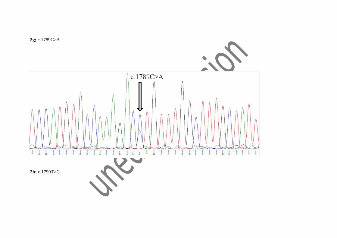
2g; c.1789C>A
2h; c.1790T>C

2i; c.1849C>A


Tables
Table 1A: All pathogenic and likely pathogenic DNA sequence variants found in polycystic kidney disease patients
EX/IV
S
CDS/Gene
Position
Protein/I
VS
Type of
Variant
In-silico analysis
rs ID PKDB Remark Patient Id Polyphe
n-2
PROVE
AN SIFT
Mutation
Taster
IVS1 g.11732A>G IVS1-
59A>G Intronic
Pathogeni
c
Novel/Lik
ely
Pathogenic
80.1
EX2 c.646T>C p.Y216H Missense
Probabl
y
damagin
g
Deleterio
us
Tolerate
d
Pathogeni
c
Novel/Lik
ely
Pathogenic
82.1
EX4 c.854_854del
G
p.G285Afs
*32
Protein
Truncatin
g
Pathogeni
c
Novel/Path
ogenic 39.1
EX4 c.915C>A p.N305K Missense
Possibly
damagin
g
Deleterio
us
Damagi
ng
Pathogeni
c
Novel/Path
ogenic 81.1
EX4 c.973C>T p.R325X
Protein
Truncatin
g
Pathogeni
c
Reported/P
athogenic 72.1
EX4 c.1050_1050d
elC
p.S351Vfs
*24
Protein
Truncatin
g
Pathogeni
c
Novel/Path
ogenic 64.1
EX6 c.1354A>G p.I452V Missense Benign Neutral Tolerate
d
Pathogeni
c rs1801612
Likely
neutral
Reported
/Likely
Pathogenic
38.1
IVS6 g.39212G>C IVS6+9G>
C Intronic
Pathogeni
c
rs3769016
84
Reported
/Likely
Pathogenic
37.1,42.1,44.1
EX7 c.1604_1604d
elT
p.L535Hfs
*27
Protein
Truncatin
Pathogeni
c
Novel/Path
ogenic 85.1

g
EX8 c.1789C>A p.L597M Missense
Probabl
y
damagin
g
Neutral Damagi
ng
Pathogeni
c
Novel/Lik
ely
Pathogenic
4.1,5.1,22.1
EX8 c.1790T>C p.L597P Missense
Probabl
y
damagin
g
Deleterio
us
Damagi
ng
Pathogeni
c
Novel/Path
ogenic 54.1
EX8 c.1849C>A p.L617I Missense Benign Neutral Damagi
ng
Pathogeni
c
Novel/Lik
ely
Pathogenic
4.1,5.1,22.1
EX11 c.2164G>T p.V722L Missense Benign Neutral Tolerate
d
Pathogeni
c
rs5299454
69
Novel/Lik
ely
Pathogenic
32.1,34.1,81.1,7
9.1
EX11 c.2182_2183d
elAG
p.L729Afs
*10
Protein
Truncatin
g
Pathogeni
c
Definitel
y
pathogeni
c
Reported/P
athogenic
5.1,27.1,28.1,43
.1
EX11 c.2224C>T p.R742X
Protein
Truncatin
g
Pathogeni
c
rs1219180
40
Reported/P
athogenic 77.1
EX13 c.2494A>G p.S832G Missense Benign Neutral Tolerate
d
Pathogeni
c
rs1455745
34
Reported
/Likely
Pathogenic
60.1

Table 1B: All polymorphism and likely neutral DNA sequence variants found in polycystic kidney disease patients
EX/IVS CDS/Gene
Position Protein/IVS
Type of
Variant
In-silico analysis
rs ID PKDB Remark Polyphen-
2 PROVEAN SIFT
Mutation
Taster
EX1 c.83G>C p.R28P Missense Benign Neutral Damaging Polymorphism rs1805044 Likely
neutral Reported/polymorphism
IVS1 g.11761T>C IVS1-32T>C Intronic
Polymorphism rs372764946
Reported/polymorphism
IVS1 g.11775C>T IVS1-16C>T Intronic
Polymorphism rs62310565 Likely
neutral Reported/polymorphism
IVS3 g.28713C>A IVS3+27C>A Intronic
Polymorphism
Novel/polymorphism
IVS3 g.28743T>C IVS3+57T>C Intronic
Polymorphism rs17786456
Reported/polymorphism
IVS3 g.30562G>A IVS3-22G>A Intronic
Polymorphism rs2725221 Likely
neutral Reported/polymorphism
IVS4 g.30896C>T IVS4+62C>T Intronic
Polymorphism rs373213485
Reported/polymorphism
EX6 c.1359A>G p.P453P Silent
Neutral Tolerated Pathogenic rs107013754 Likely
neutral
Reported/ Likely
neutral
IVS7 g.48386C>T IVS7-33C>T Intronic
Polymorphism rs372552957
Reported/polymorphism
EX13 c.2398A>C p.M800L Missense Benign Neutral Tolerated Polymorphism rs2234917 Likely
neutral Reported/polymorphism
EX13 c.2460C>T p.S820S Silent
Neutral Tolerated Pathogenic rs572822238 Likely
neutral
Reported/ Likely
neutral
IVS14 g.67319G>A IVS14+27G>A Intronic
Polymorphism rs113117728
Reported/polymorphism
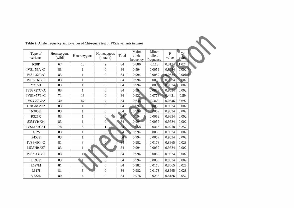
Table 2: Allele frequency and p-values of Chi-square test of PKD2 variants in cases
Type of
variants
Homozygous
(wild) Heterozygous
Homozygous
(mutant) Total
Major
allele
frequency
Minor
allele
frequency
P
value
X2
value
R28P 67 15 2 84 0.886 0.113 0.3114 1.024
IVS1-59A>G 83 1 0 84 0.994 0.0059 0.9634 0.002
IVS1-32T>C 83 1 0 84 0.994 0.0059 0.9634 0.002
IVS1-16C>T 83 1 0 84 0.994 0.0059 0.9634 0.002
Y216H 83 1 0 84 0.994 0.0059 0.9634 0.002
IVS3+27C>A 83 1 0 84 0.994 0.0059 0.9634 0.002
IVS3+57T>C 71 13 0 84 0.922 0.0773 0.4421 0.59
IVS3-22G>A 30 47 7 84 0.636 0.363 0.0546 3.692
G285Afs*32 83 1 0 84 0.994 0.0059 0.9634 0.002
N305K 83 1 0 84 0.994 0.0059 0.9634 0.002
R325X 83 1 0 84 0.994 0.0059 0.9634 0.002
S351Vfs*24 83 1 0 84 0.994 0.0059 0.9634 0.002
IVS4+62C>T 78 5 1 84 0.958 0.0416 0.0218 5.257
I452V 83 1 0 84 0.994 0.0059 0.9634 0.002
P453P 83 1 0 84 0.994 0.0059 0.9634 0.002
IVS6+9G>C 81 3 0 84 0.982 0.0178 0.8665 0.028
L535Hfs*27 83 1 0 84 0.994 0.0059 0.9634 0.002
IVS7-33C>T 83 1 0 84 0.994 0.0059 0.9634 0.002
L597P 83 1 0 84 0.994 0.0059 0.9634 0.002
L597M 81 3 0 84 0.982 0.0178 0.8665 0.028
L617I 81 3 0 84 0.982 0.0178 0.8665 0.028
V722L 80 4 0 84 0.976 0.0238 0.8186 0.052

L729Afs*10 80 4 0 84 0.976 0.0238 0.8186 0.052
R742X 83 1 0 84 0.994 0.0059 0.9634 0.002
M800L 83 1 0 84 0.994 0.0059 0.9634 0.002
S820S 83 1 0 84 0.994 0.0059 0.9634 0.002
S832G 83 1 0 84 0.994 0.0059 0.9634 0.002
IVS14+27G>A 83 1 0 84 0.994 0.0059 0.9634 0.002
P value less than <0.05 is not consistent with Hardy-Weinberg equilibrium
TABLE 3: Association of: IVS3-22G>A and IVS4-62C>T polymorphism in cases and controls
IVS3-22G>A
Cases
(N=84)
Controls
(N=80)
Odds ratio
(OR)
95% Confidence
Interval (CI) p value
GG 30 23 Referent
GA 47 37 0.9739 0.4868-1.948 0.9403
AA 7 20
G allele frequency 0.637 0.518
A allele frequency 0.363 0.481
Dominant model 0.7263 0.3754-1.403 0.4318
Recessive Model 0.2727 0.1082-0.6876 0.0056
IVS4-62C>T
CC 78 73 Referent
CT 5 7 0.6685 0.0231-2.201 0.5604
TT 1 0
C allele frequency 0.958 0.956
T allele frequency 0.042 0.043
Dominant model 0.8022 0.2575-2.499 0.7776
Recessive Model 2.892 0.116-72.099 1