research article occlusal characteristics and spacing...
TRANSCRIPT

Research ArticleOcclusal Characteristics and Spacing in Primary Dentition:A Gender Comparative Cross-Sectional Study
Madhuri Vegesna, R. Chandrasekhar, and Vinay Chandrappa
Department of Pedodontics and Preventive Dentistry, Vishnu Dental College, Vishnupur, Bhimavaram,Andhra Pradesh 534202, India
Correspondence should be addressed to Madhuri Vegesna; [email protected]
Received 27 July 2014; Accepted 24 September 2014; Published 29 October 2014
Academic Editor: Chun-Pin Lin
Copyright © 2014 Madhuri Vegesna et al. This is an open access article distributed under the Creative Commons AttributionLicense, which permits unrestricted use, distribution, and reproduction in any medium, provided the original work is properlycited.
Context. Occlusion in primary teeth varies among children of different populations and races. Aim. To assess and compare theocclusal characteristics and spacing in primary dentition among 3–6-year-oldDravidian children.Materials andMethods.The studyincluded 2281 school going children. The primary molar relation, canine relation, overjet, and overbite were assessed using Fosterand Hamilton criteria. Spacing conditions were registered according to Kisling and Krebs criteria. Results.The flush terminal planemolar relation (80.3%) was the most common primary molar relation. The distal step molar relation was more frequently foundin female children (12.8%) than in males (8.6%). Class 1 canine relation was the most prevalent canine relation (81.3%) amongmales and females. Ideal overjet (84.3%) and overbite (72.7%) were observed among the majority of the children. Spaced type ofarches occurred more frequently than closed arches in this sample. The incidence of primate spaces was more in males than infemales. Conclusion. The study population has fewer deviations from normal occlusion which indicates decreased tendency formalocclusion in permanent dentition. However, further longitudinal studies are necessary to identify the potential limitations of aclinical approach relying on early orthodontic diagnosis and intervention.
1. Introduction
Childhood is the mirror in which the propensities of adult-hood are reflected; similarly the type of occlusion in primarydentition predicts the occlusion of the permanent dentition[1]. The understanding of the anteroposterior changes thatoccur in the occlusion between the primary and perma-nent dentition is crucial for the clinicians involved in earlyorthodontic treatment [2]. Normal occlusion in primaryteeth has the following characteristics: spacing betweenanterior teeth, primate spaces, low overjet and overbite, flushterminal plane molar relation, and ovoid arch form [3, 4].The deviations in occlusion in primary dentition would becarried to succeeding permanent dentition and to a morepronounced degree [5].
Spacing is a common condition in the primary dentitionand constitutes a very important feature of the dentitionas it is an indicator of favorable development of perma-nent occlusion. Spacing often presents between all anterior
primary teeth with the most marked spaces present beingmesial to canines in the maxilla and distal to canines in themandible. These are called primate spaces. The secondary ordevelopmental spaces which are commonly found betweenthe incisors are termed physiological spaces [6, 7]. Theincidence of spacing in primary dentition varies from 42.9%to 98%. Perhaps lack of spacing suggests severe risk forcrowding in the permanent dentition [6–8]. Spacing is morecommon in the maxilla than in the mandible and spaces areobserved more among boys rather than girls [8].
Many observational studies relating to the spacing andocclusion of the primary dentition have confirmed that theocclusal characteristics vary among populations and ethnicgroups. The present study documents the nature of occlusalrelationships comprising molar relation, canine relation,overjet, overbite, and spacing of the primary dentition andalso assesses gender variations among 3–6-year-old Dravid-ian children.
Hindawi Publishing CorporationInternational Scholarly Research NoticesVolume 2014, Article ID 512680, 7 pageshttp://dx.doi.org/10.1155/2014/512680
2 International Scholarly Research Notices
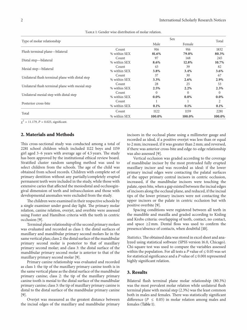
Table 1: Gender wise distribution of molar relation.
Type of molar relationship Sex TotalMale Female
Flush terminal plane—bilateral Count 916 916 1832% within SEX 81.6% 79.0% 80.3%
Distal step—bilateral Count 97 148 245% within SEX 8.6% 12.8% 10.7%
Mesial step—bilateral Count 43 39 82% within SEX 3.8% 3.4% 3.6%
Unilateral flush terminal plane with distal step Count 37 30 67% within SEX 3.3% 2.6% 2.9%
Unilateral flush terminal plane with mesial step Count 28 25 53% within SEX 2.5% 2.2% 2.3%
Unilateral mesial step with distal step Count 0 0 0% within SEX 0.0% 0.0% 0.0%
Posterior cross-bite Count 1 1 2% within SEX 0.1% 0.1% 0.1%
Total Count 1122 1159 2281% within SEX 100.0% 100.0% 100.0%
𝜒2= 11.179, 𝑃 = 0.025, significant.
2. Materials and Methods
This cross-sectional study was conducted among a total of2281 school children which included 1122 boys and 1159girl aged 3–6 years with average age of 4.5 years. The studyhas been approved by the institutional ethical review board.Stratified cluster random sampling method was used toselect children from the schools. The age of the child wasobtained from school records. Children with complete set ofprimary dentition without any partially/completely eruptedpermanent teeth were included in the study, while those withextensive caries that affected the mesiodistal and occlusogin-gival dimension of teeth and infraocclusion and those withdevelopmental anomalies were excluded from the study.
The childrenwere examined in their respective schools bya single examiner under good day light. The primary molarrelation, canine relation, overjet, and overbite were assessedusing Foster and Hamilton criteria with the teeth in centricocclusion [9].
Terminal plane relationship of the second primarymolarswas evaluated and recorded as class 1: the distal surfaces ofmaxillary and mandibular primary second molars lie in thesame vertical plan; class 2: the distal surface of themandibularprimary second molar is posterior to that of maxillaryprimary second molar; and class 3: the distal surface of themandibular primary second molar is anterior to that of themaxillary primary second molar [9].
Primary canine relationship was evaluated and recordedas class 1: the tip of the maxillary primary canine tooth is inthe same vertical plane as the distal surface of themandibularprimary canine; class 2: the tip of the maxillary primarycanine tooth is mesial to the distal surface of the mandibularprimary canine; class 3: the tip of maxillary primary canine isdistal to the distal surface of the mandibular primary canine[9].
Overjet was measured as the greatest distance betweenthe incisal edges of the maxillary and mandibular primary
incisors in the occlusal plane using a millimeter gauge andrecorded as ideal, if a positive overjet was less than or equalto 2mm; increased, if it was greater than 2mm; and reversed,if there was anterior cross-bite and edge-to-edge relationshipwas also assessed [9].
Vertical occlusion was graded according to the coverageof mandibular incisor by the most protruded fully eruptedmaxillary incisor and was recorded as ideal: if the lowerprimary incisal edges were contacting the palatal surfacesof the upper primary central incisors in centric occlusion,increased, if the mandibular incisors were touching thepalate, open bite, when a gap existed between the incisal edgesof incisors along the occlusal plane, and reduced, if the incisaltips of the lower primary incisors were not contacting theupper incisors or the palate in centric occlusion but withpositive overbite [9].
Spacing conditions were registered between all teeth inthe mandible and maxilla and graded according to Kislingand Krebs criteria: overlapping of teeth, contact, no contact,and space ≥2mm. Dental floss was used to confirm thepresence/absence of contacts, when doubtful [10].
Statistics.The obtained data was stored in excel sheet and ana-lyzed using statistical software (SPSS version 16.0, Chicago).Chi-square test was used to compare the variables assessedwithin the population. For all tests a 𝑃 value of ≤ 0.05 was setfor statistical significance and a𝑃 value of≤ 0.001 representedhighly significant relation.
3. Results
Bilateral flush terminal plane molar relationship (80.3%)was the most prevalent molar relation while unilateral flushterminal plane with mesial step (2.3%) was the least commonboth in males and females. There was statistically significantdifference (𝑃 ≤ 0.05) in molar relation among males andfemales (Table 1).

International Scholarly Research Notices 3
Table 2: Gender wise distribution of canine relationship.
Type of canine relationship Sex TotalMale Female
Class 1—bilateral Count 918 936 1854% within SEX 81.8% 80.8% 81.3%
Class 2—bilateral Count 78 56 134% within SEX 7.0% 4.8% 5.9%
Class 3—bilateral Count 50 82 132% within SEX 4.5% 7.1% 5.8%
Unilateral class 1 with class 2 Count 50 46 96% within SEX 4.5% 4.0% 4.2%
Unilateral class 1 with class 3 Count 24 38 62% within SEX 2.1% 3.3% 2.7%
Unilateral class 2 with class 3 Count 1 0 1% within SEX 0.1% 0.0% 0.0%
Posterior cross-bite Count 1 1 2% within SEX 0.1% 0.1% 0.1%
Total Count 1122 1159 2281% within SEX 100.0% 100.0% 100.0%
𝜒2= 15.301, 𝑃 = 0.009, significant.
Table 3: Distribution of overjet variations.
Overjet Sex TotalMale Female
Ideal Count 949 975 1924% within SEX 84.6% 84.1% 84.3%
Increased Count 111 91 202% within SEX 9.9% 7.9% 8.9%
Edge-to-edge Count 33 47 80% within SEX 2.9% 4.1% 3.5%
Reversed Count 11 27 38% within SEX 1.0% 2.3% 1.7%
Others Count 18 19 37% within SEX 1.6% 1.6% 1.6%
Total Count 1122 1159 2281% within SEX 100.0% 100.0% 100.0%
𝜒2= 10.986, 𝑃 = 0.027, significant.
The most common type of canine relation was bilateralclass 1 (81.3%) whereas the least frequent was unilateral class1 with class 3 (2.7%); similar trend was observed in bothgenders (Table 2). Statistical significant difference was foundamong the sexes with respect to canine relation (𝑃 ≤ 0.05).
An ideal overjet was observed among 84.3% childrenfollowed by increased overjet (8.9%) and edge-to-edge bite(3.5%), while the least frequent type was reverse overjet(1.7%) (Table 3). The evaluation of overbite showed that72.7% children had ideal overbite, 19.4% had increased bitewhile 1.5% had anterior open bite, and 1% had reduced bite(Table 4). Similar trend of prevalence was observed among
both the sexes with respect to overjet and overbite. Statisticalsignificant difference was found among the sexes with respectto overjet (𝑃 ≤ 0.05) while no significance was found withregard to overbite (𝑃 = 0.781).
The most frequent site of spacing (Table 5) in the maxil-lary arch coincided with the anthropoid space between thelateral incisor and canine (71.8%); however in mandibulararch it did not coincide with primate spaces; instead, spacingwas found at two sites, that is, between canine and lateralincisor (31.1%) and lateral incisor and central incisor (31.0%).The spaces greater than or equal to 2mm were found mostcommonly in relation tomaxillary primate spaces (1.8%).The

4 International Scholarly Research Notices
Table 4: Distribution of overbite variations.
Overbite Sex TotalMale Female
Ideal Count 826 832 1658% within SEX 73.6% 71.8% 72.7%
Increased Count 225 217 442% within SEX 20.1% 18.7% 19.4%
Anterior open bite Count 16 18 34% within SEX 1.4% 1.6% 1.5%
Reduced Count 9 13 22% within SEX 0.8% 1.1% 1.0%
Others Count 46 79 125% within SEX 4.1% 6.8% 5.5%
Total Count 1122 1159 2281% within SEX 100.0% 100.0% 100.0%
𝜒2= 1.756, 𝑃 = 0.781, not significant.
Table 5: Arch wise prevalence of spacing.
Site Normal contacts No contacts Overlapping Spacing ≥ 2mm 𝜒2 Significance
𝑃 value
Second molar—first molarMaxilla 4531 30 0 1
1.1580.763Notsignificant
99.2% 0.7% 0% 0.1%
Mandible 4534 27 0 199.3% 0.6% 0% 0.1%
First molar—canineMaxilla 3938 624 0 0
198.9630.001Highlysignificant
86.3% 13.7% 0% 0%
Mandible 3406 1152 2 274.6% 25.2% 0.1% 0.1%
Canine—lateral incisorMaxilla 1270 3190 16 86
1668.0840.001Highlysignificant
27.8% 70.0% 0.4% 1.8%
Mandible 2736 1396 420 1059.7% 30.9% 9.2% 0.2%
Lateral incisor—centralincisor
Maxilla 2853 1649 52 8
99.3060.001Highlysignificant
62.5% 36.2% 1.1% 0.2%
Mandible 2959 1413 187 364.8% 30.9% 4.1% 0.1%
Between central incisorsMaxilla 1779 485 8 9
124.7110.001Highlysignificant
38.9% 10.7% 0.2% 0.2%
Mandible 1519 660 97 533.2% 14.5% 2.2% 0.1%
sites of contact of teeth were found most frequently betweenfirst and second primary molar in both maxilla (99.2%) andmandible (99.3%).
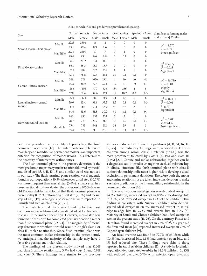
Statistical significant difference was found between thesexes (Table 6) with respect to spaces between first molar andcanine (𝑃 ≤ 0.05), canine and lateral incisor (𝑃 ≤ 0.001), andcentral and lateral incisor (𝑃 ≤ 0.001).
4. Discussion
The occlusion of the primary dentition is completely estab-lished by the age of 3 years and lasts until about 6 years of agewhen the first permanent tooth begins to erupt [11]. Under-standing the association between morphological aspects inthe primary dentition and its transition to the permanent
International Scholarly Research Notices 5
Table 6: Arch wise and gender wise prevalence of spacing.
Site Normal contacts No contacts Overlapping Spacing > 2mm Significance [among malesand females] 𝑃 valueMale Female Male Female Male Female Male Female
Second molar—first molarMaxilla 2228 2304 16 14 0 0 0 0
𝜒2= 1.270
𝑃 = 0.530
Not significant
99.1 99.4 0.9 0.6 0 0 0 0
Mandible 2234 2300 10 17 0 1 0 099.4 99.1 0.6 0.8 0 0.1 0 0
First Molar—canineMaxilla 1926 2012 318 306 0 0 0 0
𝜒2= 9.077
𝑃 = 0.028
Significant
86.1 86.3 13.9 13.7 0 0 0 0
Mandible 1625 1781 317 536 1 1 1 072.4 76.8 27.4 23.1 0.1 0.1 0.1 0
Canine—lateral incisorMaxilla 568 701 1630 1561 6 10 40 46
𝜒2= 38.799
𝑃 = 0.001
Highlysignificant
25.4 30.2 72.5 67.4 0.2 0.5 1.9 1.9
Mandible 1286 1450 770 626 184 236 4 657.0 62.4 34.6 27.1 8.2 10.2 0.2 0.3
Lateral incisor—centralincisor
Maxilla 1329 1424 880 769 34 17 1 8𝜒2= 16.304
𝑃 = 0.001
Highlysignificant
59.6 65.4 38.8 33.5 1.5 0.8 0.1 0.3
Mandible 1438 1421 714 699 90 97 2 164.0 65.6 31.8 30.2 4.1 4.1 0.1 0.1
Between central incisorsMaxilla 883 896 232 253 6 2 1 8
𝜒2= 5.480
𝑃 = 0.140
Not significant
78.7 77.3 20.7 21.8 0.5 0.2 0.1 0.7
Mandible 734 785 348 312 38 59 2 365.4 67.7 31.0 26.9 3.4 5.1 0.2 0.3
dentition provides the possibility of predicting the finalpermanent occlusion [12]. The anteroposterior relation ofmaxillary andmandibular permanent molars is an importantcriterion for recognition of malocclusions. This determinesthe necessity of interceptive orthodontics.
The flush terminal plane in the primary dentition is themost predominant primarymolar relation followed bymesialand distal step [5, 6, 11, 13–18] and similar trend was noticedin our study.The flush terminal plane relation was frequentlyfound in our population (80.3%); however distal step (10.7%)was more frequent than mesial step (3.6%). Yilmaz et al. in across-sectional study evaluated the occlusion in 205 3–6-year-old Turkish children and found that flush terminal plane waspresented by 88.29% followed by distal step (7.31%) andmesialstep (4.4%) [19]. Analogous observations were reported inFinnish and Iranian children [20, 21].
The flush terminal plane was found to be the mostcommon molar relation and considered ideal for transitionto class 1 in permanent dentition. However, mesial step wasfound to be the norm for completed primary dentition ratherthan flush terminal plane [22–24]. The magnitude of mesialstep determines whether it would result in Angle’s class I orclass III molar relationship. Since flush terminal plane wasthe most common molar relationship in the present studywe anticipate that the majority of the sample may have afavorable permanent molar relation.
The findings of the present study showed that 81.3%had class 1 canine relationship, 5.9% had class 2, and 5.8%had class 3. These findings were similar to the previous
studies conducted in different populations [4, 11, 14, 16, 17,19, 25]. Contradictory findings were reported in Finnishchildren among whom class II relation (52.4%) was themost prominent followed by class I (46.1%) and class III(1.5%) [20]. Canine and molar relationship together can bea diagnostic aid to predict changes in occlusal relationship.In clinical situations like flush terminal plane with class IIcanine relationship indicates a higher risk to develop a distalocclusion in permanent dentition. Therefore both the molarand canine relationships are taken into consideration tomakea reliable prediction of the intermaxillary relationship in thepermanent dentition [20].
The results of our investigation revealed ideal overjet in84.3% children, increased overjet in 8.9%, edge-to-edge bitein 3.5%, and reversed overjet in 1.7% of the children. Thisfinding is consistent with Nigerian children who demon-strated ideal overjet in 68.6%, increased overjet in 14.7%,edge-to-edge bite in 9.7%, and reverse bite in 7.0% [5].Majority of Saudi and Chinese children had ideal overjet asseen in the present study [11, 26]. On the contrary, Foster andHamilton found increased overjet in 72% of 21/2-3-year-oldchildren and Ravn [27] reported increased overjet in 27% ofCopenhagen children [9].
An ideal overbite was found in 72.7% of children while19.4% had increased bite, 1.5% had anterior open bite, and1% had reduced bite. These findings were akin to thosereported in Saudi Arabian children [11]. A study in Jordanianpopulation demonstrated overbite in 44.3% children, 21.85%with reduced overbite, 5.7% with anterior open bite, and

6 International Scholarly Research Notices
28.2%with increased bite [7]. Anterior open bitewas reportedin 8% of AfricanAmerican children [23].Majority of Chinesechildren had increased bite [26], whereas Belgian childrenexhibited open bite in 32% of the studied population [28].Increased open bite could be attributed to oral habits suchas dummy sucking and finger sucking [5]. Presently, nodata indicates a definitive threshold value for overbite oroverjet that could be applied in early diagnostics [20]. Thehigher prevalence of ideal overjet and overbite observed inour population may be conducive to achieve ideal anteriorrelation in permanent dentition.
Spaces in the primary teeth are described as physiologicalor developmental spaces. The spacing around the canines istermed as the simian gap [6], primate space [13], or anthro-poid space [9], since they are prominent in dentitions ofcertain lower primates. Based on spacing between the teeth,Baume has classified the arrangement of primary dentitioninto two forms: open or type I and closed or type II [6].
Anterior spacing appears to be a common finding inour study population. Primate spaces were frequently foundin the maxilla than mandible [3, 7, 29, 30] as observedin the present study. Male children demonstrated morefrequency of primate spaces than females both in the maxillaand mandible. In contrast, primate spaces did not showsignificant difference between the genders in Tehran andSaudi children [21, 31]. Extreme spacing of ≥2mm in ourstudy was associated with anthropoid spaces in the maxilla(1.8%) akin to observations made by Otuyemi [5].
The incidence of crowding or overlapping of teeth inour sample was more prevalent in the mandible than in themaxilla. In mandible, crowding was predominant betweenthe lateral incisor and canine (9.2%) and similar findingswerereported in Nigerian children [5]. This study illustrated thatcrowding was more prevalent in females than in males inboth maxilla and mandible. Majority of the study populationdemonstrated spaced arches which would lead to favorablepermanent occlusion.
5. Conclusion
Occlusal characteristics vary among populations. To summa-rize, flush terminal plane, class I canine relation, ideal overjet,ideal overbite, and spaced arches prevailed among majorityof the study population without any gender variations.These findings suggest favorable occlusal characteristics andspacing in primary dentition. However, future longitudinalstudies are needed to observe whether the transition of theseocclusal characteristics will lead to favorable occlusion in thepermanent dentition.
Disclosure
R. Chandrasekhar and Vinay Chandrappa are coauthors.
Conflict of Interests
Theauthors hereby declare that there is no conflict of interestsand the study has not been funded by any source or agency.
Acknowledgment
All the authors are associated with the Department ofPedodontics and Preventive Dentistry, Vishnu Dental Col-lege.
References
[1] G. Z. Wright and D. B. Kennedy, “Space control in the primaryand mixed dentitions,” Dental Clinics of North America, vol. 22,no. 4, pp. 579–601, 1978.
[2] B. S. Arya, B. S. Savara, and D. R. Thomas, “Prediction of firstmolar occlusion,” American Journal of Orthodontics, vol. 63, no.6, pp. 610–621, 1973.
[3] M. R. Joshi and P. G. Makhija, “Some observations on spacingin the normal deciduous dentition of 100 Indian children fromGujarat,” The British journal of orthodontics, vol. 11, no. 2, pp.75–79, 1984.
[4] K.M. E.MotayamandA. Elbardissy, “Occlusal characteristics ofprimary dentition in preschool Egyptian children,”CairoDentalJournal, vol. 23, pp. 217–226, 2007.
[5] O. D. Otuyemi, E. O. Sote, M. C. Isiekwe, and S. P. Jones,“Occlusal relationships and spacing or crowding of teeth inthe dentitions of 3-4-year-old Nigerian children,” InternationalJournal of Paediatric Dentistry, vol. 7, no. 3, pp. 155–160, 1997.
[6] L. J. Baume, “Physiological tooth migration and its significancefor the development of occlusion. I.The biogenetic course of thedeciduous dentition,” Journal of Dental Research, vol. 29, no. 2,pp. 123–132, 1950.
[7] E. S. J. Abu Alhaija and M. A. Qudeimat, “Occlusion andtooth/arch dimensions in the primary dentition of preschoolJordanian children,” International Journal of Paediatric Den-tistry, vol. 13, no. 4, pp. 230–239, 2003.
[8] N. Gkantidis, S. Psomiadis, and N. Topouzelis, “Teeth spacing:etiology and treatment,” Hellenic Orthodontic Review, vol. 10,pp. 75–92, 2007.
[9] T. D. Foster and M. C. Hamilton, “Occlusion in the primarydentition,” British Dental Journal, vol. 126, no. 2, pp. 76–79, 1969.
[10] E. Kisling and G. Krebs, “Patterns of occlusion in 3 year oldDanish children,” Community Dentistry and Oral Epidemiology,vol. 4, no. 4, pp. 152–159, 1976.
[11] N. M. A. Farsi and F. S. Salama, “Characteristics of primarydentition occlusion in a group of Saudi children,” InternationalJournal of Paediatric Dentistry, vol. 6, no. 4, pp. 253–259, 1996.
[12] A. L. T. Dutra, P. M. Berto, L. D. S. Vieira, and O. A. D. Toledo,“Longitudinal changes in the molar relationship from primaryto permanent dentition,” Conscientiae Saude, vol. 8, pp. 171–176,2009.
[13] D. J. Boyko, “The incidence of primate spaces in fifty 3-year-old children of the Burlington study,” American Journal ofOrthodontics, vol. 54, no. 6, pp. 462–465, 1968.
[14] R. S. Nanda, I. Khan, andR. Anand, “Age changes in the occlusalpattern of deciduous dentition.,” Journal of Dental Research, vol.52, no. 2, pp. 221–224, 1973.
[15] S. Alexander and N. T. Prabhu, “Profiles, occlusal plane rela-tionships and spacing of teeth in the dentitions of 3 to 4 yearold children,” Journal of Clinical Pediatric Dentistry, vol. 22, no.4, pp. 329–334, 1998.
[16] S. Prabhakaran, C. Sriram, M. Muthu, C. Rao, and N. Sivaku-mar, “Dental arch dimensions in primary dentition of children

International Scholarly Research Notices 7
aged three to five years in Chennai and Hyderabad,” IndianJournal of Dental Research, vol. 17, no. 4, pp. 185–189, 2006.
[17] S. Hegde, S. Panwar, D. R. Bolar, and M. B. Sanghavi, “Charac-teristics of occlusion in primary dentition of preschool childrenof Udaipur, India,” European Journal of Dentistry, vol. 6, no. 1,pp. 51–55, 2012.
[18] R. Khan, N. Singh, S. Govil, and S. Tandon, “Occlusion andocclusal characteristics of primary dentition in North Indianchildren of East Lucknow region,” European Archives of Paedi-atric Dentistry, vol. 15, no. 5, pp. 293–299, 2014.
[19] Y. Yilmaz, T.Gurbuz, S. Simsek, andA.Dalmis, “Primary canineand molar relationships in centric occlusion in three to sixyear-old Turkish children: a cross-sectional study,” Journal ofContemporary Dental Practice, vol. 7, no. 3, pp. 59–66, 2006.
[20] K. Keski-Nisula, R. Lehto, V. Lusa, L. Keski-Nisula, and J.Varrela, “Occurrence of malocclusion and need of orthodontictreatment in early mixed dentition,” The American Journal ofOrthodontics and Dentofacial Orthopedics, vol. 124, no. 6, pp.631–638, 2003.
[21] J. Mahmoodian, H. Afshar, and M. Hadjhashem, “Determina-tion of primate Space on 4 to 5 years old children of Tehran’sKindergarten in 2000,” Journal of Dentistry (Tehran, Iran), vol.1, no. 1, pp. 21–26, 2004.
[22] S. E. Bishara, B. J. Hoppens, J. R. Jakobsen, and F. J. Kohout,“Changes in the molar relationship between the deciduous andpermanent dentitions: a longitudinal study,” American Journalof Orthodontics and Dentofacial Orthopedics, vol. 93, no. 1, pp.19–28, 1988.
[23] M. L. Jones, A. P. Mourino, and T. A. Bowden, “Evaluation ofocclusion, trauma, and dental anomalies in African-Americanchildren of metropolitan Headstart programs,” The Journal ofClinical Pediatric Dentistry, vol. 18, no. 1, pp. 51–54, 1993.
[24] A. A. Andersona, “Occlusal development in children of AfricanAmerican descent: types of terminal plane relationships in theprimary dentition,” Angle Orthodontist, vol. 76, no. 5, pp. 817–823, 2006.
[25] L. P. da Silva and R. Gleiser, “Occlusal development betweenprimary and mixed dentitions: a 5-year longitudinal study,”Journal of Dentistry for Children, vol. 75, no. 3, pp. 287–294,2008.
[26] S. Imudom, “Occlusal characteristics of 5-year-old southernChinese children,” European Journal of Orthodontics, vol. 16, pp.456–457, 1994.
[27] J. J. Ravn, “Longitudinal study of occlusion in the primarydentition in 3- to 7-year-old children,” Scandinavian Journal ofDental Research, vol. 88, no. 3, pp. 165–170, 1980.
[28] J. C. Carvalho, F. Vinker, and D. Declerck, “Malocclusion,dental injuries and dental anomalies in the primary dentition ofBelgian children,” International Journal of Paediatric Dentistry,vol. 8, no. 2, pp. 137–141, 1998.
[29] A. Kaufman and E. Koyoumdjisky, “Normal occlusal patterns inthe deciduous dentition in preschool children in Israel,” Journalof Dental Research, vol. 46, no. 3, pp. 478–482, 1967.
[30] N. Ohno, K. Kashima, and T. Sakai, “A study on interdentalspaces of the deciduous dental arch in Indian sample,” AichiGakuin Daigaku Shigakkai shi, vol. 28, no. 1, part 1, pp. 79–91,1990.
[31] N. Salako, N. Alamoudi, R. Fateih, and I. Masoud, “Prevalenceand distribution pattern of interdental spaces in the primarydentition of Saudi Arabian children,” Journal of King AbdulazizUniversity—Medical Sciences, vol. 7, pp. 107–113, 1999.

Submit your manuscripts athttp://www.hindawi.com
Hindawi Publishing Corporationhttp://www.hindawi.com Volume 2014
Oral OncologyJournal of
DentistryInternational Journal of
Hindawi Publishing Corporationhttp://www.hindawi.com Volume 2014
Hindawi Publishing Corporationhttp://www.hindawi.com Volume 2014
International Journal of
Biomaterials
Hindawi Publishing Corporationhttp://www.hindawi.com Volume 2014
BioMed Research International
Hindawi Publishing Corporationhttp://www.hindawi.com Volume 2014
Case Reports in Dentistry
Hindawi Publishing Corporationhttp://www.hindawi.com Volume 2014
Oral ImplantsJournal of
Hindawi Publishing Corporationhttp://www.hindawi.com Volume 2014
Anesthesiology Research and Practice
Hindawi Publishing Corporationhttp://www.hindawi.com Volume 2014
Radiology Research and Practice
Environmental and Public Health
Journal of
Hindawi Publishing Corporationhttp://www.hindawi.com Volume 2014
The Scientific World JournalHindawi Publishing Corporation http://www.hindawi.com Volume 2014
Hindawi Publishing Corporationhttp://www.hindawi.com Volume 2014
Dental SurgeryJournal of
Drug DeliveryJournal of
Hindawi Publishing Corporationhttp://www.hindawi.com Volume 2014
Hindawi Publishing Corporationhttp://www.hindawi.com Volume 2014
Oral DiseasesJournal of
Hindawi Publishing Corporationhttp://www.hindawi.com Volume 2014
Computational and Mathematical Methods in Medicine
ScientificaHindawi Publishing Corporationhttp://www.hindawi.com Volume 2014
PainResearch and TreatmentHindawi Publishing Corporationhttp://www.hindawi.com Volume 2014
Preventive MedicineAdvances in
Hindawi Publishing Corporationhttp://www.hindawi.com Volume 2014
EndocrinologyInternational Journal of
Hindawi Publishing Corporationhttp://www.hindawi.com Volume 2014
Hindawi Publishing Corporationhttp://www.hindawi.com Volume 2014
OrthopedicsAdvances in